ICU · Haematology / oncology
Oncologic Emergencies — TLS, Hypercalcaemia, Hyperviscosity, SVC, Cord Compression, Neutropenic Sepsis & Checkpoint Toxicity
Also known as Oncologic emergency · Tumour lysis syndrome · TLS · Hypercalcaemia of malignancy · Superior vena cava syndrome · SVC syndrome · Malignant spinal cord compression · MSCC · Rasburicase · Allopurinol · Zoledronic acid · PTHrP · Hyperviscosity syndrome · Waldenström macroglobulinemia · Plasmapheresis · Neutropenic sepsis · Febrile neutropenia · Immune-related adverse event · irAE · Checkpoint inhibitor pneumonitis · Immune checkpoint inhibitor colitis · Cairo-Bishop classification · Cord compression · Denosumab
The oncologic emergencies in the ICU: (1) tumour lysis syndrome (TLS — cell lysis to hyperkalaemia, hyperphosphataemia, hypocalcaemia, hyperuricaemia, AKI; Cairo-Bishop classification; prophylaxis hydration plus allopurinol or rasburicase; treat rasburicase plus RRT), (2) hypercalcaemia of malignancy (PTHrP, osteolytic, ectopic vitamin D; shortened QT; treat IV saline plus bisphosphonates zoledronic acid plus calcitonin plus denosumab), (3) hyperviscosity syndrome (Waldenström IgM, serum hyperviscosity; plasmapheresis urgent), (4) superior vena cava syndrome (facial and arm swelling; treat the tumour — RT, chemo, endovascular stent), (5) malignant spinal cord compression (back pain, weakness, sensory level; dexamethasone 16 mg plus urgent MRI plus RT or surgery within 48 h; Patchell trial), (6) neutropenic sepsis (the 1-hour rule — empiric broad-spectrum antibiotic within 1 hour of presentation), and (7) immune-related adverse events from checkpoint inhibitors (colitis, pneumonitis, hepatitis — corticosteroids, infliximab). The time-critical conditions — the TLS, the cord compression, the neutropenic sepsis — demand early recognition and treatment.
On this page & tools
Your progress
Saved locally on this device.
8 MCQs with explanations
Target exams
Overview & definition
The oncologic emergencies in the ICU are the time-critical conditions from the malignancy or the treatment. The four: the tumour lysis syndrome (TLS), the hypercalcaemia of malignancy, the superior vena cava (SVC) syndrome, and the malignant spinal cord compression (MSCC). Each has the specific the management and the time-sensitivity — the TLS the hours, the cord compression the 48 hours.[1]
The expanded intensive-care list of seven time-critical oncologic emergencies: the four classical structural/metabolic crises above, plus the hyperviscosity syndrome (Waldenström macroglobulinemia — urgent plasmapheresis), the neutropenic sepsis (the 1-hour antibiotic rule), and the immune-related adverse events (irAEs) from checkpoint inhibitors (colitis, pneumonitis, hepatitis). Two further categories deserve a low threshold for recognition: the febrile neutropenia and the immune effector cell-associated cytokine release syndrome (CRS) and neurotoxicity (ICANS) from CAR-T and bispecific antibody therapy, which now share management principles with septic shock.[1]
The seven ICU oncologic emergencies — the time-critical recognition
| Emergency | The cardinal clue | The first drug / act | The clock |
|---|---|---|---|
| Tumour lysis syndrome | Hyperkalaemia + hyperphosphataemia + hypocalcaemia + hyperuricaemia 12-72 h post-chemotherapy | Rasburicase 0.15-0.2 mg/kg + aggressive hydration | Monitor labs q4-6h for 48-72 h |
| Hypercalcaemia of malignancy | Calcium over 3.0 + shortened QT + polyuria/altered mental state | IV saline + zoledronic acid 4 mg + calcitonin (rapid bridge) | Bisphosphonate onset 48-72 h |
| Hyperviscosity syndrome | IgM paraprotein + visual disturbance, headache, mucosal bleeding | Urgent plasmapheresis | Hours — before stroke or MI |
| SVC syndrome | Facial/arm plethora, JVD, dyspnoea; collateral veins | Elevate head + endovascular stent (severe) + treat the tumour | Stent gives relief within 24-48 h |
| Cord compression (MSCC) | Back pain + limb weakness/sensory level in a cancer patient | Dexamethasone 16 mg IV before the MRI | Definitive therapy within 24-48 h |
| Neutropenic sepsis | Neutrophils under 0.5 + fever over 38.3 °C | Empiric anti-pseudomonal β-lactam within 1 hour | The golden hour |
| Checkpoint inhibitor toxicity | Diarrhoea/dyspnoea/ AST-ALT rise weeks-months after a PD-1/CTLA-4 dose | Withhold ICI + corticosteroids; infliximab/vedolizumab if refractory | Corticosteroids for grade ≥2 |
Tumour lysis syndrome (TLS)
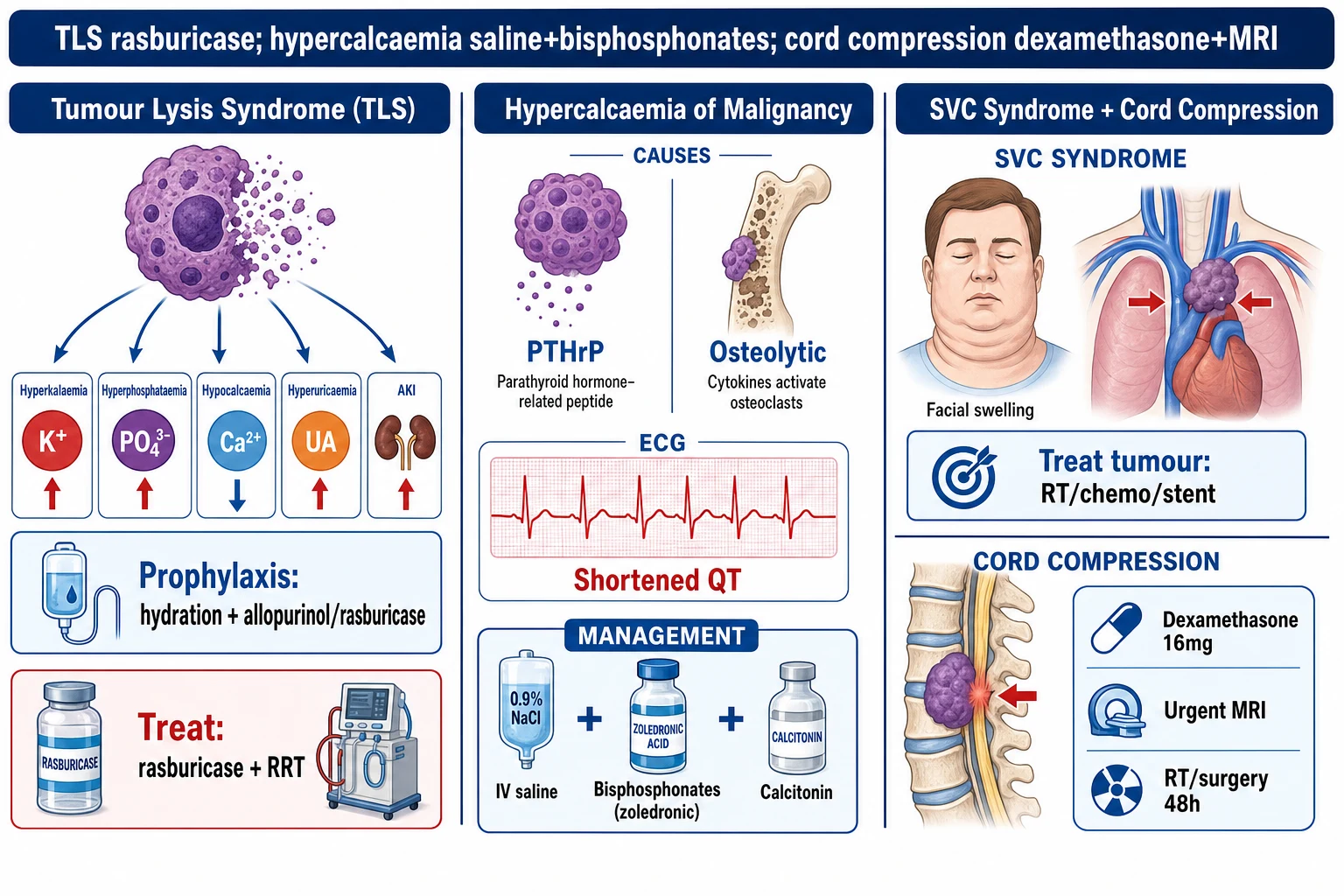
- The mechanism — the massive tumour cell lysis (after the chemotherapy initiation) → the release of the intracellular contents (the potassium, the phosphate, the uric acid, the purines).[1]
- The consequences:[1]
- The risk — the high-grade the lymphoma (the Burkitt), the AML (the high the WCC), the ALL. The after the chemotherapy the initiation (the 12 to 72 hours).[1]
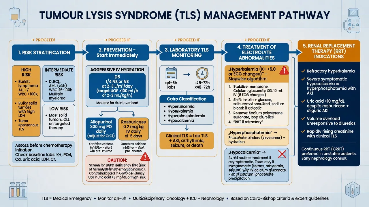
- The prophylaxis — the aggressive the hydration (the 3 L per day; the keep the urine output over 100 mL per h), the allopurinol (the reduce the uric acid production), the rasburicase (the high-risk — the directly the degrades the uric acid).[1]
- The treatment — the rasburicase (the 0.15 to 0.2 mg per kg), the treat the hyperkalaemia (the calcium the gluconate, the insulin-dextrose), the RRT if the severe.[1]
The Cairo-Bishop classification (2004) — the diagnostic standard
The Cairo-Bishop classification formally defines TLS as laboratory TLS (LTLS) within 3 days before to 7 days after cytotoxic therapy, OR clinical TLS (CTLS) when laboratory derangement produces a symptomatic complication. Laboratory TLS requires two or more of the following, present between 3 days before and 7 days after the start of therapy (or a 25 per cent change from baseline):[1]
- Uric acid over 476 µmol/L (8 mg/dL) or a 25 per cent rise
- Potassium over 6.0 mmol/L or a 25 per cent rise
- Phosphate over 1.45 mmol/L (4.5 mg/dL in adults, age-adjusted up to 6.5 in children) or a 25 per cent rise
- Calcium under 1.75 mmol/L (corrected) or a 25 per cent fall [1]
Clinical TLS = laboratory TLS plus one of: AKI (creatinine over 1.5× age/sex upper limit), cardiac arrhythmia/sudden death, or seizure (the consequences of hyperkalaemia, hypocalcaemia, and urate nephropathy).[1]
TLS risk stratification (Cairo-Bishop) — choosing the prophylaxis
| Risk | Tumour / clinical features | Prophylaxis | Monitoring |
|---|---|---|---|
| Low | CLL on fludarabine, indolent lymphoma, low-bulk disease, normal LDH | Hydration + observe | Daily labs |
| Intermediate | AML with WBC 25-100, ALL with WCC under 100, intermediate-grade NHL, myeloma on novel agents | Hydration + allopurinol 300 mg/day (start 24 h pre-chemo) | Labs q12-24h for 72 h |
| High | Burkitt/lymphoblastic lymphoma, AML WCC over 100, ALL WCC over 100, bulky chemo-sensitive disease, high LDH, pre-existing renal failure, oliguria, uric acid or phosphate already raised at baseline | Hydration + rasburicase 0.15-0.2 mg/kg/day (single dose often sufficient; weight-based; reduce dose in renal failure) | Labs q4-6h for 48-72 h; continuous cardiac monitoring |
Allopurinol vs rasburicase — the two urate-lowering agents
| Feature | Allopurinol | Rasburicase |
|---|---|---|
| Mechanism | Xanthine oxidase inhibitor — blocks the production of new uric acid (from purines) | Recombinant urate oxidase — degrades existing uric acid to soluble allantoin |
| Effect on existing uric acid | None — levels fall only as old uric acid is renally cleared | Immediate — enzymatic conversion within hours |
| Onset | Days to lower uric acid | Within 4 hours, peak effect at 24 h |
| Effect on xanthine/hypoxanthine | Causes accumulation → can precipitate xanthine nephropathy | No xanthine accumulation |
| Indication | Intermediate-risk TLS prophylaxis | High-risk TLS; established TLS; any patient with hyperuricaemic AKI |
| Dose | 300 mg PO daily (start ≥24 h pre-chemo); reduce in renal failure | 0.15-0.2 mg/kg IV once daily (often a single dose suffices); lower dose 3 mg fixed often used in adults |
| Critical caution | Do NOT rely on allopurinol once uric acid is already very high | Contraindicated in G6PD deficiency (methaemoglobinaemia + haemolysis — screen all patients first) |
| Sample handling | Routine | Send on ice and spin immediately — rasburicase continues to degrade uric acid in the collection tube at room temperature, giving falsely low readings |
Goldman 2001 (Blood) — Rasburicase vs allopurinol in children at high risk of TLS (PMID 11342423)
Source
Blood — randomised open-label trial, 52 children with lymphoma or leukaemia at high TLS risk
Intervention
Rasburicase 0.2 mg/kg/day for 5 days vs oral allopurinol
Primary outcome
Plasma uric acid area-under-the-curve over the first 5 days — 128 mg·h/dL (rasburicase) vs 866 mg·h/dL (allopurinol), p=0.001
Result
Uric acid fell to normal within 24 hours in nearly all rasburicase patients; response rate 94 per cent vs 1 per cent on day 1
Significance
Established recombinant urate oxidase as the standard for high-risk TLS — the agent that allows aggressive chemotherapy in bulky tumours
Cortes 2010 (JCO) — Rasburicase for adults at risk of TLS (PMID 20713865)
Source
Journal of Clinical Oncology — pooled analysis of three prospective open-label trials, 267 adults with haematological malignancy at TLS risk
Intervention
Rasburicase alone vs rasburicase followed by allopurinol vs allopurinol alone
Primary outcome
Proportion achieving a plasma uric acid response (within normal limits during days 3-7) — 87 per cent (rasburicase) vs 78 per cent (rasburicase + allopurinol) vs 66 per cent (allopurinol)
Result
Rasburicase-based strategies superior to allopurinol alone in adults; the combination offered no clear advantage over rasburicase alone
Significance
Confirmed that rasburicase is the agent of choice for adults at intermediate-to-high TLS risk
Established tumour lysis syndrome — the ICU management bundle
- Recognise and stratify — high-risk tumour (Burkitt, AML/ALL with WCC over 100, bulky lymphoma, high LDH) within 12-72 h of cytotoxic therapy. Send K, phosphate, calcium, uric acid, creatinine, LDH at baseline then every 4-6 hours for 48-72 hours. Continuous cardiac monitoring
- Aggressive intravenous hydration — isotonic saline 3 L/m²/day (roughly 100-150 mL/h in adults; 2-3× maintenance in children). Aim for urine output over 100 mL/h. Do NOT add potassium to the fluids. Alkalinisation of the urine is no longer recommended — it worsens calcium phosphate deposition and xanthine crystallisation
- Lower uric acid — rasburicase 0.15-0.2 mg/kg IV (single dose, may repeat at 24 h). Screen for G6PD first; if unknown, do not delay. Send samples on ice. Allopurinol is prophylactic — it does nothing for uric acid already formed and is second-line in established TLS
- Treat the hyperkalaemia (the #1 killer) — calcium gluconate 10 mL of 10 per cent for membrane stabilisation if K over 6.5 or ECG changes; insulin 10 units + 25 g dextrose; salbutamol; sodium bicarbonate if acidotic. Avoid potassium-binding resins as sole therapy (too slow)
- Treat the hypocalcaemia only if symptomatic — avoid reflex calcium replacement; raising calcium with high phosphate precipitates calcium phosphate in the renal tubules and worsens AKI. Give calcium only for tetany, seizures, a prolonged QT, or severe hyperkalaemia with ECG change
- Renal replacement therapy — start early for refractory hyperkalaemia, volume overload, symptomatic uraemia, metabolic acidosis, or calcium × phosphate product over 4.6 mmol²/L² (precipitation risk). Continuous modalities (CVVHDF) give smoother metabolic control than intermittent HD in the haemodynamically unstable patient
Hypercalcaemia of malignancy
- The causes — the PTHrP (the paraneoplastic — the squamous cell, the renal, the breast), the osteolytic metastases (the myeloma, the breast), the ectopic vitamin D (the lymphoma).[1]
- The symptoms — the "stones, bones, groans, moans" (the renal stones, the bone pain, the constipation, the psychiatric). The severe — the coma, the arrhythmias.[1]
- The ECG — the shortened QT.[1]
- The treatment:[1]
- The aggressive IV hydration (the normal saline — the 3 to 6 L per day; the calciuria).[1]
- The bisphosphonates (the zoledronic acid — the inhibit the osteoclasts; the take the 2 to 4 days).[1]
- The calcitonin (the rapid but the transient — the 4 to 6 h; the while the bisphosphonate the works).[1]
- The denosumab (the anti-RANKL — the refractory).[1]
- The glucocorticoids (if the granuloma or the lymphoma — the inhibit the vitamin D activation).[1]
The hypercalcaemia of malignancy — three mechanisms
| Mechanism | Typical tumours | The mediator | Distinguishing biochemistry |
|---|---|---|---|
| Humoral (PTHrP-mediated) — 80 per cent | Squamous cell (lung, head & neck), renal, breast, bladder, ovary, endometrium | PTH-related peptide (PTHrP) acts on the PTH-1 receptor — systemic osteoclast activation and distal tubular calcium reabsorption | Low phosphate, low 1,25-(OH)₂-vit-D, PTH suppressed, PTHrP elevated — mimics primary hyperparathyroidism biochemically except PTH itself is low |
| Local osteolytic — 20 per cent | Multiple myeloma, breast cancer bone metastases, lymphoma | Cytokines (RANKL, MIP-1, IL-6) and PTHrP from tumour cells in bone marrow → focal osteoclast activation | Often multifocal; alkaline phosphatase may be high; a myeloma screen (serum free light chains, protein electrophoresis) is mandatory |
| Ectopic vitamin D (calcitriol) — rare | Lymphoma (especially dysgerminoma), some ovarian Sertoli-Leydig tumours | Tumour expresses 1α-hydroxylase → unregulated conversion of 25-OH-D to active 1,25-(OH)₂-D → intestinal calcium absorption | Suppressed PTH, elevated 1,25-(OH)₂-D, hypercalciuria — and uniquely steroid-responsive |
The therapeutic agents for hypercalcaemia of malignancy — onset, duration, role
| Agent | Mechanism | Onset | Duration | Dose / role |
|---|---|---|---|---|
| IV 0.9 per cent saline | Volume expansion → GFR up → filtered calcium load up → forced calciuria | Immediate | While infused | 3-6 L/day; the cornerstone — restores volume (polyuria from nephrogenic DI of hypercalcaemia dehydrates) |
| Furosemide (only after rehydration) | Inhibits Na-K-2Cl in the loop → the positive lumen potential falls → calcium is no longer reabsorbed | Minutes | While infused | 20-40 mg IV; only once euvolaemic; largely superseded but useful in volume overload |
| Calcitonin | Inhibits osteoclasts; increases renal calcium excretion | 2-4 hours | 48 hours (then tachyphylaxis) | 4 IU/kg SC q12h — the bridge for symptomatic/severe hypercalcaemia while the bisphosphonate works |
| Zoledronic acid | Nitrogen-bisphosphonate → inhibits farnesyl pyrophosphate synthase in osteoclasts → apoptosis | 2-4 days | 3-4 weeks | 4 mg IV over 15 min (renally dose-adjusted); the first-line definitive agent |
| Denosumab | Monoclonal anti-RANKL — blocks osteoclast maturation | 1-2 days | Months | 120 mg SC q4 weeks; for bisphosphonate-refractory disease or renal failure (not renally cleared — safer than bisphosphonates when eGFR under 30) |
| Glucocorticoids | Reduce 1,25-(OH)₂-D synthesis (inhibit 1α-hydroxylase) | Days | Variable | Prednisolone 40-60 mg — only for vitamin-D-mediated (lymphoma) or granulomatous disease; useless in PTHrP/osteolytic |
| Dialysis | Removes calcium directly | Immediate | While running | The last-resort for life-threatening hypercalcaemia (calcium over 4.5, coma, arrhythmia) with renal failure or fluid overload |
STONESHypercalcaemia — STONES, BONES, GROANS, MOANS, and PSYCHIATRIC OVERTONES
Superior vena cava (SVC) syndrome
- The obstruction of the SVC → the facial and arm swelling, the jugular distension, the dyspnoea, the cerebral the oedema (the severe).[1]
- The causes — the malignancy (the NSCLC, the lymphoma) or the thrombosis (the catheter-related).[1]
- The management:[1]
Malignant vs benign SVC syndrome — the modern split
| Feature | Malignant SVC syndrome | Catheter-related (benign) SVC syndrome |
|---|---|---|
| Cause | Extrinsic compression or direct invasion by tumour (NSCLC ~50 per cent, lymphoma, germ-cell, metastatic) or secondary thrombosis around tumour | Indwelling central venous catheter (PICC, port, haemodialysis line, pacemaker/ICD lead) → endothelial injury and stasis → thrombus |
| Trend | Stable proportion of cases | Increasing — rising use of long-term catheters and devices |
| Onset | Days to weeks | Sudden, often on catheter manipulation |
| Imaging | CT chest with contrast (the venous phase) — mass, collateral circulation | Doppler ultrasound of the upper-limb veins; CT venography for the SVC itself |
| Definitive treatment | Treat the tumour (chemo/RT) plus endovascular stent for symptomatic/severe | Remove the catheter (if feasible) plus anticoagulation; stent for refractory |
| Steroids | Useful for lymphoma/germ-cell (chemosensitive, oedematous) | Not indicated |
Definitive treatments for SVC syndrome — when to use each
| Modality | Onset of relief | Indication | Limitation |
|---|---|---|---|
| Endovascular stent (self-expanding metal, often with thrombolysis/angioplasty) | 24-48 hours (fastest) | Severe symptoms (cerebral or laryngeal oedema, stridor); chemo-resistant tumours; diagnosis not yet tissue-confirmed; rapid relief while awaiting RT/chemo | Requires interventional radiology; thrombus may need lytic pretreatment; re-occlusion possible |
| Radiotherapy | 1-2 weeks | Chemo-resistant solid tumours (squamous cell carcinoma); the historical standard | Slow; a tissue diagnosis is required first; field may include lung |
| Chemotherapy | Days | Chemosensitive tumours — small-cell lung cancer, lymphoma, germ-cell | Only for chemo-responsive disease |
| Glucocorticoids | Hours | Lymphoma, germ-cell, inflammation-mediated; useful as a bridge | Largely ineffective in solid tumours; may obscure tissue diagnosis |
Rowell & Gleeson 2002 (Clin Oncol) — Steroids, RT, chemo and stents for SVC obstruction (PMID 12555872)
Source
Clinical Oncology — systematic review of 2 English-language RCTs plus observational series of malignant SVC obstruction (mostly bronchial carcinoma)
What it found
Insufficient evidence from randomised trials to show that chemotherapy or steroids add to radiotherapy; stent placement provided the most rapid symptomatic relief of all modalities
Stent evidence
Small-chemo-sensitive-cell tumours do better with primary chemotherapy; stenting gives the fastest relief in the severely symptomatic and in non-small-cell carcinoma
Significance
Set the contemporary standard: stent first for the severely symptomatic, definitive anti-tumour therapy (chemo/RT) for chemo-responsive disease
Malignant spinal cord compression (MSCC)
- The metastatic disease (the breast, the prostate, the lung, the myeloma) → the compression of the spinal cord.[1]
- The presentation — the back pain (the 95 per cent, the often the preceding the days to the weeks), the weakness, the sensory the level, the sphincter the dysfunction (the late, the poor the prognosis).[1]
- The diagnosis — the urgent the MRI whole the spine.[1]
- The management — the time-critical (the under 24 to 48 hours for the best the neurological the outcome):[1]
Patchell 2005 (Lancet) — Surgery + radiotherapy vs radiotherapy alone for MSCC (PMID 16112300)
Source
Lancet — randomised multi-institutional trial, 101 patients with a single area of metastatic spinal cord compression
Intervention
Direct decompressive surgery plus post-operative radiotherapy (30 Gy in 10 fractions) vs radiotherapy alone
Primary outcome
Ambulation — 84 per cent (surgery+RT) vs 57 per cent (RT alone), p=0.001; surgery patients retained ambulation longer (122 vs 13 days)
Other results
Significantly more regained the ability to walk (62 per cent vs 19 per cent among non-ambulatory at entry); less steroid use, fewer narcotics; median survival 126 vs 100 days
Caveats
Trial limited to a single compressive site and a reasonable prognosis (expected survival over 3 months); not generalisable to multi-level disease or poor-prognosis patients
Significance
Established surgery followed by RT as the standard for the single-level, surgically-fit MSCC patient; RT remains the definitive treatment for the majority
Radiotherapy vs surgery for MSCC — the decision
| Factor | Radiotherapy alone | Surgery then radiotherapy |
|---|---|---|
| Indication | The majority of patients; multi-level disease; poor surgical candidate; very short prognosis; radiosensitive tumour (myeloma, lymphoma, small-cell, breast, prostate) | Single compressive level + spinal instability + bony retropulsion into canal + good functional status + expected survival over 3-6 months |
| Onset of neurological benefit | Days to weeks | Hours (immediate decompression) |
| Patchell evidence (2005) | 57 per cent ambulated; 19 per cent regained ambulation | 84 per cent ambulated; 62 per cent regained ambulation — significantly superior in the single-site, fit patient[4] |
| Timing window | Within 24-48 hours of neurological deficit | Same; earlier is better — the ambulatory patient at presentation tends to remain ambulatory |
| Limits | Slow; ineffective for unstable spine | Major surgery in a cancer patient; not suitable for multi-level or posterior-element destruction with instability alone |
Suspected malignant spinal cord compression — the time-critical pathway
- Suspect at the bedside — any new back pain with a neurological deficit (limb weakness, sensory level, gait disturbance) in a patient with known or suspected cancer is MSCC until proven otherwise. 95 per cent present with pain; the neurological deficit often follows by days
- Dexamethasone 16 mg IV immediately, before imaging — reduces vasogenic cord oedema and arrests neurological deterioration while the workup proceeds. Do not wait for the MRI
- Urgent MRI of the WHOLE spine (T1 and T2, with sagittal and axial sequences) — not a focused study; skip-lesions are common and change the radiation field. Plain films and CT are inadequate (they miss soft-tissue cord compression). If MRI is contraindicated, a CT myelogram is the alternative
- Definitive therapy within 24-48 hours — radiotherapy (typically 8 Gy single fraction or 20 Gy in 5 fractions for prognosis under 6 months; 30 Gy in 10 fractions for better prognosis) OR surgical decompression + stabilisation followed by RT for the single-level, surgically-fit patient (Patchell criteria)
- Determine the prognosis and tailor intensity — ambulatory at presentation, single site, radiosensitive tumour, good performance status → best outcome (over 80 per cent retain ambulation). Non-ambulatory, sphincter dysfunction, multi-level disease, or very short prognosis → lower-intensity palliative RT and symptom-focused care
- Continue dexamethasone 16 mg/day in divided doses, tapering over 2-3 weeks during/after definitive therapy. Add a PPI for gastric protection; monitor glucose (steroid hyperglycaemia in the cancer patient)
Hyperviscosity syndrome
The hyperviscosity syndrome is the clinical consequence of a pathologically raised serum viscosity from a paraprotein — almost always IgM in Waldenström macroglobulinemia (the prototype, 30 per cent of cases become symptomatic), but also IgG or IgA myeloma (10-15 per cent), polyclonal hypergammaglobulinaemia, and the polycythaemic/leukaemic hypercellular states. The serum relative viscosity is normally 1.4-1.8 (water = 1); symptoms generally appear above 4, and the syndrome is established above 6-7.[7]
- The mechanism — high-molecular-weight pentameric IgM raises plasma viscosity → sluggish capillary flow → microvascular sludging, rouleaux, and impaired oxygen delivery. The IgM is predominantly intravascular (80 per cent), which is why plasmapheresis works immediately — it physically removes the protein.
- The triad — mucosal bleeding (epistaxis, gingival, GI — from platelet dysfunction and coating of clotting factors), visual disturbance (the classic "sausage" retinal veins, retinal haemorrhages, flame haemorrhages, papilloedema on fundoscopy), and neurological symptoms (headache, vertigo, ataxia, somnolence, hearing loss, seizures — "Bing-Neel syndrome"). Volume overload and a high-output state may precipitate cardiac failure.
- The diagnosis — a serum viscosity over 4 (relative units), with the clinical triad, in a patient with a monoclonal IgM (or rarely IgG/IgA). Fundoscopy is the bedside equivalent of a serum viscosity — engorged tortuous retinal veins ("string of sausages") confirm hyperviscosity.
- The treatment — urgent therapeutic plasma exchange (TPE), 1-1.5 plasma volumes, daily until symptomatic and viscosity below 4; definitive therapy of the underlying clone (rituximab-based chemo for Waldenström; bortezomib/cyclophosphamide for myeloma). [1]
IgM vs IgG/Ig hyperviscosity — why plasmapheresis works fast for IgM
| Feature | IgM (Waldenström) | IgG or IgA (myeloma) |
|---|---|---|
| Molecular weight / structure | 970 kDa pentamer | 150 kDa (IgG) or 160 kDa (IgA) monomer |
| Distribution | ~80 per cent intravascular | ~50 per cent intravascular |
| Relative viscosity threshold for symptoms | Lower (per molecule) — pentamer has outsized rheological effect | Higher — needs higher absolute concentration |
| Plasmapheresis efficiency | High — most of the pathogenic protein is removed in one session | Lower — substantial extravascular re-equilibration; slower, less complete removal |
| Definitive therapy | Rituximab ± bendamustine or bortezomib-dexamethasone | Bortezomib/lenalidomide-based for myeloma |
Hyperviscosity syndrome — the acute management
- Recognise the triad — mucosal bleeding, visual disturbance, neurological symptoms, in a patient with a monoclonal IgM (Waldenström) or, less commonly, IgG/IgA myeloma. Fundoscopy shows sausage-shaped retinal veins
- Send the confirmatory labs — serum viscosity, serum electrophoresis with quantification of the M-protein, IgM level, full blood count, coagulation, type and crossmatch (anticipate transfusion needs). A relative viscosity over 4 with symptoms confirms the diagnosis
- Urgent therapeutic plasma exchange — 1-1.5 plasma volumes exchanged with albumin (or plasma if bleeding/ Factor deficiency), daily until symptoms resolve and viscosity below 4. Large-bore peripheral or central access. Symptomatic improvement often within one session
- Definitive treatment of the clone — rituximab-based therapy for Waldenström (bendamustine-rituximab or bortezomib-dexamethasone-rituximab); bortezomib-based for myeloma. Rituximab can cause an IgM "flare" in the first 4 weeks — pre-treat or defer rituximab until viscosity controlled
- Avoid packed-cell transfusion until after plasmapheresis — anaemia is protective against viscosity; transfuse one unit at a time, slowly, for symptomatic anaemia only
- Monitor — daily serum viscosity and IgM during exchange; fundoscopy before and after; serial neurology and haemoglobin. Long-term: maintenance therapy to suppress the clone
Neutropenic sepsis (febrile neutropenia) — the 1-hour rule
The febrile neutropenia — oral temperature over 38.3 °C once, or over 38.0 °C sustained over one hour, in a patient with a neutrophil count under 0.5 × 10⁹/L (or under 1.0 and falling) — is a time-critical medical emergency. The mortality of untreated neutropenic sepsis is up to 50 per cent; the single most evidence-based intervention is empiric broad-spectrum antibiotic therapy within one hour of presentation (the "1-hour rule", now embedded in the Surviving Sepsis guidelines, NICE, and the ESMO/MASCC consensus).[6]
- The pathophysiology — chemotherapy-induced mucosal damage and profound neutropenia allow translocation of gut and skin organisms; the absence of neutrophils means the patient may be bacteraemic without pus, without localising signs, and with subtle inflammatory markers. Coagulase-negative staphylococci, viridans streptococci, enteric Gram-negatives (E. coli, Klebsiella, Pseudomonas), and pneumococci are common; invasive fungal infection (Aspergillus, Candida) complicates prolonged neutropenia.
- The clinical trap — the classical signs of infection (induration, purulence, rising CRP, dense infiltrate on CXR) are all neutrophil-dependent and may be ABSENT. A fever in a neutropenic patient is sepsis until proven otherwise.
- The antibiotic choice — an anti-pseudomonal β-lactam as monotherapy: piperacillin-tazobactam 4.5 g IV q6h, ceftazidime 2 g IV q8h, cefepime 2 g q8h, or meropenem 1 g q8h. Add vancomycin (or linezolid) for suspected catheter infection, mucositis, haemodynamic instability, or known MRSA colonisation. Add a macrolide/fluoroquinolone and consider atypical cover if a respiratory source.
- The risk stratification — the MASCC risk index (over 21 = low risk; 15 or under = high risk) integrates burden of illness, age, site of infection, and dynamic signs to identify the patient who may be managed with oral antibiotics and early discharge. [1]
High-risk vs low-risk febrile neutropenia (MASCC criteria)
| Feature | High-risk (inpatient, IV antibiotics) | Low-risk (consider oral, outpatient) |
|---|---|---|
| Expected neutropenia duration | Over 7 days | Under 7 days |
| Absolute neutrophil count | Under 100/µL (0.1 × 10⁹/L) | Over 100/µL, expected to recover |
| Clinical stability | Haemodynamic instability, organ dysfunction, comorbidity, mucositis, pneumonia, line infection | Clinically stable, no focus identified, no comorbidity |
| MASCC risk index | 15-21 (or below) | Over 21 |
| Antibiotic route | IV anti-pseudomonal β-lactam ± vancomycin | Oral ciprofloxacin + amoxicillin-clavulanate (after observation) |
| Setting | Hospital; ICU if unstable | Outpatient, with daily review and clear safety-netting |
Klastersky 2018 (BMC Cancer / ESMO-MASCC) — Febrile neutropaenia ESMO guidelines (PMID 30249215)
Source
ESMO/MASCO consensus guidelines — the European standard for the management of febrile neutropaenia in solid tumours and haematological malignancy
Key recommendations
Empiric anti-pseudomonal β-lactam within one hour of presentation; de-escalation after 48-72 h guided by culture; vancomycin/teicoplanin only for specific indications (catheter, mucositis, haemodynamic instability, known MRSA); G-CSF for high-risk prophylaxis and selected treatment
Risk stratification
MASCC index validated to separate low-risk (under 5 per cent complications) from high-risk patients eligible for outpatient oral therapy
Anti-fungal / anti-viral
Empiric antifungal ( mould-active) for persistent fever after 4-7 days of broad-spectrum antibiotics in prolonged neutropenia; imaging-driven (CT chest, galactomannan) diagnosis
Febrile neutropenia — the 1-hour bundle
- Recognise the trigger — fever over 38.3 °C (single oral reading) or over 38.0 °C sustained over 1 hour, in a patient with neutrophils under 0.5 × 10⁹/L (or under 1.0 and falling). Do NOT wait for cultures to start antibiotics
- Within the first hour — two sets of blood cultures (peripheral + from each lumen of any central line), urinalysis and culture, chest radiograph, and a single dose of empiric anti-pseudomonal β-lactam within 60 minutes. The antibiotic is the only step that cannot be deferred
- Resuscitate as for any sepsis — 30 mL/kg crystalloid for hypotension or lactate over 2; vasopressors (noradrenaline first-line) for persistent hypotension; intubate early if shock or work-of-breathing rises. Steroids are NOT routinely indicated (no neutrophils to recruit)
- Source assessment and de-escalation — examine the mouth, perianal area, line exit site, and skin; add vancomycin/linezolid for suspected line, severe mucositis, or haemodynamic instability; review cultures at 48-72 h and narrow the spectrum
- Add antifungal cover for persistent fever beyond 4-7 days of broad-spectrum therapy in prolonged neutropenia — CT chest and serum galactomannan to drive mould-active (voriconazole, liposomal amphotericin, caspofungin) treatment
- G-CSF is NOT routine — reserve for high-risk prophylaxis, severe sepsis with prolonged expected neutropenia, or specific indications; routine therapeutic G-CSF has not shown a clear mortality benefit
Immune-related adverse events (irAEs) from checkpoint inhibitors
The immune checkpoint inhibitors (ICIs) — anti-PD-1 (nivolumab, pembrolizumab), anti-PD-L1 (atezolizumab, durvalumab), and anti-CTLA-4 (ipilimumab, tremelimumab) — release the physiological brake on T-cell activation and can produce a unique spectrum of immune-related adverse events (irAEs). These can affect any organ, typically onset weeks to months after the first dose (but reported years later), and are managed by withholding the ICI, immunosuppression with corticosteroids, and targeted biologicals (infliximab, vedolizumab, rituximab) for refractory disease. ICU admission is driven by organ failure — pneumonitis with hypoxia, colitis with perforation or megacolon, hepatitis with coagulopathy, myocarditis, and encephalitis.[8][9]
- The mechanism — loss of self-tolerance → autoreactive T-cell activation against normal tissues. The CTLLA-4 agents (ipilimumab) cause more frequent and more severe irAEs than the PD-1/PD-L1 agents; combination therapy is the most toxic.
- The organ-specific patterns — colitis (diarrhoea, abdominal pain, bleeding; risk of perforation and toxic megacolon), pneumonitis (cough, dyspnoea, hypoxia; bilateral ground-glass infiltrates), hepatitis (asymptomatic transaminitis to fulminant hepatitis), endocrinopathy (thyroiditis, hypophysitis, type 1 diabetes, primary adrenal insufficiency — the latter two can present as shock), myocarditis (often fulminant, high mortality), nephritis (acute interstitial nephritis), skin (rash, Stevens-Johnson), and neurological (Guillain-Barré, myasthenia, encephalitis).
- The grading (CTCAE 1-4) drives management — grade 1 (mild, continue ICI with close monitoring), grade 2 (withhold ICI, topical or low-dose oral steroids), grade 3-4 (withhold/permanently discontinue ICI, high-dose IV steroids 1-2 mg/kg/day methylprednisolone, add biological for refractory). [1]
The five ICU-relevant irAEs — recognition and the first-line immunosuppression
| Organ / syndrome | Presentation | Diagnosis | First-line immunosuppression |
|---|---|---|---|
| Immune colitis | Diarrhoea (over 6/day or nocturnal), abdominal pain, bleeding; risk of perforation/toxic megacolon | Stool culture (exclude C. difficile, CMV), flexible sigmoidoscopy with biopsy (lymphocytic colitis), CT abdomen if toxic | Withhold ICI; methylprednisolone 1-2 mg/kg/day; infliximab 5 mg/kg or vedolizumab 300 mg if refractory or steroid-dependent at 3-5 days |
| Immune pneumonitis | Cough, dyspnoea, hypoxia; bilateral ground-glass / organising pneumonia on CT | CT chest (ground-glass, reticular, OP pattern); exclude infection (BAL); biopsy if uncertain | Withhold ICI; methylprednisolone 1-2 mg/kg/day (2-4 mg/kg for severe); infliximab, MMF, or rituximab for refractory; ventilate for respiratory failure |
| Immune hepatitis | Asymptomatic transaminitis; fulminant hepatitis with coagulopathy | ALT/AST over 3× ULN; exclude viral/drug hepatitis; liver biopsy if severe | Withhold ICI; methylprednisolone 1-2 mg/kg/day; mycophenolate for refractory (avoid infliximab — hepatotoxic) |
| Immune myocarditis | Chest pain, heart failure, arrhythmia, AV block; can be fulminant | Troponin + ECG + echocardiogram + cardiac MRI; biopsy if uncertain; mortality up to 50 per cent | Withhold ICI; methylprednisolone 1 g/day for 3-5 days; MMF; consider Abetimus/rituximab/plasma exchange; supportive heart failure therapy; mechanical support |
| Endocrinopathy (thyroid, hypophysitis, T1DM, adrenal insufficiency) | Fatigue, hypotension, hyponatraemia, hypoglycaemia/hyperglycaemia, headache (hypophysitis) | TSH/free-T4, cortisol, ACTH, FSH/LH, prolactin, morning cortisol; pituitary MRI for hypophysitis | Hormone replacement is primary (hydrocortisone, levothyroxine, insulin); steroids for hypophysitis mass effect; usually DO NOT need to permanently stop ICI |
Haanen (SITOX) 2017 (J Immunother Cancer / Ann Oncol) — Consensus guidelines for CPI toxicity management (PMID 29162153)
Source
Multidisciplinary expert consensus (Society for Immunotherapy of Cancer Toxicity Management / ESMO) — the international standard for managing irAEs
Core principle
Suspect irAEs in any patient on PD-1/PD-L1/CTLA-4 therapy presenting with new organ dysfunction; grade severity by CTCAE; treat with corticosteroids for grade ≥2, biologicals for steroid-refractory disease
Colitis
Infliximab 5 mg/kg or vedolizumab 300 mg for steroid-refractory colitis after 3-5 days; consider early biological for severe colitis (perforation risk)
Pneumonitis
Empiric corticosteroids 1-2 mg/kg for grade ≥3; broad-spectrum antibiotics until infection excluded; infliximab/MMF/cyclophosphamide/rituximab for refractory
Myocarditis
High-dose methylprednisolone (1 mg/kg/day to 1 g/day pulse) plus MMF; early mechanical support; permanently discontinue ICI
Steroid taper
Taper slowly over at least 4-6 weeks after resolution; rebound irAEs common if tapered too fast
Suspected immune-related adverse event — the ICU pathway
- Suspect the drug — any new organ dysfunction in a patient on (or recently on) a checkpoint inhibitor is an irAE until excluded. The median onset is 4-8 weeks but can be days to years; CTLA-4 and combination therapy are the most toxic
- Grade the severity (CTCAE 1-4) and exclude infection — grade determines management. Send infection screens (including opportunistic — CMV, PJP, fungal) before immunosuppression; biopsy the affected organ (colon, lung, liver, myocardium) if severity permits, because the histology drives the biological choice
- Withhold the ICI for grade ≥2 — permanently discontinue for grade 4 (and most grade 3) pneumonitis, myocarditis, colitis, hepatitis. Coordinate with the oncologist — never restart without their input
- Corticosteroids are first-line — methylprednisolone 1-2 mg/kg/day for grade 2-3; 2-4 mg/kg/day or 1 g/day pulses for grade 4 (myocarditis, severe pneumonitis/encephalitis). Cover for opportunistic infection (PJP prophylaxis with cotrimoxazole), monitor glucose
- Add a biological for steroid-refractory disease at 3-5 days — infliximab 5 mg/kg for colitis (avoid in uncontrolled sepsis/TB); vedolizumab 300 mg as gut-selective alternative (safer in sepsis); mycophenolate or tacrolimus for hepatitis; MMF or rituximab for pneumonitis; MMF ± plasma exchange for myocarditis
- Supportive care and the slow taper — ventilatory, haemodynamic, renal support as needed; treat the iatrogenic immunosuppression (PJP prophylaxis, stress-ulcer prophylaxis). Taper corticosteroids over at least 4-6 weeks after resolution; rebound and secondary irAEs are common
Exam practice — SAQs
SAQ — Tumour lysis syndrome after chemotherapy for Burkitt lymphoma
10 minutes · 10 marks
A 22-year-old man with newly diagnosed Burkitt lymphoma (LDH 4200, bulky abdominal mass) received his first cycle of R-CODOX-M chemotherapy 36 hours ago. He is now admitted with weakness and palpitations. Labs: potassium 7.2 mmol/L (ECG shows peaked T waves and widened QRS), phosphate 2.8 mmol/L, corrected calcium 1.6 mmol/L, uric acid 620 micromol/L, creatinine 180 micromol/L (baseline 80). Urine output 25 mL/h.
SAQ — Neutropenic sepsis and malignant spinal cord compression
10 minutes · 10 marks
A 60-year-old woman with AML, day 12 post-induction chemotherapy, presents with fever 38.8 degrees C and rigors. Neutrophils 0.2 x 10⁹/L. She also complains of 3 days of severe mid-thoracic back pain and new leg weakness; on examination she has a T6 sensory level and cannot lift her legs against gravity. The registrar is unsure what to prioritise.
SAQ — Tumour lysis syndrome with refractory hyperkalaemia and G6PD deficiency
10 minutes · 10 marks
A 58-year-old man with newly diagnosed AML (WCC 142 x 10⁹/L, LDH 3100, uric acid 520 micromol/L, creatinine 95) was started on hydroxycarbamide 12 hours ago in preparation for induction. He is admitted with weakness, cramps and a slow pulse. Labs: potassium 8.1 mmol/L, phosphate 3.4 mmol/L, corrected calcium 1.5 mmol/L, uric acid 780 micromol/L, creatinine 240 micromol/L, urine output 15 mL/h. ECG shows sine-wave QRS. He is of West African ancestry and his G6PD level is pending.
SAQ — Neutropenic enterocolitis (typhlitis) post-induction for AML
10 minutes · 10 marks
A 42-year-old woman, day 14 post-induction chemotherapy for AML, presents with fever 39.1 degrees C, severe right-lower-quadrant abdominal pain, distension, and bloody diarrhoea. Neutrophils 0.1 x 10⁹/L, platelets 18, lactate 3.2 mmol/L, BP 95/60. CT abdomen shows marked caecal and ascending-colon wall thickening (12 mm) with peri-caecal fat stranding and a small volume of free fluid; no pneumoperitoneum.
Red flags
[1] [1]References
- [1]Cairo MS, Bishop M Tumour lysis syndrome: new therapeutic strategies and classification Br J Haematol, 2004.PMID 15384972
- [2]Goldman SC, Holcenberg JS, Finklestein JZ, et al A randomized comparison between rasburicase and allopurinol in children with lymphoma or leukemia at high risk for tumor lysis Blood, 2001.PMID 11342423
- [3]Cortes J, Moore JO, Maziarz RT, et al Control of plasma uric acid in adults at risk for tumor Lysis syndrome: efficacy and safety of rasburicase alone and rasburicase followed by allopurinol compared with allopurinol alone--results of a multicenter phase III study J Clin Oncol, 2010.PMID 20713865
- [4]Patchell RA, Tibbs PA, Regine WF, et al Direct decompressive surgical resection in the treatment of spinal cord compression caused by metastatic cancer: a randomised trial Lancet, 2005.PMID 16112300
- [5]Rowell NP, Gleeson FV Steroids, radiotherapy, chemotherapy and stents for superior vena caval obstruction in carcinoma of the bronchus: a systematic review Clin Oncol (R Coll Radiol), 2002.PMID 12555872
- [6]Klastersky J, de Naurois J, Rolston K, et al Febrile neutropenia (FN) occurrence outside of clinical trials: occurrence and predictive factors in adult patients treated with chemotherapy and an expected moderate FN risk. Rationale and design of a real-world prospective, observational, multinational study BMC Cancer, 2018.PMID 30249215
- [7]Kyle RA, Gertz MA, Witzig TE, et al Prognostic markers and criteria to initiate therapy in Waldenstrom's macroglobulinemia: consensus panel recommendations from the Second International Workshop on Waldenstrom's Macroglobulinemia Semin Oncol, 2003.PMID 12720119
- [8]Postow MA, Sidlow R, Hellmann MD Immune-Related Adverse Events Associated with Immune Checkpoint Blockade N Engl J Med, 2018.PMID 29320654
- [9]Haanen JBAG, Carbonnel F, Robert C, et al Managing toxicities associated with immune checkpoint inhibitors: consensus recommendations from the Society for Immunotherapy of Cancer (SITC) Toxicity Management Working Group J Immunother Cancer, 2017.PMID 29162153
- [10]Gertz MA Contemporary management of superior vena cava syndrome J Vasc Surg Venous Lymphat Disord, 2026.PMID 41866116