ICU · Haematology / haemoglobinopathy
Sickle-Cell Crisis — Vaso-Occlusive, Acute Chest & Exchange Transfusion
Also known as Sickle-cell disease · SCD · Sickle-cell crisis · Vaso-occlusive crisis · VOC · Acute chest syndrome · ACS · Splenic sequestration · Aplastic crisis · Exchange transfusion · Hydroxyurea · Hydroxycarbamide · Haemoglobin S · HbS polymerisation · Incentive spirometry · Crizanlizumab · Voxelotor · Transcranial Doppler
The sickle-cell crisis in the ICU: the vaso-occlusive crisis (the commonest — pain from the microvascular occlusion by the sickled cells), the acute chest syndrome (the leading cause of death — the new infiltrate, the hypoxia), the splenic sequestration (children), the aplastic crisis (parvovirus B19), the haemolytic crisis. The management: oxygen (reduce the sickling), IV hydration (hypotonic — reduce the viscosity), analgesia (the opioids — patient-controlled), the transfusion (simple or exchange), the hydroxyurea (the prevention). The acute chest syndrome — the antibiotics (the infection), the incentive spirometry (the atelectasis prevention), the transfusion, the bronchodilators. The exchange transfusion for the acute chest, the stroke, the severe.
On this page & tools
Your progress
Saved locally on this device.
Target exams
Red flags
Overview & definition
The sickle-cell disease (SCD) is the haemoglobinopathy (the HbS — the glutamic-acid-to-valine substitution at the beta-6 position) → the deoxygenated HbS the polymerises → the sickle-shaped RBC → the vaso-occlusion (the pain, the organ damage) and the haemolysis (the anaemia). The crisis the triggered by the hypoxia, the dehydration, the infection, the cold, the stress. The acute chest syndrome (ACS) is the leading cause of death. The ICU manages the severe crisis, the ACS, the stroke, the multi-organ failure.[1]
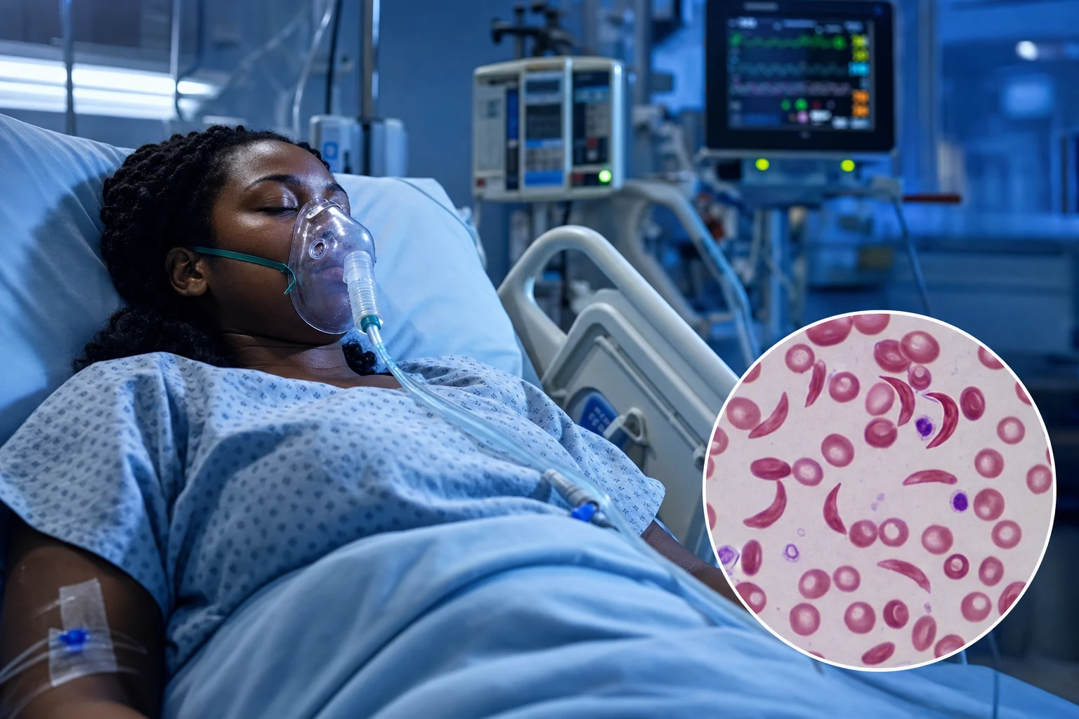
SCD is the most common severe monogenic disorder worldwide and is caused by a single point mutation in the β-globin gene (HBB) on chromosome 11 — a GAG→GTG transversion producing glutamic-acid-to-valine at the sixth position of β-globin (β6 Glu→Val, E6V). The disease is inherited autosomal recessively. The genotype determines the clinical severity: HbSS (sickle cell anaemia, most severe), HbSβ⁰-thalassaemia (no normal β-chain, severe), HbSβ⁺-thalassaemia (reduced normal β-chain, moderate), and HbSC (milder but can still develop ACS and stroke). The sickle-cell trait (HbAS, ~40 per cent HbS) is asymptomatic under normal conditions — sickling only with extreme hypoxia (severe altitude, unpressurised aircraft, overwhelming sepsis) — and is protective against falciparum malaria, which explains the gene frequency across sub-Saharan Africa, the Mediterranean, the Middle East, and parts of India.[1][6]
The clinical hallmark is the episodic acute crisis superimposed on chronic haemolytic anaemia and progressive end-organ damage. The crises are: vaso-occlusive pain (commonest), acute chest syndrome (leading cause of death), splenic sequestration (children), aplastic crisis (parvovirus B19), haemolytic crisis, stroke, priapism, and hepatic sequestration. Chronic complications include functional asplenia (by age 5 in HbSS — encapsulated organism sepsis risk), pulmonary hypertension (an independent risk factor for death), chronic kidney disease, avascular necrosis of the femoral head, leg ulcers, retinopathy (HbSC), and silent cerebral infarcts (cognitive decline). The modern ICU goal is not only to manage the acute crisis but to recognise and reverse the triggers and to institute disease-modifying therapy (hydroxycarbamide, and where appropriate the newer anti-P-selectin and HbS-polymerisation-inhibitor drugs).[1][6]
Pathophysiology — HbS polymerisation, sickling, vaso-occlusion, haemolysis, endothelial dysfunction
The entire pathophysiology of SCD flows from a single molecular event — the polymerisation of deoxygenated HbS. Understanding the cascade explains every crisis type and every management decision (oxygen reduces polymerisation; hydration reduces viscosity; exchange transfusion reduces the HbS fraction; hydroxycarbamide raises HbF which inhibits polymerisation).[1][6]
The sickle cascade — from point mutation to organ failure
| Step | Event | Consequence | Clinical correlate |
|---|---|---|---|
| 1. The mutation | β6 Glu→Val (HBB gene, chromosome 11) — a hydrophilic glutamate replaced by a hydrophobic valine on the β-globin surface | A hydrophobic patch forms on deoxy-HbS that fits into a complementary pocket on an adjacent β-chain | The molecular basis of the entire disease |
| 2. Polymerisation (the key event) | On deoxygenation, HbS forms rigid tacticoidal polymers (14-strand helical fibres) that stretch the RBC into a sickle shape. Polymerisation is time-, concentration-, and deoxygenation-dependent (delay time ∝ 1/[HbS]³⁰ — exquisitely sensitive to HbS concentration) | Rigid, deformed, poorly deformable sickled RBCs | Explains why hypoxia, acidosis, and dehydration (which concentrates HbS) all trigger crises; why HbF (which does not enter the polymer) is protective |
| 3. Reversible → irreversible sickling | With repeated sickling cycles the RBC membrane is damaged (calcium influx, potassium loss, dehydration, phosphatidylserine externalisation). After ~7 sickling cycles the cell is irreversibly sickled (ISC) even when reoxygenated | Irreversibly sickled cells persist, are rapidly cleared, and adhere to endothelium | The reticulocyte count is high (marrow compensates); ISCs are the most adherent cells |
| 4. Vaso-occlusion (the clinical crisis) | Sickled RBCs + leukocytes + platelets adhere to the post-capillary venular endothelium (a leukocyte-initiated, P- and E-selectin–mediated process), forming a heterocellular occlusive plug. Rigid cells cannot pass through capillaries | Microvascular ischaemia → pain, organ infarction | The painful crisis (bone, chest, abdomen), the dactylitis (children), the stroke, the splenic infarction, the avascular necrosis |
| 5. Haemolysis (the chronic anaemia) | Sickled RBCs are mechanically fragile and are destroyed extravascularly by the spleen (and intravascularly). RBC lifespan falls from 120 to ~17 days | Chronic haemolytic anaemia (Hb typically 70-90 g/L in HbSS); high reticulocytes; high LDH, indirect bilirubin, low haptoglobin | The baseline anaemia; the haemolytic crisis; pigment gallstones; the haemolytic component of endothelial dysfunction |
| 6. Endothelial dysfunction (the chronic vasculopathy) | Free haemoglobin scavenges nitric oxide (NO); arginase release from lysed RBCs depletes arginine (NO substrate); haem breakdown → reactive oxygen species. Endothelium is chronically activated (high VCAM-1, vWF) | Reduced NO bioavailability → vasoconstriction, platelet activation, proliferation → pulmonary hypertension, chronic vasculopathy | Pulmonary hypertension (independent risk factor for death — TRV over 2.5 m/s doubles mortality); priapism; leg ulcers; chronic kidney disease |
| 7. The triggers (tipping the balance) | Anything that increases deoxygenation (hypoxia, sleep apnoea), reduces RBC hydration (dehydration, osmolar stress, renal concentrating defect), slows transit time (acidosis, fever), or inflames the endothelium (infection) shortens the polymerisation delay time | A crisis is precipitated | Explains why crises cluster with infection, cold, altitude, surgery, dehydration, pregnancy |
Why polymerisation is the master switch
The kinetics of HbS polymerisation are central to understanding (and treating) SCD. The polymerisation delay time is inversely proportional to roughly the 15th to 30th power of the deoxy-HbS concentration — meaning a small change in HbS concentration, intracellular hydration, or oxygenation produces a huge change in the rate of sickling. This is the rationale for three of the four pillars of management:[1]
- Oxygen keeps HbS in the oxygenated (R-state) form — it cannot polymerise.
- Hydration dilutes plasma and (with hypotonic fluids) drives water into the dehydrated sickle RBC, lowering the intracellular HbS concentration below the polymerisation threshold.
- Exchange transfusion physically removes HbS-containing cells and replaces them with normal HbA cells — the HbS fraction (not the total Hb) is what matters.
- Hydroxycarbamide raises fetal haemoglobin (HbF, α₂γ₂), and the γ-chain does not carry the β6 valine, so HbF cannot enter the HbS polymer — it is the most powerful natural inhibitor of sickling. It also reduces the leukocyte count (less vaso-occlusion) and improves RBC hydration. [1]
The role of HbF — and why hydroxycarbamide works
Fetal haemoglobin (HbF) is the major genetic modifier of SCD severity. Individuals with high HbF (hereditary persistence of fetal haemoglobin, or the Senegal/Arab-Indian haplotypes) have milder disease because HbF dilutes the intracellular HbS concentration and physically disrupts the polymer. Hydroxycarbamide (hydroxyurea) is the disease-modifying mainstay precisely because it re-induces HbF synthesis (via inhibition of ribonucleotide reductase → stress erythropoiesis → F-cells) — the MSH trial (Charache 1995) showed it roughly halved the annual crisis rate.[2]
The two phenotypes of SCD — vaso-occlusive vs haemolytic
An increasingly important concept is that SCD has two overlapping clinical phenotypes driven by the dominant mechanism:[5]
The vaso-occlusive vs the haemolytic phenotype of SCD
| Feature | Vaso-occlusive phenotype | Haemolytic phenotype |
|---|---|---|
| Dominant mechanism | Viscosity / vaso-occlusion (high HbS, polymerisation, adhesion) | Haemolysis-driven endothelial dysfunction (NO scavenging) |
| Typical Hb | Higher (~90 g/L) — less haemolysis | Lower (~65 g/L) — severe haemolysis; high LDH, low haptoglobin |
| Clinical features | Frequent painful crises, ACS, osteonecrosis | Pulmonary hypertension, leg ulcers, priapism, stroke, chronic kidney disease |
| ** responds to transfusion** | Yes (reduces HbS, viscosity) | Yes, but the vasculopathy persists — needs targeted PH therapy |
| TRV (tricuspid regurgitant velocity) | Normal | Elevated (over 2.5 m/s → PH → doubles mortality) |
This matters in the ICU: a patient with the haemolytic phenotype and pulmonary hypertension is a higher-risk patient who needs a lower threshold for echocardiography, BNP, and aggressive management.[5]
The crisis types
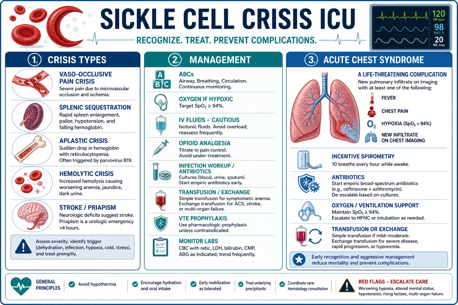
- Vaso-occlusive crisis (VOC) — the commonest. The pain from the microvascular occlusion (the chest, the back, the abdomen, the limbs, the dactylitis — the hand-foot in children). The triggered by the hypoxia, the dehydration, the infection, the cold.[1]
- Acute chest syndrome (ACS) — the leading cause of death. The new pulmonary infiltrate plus the hypoxia (the infection, the fat embolus, the atelectasis). The progresses rapidly to the ARDS.[1]
- Splenic sequestration — the sickled cells the sequester in the spleen → the splenomegaly, the severe anaemia, the hypovolaemia. In the children (before the autoinfarction).[1]
- Aplastic crisis — the parvovirus B19 → the temporary the marrow the shutdown → the severe anaemia (the reticulocytes the absent).[1]
- Haemolytic crisis — the accelerated haemolysis (the Hb the falls, the LDH the rises, the bilirubin the rises).[1]
- Stroke — ischaemic (children) and haemorrhagic (adults); the children are screened with transcranial Doppler (TCD) and the primary prevention is the chronic transfusion.[3][6]
- Priapism — the painful sustained erection (the cavernous sickling); the emergency — the aspirate + the phenylephrine within the hours (over 24 h → the erectile dysfunction).[6]
- Hepatic sequestration / intrahepatic cholestasis — the sickled cells the sequester in the liver → the acute hepatomegaly, the right upper quadrant pain, the hyperbilirubinaemia (the rare — the severe).[1]
1. Vaso-occlusive pain crisis (VOC) — the commonest
VOC is the hallmark of SCD and the most frequent reason for ICU admission. The pathophysiology is the post-capillary venular occlusion by a heterocellular plug of sickled RBCs, leukocytes (primarily neutrophils), and platelets, mediated by endothelial adhesion molecules (P-selectin, E-selectin, VCAM-1). The pain is ischaemic and infarctive in nature — comparable in severity to surgical pain or a myocardial infarction — and the most common sites are the long bones (marrow infarction), the lumbar spine, the chest wall, and the abdomen. In infants and young children the first presentation is often dactylitis (hand-foot syndrome) — the painful symmetric swelling of the hands and feet from infarction of the small tubular bones.[1]
The pain of VOC drives a vicious cycle: pain → splinting and hypoventilation → atelectasis → hypoxia → more sickling → more pain. This is why under-treatment of pain is dangerous, not just inhumane — it converts a painful crisis into acute chest syndrome. The triggers are the same that shorten the polymerisation delay: infection (commonest — identify and treat), dehydration, hypoxia, acidosis, fever, cold exposure, stress, menstruation, and sleep apnoea. A specific precipitant worth knowing is fat embolism syndrome following marrow necrosis — a VOC in a long bone releases necrotic marrow fat into the circulation, producing fever, hypoxia, a fall in Hb and platelets, and a petechial rash; it is a common trigger for ACS.[4]
2. Acute chest syndrome (ACS) — the leading cause of death
ACS is defined as a new pulmonary infiltrate on chest radiograph involving at least one complete lung segment, consistent with alveolar consolidation but not atelectasis, accompanied by one or more of: fever (over 38.5°C), chest pain, tachypnoea, wheeze, or cough, and/or hypoxaemia (a fall from baseline SpO₂ of over 4 per cent, or PaO₂ under 60 mmHg). It is the leading cause of death in SCD and the commonest cause of ICU admission. The National ACS Study (Vichinsky 2000) defined its causes:[4]
The causes of acute chest syndrome (Vichinsky 2000 — National ACS Study)
| Cause | Frequency | Mechanism / distinguishing feature |
|---|---|---|
| Infection (pneumonia) | ~30 per cent | Chlamydia pneumoniae and Mycoplasma pneumoniae are the commonest (together over 40 per cent of identified pathogens) — hence the mandate for atypical cover (macrolide). Respiratory syncytial virus in children. S. pneumoniae, H. influenzae less common now (vaccination/penicillin prophylaxis) |
| Fat embolism | ~10 per cent (clinical) — higher histologically | From marrow necrosis during a painful crisis; classic triad of hypoxia, fall in Hb and platelets, and neurological symptoms; lipid-laden macrophages in bronchoalveolar lavage |
| Pulmonary infarction | ~15 per cent | In-situ sickling in the pulmonary microvasculature; infarction without infection |
| Atelectasis | Common | From hypoventilation (pain, opioid sedation, splinting) — the rationale for incentive spirometry |
| Pulmonary embolism | Less common | Real but over-diagnosed; the V/Q scan is unreliable in SCD (baseline abnormalities) |
| Unknown / mixed | ~25 per cent | Often multifactorial |
ACS progresses rapidly — a patient admitted for a bony pain crisis can develop a new infiltrate and hypoxia within 24-48 hours (the "infarctive spread" pattern). The predictive factors for a severe course are: a platelet count that falls (consumption), a Hb that falls (haemolysis/sequestration), a rising respiratory rate, multilobar infiltrates, and neurological symptoms (suggesting fat embolism). Mortality is highest in adults (over 9 per cent in the National ACS Study, vs 1-3 per cent in children).[4]
3. Splenic sequestration — the paediatric emergency
Splenic sequestration is primarily a disease of young children (typically 6 months to 3 years) because the spleen has not yet autoinfarcted. A pool of sickled RBCs becomes trapped in the splenic red pulp → the spleen acutely enlarges → a large fraction of the blood volume is sequestered → severe anaemia and hypovolaemic shock. The clinical picture is sudden pallor, weakness, tachycardia, tachypnoea, and a rapidly enlarging, tender spleen in a child with SCD. The Hb may fall by 20-30 g/L over hours and the reticulocyte count is high (the marrow is trying to compensate — distinguishing this from aplastic crisis). Management is volume resuscitation and transfusion (carefully — the sequestered cells can re-enter the circulation causing hyperviscosity). Recurrence is common (over 50 per cent) and chronic transfusion or splenectomy is considered after the first major episode. Functional asplenia develops by age 5 in HbSS (autoinfarction) — after which splenic sequestration becomes rare but the patient is at lifelong risk of overwhelming encapsulated-organism sepsis (pneumococcus, H. influenzae, meningococcus), mandating vaccination and penicillin prophylaxis.[1][6]
4. Aplastic crisis — parvovirus B19
Parvovirus B19 infects and destroys erythroid progenitors in the bone marrow, producing a temporary (7-10 day) arrest of erythropoiesis. In a normal person this is trivial (a minor drop in Hb, unnoticed). In SCD the RBC lifespan is only ~17 days, so even a brief marrow shutdown produces a catastrophic fall in Hb — the reticulocyte count falls to near-zero (the diagnostic clue: a low reticulocyte count in a patient who normally runs a high one). The presentation is acute worsening anaemia with fatigue, pallor, and dyspnoea, but typically without the pain, haemolysis, or jaundice of a VOC or haemolytic crisis. The marrow recovers spontaneously within 1-2 weeks and produces a wave of reticulocytes (the recovery phase). Management is transfusion to bridge the aplastic period; isolate the patient (parvovirus is contagious and dangerous to pregnant women — hydrops fetalis).[1][6]
5. Haemolytic crisis
An acute acceleration of the chronic haemolysis — a sudden fall in Hb with a rise in LDH, indirect bilirubin, and reticulocytes (distinguishing it from aplastic crisis), and a fall in haptoglobin. Triggers include infection (especially malaria in endemic areas — which is why HbS exists in the first place), oxidant drugs (the G6PD overlap), transfusion reactions, and the delayed haemolytic transfusion reaction (DHTR — antibody-mediated, 5-14 days after transfusion). A hyperhaemolysis syndrome is a feared complication where transfusion itself accelerates haemolysis of both donor and native RBCs — further transfusion worsens it; treat with steroids and IV immunoglobulin.[1]
6. Stroke — ischaemic in children, haemorrhagic in adults
Stroke is a devastating complication of SCD. Ischaemic stroke predominates in children (peak age 2-5 years, before transfusion programmes) and results from a progressive large-vessel vasculopathy of the intracranial internal carotid and the circle of Willis (the moyamoya pattern). The STOP trial (Adams 1998) revolutionised primary prevention: annual transcranial Doppler (TCD) screening identifies children with elevated blood flow velocities (time-averaged mean of the maximum over 200 cm/s in the distal ICA or MCA), and chronic transfusion to keep HbS under 30 per cent reduces first stroke by over 90 per cent. Haemorrhagic stroke predominates in adults (the moyamoya collaterals rupture) and carries a high mortality. Acute treatment of an ischaemic stroke in SCD is urgent exchange transfusion (target HbS under 30 per cent, total Hb under 100) — not thrombolysis, which is relatively contraindicated and of unproven benefit in this vasculopathy. Chronic transfusion after a first stroke prevents recurrence (recurrence rate without transfusion is over 60 per cent). The TWiTCH trial (Ware 2016) showed hydroxycarbamide was non-inferior to chronic transfusion for maintaining TCD velocities in children who had normalised their velocities on transfusion — allowing transfusion to be safely stopped and reducing iron overload. The SIT trial (DeBaun 2014) showed chronic transfusion reduced recurrent stroke in children with silent cerebral infarcts.[3][6][7][8]
7. Priapism — the urological emergency
Priapism (a prolonged, painful erection from cavernosal sickling and venous outflow obstruction) affects up to 35 per cent of males with SCD, peaking in adolescence and young adulthood. Stuttering priapism (recurrent, self-limiting episodes under 4 hours) is the precursor; a prolonged episode over 4 hours is the ischaemic emergency — beyond 12-24 hours, irreversible corporal fibrosis and permanent erectile dysfunction (>50 per cent risk) supervene. The glans is characteristically spared (the corpus spongiosum does not sickle), distinguishing the sickle/low-flow pattern from a high-flow priapism. First-line management is urological — corporal aspiration and intracavernosal phenylephrine (a pure α1-agonist) — alongside the standard supportive measures (oxygen, hydration, analgesia). Urgent exchange transfusion (HbS under 30 per cent) is for the refractory case. Beware the post-priapism ACS: the trapped, deoxygenated sickled blood re-enters the systemic circulation as detumescence occurs, triggering a pulmonary crisis 24-72 hours later. The full stepwise protocol is detailed in the dedicated Priapism section below.[1][6]
8. Hepatic sequestration and intrahepatic cholestasis
Less common than the splenic form but important: sickled RBCs can pool in the hepatic sinusoids (hepatic sequestration — rapidly enlarging tender liver, falling Hb, high reticulocytes, managed like splenic sequestration with volume and transfusion) or obstruct the bile canaliculi (intrahepatic cholestasis — a catastrophic crisis with bilirubin often over 500 µmol/L, a surprisingly normal ALT/AST, coagulopathy, and encephalopathy; managed with urgent exchange transfusion and hepatic failure support). The common acute sickle hepatic crisis (sinusoidal sickling with RUQ pain, low-grade fever, and modest bilirubin) is self-limiting with hydration. The diagnostic trap is intrahepatic cholestasis masquerading as "just haemolysis" — a deepening jaundice with a normal-looking transaminase profile in a sickle patient is cholestasis until proven otherwise. Pigment gallstones (from chronic haemolysis) are the commonest surgical cause of jaundice and must be distinguished. The three syndromes are compared in the dedicated Sickle hepatopathy section below.[1]
The management
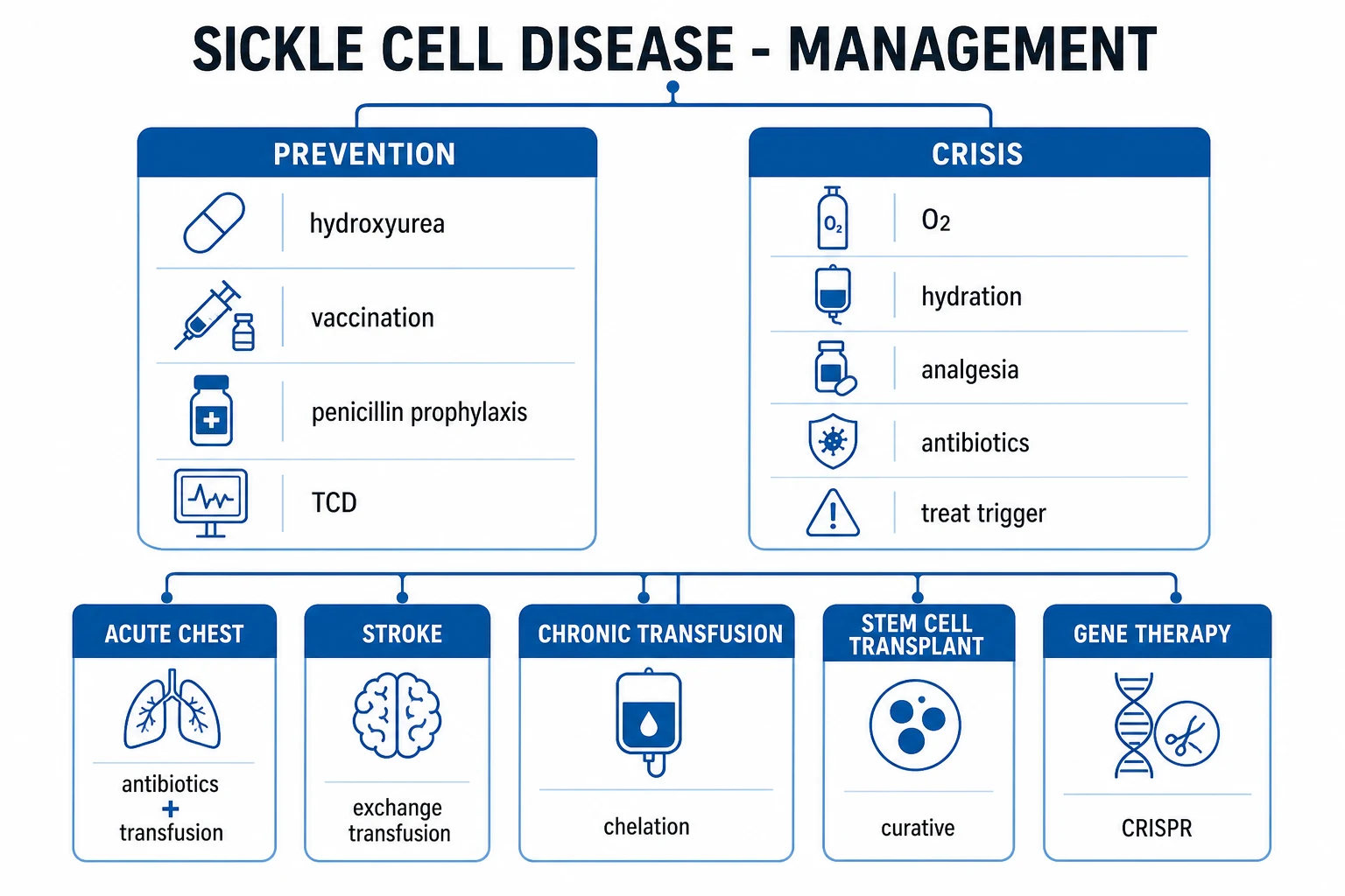
The management of an acute sickle-cell crisis rests on five pillars: (1) oxygen (reduce the sickling), (2) IV hydration (reduce the viscosity, dilute the HbS), (3) analgesia (opioid PCA — do NOT under-treat), (4) transfusion (simple for symptomatic anaemia; exchange for ACS, stroke, severe), and (5) disease-specific therapy (antibiotics for ACS, incentive spirometry, bronchodilators, and hydroxycarbamide / newer agents for prevention).[1][6]
1. Oxygen (reduce the sickling)
- The supplemental oxygen to keep the SpO2 over 94 to 96 per cent. The reduce the hypoxic sickling.[1]
Oxygen is the most fundamental therapy because oxygenated HbS cannot polymerise. Target an SpO₂ of 94-96 per cent (or the patient's baseline, which may be lower in chronic lung disease). The mask or nasal specs; escalate to high-flow nasal cannula or NIV for the evolving ACS. Pulse oximetry can be slightly inaccurate in SCD (the carboxyhaemoglobin from chronic haemolysis can falsely elevate readings) — confirm with an arterial gas if there is any doubt. There is no role for hyperbaric oxygen in the routine crisis.[1]
2. IV hydration
- The hypotonic or isotonic fluid (the 0.9 per cent saline or the 5 per cent dextrose — the reduce the viscosity, the improve the flow). The 1.5 times the maintenance. The avoid the overload (the pulmonary oedema — the worsens the ACS).[1]
Hydration lowers the plasma viscosity and (with hypotonic solutions) drives water into the dehydrated sickle RBC, reducing the intracellular HbS concentration below the polymerisation threshold. The standard regimen is 1-1.5 times maintenance (typically 3-4 L/day in an adult, or 1.5× maintenance in a child) of 0.9 per cent saline or 5 per cent dextrose with added electrolytes. Avoid fluid overload — it precipitates pulmonary oedema and is a recognised iatrogenic trigger for ACS. Use a balanced crystalloid rather than 0.9 per cent saline if large volumes are needed (less hyperchloraemic acidosis). The sickle patient with chronic kidney disease (common) needs careful balance — over-hydration causes pulmonary oedema, under-hydration perpetuates sickling.[1][6]
3. Analgesia
- The opioids (the morphine, the fentanyl) — the pain the severe. The patient-controlled analgesia (PCA). The titrate to the pain. The not the under-treat (the pain the drives the tachycardia, the increased the oxygen demand). The avoid the meperidine (pethidine — the seizure risk).[1]
Analgesia in the vaso-occlusive crisis — the opioid options
| Drug | Dose | Pros | Cons / cautions |
|---|---|---|---|
| Morphine (first-line) | PCA: bolus 1-2 mg, lockout 10 min, no background infusion (background increases respiratory depression without better analgesia). Or 0.1 mg/kg q3-4h | Familiar, reversible with naloxone, inexpensive | Histamine release (itch — common, treat with ondansetron or low-dose naloxone infusion, NOT antihistamines which sedate); accumulates in renal failure (M6G metabolite) |
| Fentanyl | PCA: bolus 20-50 mcg, lockout 6 min. Or infusion 25-100 mcg/h | No active metabolites (safe in renal impairment); less histamine | Short half-life (need infusion or PCA); tolerance develops faster |
| Hydromorphone | 0.2-0.4 mg q3-4h; PCA bolus 0.2 mg | Less histamine, less itch | Longer half-life |
| Pethidine / meperidine | AVOID | — | Normeperidine metabolite accumulates → tremor, myoclonus, seizures (especially with renal impairment or repeated dosing) |
The pain of a VOC is severe and comparable to surgical pain — under-treatment is the commonest error and is dangerous: it drives splinting, hypoventilation, atelectasis, and converts a painful crisis into ACS. The principle is rapid assessment, rapid analgesia, and reassessment every 15-30 minutes until the pain is controlled. A loading dose (e.g. morphine 0.1 mg/kg) is given, then the PCA. Avoid the background (basal) infusion on the PCA — it increases respiratory depression without improving pain scores. Monitor sedation score and respiratory rate (the SCD patient has chronic tachypnoea, so a "normal" rate may be a danger sign). Add a non-opioid baseline — paracetamol 1 g q6h, and an NSAID (ibuprofen, ketorolac short course) if renal function permits — for opioid-sparing and anti-inflammatory effect. Ketamine (subanaesthetic infusion, 0.1-0.3 mg/kg/h) is a useful opioid-sparing adjunct in refractory pain and has the bonus of bronchodilation. Lidocaine infusion has been studied. Transition to oral opioids (oxyCODONE, morphine) when the patient is improving and the IV requirements fall.[1][6]
4. Transfusion
- The simple transfusion — if the Hb under 90 (or the symptomatic). The target the Hb the under 100 (the avoid the hyperviscosity — the HbS the percentage the matters).[1]
- The exchange transfusion — for the acute chest syndrome, the stroke, the severe multi-organ, the refractory VOC. The reduce the HbS the percentage to under 30 per cent (the apheresis the removes the sickled cells and the replaces with the normal).[1]
Transfusion is the most specific therapy in SCD — it reduces the HbS fraction and improves oxygen delivery. The two modalities have different indications and different targets:[1][6]
Simple vs exchange transfusion in SCD
| Feature | Simple transfusion | Exchange transfusion (erythrocytapheresis) |
|---|---|---|
| What it does | Adds HbA cells on top of the patient's own — raises the Hb and dilutes HbS | Removes the patient's HbS cells and replaces them with HbA cells — actively lowers HbS fraction |
| Indication | Symptomatic anaemia (Hb under 90 or a fall of over 20 from baseline), splenic sequestration, aplastic crisis, pre-operatively | ACS (moderate-severe), acute stroke, refractory VOC, severe multi-organ failure, severe priapism, hepatic sequestration |
| HbS target | Not the goal (HbS falls only by dilution) | Under 30 per cent (acute); under 30 per cent maintained for chronic transfusion programmes |
| Total Hb target | Under 100 g/L (avoid hyperviscosity) | Under 100 g/L (CRITICAL — the HbS percentage matters, not the total Hb) |
| Volume | 1 unit raises Hb ~10 g/L | Automated apheresis (or manual 2-volume exchange) — requires large-bore central access |
| Iron overload | Yes (every unit adds ~200 mg iron) | Less (iron-neutral — removes iron with the patient's cells) |
| Risks | Alloimmunisation (common — Rh, Kell), hyperviscosity, infection, TRALI | Same plus central-line and apheresis risks (citrate hypocalcaemia, donor exposure 4-6 units) |
The single most important transfusion rule in SCD: keep the total Hb under 100 g/L. Raising the total Hb above this (especially above 110) without lowering HbS produces hyperviscosity — the transfused cells increase the haematocrit, the sickled cells remain, and the blood becomes too viscous to perfuse the microcirculation — precipitating or worsening a stroke, ACS, or multi-organ failure. The HbS fraction is what matters, not the total Hb.[1][6]
Exchange transfusion for severe ACS or acute stroke — the practical protocol
- Confirm the indication — severe or progressive ACS (multilobar, rising oxygen requirement, falling Hb/platelets), suspected/confirmed acute stroke, refractory VOC, severe multi-organ failure
- Crossmatch and obtain phenotype-matched units (Rh, Kell — alloimmunisation is common in SCD; request antigen-matched blood). Target HbS under 30 per cent; estimated 4-6 units
- Large-bore vascular access — apheresis requires a central double-lumen catheter; manual exchange can be done through two large peripheral cannulae
- Calculate the exchange volume — typically a 2-volume exchange replaces ~85 per cent of the patient's RBC mass; the apheresis machine calculates this from the Hct, weight, and target HbS
- Monitor during the procedure — calcium (citrate anticoagulant chelates calcium → hypocalcaemia — give calcium gluconate), blood pressure, temperature (hypothermia), potassium
- Check the post-exchange HbS — target under 30 per cent; if not achieved, extend the exchange. Keep the total Hb under 100 g/L
- For acute stroke — do NOT delay for imaging if the clinical diagnosis is clear — the window for neurological salvage is hours. Confirm with CT/MRI as soon as possible but start the exchange immediately on clinical grounds
- Plan the chronic transfusion programme after a stroke (lifelong or until transition to another therapy; monthly transfusions to keep HbS under 30 per cent; chelation with deferasirox from the start)
- Watch for the hyperhaemolysis / delayed haemolytic transfusion reaction — a fall in Hb 5-14 days after transfusion with a rise in LDH/bilirubin; further transfusion can worsen it (treat with steroids, IVIG, eculizumab in severe cases)
5. The specific
- The antibiotics for the ACS (the infection the common the trigger — the ceftriaxone, the macrolide).[1]
- The incentive spirometry for the ACS and the chest VOC (the prevent the atelectasis).[1]
- The bronchodilators if the wheeze.[1]
- The hydroxyurea for the prevention (the reduce the crisis frequency).[1]
The acute chest syndrome (the leading cause of death)
- The new pulmonary infiltrate plus the hypoxia (the fever, the cough, the chest pain, the tachypnoea).[1]
- The triggers — the infection (the pneumonia), the fat embolus (from the bone marrow necrosis), the atelectasis (the hypoventilation from the pain).[1]
- The management:[1]
- The oxygen and the ventilation (the NIV or the invasive — the for the progressive hypoxia).[1]
- The antibiotics (the ceftriaxone plus the macrolide — the cover the typical and the atypical).[1]
- The incentive spirometry every 2 hours (the prevent the atelectasis — the most effective for the early).[1]
- The simple or exchange transfusion (the exchange for the severe or the progressive).[1]
- The bronchodilators if the wheeze.[1]
ACS is the most feared complication of SCD and the leading cause of death. Every patient admitted with a painful crisis must be treated as a potential ACS in evolution — the daily chest examination, the regular vital signs (a rising respiratory rate is the earliest sign), and the low threshold for a repeat chest radiograph. The diagnostic trap is that the infiltrate may not be present on the initial film — it commonly appears 24-72 hours after admission as the atelectasis or pneumonia declares. The following is the bundle of care that has reduced ACS mortality:[4][6]
Acute chest syndrome — the complete ICU management bundle
- Recognise early — new infiltrate + hypoxia OR a rising respiratory rate and falling SpO₂ in a patient admitted for VOC. A daily CXR and continuous SpO₂ monitoring for any VOC admission. Do NOT attribute tachypnoea to "pain" alone
- Oxygen — escalate from mask to high-flow nasal cannula to NIV as needed; target SpO₂ 94-96 per cent (or baseline). Invasive ventilation for the progressive hypoxia, fatigue, or ARDS — lung-protective ventilation (6 mL/kg tidal volume, plateau under 30)
- Broad-spectrum antibiotics IMMEDIATELY — ceftriaxone 2 g IV daily PLUS a macrolide (azithromycin 500 mg day 1 then 250 mg, OR clarithromycin). Chlamydia and Mycoplasma together cause over 40 per cent of infectious ACS — atypical cover is mandatory. Add vancomycin/linezolid if MRSA or severe. Send blood cultures, sputum, and a respiratory viral panel (RSV in children); do not delay antibiotics for results
- Incentive spirometry every 2 hours while awake (10 maximal inspirations each session) — the single most effective measure to prevent the atelectasis that drives early ACS. Add chest physiotherapy and early mobilisation. This is the evidence-based intervention most often omitted
- Bronchodilators — nebulised salbutamol if any wheeze or a history of asthma/reactive airways (common in SCD)
- Pain control that does not splint — opioid PCA, but titrate carefully (over-sedation worsens hypoventilation and atelectasis). Use ketamine (bronchodilator) as an adjunct
- Transfusion strategy —
- Mild ACS (single lobe, SpO₂ stable, Hb stable): observe, no transfusion unless Hb under 90 or falling
- Moderate ACS (multilobar, SpO₂ falling, Hb falling): simple transfusion to Hb under 100
- Severe or progressive ACS (rapidly progressive infiltrates, rising oxygen requirement, falling Hb/platelets, neurological symptoms suggesting fat embolism): urgent exchange transfusion to HbS under 30 per cent. Do not wait for intubation
- Investigate for fat embolism if there are neurological symptoms, a fall in Hb and platelets, or a petechial rash — lipid-laden macrophages on BAL (if intubated)
- Consider pulmonary hypertension and chronic lung disease in the adult with recurrent ACS — echocardiography (TRV), BNP
- Do NOT use diuretics for the infiltrate unless there is clear volume overload — the infiltrate is not pulmonary oedema; over-diuresis worsens sickling. Balance is everything
- Disease-modifying therapy — start (or optimise) hydroxycarbamide during the admission if the patient is not on it (reduces recurrence). Consider crizanlizumab (anti-P-selectin) or voxelotor (HbS polymerisation inhibitor) for the patient with recurrent ACS
- Venous thromboembolism prophylaxis — SCD patients are prothrombotic; give LMWH unless contraindicated. Do NOT anticoagulate therapeutically for the infiltrate unless PE is confirmed (CTPA preferred over V/Q — the V/Q is unreliable in SCD)
Differential diagnosis — what the crisis might NOT be
Not every acute presentation in a patient with SCD is a sickle crisis. Several mimics must be considered, and some are life-threatening if missed.[1][6]
The differential diagnosis of acute pain or dyspnoea in SCD
| Presentation | Sickle crisis (the diagnosis of exclusion in a known SCD patient) | The mimics to exclude |
|---|---|---|
| Acute chest pain + infiltrate | Acute chest syndrome | Bacterial pneumonia (in a non-sickler), pulmonary embolism, heart failure, aortic dissection, ACS + PE coexisting |
| Acute abdominal pain | Mesenteric vaso-occlusion, hepatic sequestration | Appendicitis, cholecystitis (pigment stones — common in SCD), pancreatitis, sickle hepatopathy, splenic infarction, ruptured spleen, ectopic pregnancy |
| Acute back pain | Lumbar VOC | Discitis, epidural abscess, vertebral osteomyelitis (Salmonella classically but Staphylococcus more common), renal colic, pyelonephritis |
| Acute limb pain | Long-bone VOC, dactylitis | Osteomyelitis (differentiate from infarction — both can be culture-negative early, both raise inflammatory markers, MRI differentiates), deep vein thrombosis, septic arthritis |
| Acute severe anaemia | Aplastic crisis (parvovirus B19), splenic sequestration (children), hyperhaemolysis | Blood loss (GI bleed), delayed haemolytic transfusion reaction, parvovirus in a non-sickle context |
| Acute neurological deficit | Stroke (ischaemic in children) | Meningitis (functional asplenia!), subarachnoid haemorrhage, metabolic encephalopathy (opioid, hypoxia), seizure/post-ictal |
| Fever without a source | Pain crisis with fever (infarction is inflammatory) | Sepsis (overwhelming pneumococcal in the asplenic patient — treat empirically NOW), osteomyelitis, malaria (endemic areas) |
The cardinal rule: a fever in an asplenic sickle patient is sepsis until proven otherwise — blood cultures and empirical ceftriaxone (covering pneumococcus) within one hour. The pain of a VOC is inflammatory and can cause a low-grade fever, but a fever over 38.5°C mandates an infection workup.[1]
Prevention and disease-modifying therapy
Prevention is now central to SCD management — the goal is to reduce the crisis frequency, prevent end-organ damage, and prolong life. The disease-modifying options are:[2][6][9][10]
Disease-modifying and preventive therapy in SCD
| Therapy | Mechanism / dose | Evidence / indication | Notes |
|---|---|---|---|
| Hydroxycarbamide (hydroxyurea) | Inhibits ribonucleotide reductase → raises HbF, lowers leukocyte count, improves RBC hydration; 15-35 mg/kg/day, titrate to max tolerated dose (keep neutrophils over 2.0) | MSH trial (Charache 1995) — halved the annual pain crisis rate and reduced ACS and transfusion. Now first-line for all HbSS from age 9 months | The mainstay. Monitor FBC every 4-12 weeks. Safe in pregnancy? — traditionally avoided, modern data emerging |
| Chronic transfusion programme | Keeps HbS under 30 per cent | Primary stroke prevention (STOP — Adams 1998) for children with abnormal TCD; secondary prevention after a first stroke; for recurrent ACS | Iron overload → chelate (deferasirox) from the start; alloimmunisation; venous access. TWiTCH (Ware 2016) showed hydroxycarbamide non-inferior to transfusion for stroke prevention in selected children |
| Crizanlizumab (anti-P-selectin monoclonal) | Blocks P-selectin–mediated cell adhesion → prevents the vaso-occlusive plug; 5 mg/kg IV every 4 weeks | SUSTAIN trial (Ataga 2017) — reduced the annual crisis rate by 45 per cent in the high-dose arm; approved for VOC prevention | For patients with recurrent VOC despite hydroxycarbamide; expensive; does not replace hydroxycarbamide |
| Voxelotor (HbS polymerisation inhibitor) | Allosterically stabilises the oxygenated R-state of HbS → prevents polymerisation; 1500 mg PO daily | HOPE trial (Vichinsky 2019) — raised Hb by over 10 g/L in ~50 per cent; approved for chronic haemolytic anaemia of SCD | The first drug to target the root molecular lesion; add to hydroxycarbamide |
| L-glutamine (Endari) | Reduces oxidative stress in the RBC; 0.3 mg/kg BD | Reduced crisis frequency in a 2018 trial; approved as an adjunct | Modest effect; well tolerated |
| Penicillin prophylaxis | Prevents pneumococcal sepsis in the asplenic child | From 2 months to at least 5 years (some to lifelong in HbSS) | The basis of the survival revolution in SCD |
| Vaccination | Pneumococcal (PCV13 + PPSV23), H. influenzae type b, meningococcal (MenACWY + MenB), influenza annual, hepatitis B | Lifelong — functional asplenia | The asplenic patient needs a vaccination card and standing orders for empirical antibiotics |
| Haematopoietic stem cell transplant (HSCT) | Curative — replaces the defective HSC clone | Matched-sibling donor HSCT: over 90 per cent disease-free survival in children; gene therapy (CRISPR/Cas9 — exa-cel, lovo-cel) approved 2023-2024 | The only cure; limited by donor availability (matched sibling) and transplant risk; gene therapy expanding rapidly |
| TCD screening (children) | Annual transcranial Doppler from age 1-2 to 16 | Identifies the high-risk child (TAMMV over 200 cm/s) for primary stroke prevention transfusion | STOP and TWiTCH underpin this |
Exam practice — SAQs
SAQ — Acute chest syndrome in sickle-cell disease
10 minutes · 10 marks
A 24-year-old man with HbSS sickle-cell disease is admitted with a vaso-occlusive pain crisis affecting his chest and limbs. He was treated with IV morphine via PCA and IV fluids. On day 2 of admission he develops worsening dyspnoea, a cough productive of yellow sputum, and pleuritic chest pain. T 38.6 degrees C, RR 32, SpO2 90 percent on room air dropping to 88 percent on 4 L nasal specs. Chest X-ray shows new bilateral lower lobe infiltrates. Hb 66 g/L (baseline 78), reticulocytes 12 per cent, WCC 18. SaO2 92 percent on 6 L HFNC.
SAQ — Acute stroke in a child with sickle-cell disease
10 minutes · 10 marks
A 7-year-old girl with HbSS sickle-cell disease is brought to the ED with acute right-sided weakness and aphasia that began 2 hours ago. CT brain shows no haemorrhage; MRA shows occlusion of the left middle cerebral artery. Hb 62 g/L (baseline 70). She is afebrile, BP 100/60.
Red flags
[1]Priapism — the urological emergency of SCD
Priapism — the prolonged, painful, unwanted erection — affects up to 35 per cent of males with SCD (mostly HbSS), with a peak incidence in the second and third decades. It is caused by sickling in the corpora cavernosa, which obstructs venous outflow and traps deoxygenated blood, producing a vicious cycle of stasis, further sickling, and ischaemia. Stuttering priapism (episodes lasting under 4 hours, recurring over days to weeks) is the common precursor; a prolonged episode (over 4 hours) is the ischaemic emergency. The glans is typically NOT engorged (the corpus spongiosum is spared), distinguishing a sickle priapism from a high-flow (arterial) priapism.[1][6]
The urgency: cavernosal ischaemia beyond 4-6 hours threatens the corporal smooth muscle; beyond 12-24 hours, irreversible fibrosis and permanent erectile dysfunction supervene (over 50 per cent of prolonged episodes over 24 hours result in permanent ED). The first-line management is urological — corporal aspiration and intracavernosal phenylephrine — NOT simply hydration and analgesia. Exchange transfusion is for the refractory case or as an adjunct.[6]
Acute priapism in SCD — the stepwise management
- Recognise and time it — any unwanted erection over 2 hours is a priapism; over 4 hours is the ischaemic emergency. Confirm the glans is soft (corpus spongiosum spared — the sickle/low-flow pattern). Distinguish from high-flow priapism (painless, post-traumatic, glans engorged — the cavernosal blood gas is bright red, not dark)
- First aid and supportive — analgesia (opioid), hydration (1.5× maintenance), oxygen, warmth. These reduce ongoing sickling but will NOT resolve an established priapism alone — escalate the urological steps in parallel
- Corporal aspiration (urology at the bedside) — aspirate 20-30 mL of the dark, deoxygenated cavernosal blood (send a gas: a low pH, low pO₂, high pCO₂ confirms the ischaemic/low-flow pattern). This decompresses the corpora
- Intracavernosal phenylephrine — a pure α1-adrenergic agonist (100-500 mcg diluted 1:1 with saline), injected into the corpora every 5-10 minutes for up to 1 hour (monitor for hypertension, reflex bradycardia, headache). Phenylephrine is preferred over adrenaline because it is a pure α1-agonist with minimal β-mediated tachyarrhythmia. Avoid in severe hypertension or cardiovascular disease
- If detumescence is achieved — admit, observe for recurrence (the stuttering pattern commonly recurs within 24 hours), start an oral sympathomimetic (etilefrine or pseudoephedrine) for stuttering prevention, and arrange outpatient urology follow-up
- If priapism persists beyond 6-12 hours despite aspiration and phenylephrine — urgent exchange transfusion to HbS under 30 per cent (lower the circulating HbS fraction to halt ongoing sickling). A surgical shunt (Winter corporoglanular T-shunt, or the Al-Ghorab corporospongiosal shunt) is the urological salvage for the truly refractory case
- Warn about the post-priapism ACS — the trapped, deoxygenated sickled blood is released into the systemic circulation as detumescence occurs, triggering an acute chest syndrome 24-72 hours later in a significant proportion. Monitor the SpO₂ and chest for several days; have a low threshold for a chest radiograph
- Long-term — a self-administered intracavernosal phenylephrine kit (taught in clinic) for recurrent stuttering priapism; consideration of a chronic transfusion programme or hydroxycarbamide optimisation for the recurrent sufferer; counselling on erectile dysfunction and fertility preservation
Sickle hepatopathy — acute hepatic crisis, sequestration, and intrahepatic cholestasis
The liver is a frequent, often under-recognised target in SCD. Three distinct — but overlapping — acute hepatopathic syndromes are encountered, and distinguishing them changes the management:[1]
The three acute sickle hepatopathic syndromes
| Feature | Acute sickle hepatic crisis | Hepatic sequestration | Intrahepatic cholestasis |
|---|---|---|---|
| Mechanism | Sinusoidal sickling → hepatic ischaemia | Pooling of sickled RBCs in hepatic sinusoids (analogous to splenic sequestration) | Massive sinusoidal sickling obstructs bile canaliculi |
| Clinical | RUQ pain, low-grade fever, tender hepatomegaly | Rapidly enlarging, tender liver; falling Hb | Jaundice, RUQ pain, coagulopathy, encephalopathy |
| Bilirubin | Modest (50-85 µmol/L) — mostly indirect (haemolysis) | Variable | MASSIVE (over 500 µmol/L), mixed direct and indirect |
| ALT/AST | Mildly raised (under 5× ULN) | Normal | Surprisingly normal or low (the failure is cholestatic, not hepatocellular) |
| Hb | Stable (at baseline) | FALLING (the sequestered cells are out of the circulation) — reticulocytes high | Variable |
| Management | Supportive — hydration, analgesia; self-limiting over days | Volume resuscitation, transfusion (careful — re-entry of sequestered cells); like splenic sequestration | Urgent exchange transfusion; correct coagulopathy (vitamin K, FFP); full hepatic failure support |
| Prognosis | Good | Good with transfusion | Poor — high mortality despite exchange |
The diagnostic trap: a sickle patient who develops deepening jaundice with a "normal-looking" ALT and AST has intrahepatic cholestasis until proven otherwise — do not be reassured by the modest transaminases. The bilirubin (often over 500 µmol/L), the coagulopathy, and the encephalopathy mark this as a hepatic emergency. Exchange transfusion is the definitive therapy. Exclude the surgical causes (cholecystitis from pigment stones — common in SCD; biliary obstruction; Budd-Chiari syndrome) with an ultrasound and MRCP.[1]
Renal manifestations — papillary necrosis, sickle nephropathy, chronic kidney disease
The renal medulla is, by design, the perfect sickling environment: it is hypoxic (pO₂ 10-20 mmHg), hypertonic (up to 1200 mOsm/kg), and acidotic (pH ~7.2) — exactly the conditions that shorten the polymerisation delay time. Sickling in the vasa recta produces medullary ischaemia, papillary infarction, and a progressive tubulointerstitial nephropathy. The renal manifestations span the entire nephron:[1]
The renal manifestations of SCD across the nephron
| Site | Lesion | Presentation | Notes |
|---|---|---|---|
| Medullary vasa recta | Papillary infarction / necrosis | Gross or microscopic haematuria (classically painless); renal colic if a sloughed papilla obstructs | Usually left-sided (longer left renal vein → slower flow → more sickling). Conservative (hydration, rest); selective embolisation for refractory bleeding. Renal medullary carcinoma (almost exclusive to HbAS trait) is the sinister mimic in a young patient with a renal mass |
| Distal tubule (collecting duct) | Impaired concentrating ability | Isosthenuria (max UOsm ~400-450 mOsm/kg), polyuria, nocturia, enuresis in children | Present from childhood; the basis of the dehydration susceptibility. Not corrected by desmopressin (nephrogenic, not central) |
| Proximal tubule / glomerulus | Hyperfiltration, then focal segmental glomerulosclerosis (FSGS) | Albuminuria → nephrotic-range proteinuria → progressive CKD | The leading cause of SCD-related end-stage renal disease. Screen with annual urinary albumin:creatinine ratio from age 10; ACE-inhibitor or ARB for albuminuria |
| Whole kidney | Sickle nephropathy / CKD | Progressive fall in eGFR; anaemia disproportionate to the haemolysis (low EPO) | A common cause of death in the older SCD patient. Avoid nephrotoxins (NSAIDs — caution in the VOC), dose-adjust drugs, transfuse to a renal-appropriate target |
| Acute | Acute tubular necrosis (ATN) | Oliguria, rising creatinine in a severe crisis (ACS, sepsis, MOSF) | Volume-resuscitate, avoid nephrotoxins, renal replacement if needed. CRRT preferred for haemodynamic stability |
A practical pearl: the chronic inability to concentrate the urine (isosthenuria) means the sickle patient is permanently mildly volume-deplete and highly sensitive to dehydration — a perioperative fast, diuretics, or radiocontrast can tip them into a crisis. Keep them hydrated (oral or IV) through any physiological stress, and dose-adjust all renally-cleared drugs.[1]
Multi-organ failure syndrome (MOSF) in SCD
A rare but devastating and frequently fatal complication, the SCD multi-organ failure syndrome is the visceral and haematological collapse that complicates a severe crisis — typically a fulminant VOC with fat embolism, a catastrophic ACS, or a triggering infection. The pathophysiology is diffuse intravascular sickling, endothelial activation, and a cascade of microvascular ischaemia, haemolysis, and DIC-like consumption that overwhelms multiple organs in parallel.[1][4]
Multi-organ failure syndrome (MOSF) in SCD — the aggressive bundle
- Recognise the syndrome — the constellation: fever (often high), a rapidly falling Hb and platelet count, rising LDH and bilirubin, and progressive failure of two or more organs (lungs → ACS/ARDS; liver → cholestasis/coagulopathy; kidneys → ATN/oliguria; brain → encephalopathy). A severe VOC that "declines" systemically is MOSF
- Urgent exchange transfusion (the definitive therapy) — HbS under 30 per cent, total Hb under 100. Mobilise apheresis and phenotype-matched units immediately. The earlier the exchange, the better the outcome
- Empirical broad-spectrum antibiotics — the trigger is often infectious; cover broadly (e.g. piperacillin-tazobactam PLUS vancomycin PLUS a macrolide, tailored to the likely source). Send blood, urine, sputum cultures and a respiratory viral panel before the first dose
- Full organ support — lung-protective ventilation (6 mL/kg, plateau under 30) for ARDS; vasopressors (noradrenaline first-line) for shock; renal replacement therapy (CRRT preferred) for AKI; correct coagulopathy (vitamin K, FFP, cryoprecipitate for the fibrinogen) but do NOT over-anticoagulate therapeutically
- Investigate for fat embolism — the classic triad (hypoxia, falling Hb and platelets, neurological signs) plus the petechial rash; lipid-laden macrophages on BAL if intubated
- Early haematology and apheresis-team involvement — the exchange transfusion is logistically complex; do not delay it for a definitive diagnosis
- Prognosis and family discussion — mortality is high (over 20-30 per cent) despite maximal support. Survivors should be enrolled in a chronic transfusion programme and evaluated for HSCT/gene therapy
Sickle cell trait (HbAS) — not always benign
The sickle cell trait (HbAS — one βS and one βA allele, ~40 per cent HbS) is carried by ~300 million people worldwide and is, in the vast majority, an asymptomatic carrier state: the erythrocyte lifespan is near-normal and sickling does not occur under physiological conditions. The trait is protective against severe falciparum malaria (the heterozygote advantage that maintains the gene frequency in endemic regions). However, four trait-related emergencies are well-described and must be known to the intensivist:[1]
The four emergencies of sickle cell TRAIT (HbAS)
| Emergency | Setting | Mechanism | Key point |
|---|---|---|---|
| Exertional sickling / collapse | Intense exertion, heat, dehydration, new-onset training (military recruits, elite athletes, football) | Extreme metabolic demand + acidosis + dehydration concentrates HbS enough to sickle trait cells | Presents as exertional rhabdomyolysis (CK in the tens of thousands), acute kidney injury, disseminated intravascular coagulation, and sudden death. Recognise, cool, hydrate aggressively, treat the rhabdomyolysis |
| Splenic (and renal) infarction at altitude | Altitude above ~2,500 m (mountains, unpressurised aircraft, hypobaric chambers) | The alveolar pO₂ is low enough to sickle trait cells, infarcting the spleen (left upper quadrant pain) and the renal papillae | Symptomatic splenic infarction in a young person at altitude is near-pathognomonic of trait. Descend, oxygenate, hydrate; the infarcted spleen usually recovers |
| Renal medullary carcinoma | Young adults (teens to 30s), almost exclusively in HbAS | An aggressive epithelial tumour of the renal medulla, near-pathognomonic of trait | Suspect in a young person with haematuria and a central renal mass; the prognosis is poor (median survival under a year); refer urgently to oncology/urology |
| Renal concentrating defect & haematuria | Universal in trait | Medullary sickling (hypertonic, hypoxic) impairs the countercurrent multiplier → isosthenuria; papillary infarction → microscopic or gross haematuria | Isosthenuria and microscopic haematuria are NORMAL findings in trait; they are not, by themselves, a cause for alarm. Macroscopic haematuria warrants a single imaging study to exclude papillary necrosis or tumour |
The cardinal principle: a true trait patient does not have the classic painful vaso-occlusive crisis. If a patient labelled "trait" presents with a painful crisis, ACS, or stroke, the diagnosis is almost certainly wrong — they may be HbSS, HbSβ⁺-thalassaemia, or HbSC — and a haemoglobin electrophoresis (or HPLC) should be performed to clarify. Trait sickling is reserved for the extreme-stress events above.[1]
Pregnancy and sickle cell disease
Pregnancy in SCD is high-risk for both mother and fetus. The hypercoagulable, hypervoalaemic, hyperdynamic physiology of pregnancy, combined with the pregnancy-induced renal concentrating defect (which worsens the baseline isosthenuria), increases the rate of sickling and its complications. The maternal mortality is elevated (particularly in low-resource settings), and the rates of VOC, ACS, pre-eclampsia, eclampsia, venous thromboembolism, sepsis, fetal growth restriction, preterm birth, and perinatal mortality are all higher than in the non-sickle population.[6]
Pregnancy in SCD — the elevated risks and the management response
| Domain | The elevated risk | The management response |
|---|---|---|
| Vaso-occlusive crisis | More frequent and severe (especially in the third trimester and peripartum) | Early and aggressive management of any crisis (oxygen, hydration — cautiously, opioids); a low threshold for transfusion; do NOT withhold opioids |
| Acute chest syndrome | More frequent; a leading cause of maternal mortality | Standard ACS bundle (ceftriaxone + macrolide, incentive spirometry, oxygen); exchange transfusion for severe; the obstetric team involved early |
| Pre-eclampsia / eclampsia | 2-3× the background rate | Low-dose aspirin from 12 weeks; close BP surveillance; magnesium sulphate for eclampsia prophylaxis; note the fluid-balance challenge (pre-eclampsia + SCD hydration = pulmonary oedema risk) |
| Venous thromboembolism | Markedly elevated (pregnancy + SCD pro-thrombosis) | Prophylactic LMWH throughout pregnancy and for 6 weeks postpartum (all SCD pregnancies unless contraindicated); mechanical prophylaxis in labour |
| Hydroxycarbamide | Teratogenic risk (animal data); generally STOPPED pre-conception or on diagnosis of pregnancy | Stop and switch to a prophylactic transfusion programme if the crisis history is severe; resume postpartum (avoid breast-feeding if on hydroxycarbamide) |
| Fetal | Growth restriction, preterm birth, perinatal loss | Serial growth scans (every 4 weeks from 24 weeks); a low threshold for delivery in the deteriorating mother; a planned delivery at 38-40 weeks |
| Anaemia | Worsening (pregnancy dilution + haemolysis + folate demand) | Folate 5 mg daily; iron ONLY if documented deficiency (iron overload risk); transfuse to keep Hb over 70-80 or if symptomatic |
The intensive care principle: manage a crisis in pregnancy exactly as in the non-pregnant (oxygen, hydration, opioids, transfuse), but with three modifications — (i) avoid fluid overload (the pre-eclampsia overlap and the reduced colloid osmotic pressure make pulmonary oedema more likely); (ii) use the lowest effective opioid dose (the neonate may develop withdrawal if the mother is on a high-dose opioid at delivery — have naloxone and a neonatal plan ready); and (iii) involve the obstetric and neonatal teams from the outset, and deliver (often by caesarean, for obstetric indications) if the maternal condition is deteriorating.[6]
Advanced respiratory support — when ACS progresses to ARDS
Most ACS responds to the bundle (oxygen, antibiotics, spirometry, transfusion). A minority progresses to severe hypoxaemic respiratory failure and an ARDS-like picture — the scenario that consumes the most ICU resources and carries the highest mortality. The principles of escalation are an adaptation of the general ARDS paradigm, with several SCD-specific modifications.[4]
Escalating respiratory support in severe ACS / SCD-ARDS
- Escalate FiO₂ and flow — high-flow nasal cannula (HFNC, 50-60 L/min, FiO₂ up to 1.0) is the first escalation; it reduces the work of breathing, washes out dead space, and improves oxygenation. If the work of breathing or the gas exchange does not improve, proceed to NIV or intubation
- Non-invasive ventilation (NIV) — CPAP or BiPAP can buy time and avoid intubation in the cooperative, haemodynamically stable patient, but DO NOT persist if the patient is tiring, unable to protect the airway, or becoming encephalopathic (a feature of fat embolism) — delay to intubation in the fatiguing SCD patient is dangerous
- Intubation and lung-protective ventilation — intubate early for progressive hypoxia (PaO₂ under 60 on FiO₂ over 0.6), fatigue, or a falling GCS. Use a low tidal volume (6 mL/kg ideal body weight), plateau pressure under 30 cmH₂O, and titrate PEEP to oxygenation. The SCD lung is stiff from infiltrate and atelectasis; a recruitment manoeuvre may help but avoid overdistension. Use the lowest FiO₂ compatible with an SpO₂ over 92 (balancing oxygenation against absorption atelectasis and oxygen toxicity)
- Prone ventilation — for a PaO₂/FiO₂ under 150 despite optimised ventilation, prone positioning (16 hours/day) improves oxygenation and mortality in classical ARDS; it is used in SCD-ARDS, with the caveat that the prone position complicates any ongoing exchange transfusion and the vascular access
- Urgent exchange transfusion (do NOT wait for intubation) — the HbS fraction must be lowered; in the intubated, severely hypoxic patient, an exchange to HbS under 30 per cent (even under 20 per cent in the most severe) is the single most SCD-specific intervention. Coordinate with the apheresis team — the exchange can run through a dialysis-type central line in parallel with ventilation
- Inotropes and vasopressors — noradrenaline for shock (the SCD patient in MOSF may have distributive shock from fat embolism or sepsis); vasopressin as a second agent; avoid pure α-vasoconstriction that worsens the microcirculation
- Consider ECMO (veno-venous) — for the refractory hypoxaemia (PaO₂/FiO₂ under 80 despite prone ventilation and optimised PEEP) that is potentially reversible. V-V ECMO has been used successfully in SCD-ARDS; the exchange transfusion can run in parallel. Early referral to an ECMO centre is appropriate for the young, previously-well patient with a reversible precipitant
- Neuromuscular blockade — cisatracurium infusion for the first 48 hours in the most severe (PaO₂/FiO₂ under 150) improves oxygenation and, in classical ARDS, mortality; used judiciously in SCD-ARDS
- Address the precipitant — treat the infection (broaden antibiotics), the fat embolism (supportive; the exchange transfusion helps), and the volume status (the SCD lung is exquisitely sensitive to both overload and under-resuscitation — aim for euvolaemia)
- Wean and extubate to HFNC — extubate to high-flow nasal cannula or NIV; resume incentive spirometry as soon as the patient is awake; continue the antibiotics to a full course
Transfusion medicine in depth — alloimmunisation, hyperhaemolysis, and iron overload
Transfusion is the most specific therapy in SCD — but it is also the source of the most complex, lifelong transfusion-medicine complications. Three problems dominate and each can be fatal if mismanaged:[1]
The three transfusion-medicine complications of SCD
| Complication | The mechanism | The clinical picture | The management |
|---|---|---|---|
| Alloimmunisation | The SCD patient (often of African ancestry) is transfused with blood from a donor pool of predominantly Caucasian ancestry → exposure to non-self RBC antigens (Rh, Kell, Duffy, Kidd, MNS) → antibody formation | A positive antibody screen; difficulty cross-matching; a delayed haemolytic transfusion reaction on the NEXT transfusion; future transfusions restricted to antigen-negative units (which may be rare) | Prevent by extended-phenotype-matched blood (match for Rh C/c, E/e and Kell at minimum, often extended to Duffy, Kidd, MNS) from the FIRST transfusion. Communicate the antibody history across hospitals; issue a transfusion card |
| Delayed haemolytic transfusion reaction (DHTR) / hyperhaemolysis | An anamnestic antibody response 5-14 days after transfusion destroys the transfused cells AND, via bystander haemolysis, the patient's OWN cells | Falling Hb 5-14 days post-transfusion, jaundice, dark urine, a positive DAT, a new antibody; reticulocytopenia (the bystander haemolysis mimics aplastic crisis) | AVOID further transfusion if at all possible (more blood worsens the haemolysis); corticosteroids, IVIG; eculizumab (terminal complement blockade) for the life-threatening case. The trap: a "falling Hb needing more blood" that is, in fact, a hyperhaemolysis |
| Transfusional iron overload | Every unit adds ~200 mg of iron; SCD has no iron-excretion mechanism → hepatic, cardiac, endocrine iron deposition | Rising ferritin (over 1000 µg/L); hepatic fibrosis; endocrine failure (diabetes, hypothyroidism, hypogonadism — short stature and delayed puberty in children); cardiomyopathy (the lethal late complication) | Deferasirox (oral, once daily) from the start of a chronic transfusion programme; deferoxamine (SC infusion) for intolerance or severe overload; monitor ferritin every 3 months and hepatic/cardiac iron by MRI (T2*) periodically. The rationale for switching chronic-transfusion children to hydroxycarbamide (TWiTCH) is, in part, iron-overload avoidance |
The single most transfusion-safe principle: phenotype-match from the first transfusion, watch for the DHTR in the 2 weeks after every transfusion, and chelate from the start of any chronic programme.[1]
Exam practice — SAQs
SAQ — Acute chest syndrome complicating a vaso-occlusive crisis
10 minutes · 10 marks
A 24-year-old woman with HbSS sickle cell disease is admitted to the ICU with a severe vaso-occlusive pain crisis affecting her chest wall and lumbar spine. She is treated with IV hydration (1.5 times maintenance) and morphine PCA. Thirty-six hours after admission her respiratory rate rises from 22 to 34, her SpO2 falls from 97 to 89 per cent on 4 L nasal cannulae, her temperature is 39.0 degrees C, and a chest radiograph shows a new right lower lobe infiltrate. Hb has fallen from 78 to 62 g/L and platelets from 280 to 140. She is functionally asplenic.
SAQ — Exchange transfusion for acute stroke in sickle cell disease
10 minutes · 10 marks
A 9-year-old boy with HbSS sickle cell disease (baseline Hb 72 g/L, on hydroxycarbamide 20 mg/kg/day) is brought to the emergency department with acute onset of right hemiparesis and expressive aphasia. CT brain confirms an acute left middle cerebral artery territory infarct with no haemorrhage. His transcranial Doppler 6 months ago showed elevated velocities (time-averaged mean of the maximum 215 cm/s in the left MCA). You are the ICU consultant asked to manage the exchange transfusion.
Clinical pearls
Key trials and evidence
MSH — Charache 1995 — Hydroxyurea reduces painful crises in sickle cell anaemia (PMID 7715639)
Source
New England Journal of Medicine — Multicentre double-blind RCT, 299 adults with HbSS and ≥3 crises/year
Intervention
Hydroxyurea (initially 15 mg/kg/day, titrated) vs placebo
Primary outcome
Annual rate of painful crises — median 2.5 vs 4.5 (p<0.001); crisis rate roughly HALVED
Secondary
Reduced ACS, transfusion requirements, and hospitalisation; raised HbF from ~5% to ~20%
Significance
The landmark trial establishing hydroxycarbamide as the disease-modifying mainstay of SCD
Clinical bottom line
Hydroxycarbamide is first-line for all HbSS from age 9 months — it halves the crisis rate and reduces ACS
STOP — Adams 1998 — Transfusions prevent first stroke in children with abnormal TCD (PMID 9647873)
Source
New England Journal of Medicine — multicentre RCT, 130 children with HbSS and abnormal transcranial Doppler (TAMMV over 200 cm/s)
Intervention
Chronic transfusion (to keep HbS under 30 per cent) vs standard care
Result
Stopped early — stroke rate 1 per cent (transfusion) vs 11 per cent (standard), a 90 per cent relative reduction
Significance
Established annual TCD screening (age 1-2 to 16) and chronic transfusion for primary stroke prevention — transformed the natural history of childhood SCD
Follow-on
STOP II (2005) showed stopping transfusion after normalisation of TCD led to reversion and stroke — transfusion cannot simply be stopped
Clinical bottom line
TCD screening + chronic transfusion for abnormal TCD reduces first stroke by over 90 per cent in children with SCD
Vichinsky 2000 — Causes and outcomes of acute chest syndrome (PMID 10861320)
Source
New England Journal of Medicine — National Acute Chest Syndrome Study, 671 episodes in 538 patients
What it did
Prospectively characterised the causes, clinical course, and outcomes of ACS using bronchoalveolar lavage and serology
Causes identified
Infection ~30 per cent (Chlamydia and Mycoplasma commonest, together over 40 per cent of pathogens); fat embolism ~10 per cent (clinical, higher histologically); infarction ~15 per cent; atelectasis; unknown ~25 per cent
Mortality
Overall ~3 per cent; adults ~9 per cent (ACS is more lethal in adults)
Key clinical point
The mandate for atypical antibiotic cover (macrolide) and the recognition of fat embolism as a cause
Clinical bottom line
The definitive study of ACS — Chlamydia/Mycoplasma are common, hence macrolide cover is mandatory
TWiTCH — Ware 2016 — Hydroxycarbamide vs chronic transfusion for stroke prevention (PMID 26670617)
Source
Lancet — multicentre open-label phase 3 non-inferiority trial, 121 children with HbSS and abnormal TCD normalised on chronic transfusion
Intervention
Switch from chronic transfusion to hydroxycarbamide (max tolerated dose) vs continued transfusion
Primary outcome
Annualised TCD velocity — hydroxycarbamide NON-INFERIOR to transfusion at 24 months
Significance
Children who have normalised their TCD on transfusion can be safely switched to hydroxycarbamide — avoiding the iron overload, alloimmunisation, and venous access burden of lifelong transfusion
Caveat
Only for children with NO prior stroke and a normal MRI; transfusion remains standard after a stroke
Clinical bottom line
Hydroxycarbamide can replace chronic transfusion for PRIMARY stroke prevention in selected children — a major advance
SIT — DeBaun 2014 — Transfusions for silent cerebral infarcts (PMID 25372094)
Source
New England Journal of Medicine — multicentre RCT, 196 children with SCD and silent cerebral infarcts
Intervention
Chronic transfusion vs observation for 3 years
Primary outcome
Recurrent stroke or new/enlarging silent infarct — 6 per cent (transfusion) vs 14 per cent (observation), relative risk reduction 58 per cent
Significance
Silent cerebral infarcts (the commonest neurological complication of SCD, causing cognitive decline) are reduced by chronic transfusion
Clinical bottom line
Silent cerebral infarcts are not benign — chronic transfusion reduces their progression
SUSTAIN — Ataga 2017 — Crizanlizumab for prevention of pain crises (PMID 27959701)
Source
New England Journal of Medicine — multicentre double-blind RCT (SUSTAIN), 198 patients with SCD
Intervention
Crizanlizumab (anti-P-selectin monoclonal antibody, 5 mg/kg IV every 4 weeks) vs placebo, WITH OR WITHOUT hydroxycarbamide
Mechanism
Blocks P-selectin–mediated adhesion of sickled cells and leukocytes to the endothelium — prevents the vaso-occlusive plug
Primary outcome
Annual rate of sickle pain crises — 1.63 vs 2.98 (high-dose), a 45 per cent reduction
Significance
The first targeted anti-adhesion therapy in SCD; approved for VOC prevention in patients with recurrent crises despite hydroxycarbamide
Clinical bottom line
An add-on to hydroxycarbamide for the patient with recurrent VOC — does not replace it; expensive; IV every 4 weeks
HOPE — Vichinsky 2019 — Voxelotor (HbS polymerisation inhibitor) phase 3 (PMID 31199090)
Source
New England Journal of Medicine — multicentre double-blind RCT (HOPE), 274 patients with SCD
Intervention
Voxelotor 1500 mg or 900 mg PO daily vs placebo — allosterically stabilises the oxygenated R-state of HbS, preventing polymerisation
Primary outcome
Hb response (over 10 g/L rise) at 24 weeks — 51 per cent (1500 mg) vs 6 per cent (placebo)
Significance
The first drug to target the ROOT molecular lesion (HbS polymerisation); raises Hb and reduces haemolysis markers
Clinical bottom line
Approved for the chronic haemolytic anaemia of SCD; an add-on to hydroxycarbamide. (Note: post-marketing safety reviews ongoing — check current guidance.)
Gladwin 2004 — Pulmonary hypertension as a risk factor for death in SCD (PMID 14985486)
Source
New England Journal of Medicine — prospective cohort, 195 adults with SCD, echocardiographic screening
What it did
Measured the tricuspid regurgitant velocity (TRV) as a non-invasive marker of pulmonary artery pressure
Key finding
A TRV over 2.5 m/s identified pulmonary hypertension and was an INDEPENDENT risk factor for death (relative risk 10.6 at TRV over 3.0)
Significance
Established pulmonary hypertension as a major, previously under-recognised cause of mortality in SCD; supports echocardiographic screening of adults
Clinical bottom line
Screen the adult SCD patient with echocardiography — a TRV over 2.5 m/s doubles mortality and warrants right-heart catheterisation
Yawn 2014 — NHLBI Expert Panel Report: Management of SCD (PMID 25203083)
Source
JAMA — the NHLBI 2014 evidence-based guideline summary (the US standard of care)
What it did
Systematic evidence review and consensus guideline for the management of SCD across the lifespan
Key recommendations
Hydroxycarbamide for all HbSS from age 9 months; annual TCD screening age 1-16 with transfusion for abnormal TCD; transfusion for symptomatic anaemia and exchange for severe ACS/stroke; penicillin prophylaxis to age 5; comprehensive vaccination
Clinical bottom line
The framework guideline examiners expect you to know — hydroxycarbamide first, TCD screening of children, vaccination and penicillin prophylaxis for the asplenic
Bellet 1995 — Incentive spirometry prevents acute pulmonary complications in SCD (PMID 7637747)
Source
New England Journal of Medicine — prospective randomised trial, 29 patients with SCD admitted with chest or back pain above the diaphragm
Intervention
Maximal incentive spirometry (10 inspirations every 2 hours while awake) vs non-q-2-hourly spirometry
Primary outcome
Incidence of acute chest syndrome — 5 per cent (spirometry) vs 26 per cent (control), an 80 per cent relative reduction
Mechanism
Reverses the atelectasis that converts pain- and opioid-driven hypoventilation into a radiographic infiltrate
Significance
The single most evidence-based, cheapest, safest preventive intervention for early ACS — and, in practice, the one most often omitted from admission orders
Clinical bottom line
Every SCD patient admitted with chest or back pain above the diaphragm goes on supervised incentive spirometry every 2 hours while awake, day one
Steinberg 2010 — MSH 17.5-year follow-up: long-term hydroxyurea is safe and reduces mortality (PMID 20513116)
Source
American Journal of Hematology — long-term observational follow-up of the Multicentre Study of Hydroxyurea (MSH) randomised cohort, 233 adults, median 9 years
What it did
Reported the long-term risks, benefits, and survival of hydroxyurea in the original MSH cohort after 17.5 years of follow-up
Key finding
Hydroxyurea reduced mortality by roughly 40 per cent (hazard ratio ~0.58); the survival benefit was greatest in those with the highest HbF induction and the most marked fall in crisis rate
Safety
No excess of malignancy or clinically significant myelosuppression over nearly two decades — the long-term safety fears were unfounded
Significance
Established that hydroxyurea is not merely symptom-modifying but LIFE-PROLONGING — the case for early, lifelong therapy in all HbSS
Clinical bottom line
Hydroxyurea reduces long-term mortality in SCD by about 40 per cent, is safe over decades, and should be continued lifelong in HbSS — do not stop it during an acute admission
Exam technique — how to answer a sickle-cell question
The 90-second viva answer for 'Discuss sickle-cell disease in the ICU patient'
- Define — "Sickle-cell disease is the haemoglobinopathy from a single β6 Glu→Val mutation producing HbS, which polymerises on deoxygenation, causing vaso-occlusion, haemolysis, and endothelial dysfunction"
- Pathophysiology (one line) — "Deoxygenated HbS polymerises into rigid fibres that sickle the RBC — the kinetics are exquisitely sensitive to the HbS concentration, which is why oxygen (prevents polymerisation), hydration (lowers concentration), exchange transfusion (removes HbS cells), and hydroxycarbamide (raises HbF, which cannot enter the polymer) all work"
- The crisis types — "Vaso-occlusive pain (commonest); acute chest syndrome (the leading cause of death); splenic sequestration (children); aplastic crisis (parvovirus B19); haemolytic; stroke (ischaemic in children — TCD screening); priapism"
- Management (five pillars) — "Oxygen (SpO₂ over 94); IV hydration (1-1.5× maintenance, avoid overload); analgesia (opioid PCA — do NOT under-treat); transfusion (simple for Hb under 90; exchange for ACS, stroke, severe — HbS under 30 per cent, total Hb under 100); specific therapy (antibiotics, incentive spirometry, bronchodilators)"
- ACS in detail — "The leading cause of death. Ceftriaxone PLUS a macrolide (Chlamydia and Mycoplasma are common); incentive spirometry every 2 hours (the evidence-based prevention); transfusion for the moderate, exchange for the severe; escalate to NIV/invasive ventilation as needed"
- Prevention / disease-modifying — "Hydroxycarbamide is the mainstay (MSH trial); chronic transfusion for abnormal TCD / post-stroke (STOP, TWiTCH); newer agents — crizanlizumab (anti-P-selectin, SUSTAIN), voxelotor (HbS polymerisation inhibitor, HOPE); penicillin prophylaxis and vaccination for the asplenic; HSCT and gene therapy are curative"
- The red flags — "ACS is the leading cause of death; fever in the asplenic patient is sepsis until proven otherwise (ceftriaxone within 1 hour); keep the total Hb under 100 (hyperviscosity kills); acute stroke = urgent exchange transfusion, not thrombolysis; avoid pethidine"
- Prognosis — "Survival has improved dramatically with hydroxycarbamide, penicillin prophylaxis, vaccination, and transfusion programmes; pulmonary hypertension (TRV over 2.5) is an independent risk factor for death; gene therapy is now curative for selected patients"
Common exam pitfalls in sickle-cell crisis
| Pitfall | The error | The correct answer |
|---|---|---|
| "Give ceftriaxone alone for ACS" | Misses Chlamydia and Mycoplasma (over 40 per cent of infectious ACS) | Ceftriaxone PLUS a macrolide (azithromycin) |
| "Withhold opioids to avoid sedation" | Under-treatment drives splinting, hypoventilation, atelectasis → ACS | Opioid PCA, titrated, reassessed — do NOT under-treat |
| "Transfuse the Hb to 120" | Hyperviscosity worsens stroke and ACS | Keep total Hb under 100; the HbS percentage (under 30 per cent for exchange) is what matters |
| "Give tPA for an acute stroke in a child with SCD" | The stroke is a vasculopathy, not an embolus; tPA is relatively contraindicated | Urgent exchange transfusion to HbS under 30 per cent |
| "Use pethidine for the pain" | Normeperidine → seizures (especially renal impairment) | Morphine (first-line) or fentanyl |
| "Fever is from the VOC" | Asplenic sepsis is rapidly fatal | Fever over 38.5°C = sepsis until proven otherwise — ceftriaxone within 1 hour |
| "Atelectasis is harmless" | Atelectasis from hypoventilation is the pathway from VOC to ACS | Incentive spirometry every 2 hours (reduces ACS from 26 per cent to 5 per cent) |
| "Stop hydroxycarbamide if the patient is in crisis" | Hydroxycarbamide prevents crises, not treats them — continue it | Continue hydroxycarbamide during admission; optimise the dose |
| "TCD is only for stroke patients" | TCD is for PRIMARY prevention in all children age 1-16 | Annual TCD; chronic transfusion if abnormal (STOP trial) |
| "All SCD patients have splenic sequestration risk" | Functional asplenia develops by age 5 in HbSS — splenic sequestration is then rare but sepsis risk is lifelong | Splenic sequestration is a paediatric emergency; sepsis prophylaxis is lifelong |
Prognosis
The prognosis of SCD has improved dramatically with hydroxycarbamide, penicillin prophylaxis, comprehensive vaccination, transfusion programmes, and better supportive care — median survival in high-income countries is now over 50 years (vs childhood death a generation ago). The ICU mortality is driven by the ACS, the stroke, the sepsis, and the multi-organ failure.[1][6]
Prognostic factors in SCD
| Factor | Effect on prognosis | Detail |
|---|---|---|
| Acute chest syndrome | Dominant acute determinant of mortality | Adults worse than children (9 per cent vs 1-3 per cent); multilobar and progressive courses have high mortality |
| Pulmonary hypertension (TRV over 2.5) | Independent risk factor for death | Doubles mortality; the haemolytic-endothelial-dysfunction phenotype |
| Stroke | High morbidity | Without transfusion, over 60 per cent recur; cognitive decline from silent infarcts |
| Hydroxycarbamide use | Improved | Halves crisis rate, reduces ACS, improves survival |
| Genotype | HbSS / HbSβ⁰ worst; HbSC / HbSβ⁺ milder | But HbSC can still develop ACS and stroke |
| HbF level / haplotype | Higher HbF = milder | Senegal / Arab-Indian haplotypes have higher HbF |
| Age | Bimodal mortality | Childhood (sepsis, splenic sequestration, stroke); adulthood (ACS, PH, renal failure) |
| Access to care / hydroxycarbamide / transfusion | Major | Survival tracks with access to comprehensive SCD care |
| Gene therapy / HSCT | Curative | The next decade will see curative therapy increasingly available |
Summary — the non-negotiables
[1]References
- [1]Piel FB, Steinberg MH, Rees DC Sickle Cell Disease N Engl J Med, 2017.PMID 28423290
- [2]Charache S, Terrin ML, Moore RD, et al. Effect of hydroxyurea on the frequency of painful crises in sickle cell anemia. Investigators of the Multicenter Study of Hydroxyurea in Sickle Cell Anemia N Engl J Med, 1995.PMID 7715639
- [3]Adams RJ, McKie VC, Hsu L, et al. Prevention of a first stroke by transfusions in children with sickle cell anemia and abnormal results on transcranial Doppler ultrasonography N Engl J Med, 1998.PMID 9647873
- [4]Vichinsky EP, Styles LA, Colangelo LH, Wright EC, Castro O, Nickerson B Causes and outcomes of the acute chest syndrome in sickle cell disease. National Acute Chest Syndrome Study Group N Engl J Med, 2000.PMID 10861320
- [5]Gladwin MT, Sachdev V, Jison ML, et al. Pulmonary hypertension as a risk factor for death in patients with sickle cell disease N Engl J Med, 2004.PMID 14985486
- [6]Yawn BP, Buchanan GR, Afenyi-Annan AN, et al. Management of sickle cell disease: summary of the 2014 evidence-based report by expert panel members JAMA, 2014.PMID 25203083
- [7]DeBaun MR, Gordon M, McKinstry RC, et al. Transfusions for silent cerebral infarcts in sickle cell anemia N Engl J Med, 2014.PMID 25372094
- [8]Ware RE, Davis BR, Schultz WH, et al. Hydroxycarbamide versus chronic transfusion for maintenance of transcranial doppler flow velocities in children with sickle cell anaemia-TCD With Transfusions Changing to Hydroxyurea (TWiTCH): a multicentre, open-label, phase 3, non-inferiority trial Lancet, 2016.PMID 26670617
- [9]Ataga KI, Kutlar A, Kanter J, et al. Crizanlizumab for the Prevention of Pain Crises in Sickle Cell Disease N Engl J Med, 2017.PMID 27959701
- [10]Vichinsky E, Hoppe CC, Ataga KI, et al. A Phase 3 Randomized Trial of Voxelotor in Sickle Cell Disease N Engl J Med, 2019.PMID 31199090
- [11]Bellet PS, Kalinyak KA, Shukla R, Gelfand MJ, Rucknagel DL Incentive spirometry to prevent acute pulmonary complications in sickle cell diseases N Engl J Med, 1995.PMID 7637747
- [12]Steinberg MH, McCarthy WF, Castro O, et al. The risks and benefits of long-term use of hydroxyurea in sickle cell anemia: A 17.5 year follow-up Am J Hematol, 2010.PMID 20513116