MedVellum
MCQsExamsAtlas
DashboardPricing
MBBS / Core medicine✳Dermatology✳ICU Fellowship (CICM)✳Anaesthesia✳Emergency Medicine✳Psychiatry Fellowship✳Paediatrics Fellowship✳Physician Medicine✳MCQs✳SAQs✳Vivas✳OSCE✳Evidence-first✳MBBS / Core medicine✳Dermatology✳ICU Fellowship (CICM)✳Anaesthesia✳Emergency Medicine✳Psychiatry Fellowship✳Paediatrics Fellowship✳Physician Medicine✳MCQs✳SAQs✳Vivas✳OSCE✳Evidence-first✳

MedVellum.

The folio

Exam-exhaustive medical education across every specialty — evidence-graded topics, engraved plates, and practice in every written and oral format. Educational content only — not medical advice.

llms.txt · psychiatry LLM catalog · sitemap

Atlas

  • Specialty atlas
  • MBBS / Core medicine
  • Dermatology
  • ICU Fellowship (CICM)
  • Anaesthesia
  • Emergency Medicine
  • Psychiatry Fellowship
  • Paediatrics Fellowship
  • Physician Medicine

Study & account

  • MCQ practice
  • Practice alias
  • Exam tools
  • Dashboard
  • Pricing
  • Sign in

© 2026 MedVellum. For education only — not a substitute for clinical judgement.

Folio edition · Set in Instrument Serif & Archivo

ICU TopicsHaematology

ICU · Haematology

Acute transfusion reactions: TRALI, TACO, allergic, haemolytic, and septic

Also known as Transfusion reaction · TRALI · Transfusion-related acute lung injury · TACO · Transfusion-associated circulatory overload · Acute haemolytic transfusion reaction · Anaphylactic transfusion reaction

Transfusion reactions range from mild (febrile, allergic) to life-threatening (TRALI, acute haemolytic, anaphylaxis, septic, TACO). CLASSIFICATION by mechanism: (1) IMMUNE: acute haemolytic (ABO mismatch — intravascular haemolysis, catastrophic, STOP immediately), delayed haemolytic (days-weeks), TRALI (donor anti-leucocyte antibodies - acute lung injury within 6h), anaphylactic (IgA deficiency in recipient — severe), febrile non-haemolytic (cytokines), allergic/urticarial (mild). (2) NON-IMMUNE: TACO (volume overload), septic (bacterial contamination of platelets — fever, shock), hyperkalaemia (old RBCs), hypocalcaemia (citrate), hypothermia. KEY ACTION for ANY reaction: STOP transfusion, maintain IV access with normal saline, assess (ABC), send blood unit + samples to blood bank, report. TRALI: new ARDS within 6h of transfusion — manage as ARDS (oxygen, ventilation), diuretics DON'T help (not volume overload). TACO: volume overload — diurese. Acute haemolytic: ABO incompatibility — STOP, fluids, support BP/kidneys, treat DIC. Anaphylactic: adrenaline IM/IV.

high16 referencesUpdated 4 July 2026
On this page & tools

Your progress

Saved locally on this device.

Target exams

CICMFFICMEDIC

Red flags

ANY transfusion reaction: STOP the transfusion, keep IV open with saline, assess ABCTRALI: ARDS within 6h of transfusion — NOT volume overload (diuretics don't help)Acute haemolytic (ABO mismatch): catastrophic — fever, back pain, hypotension, haemoglobinuriaAnaphylactic (IgA deficiency): severe — IM adrenaline immediatelySeptic (platelets): fever + shock during/shortly after — STOP, cultures, antibiotics

Your progress

Saved locally on this device.

Target exams

CICMFFICMEDIC

Red flags

ANY transfusion reaction: STOP the transfusion, keep IV open with saline, assess ABCTRALI: ARDS within 6h of transfusion — NOT volume overload (diuretics don't help)Acute haemolytic (ABO mismatch): catastrophic — fever, back pain, hypotension, haemoglobinuriaAnaphylactic (IgA deficiency): severe — IM adrenaline immediatelySeptic (platelets): fever + shock during/shortly after — STOP, cultures, antibiotics
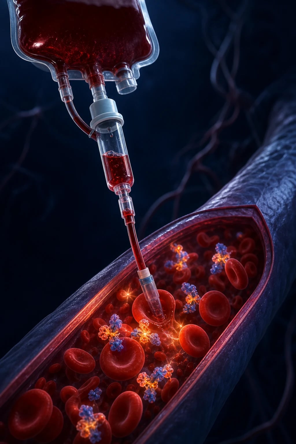
Cinematic clinical photograph of a stopped blood-transfusion unit beside a saline flush and a patient on supplemental oxygen, ICU setting, clinical-blue lighting, no text, no people
FigureStop, clip and sample — TRALI is new ARDS within six hours and does not respond to diuretics; TACO does.
Educational comparison TRALI versus TACO pathophysiology: antibody-mediated neutrophil lung injury and capillary leak versus hydrostatic volume overload, clinical-blue diagram, no faces
FigureTRALI is inflammatory capillary leak (often donor anti-HLA/HNA antibodies); TACO is hydrostatic overload. BP, JVP, echo and response to diuretics separate them at the bedside.
Management algorithm for acute transfusion reaction: stop transfusion, maintain IV access with saline, ABC support, distinguish TRALI TACO haemolytic septic allergic, report blood bank, clinical educational infographic
FigureAcute reaction algorithm — stop the unit, keep the line with saline, support ABC, send residual unit and samples to blood bank, and branch on TRALI vs TACO vs haemolytic vs septic vs allergic.

In one line

Any transfusion reaction: STOP transfusion, maintain IV with saline, assess ABC, send blood unit + samples to blood bank, report. TRALI: new ARDS within 6h of transfusion (donor anti-leucocyte antibodies) — manage as ARDS (oxygen, ventilation; diuretics DON'T help). TACO: volume overload — diurese. Acute haemolytic (ABO mismatch): catastrophic — fever, back pain, hypotension, haemoglobinuria — STOP, aggressive fluids, support BP/kidneys, treat DIC. Anaphylactic (IgA deficiency): IM adrenaline. Septic (platelets): fever + shock — STOP, cultures, antibiotics. Febrile/urticarial (mild): antipyretic/antihistamine (may continue cautiously).

[1]

Transfusion reactions compared

ReactionTimingMechanismSeverityKey featuresTreatment
Febrile non-haemolyticDuring/afterCytokines from donor leucocytesMildFever, rigorsAntipyretic; may continue
Allergic/urticarialDuring/afterPlasma protein (mild IgE)Mild-moderateUrticaria, itchAntihistamine; may continue
AnaphylacticRapidly (mls)IgA deficiency (anti-IgA)SEVEREHypotension, bronchospasm, urticariaIM adrenaline, fluids
Acute haemolytic (ABO)During (mls)ABO incompatibility (IgM/IgG)CATASTROPHICFever, back pain, hypotension, haemoglobinuria, DIC, AKISTOP, fluids, support BP/kidneys
TRALIWithin 6hDonor anti-leucocyte AbSevereARDS (hypoxaemia, bilateral infiltrates)Oxygen, ventilation (diuretics DON'T help)
TACODuring/within 6hVolume overloadModerate-severePulmonary oedema, hypertension, JVP raisedDiurese, oxygen
Septic (platelets)During/afterBacterial contaminationSevereHigh fever, rigors, shockSTOP, cultures, antibiotics
[1]

TRALI vs TACO — detailed clinical differentiation

ParameterTRALITACO
Underlying mechanismDonor anti-HLA / anti-HNA antibodies -> recipient neutrophil activation -> pulmonary capillary leak (non-cardiogenic ARDS)Hydrostatic — transfused volume exceeds cardiac reserve -> cardiogenic pulmonary oedema
Onset after transfusionWithin 6 h (usually 1-2 h)During or within 6 h (often towards end / after)
Blood pressureHYPOTENSION (transient)HYPERTENSION (raised from baseline)
JVPLow / normalRaised
Fluid balanceNeutral or negativeMarkedly positive
TemperatureFever commonAfebrile (usually)
AuscultationBilateral crackles (ARDS)Crackles + S3 gallop
EchocardiographyNormal LV systolic functionLV dysfunction / diastolic dysfunction / volume overload
NT-proBNPNormal / lowElevated (doubling from baseline strongly suggests TACO)
PCWP (if measured)Normal (<=18 mmHg)Raised (>18 mmHg)
Diuretic responseNone — and worsens hypotensionGood — rapid symptomatic improvement
Chest X-rayBilateral infiltrates (ARDS pattern)Bilateral alveolar infiltrates +/- cardiomegaly +/- effusions
Resolution48-72 hRapid (hours) with diuresis
Mortality5-10%<5%
Risk factorsMultiparous female donor; recent surgery, sepsis, alcohol misuse, massive transfusionHeart failure, renal impairment, elderly, infants, rapid/large transfusion, positive fluid balance
TreatmentOxygen / lung-protective ventilation; NO diuretics; vasopressors for shockDiurese (frusemide), sit upright, oxygen +/- NIV
PreventionMale-predominant plasma donors; screen donors for anti-HLA/HNA; leucoreductionSlow transfusion, single-unit policy, pre-transfusion diuretic in high-risk patients
[1]

Immediate management of ANY suspected transfusion reaction

  1. STOP THE TRANSFUSION — clamp the line (don't remove IV access — keep the cannula). DO NOT restart the transfusion until assessed (even if 'just a chill' — could be early haemolysis/sepsis). Keep the blood unit attached (don't discard)
  2. MAINTAIN IV ACCESS + ASSESS ABC — (a) Keep IV cannula patent with NORMAL SALINE (slow infusion). (b) AIRWAY: stridor, wheeze (anaphylaxis). (c) BREATHING: hypoxaemia, respiratory distress (TRALI, TACO, anaphylaxis). (d) CIRCULATION: hypotension (anaphylaxis, haemolytic, septic, TACO), tachycardia, JVP (TACO). (e) SKIN: urticaria (allergic), flushing. (f) PAIN: back/flank pain (haemolytic). Check vitals q5-15min
  3. INVESTIGATE — (a) CHECK IDENTITY (patient vs blood unit — ABO mismatch from clerical error is #1 cause of haemolytic reactions). (b) SEND: blood unit + giving set to blood bank. (c) SEND patient samples: FBC (haemolysis — Hb drop), U&E (AKI), coagulation (DIC), LDH + haptoglobin (haemolysis), direct antiglobulin test (DAT — positive in immune haemolysis), blood cultures (sepsis), urine (haemoglobinuria). (d) REPEAT GROUP + CROSSMATCH (rule out mismatch). (e) REPORT to blood bank + haemovigilance
  4. CLASSIFY THE REACTION + SPECIFIC TREATMENT — (a) MILD (febrile, urticarial): antipyretic (paracetamol), antihistamine (chlorphenamine). May RESTART transfusion cautiously (after review) if symptoms settle — but consider if needed. (b) ANAPHYLAXIS: IM adrenaline 0.5 mg (lateral thigh), repeat q5min, IV fluids, oxygen, intubate if airway. (c) ACUTE HAEMOLYTIC: aggressive IV fluids (maintain urine output >100 mL/hr — prevent AKI), vasopressors (noradrenaline), treat DIC (products), alkalinise urine (controversial). (d) TRALI: oxygen, HFNC/NIV/intubation (lung-protective), NO diuretics (not volume overload). (e) TACO: sit up, oxygen, diurese (frusemide), morphine if distressed. (f) SEPTIC: broad-spectrum antibiotics, vasopressors, cultures
  5. DOCUMENT + REPORT — (a) Document reaction (type, severity, intervention, outcome) in medical record. (b) REPORT to blood bank (transfusion reaction reporting — mandatory haemovigilance). (c) Blood bank investigates (re-check group/crossmatch, test for antibodies, bacterial culture of unit). (d) Inform patient (what happened — avoid future reactions — e.g., IgA deficiency needs washed products). (e) FOLLOW-UP: future transfusions (special requirements — e.g., irradiated, washed, CMV-negative, leucodepleted — depending on reaction)
  6. PREVENTION — (a) LEUCODEPLETION (all blood in most countries — reduces febrile, CMV, TRALI partially). (b) MALE-ONLY plasma (for TRALI — male donors less likely to have anti-leucocyte antibodies from pregnancy). (c) CAREFUL GROUP + CROSSMATCH (avoid ABO mismatch — clerical check). (d) IRRADIATED products (for immunocompromised — prevents transfusion-associated graft-versus-host disease). (e) WASHED products (for IgA deficiency — removes plasma). (f) RESTRICTIVE transfusion (TRICC trial — Hb threshold 70 g/L — fewer transfusions = fewer reactions)
[1]

TRALI management — step-by-step pathway

  1. STOP THE TRANSFUSION IMMEDIATELY — clamp the line; keep the unit + giving set for blood-bank investigation. Do NOT restart. Maintain IV access with normal saline.
  2. RECOGNISE — new hypoxaemia + bilateral infiltrates within 6 h of transfusion, with no left atrial hypertension (normal echo / normal NT-proBNP), satisfies the 2018-2019 TRALI consensus criteria. Differentiate from TACO at the bedside: low JVP, hypotension, normal BNP favour TRALI.[1]
  3. SUPPORTIVE — OXYGEN FIRST — escalate HFNC -> NIV (CPAP/bilevel) -> intubation as hypoxaemia dictates. Use lung-protective ventilation (Vt 6 mL/kg predicted body weight, plateau pressure <30 cmH2O, titrated PEEP) — the injured lung behaves identically to ARDS from any cause.[1]
  4. DO NOT GIVE DIURETICS — TRALI is a capillary-leak syndrome, not volume overload; frusemide worsens hypotension and provides no benefit. (A trial of diuretic may be diagnostic if TACO is in the differential, but expect no improvement in true TRALI.)
  5. MANAGE SHOCK — transient hypotension is common: cautious crystalloid (avoid large boluses — capillary leak worsens), noradrenaline for vasoplegia. Most haemodynamics normalise within 24 h.
  6. INVESTIGATE + REPORT — send donor + recipient samples to blood bank for anti-HLA / anti-HNA antibody testing and leucocyte antigen mismatch studies; FBC, coagulation, DAT (exclude haemolysis); NT-proBNP + bedside echo (exclude TACO); report to haemovigilance — mandatory.[11]
  7. RECOVERY — most cases resolve within 48-72 h with supportive care; mortality 5-10%. No proven specific therapy — steroids, plasmapheresis, and extracorporeal antibody removal are unproven. Avoid further transfusion where possible.
  8. PREVENT RECURRENCE — future plasma should come from male or never-pregnant female donors; consider washed RBCs / plasma-reduced products; the implicated donor is permanently deferred from plasma donation.[12]

TACO management — step-by-step pathway

  1. STOP THE TRANSFUSION — clamp the line; keep unit + giving set for blood-bank notification and haemovigilance reporting.[13]
  2. RECOGNISE — acute dyspnoea, hypertension, raised JVP, bibasal crackles, S3 gallop, positive fluid balance, and elevated NT-proBNP during or within 6 h of transfusion. Lung ultrasound shows multiple diffuse B-lines.
  3. SIT UPRIGHT + HIGH-FLOW OXYGEN — target SpO2 >=92% (or >=88% in COPD). Apply HFNC early in hypoxaemic patients.
  4. DIURESE — FRUSEMIDE 40-80 mg IV (double the patient's usual dose if on chronic diuretic); repeat in 30-60 min if inadequate response. Aim for net negative fluid balance (e.g., -1 L over 6 h).
  5. NIV / CPAP IF HYPOXAEMIC — non-invasive ventilation reduces work of breathing, counters auto-PEEP, and lowers LV afterload; escalate to intubation if fatigue, acidosis, or failure.
  6. REDUCE CIRCULATORY LOAD — stop all non-essential IV fluids; review total intake; consider a vasodilator (IV GTN) in the hypertensive patient with ongoing pulmonary oedema; hold or slow remaining transfusions.
  7. MONITOR — continuous SpO2, frequent BP, JVP, urine output (catheterise), repeat NT-proBNP and lung ultrasound (resolution of B-lines confirms response).
  8. REPORT + PREVENT RECURRENCE — TACO is a reportable transfusion reaction. For future transfusions in the same patient: transfuse one unit at a time, slowly (over 2-4 h, max 4 h/unit), give pre-transfusion frusemide in high-risk patients, prefer smaller-volume products, and avoid concomitant crystalloid.[13]

Exam practice — SAQs

SAQ — Acute haemolytic transfusion reaction from ABO incompatibility

10 minutes · 10 marks

A 65-year-old man receives his first unit of packed red cells for a GI bleed. Five minutes into the transfusion he develops fever, rigors, severe lower back pain, hypotension (BP 76/40), and nausea. The nurse stops the transfusion. The patient is tachycardic at 130, tachypnoeic at 28, and has dark red urine in the catheter bag.

[1]

SAQ — TACO in an elderly cardiac patient after transfusion

10 minutes · 10 marks

An 82-year-old woman with heart failure (EF 35 per cent) and chronic kidney disease receives 2 units of packed red cells over 3 hours for symptomatic anaemia (Hb 58 g/L). Four hours after completing the transfusion she develops acute dyspnoea, orthopnoea, and is unable to speak in full sentences. RR 36, SpO2 88 percent on room air, BP 172/96, JVP raised, bilateral crackles to mid-zones. NT-proBNP is markedly elevated.

[1]

SAQ — TRALI management after plasma transfusion in a perioperative patient

10 minutes · 10 marks

A 62-year-old woman, day 2 after a Whipple pancreaticoduodenectomy, receives 2 units of fresh frozen plasma for an elevated INR. Forty minutes into the second unit she develops acute dyspnoea, a fall in SpO2 to 86% on room air, fever (38.8C), and hypotension (BP 88/52). Bilateral crackles are heard. Her central venous pressure is 6 mmHg. A bedside echocardiogram shows normal LV systolic function.

[1]

SAQ — Differentiating TRALI from TACO after massive transfusion in trauma

10 minutes · 10 marks

A 55-year-old man is in ICU after a motor-vehicle crash with pelvic and femoral fractures. He has received 8 units of packed red cells, 6 units of fresh frozen plasma, and 2 pools of platelets via a massive transfusion protocol. Two hours after the transfusion he develops acute hypoxaemia (SpO2 88% on 60% FiO2 via high-flow nasal cannula). BP 102/65, HR 110, CVP 10 mmHg, fluid balance +4.2 L. Bilateral crackles are present and the chest X-ray shows new bilateral infiltrates.

[1]

Clinical pearls

High-yield transfusion reaction points for CICM/FFICM exam

  1. STOP the transfusion FIRST — every reaction. (1) The FIRST action for ANY suspected transfusion reaction (fever, rigor, hypotension, urticaria, dyspnoea, back pain) is to STOP the transfusion and clamp the line. (2) WHY: (a) The reaction may be LIFE-THREATENING (acute haemolytic, anaphylaxis, septic, TRALI) — can't tell initially. (b) Continuing the transfusion worsens the reaction (more antigen/antibody/volume). (c) Even 'mild' reactions may be early signs of severe (fever could be early septic/haemolytic). (3) KEEP IV ACCESS (don't remove cannula) — maintain with normal saline (for resuscitation). (4) KEEP the blood unit + giving set (for blood bank investigation). (5) ASSESS ABC + vitals (q5-15min). (6) THEN investigate + classify + treat. (7) The cost of stopping a transfusion that turns out to be mild (delay in transfusion) is FAR less than the cost of continuing a severe reaction (death).[6]
  2. TRALI — ARDS within 6 hours. (1) DEFINITION: Transfusion-Related Acute Lung Injury. NEW acute lung injury (ARDS — bilateral infiltrates + hypoxaemia + no left atrial hypertension) within 6 HOURS of transfusion. (2) MECHANISM: DONOR antibodies (anti-HLA or anti-neutrophil — usually from MULTIPAROUS female donors who developed antibodies during pregnancy) -> bind to recipient's neutrophils -> neutrophils sequester in pulmonary capillaries -> release proteases + reactive oxygen species -> capillary leak -> ARDS. (3) CLINICAL: hypoxaemia (SpO2 drop, respiratory distress), bilateral infiltrates (CXR), fever, hypotension. (4) DIAGNOSIS: clinical (ARDS within 6h of transfusion + no other cause). Send donor sample to blood bank (test for anti-HLA/anti-neutrophil antibodies). (5) DIFFERENTIAL: TACO (volume overload — but TACO has hypertension, raised JVP, positive fluid balance; TRALI has hypotension, low JVP). (6) MANAGEMENT: OXYGEN, HFNC/NIV/intubation (lung-protective ventilation). NO diuretics (TRALI is NOT volume overload — diuretics worsen hypotension). Supportive (vasopressors if hypotensive). Usually resolves in 48-72h. (7) MORTALITY: 5-10% (lower than other causes of ARDS). (8) PREVENTION: male-only plasma donors (men don't develop anti-HLA from pregnancy).[1]
  3. TACO — circulatory overload. (1) DEFINITION: Transfusion-Associated Circulatory Overload. Volume overload from transfusion (too much volume + too fast). (2) MECHANISM: transfused volume (blood + saline flush) exceeds circulatory capacity -> venous congestion -> pulmonary oedema. (3) RISK FACTORS: elderly, heart failure, renal failure, rapid transfusion, large volume. (4) CLINICAL: dyspnoea, orthopnoea, HYPERTENSION (unlike TRALI — hypotensive), raised JVP, bibasal crackles, S3 gallop. (5) DIAGNOSIS: clinical (pulmonary oedema + positive fluid balance + timing with transfusion). Echo (if available — LV dysfunction). NT-proBNP (elevated). (6) DIFFERENTIAL: TRALI (but TRALI: hypotension, low JVP, no fluid overload — TACO: hypertension, raised JVP, positive balance). (7) MANAGEMENT: SIT UP, oxygen, DIURESE (frusemide IV — the key treatment — offload volume), morphine (if distressed — venodilator), NIV (if severe). Slow/stop transfusion. (8) PREVENTION: transfuse SLOWLY (over 2-4h per unit — not faster), diurese between units (in heart failure patients), avoid unnecessary transfusions.[2]
  4. Acute haemolytic reaction — ABO incompatibility. (1) MECHANISM: recipient antibodies (anti-A or anti-B — usually IgM) attack transfused RBCs of WRONG ABO group -> intravascular haemolysis (complement-mediated RBC destruction) -> catastrophic. (2) CAUSE: usually CLERICAL ERROR (wrong patient, wrong unit — misidentification at sampling, labelling, or administration). (3) CLINICAL (during transfusion — within first few mL): (a) FEVER, RIGORS (inflammatory response). (b) BACK/FLANK PAIN (classic — from renal capsular distension from haemoglobin). (c) HYPOTENSION (vasodilation + cytokines). (d) HAEMOGLOBINURIA (dark/red urine — from free haemoglobin). (e) BLEEDING (DIC). (f) AKI (from haemoglobin nephrotoxicity + hypotension). (4) DIAGNOSIS: clinical + lab — Hb DROP (haemolysis), positive DAT (antibody on RBCs), low haptoglobin, high LDH, haemoglobinuria, coagulopathy (DIC). Re-check group/crossmatch (confirm mismatch). (5) MANAGEMENT: (a) STOP transfusion IMMEDIATELY (first action). (b) AGGRESSIVE IV FLUIDS (maintain urine output >100 mL/hr — flush haemoglobin through kidneys -> prevent AKI). (c) Vasopressors (noradrenaline — for hypotension). (d) Treat DIC (platelets, plasma, cryoprecipitate). (e) Alkalinise urine (bicarbonate — controversial — may reduce haemoglobin precipitation). (f) Renal replacement therapy (if AKI). (g) MORTALITY: high (up to 30-40%). (6) PREVENTION: rigorous identification (2 identifiers at sampling + administration), barcode systems, careful crossmatch.[3]
  5. Anaphylactic reaction — IgA deficiency. (1) MECHANISM: recipient with IgA DEFICIENCY (common — 1 in 500-1000) develops anti-IgA antibodies (sensitised from previous transfusion or exposure) -> transfused product contains IgA (in plasma) -> anti-IgA binds -> MASSIVE IgE-mediated anaphylaxis (mast cell degranulation). (2) CLINICAL (rapidly — within mLs of transfusion): (a) HYPOTENSION (shock). (b) BRONCHOSPASM (wheeze, stridor). (c) URTICARIA (hives). (d) ANGIOEDEMA (lip/tongue swelling). (e) GI (nausea, vomiting, cramping). (3) DIAGNOSIS: clinical (anaphylaxis during transfusion). TRYPTASE (elevated — within 1-2h — mast cell marker). IgA level + anti-IgA antibodies (confirm deficiency — post-event). (4) MANAGEMENT: (a) STOP transfusion. (b) IM ADRENALINE 0.5 mg (lateral thigh — repeat q5min). (c) IV fluids (rapid — for shock). (d) Oxygen, intubate (if airway). (e) Antihistamine (chlorphenamine), steroid (hydrocortisone — for delayed phase). (5) PREVENTION (future transfusions): WASHED RBCs/platelets (removes plasma + IgA) or IgA-deficient donor products.[4]
  6. Septic transfusion — bacterial contamination of platelets. (1) MECHANISM: bacteria contaminate the blood unit (especially PLATELETS — stored at room temperature -> bacterial growth) -> infused -> bacteraemia/sepsis. (2) ORGANISMS: Gram-negative (Serratia, Klebsiella, Pseudomonas — platelets) or Gram-positive (Staphylococcus — skin flora at donation). (3) CLINICAL (during or shortly after transfusion): (a) HIGH FEVER (rigors — abrupt, high). (b) SHOCK (hypotension — septic). (c) Nausea, vomiting. (d) DIC. (4) DIAGNOSIS: clinical (fever + shock during transfusion). CULTURE the blood unit + patient blood (same organism -> confirms). (5) MANAGEMENT: (a) STOP transfusion. (b) BROAD-SPECTRUM ANTIBIOTICS (immediately — don't wait for cultures — vancomycin + gram-negative cover). (c) Vasopressors (noradrenaline). (d) Cultures (blood — unit + patient). (e) Supportive (ventilation, renal support). (6) MORTALITY: high (especially Gram-negative). (7) PREVENTION: bacterial screening of platelets (culture before release), pathogen reduction (new technologies), proper storage (temperature monitoring), visual inspection (don't use if turbid/clumped).[5]
  7. Febrile non-haemolytic reaction — cytokines. (1) MECHANISM: cytokines (IL-1, IL-6, TNF) from DONOR LEUCOCYTES (released during storage) -> recipient inflammatory response -> fever, rigors. (2) CLINICAL: fever (>1°C rise), rigors, chills — during or shortly after transfusion. No hypotension, no haemolysis. (3) DIAGNOSIS: exclusion (rule out haemolysis, sepsis). Negative DAT, no Hb drop, normal haptoglobin. (4) MANAGEMENT: (a) STOP transfusion (initially — exclude severe causes). (b) ANTIPYRETIC (paracetamol). (c) Meperidine (for severe rigors — if available). (d) May RESTART transfusion cautiously (if settled + no haemolysis/sepsis). (5) PREVENTION: LEUCODEPLETION (removes donor leucocytes -> reduces cytokines — standard in most countries now). (6) COMMON (1-3% of transfusions) — usually mild.[6]
  8. Allergic/urticarial reaction — mild IgE. (1) MECHANISM: recipient IgE against donor PLASMA PROTEINS -> mast cell degranulation (mild, localised — unlike anaphylaxis which is severe). (2) CLINICAL: URTICARIA (hives — localised or generalised), pruritus (itch), flushing. No hypotension, no bronchospasm (if these -> anaphylaxis). (3) MANAGEMENT: (a) STOP transfusion (assess — exclude anaphylaxis). (b) ANTIHISTAMINE (chlorphenamine 10 mg IV). (c) May RESTART transfusion SLOWLY (after antihistamine + symptoms settled). (d) If recurs or progresses (hypotension, bronchospasm): STOP (anaphylaxis). (4) PREVENTION: antihistamine pre-medication (if recurrent — controversial), washed products (if severe). (5) COMMON (1-3% of transfusions) — usually mild.[4]
  9. Delayed haemolytic reaction — anamnestic response. (1) MECHANISM: recipient previously sensitised to RBC antigen (previous transfusion, pregnancy) -> antibody level low (undetectable at crossmatch) -> transfused RBCs with antigen -> anamnestic (secondary) immune response -> antibody rises -> extravascular haemolysis (spleen destroys antigen-positive RBCs). (2) TIMING: DAYS-WEEKS after transfusion (delayed — takes time for antibody to rise). (3) CLINICAL: (a) FALLING Hb (after initial rise from transfusion — haemolysis). (b) Mild jaundice (from haemolysis). (c) Fever. (d) Usually mild (not catastrophic like acute). (4) DIAGNOSIS: (a) Positive DAT (antibody on RBCs). (b) Antibody screen (identifies antibody). (c) Hb drop (after transfusion). (d) Low haptoglobin, high LDH (haemolysis markers). (5) MANAGEMENT: usually supportive (transfuse COMPATIBLE units — antigen-negative). Rarely severe (if massive haemolysis -> treat as acute). (6) PREVENTION: careful antibody screen (detect previous antibodies — record in file), antigen-matched units (if known antibody).[3]
  10. Hyperkalaemia from old RBC units. (1) MECHANISM: RBCs leak potassium into plasma during STORAGE (especially after 2-3 weeks). (2) RISK: massive transfusion (many units -> cumulative K+ load), neonates (small blood volume -> high concentration), renal failure (can't excrete K+). (3) CLINICAL: hyperkalaemia (peaked T waves, arrhythmia — cardiac arrest in severe). (4) PREVENTION: use FRESH RBCs (<7 days — for massive transfusion, paediatric, renal failure). (5) MANAGEMENT: treat hyperkalaemia (calcium gluconate for cardiac protection, insulin/dextrose, bicarbonate, dialysis if severe). (6) ALSO: hypocalcaemia from citrate (anticoagulant — chelates Ca2+) -> monitor ionised Ca2+ during massive transfusion, replace (calcium chloride/gluconate).[6]
  11. TRALI vs TACO — the key distinction. (1) BOTH cause respiratory distress after transfusion. (2) DIFFERENCES: (a) MECHANISM: TRALI = capillary leak (inflammatory — donor antibodies); TACO = volume overload (hydrostatic). (b) BP: TRALI = HYPOTENSION (vasodilation); TACO = HYPERTENSION (volume overload). (c) JVP: TRALI = LOW/normal (no overload); TACO = RAISED (volume overload). (d) Fluid balance: TRALI = neutral/negative; TACO = positive. (e) Echo: TRALI = normal LV (but pulmonary oedema from leak); TACO = LV dysfunction/overload. (f) NT-proBNP: TRALI = normal/low; TACO = high. (3) TREATMENT DIFFERS: (a) TRALI: oxygen, ventilation — NO diuretics (not volume overload — diuretics worsen hypotension). (b) TACO: diurese (frusemide — offload volume). (4) CLINICAL: if hypotensive + respiratory distress after transfusion -> TRALI (diurese WORSENS). If hypertensive + respiratory distress -> TACO (diurese HELPS). (5) ALSO: BNP/NT-proBNP helps distinguish (high in TACO, normal in TRALI).[1]
  12. Transfusion-associated graft-versus-host disease (TA-GVHD) — rare but fatal. (1) MECHANISM: donor T-lymphocytes (in transfused product) engraft in recipient (who is immunocompromised — can't reject them) -> attack recipient tissues (skin, gut, liver, marrow) -> GVHD. (2) RISK: immunocompromised (chemo, transplant, congenital immunodeficiency), recipients of directed donations (from relatives — shared HLA haplotypes -> don't recognise as foreign). (3) CLINICAL (days-weeks after transfusion): fever, rash (maculopapular), diarrhoea, liver dysfunction, pancytopenia (marrow attack). (4) MORTALITY: >90% (no effective treatment — bone marrow failure). (5) PREVENTION: IRRADIATED blood products (25-50 Gy — kills donor T-lymphocytes) for immunocompromised recipients + directed donations. (6) NOT prevented by leucodepletion (some T-cells remain — only irradiation is effective).[6]
  13. Restrictive transfusion strategy — fewer reactions. (1) TRICC trial (1998, NEJM): restrictive (Hb threshold 70 g/L) vs liberal (100 g/L) in ICU -> RESTRICTIVE was SAFE (no increase in mortality) and FEWER transfusions. (2) SUBSEQUENT trials (TRISS — sepsis; REALITY — GI bleed; others): all support RESTRICTIVE (Hb 70 threshold) in most ICU patients. (3) EXCEPTIONS (consider higher threshold): (a) Acute coronary syndrome (Hb 80 — ischaemic heart disease needs oxygen-carrying capacity). (b) Acute brain injury (TBI, stroke — Hb 90-100 — cerebral oxygenation). (c) Severe sepsis with lactate >4 (controversial). (d) Massive haemorrhage (transfuse to Hb 80+ — per massive transfusion protocol, 1:1:1). (4) PRINCIPLE: FEWER transfusions = FEWER reactions + FEWER complications (infection, TRALI, TACO, haemolysis) + cost savings. (5) CLINICAL: transfuse for ANAEMIA symptoms (not a number) — unless acute bleeding (then massive transfusion protocol).[6]
  14. Reporting + haemovigilance. (1) EVERY transfusion reaction (mild or severe) must be REPORTED to the blood bank + haemovigilance system. (2) WHY: (a) Detect CLERICAL ERRORS (misidentification — the #1 cause of acute haemolytic). (b) Detect DONOR ISSUES (TRALI from multiparous donor — exclude from plasma donation). (c) Detect PRODUCT ISSUES (bacterial contamination — recall lot). (d) NATIONAL SURVEILLANCE (track reaction rates, identify trends, improve safety). (3) DOCUMENT in patient record (reaction type, severity, intervention, outcome). (4) INFORM PATIENT (what happened — they need to tell future healthcare providers — wear a medical alert if severe allergy/IgA deficiency). (5) SPECIAL REQUIREMENTS for FUTURE transfusions (based on reaction): washed (IgA deficiency), irradiated (immunocompromised), CMV-negative (transplant/pregnant), antigen-matched (known antibodies). (6) HAEMOVIGILANCE has dramatically improved transfusion safety (reaction rates fallen since implementation).[6]

Prevention strategies — by reaction type

ReactionPrimary prevention strategy
Acute haemolytic (ABO mismatch)Two-person bedside identity check; barcode/PDA patient identification; rigorous clerical check at sampling, labelling, and administration; repeated crossmatch
TRALIMale-predominant plasma donation (defer multiparous female donors from plasma/platelet donation); screen donors for anti-HLA / anti-HNA antibodies; universal leucoreduction
TACOSlow transfusion (2-4 h/unit); single-unit policy; pre-transfusion diuretic in high-risk patients (elderly, heart/renal failure); avoid concurrent crystalloid
Anaphylactic (anti-IgA / IgA deficiency)Washed or IgA-deficient donor products; medical-alert bracelet; document in medical record
Febrile non-haemolyticUniversal leucoreduction (standard in most high-income countries) — removes donor leucocyte-derived cytokines
Allergic / urticarialAntihistamine premedication in patients with prior reactions; washed products for recurrent severe reactions
Septic (platelet bacterial contamination)Bacterial screening of all platelet units (culture or rapid test); pathogen-reduction technology; visual inspection before release; storage <=5 days
TA-GVHDIrradiated products (25-50 Gy) for immunocompromised recipients (chemo, HSCT, congenital immunodeficiency), neonates, and directed/related donations
Hyperkalaemia (stored RBCs)Use fresh RBCs (<7 days) for massive transfusion, paediatric exchange, and renal failure; consider washed RBCs
Hypocalcaemia (citrate)Monitor ionised Ca2+ during massive transfusion and apheresis; replace with calcium gluconate / chloride
All reactions (universal)Restrictive transfusion strategy (Hb threshold 70 g/L — TRICC, TRISS, FOCUS, AABB 2023); single-unit policy; transfuse only when benefit outweighs risk
[1]

TRALI / TACO / massive transfusion — advanced exam-high-yield pearls

  1. TRALI consensus definition (2018-2019). (1) The International TRALI Consensus Panel (revised by Vlaar 2019) defines TRALI as new acute lung injury (PaO2/FiO2 <300 or SpO2 <90%) with bilateral infiltrates on CXR, occurring within 6 h of transfusion, with no left atrial hypertension and no other risk factor for ARDS (or, if a competing risk factor exists but TRALI is the most likely driver, "possible TRALI").[1] (2) Type I: no pre-existing ARDS risk factor. Type II (possible TRALI): a risk factor (sepsis, aspiration, trauma, pancreatitis) is present but TRALI is the most likely driver. (3) Two-event model: recipient priming (sepsis, recent surgery) + donor antibody (or biologically active lipids in stored RBCs/platelets) -> neutrophil sequestration -> capillary leak.[14] (4) The implicated donor is identified by antibody testing and permanently deferred from plasma donation.
  2. TACO biomarkers — NT-proBNP is the discriminator. (1) A doubling of NT-proBNP from pre-transfusion baseline, in the context of post-transfusion dyspnoea, strongly supports TACO over TRALI (sensitivity ~80%, specificity ~80%).[13] (2) BNP rises with ventricular wall stress; in TRALI (normal LV) it stays flat or low. (3) Lung ultrasound (>=3 B-lines per intercostal space bilaterally) is a rapid bedside confirmatory tool when echo/BNP unavailable. (4) Caveat: chronic heart failure patients have elevated baseline BNP — use the delta (rise), not the absolute value.
  3. TRALI — the two-event ("threshold") hypothesis. (1) Event 1 (recipient priming): sepsis, recent surgery, massive transfusion, cytokine storm -> pulmonary endothelium "primed", neutrophils marginated in lung capillaries. (2) Event 2 (transfusion trigger): donor anti-HLA class I/II or anti-HNA antibodies (or bioactive lipids from stored cellular components) bind primed neutrophils -> oxidative burst, protease release -> endothelial damage and capillary leak.[14] (3) Explains why TRALI is more common in ICU/surgical patients (primed) than ambulatory recipients. (4) Implication: a primed recipient can develop TRALI from a relatively low-titre antibody — hence the threshold model rather than a simple dose-response.
  4. TRALI donor-deferral policy. (1) Donor anti-HLA/anti-HNA antibodies most commonly arise from pregnancy (fetal-maternal alloimmunisation) or prior transfusion/transplant. (2) Policy (UK, Netherlands, US, Australia): plasma for transfusion and apheresis platelets should come preferentially from male donors or never-pregnant female donors, OR from female donors screened negative for anti-HLA/HNA.[12] (3) This intervention reduced TRALI incidence by ~60-70% in jurisdictions that adopted it — one of the most successful haemovigilance interventions in modern transfusion medicine. (4) RBC and pooled platelet units are less affected (less plasma); universal leucoreduction contributes further.
  5. TACO — single-unit policy and diuretic cover. (1) The single most effective bedside prevention for TACO is the single-unit transfusion policy: transfuse ONE unit, reassess, only give the next if still indicated. (2) For high-risk patients (elderly, LV/RV dysfunction, CKD, eGFR <30, albumin <30, positive fluid balance), give pre-transfusion frusemide (e.g., 20-40 mg IV) and transfuse slowly (over 3-4 h).[13] (3) Avoid concurrent crystalloid; review total fluid status daily. (4) TACO is now the most commonly reported cause of transfusion-related mortality in the US (surpassing TRALI after donor-deferral policies reduced TRALI). (5) NT-proBNP-guided pre-transfusion risk stratification is increasingly recommended.
  6. Massive transfusion — the lethal triad. (1) Massive transfusion (>1 blood volume in 24 h, or >4 units RBCs in <1 h with ongoing haemorrhage) produces the lethal triad: hypothermia, acidosis, coagulopathy — each worsens the others and predicts death. (2) PROPPR trial (Holcomb, JAMA 2015): in severe trauma, a 1:1:1 ratio (platelets:plasma:RBCs) achieved earlier haemostasis and fewer exsanguination deaths vs 1:1:2, with no difference in 24-h or 30-d mortality.[9] (3) Specific complications: hypocalcaemia (citrate chelation — monitor ionised Ca2+, give 10 mL 10% calcium gluconate per 2 units), hyperkalaemia (use fresh RBCs <7 days), hypothermia (blood warmer — never microwave), dilutional coagulopathy (give plasma/platelets per ratio/viscoelastic testing). (4) Tranexamic acid (within 3 h — CRASH-2) reduces mortality in trauma bleeding.
  7. Restrictive vs liberal transfusion — the trial evidence. (1) TRICC (Hebert, NEJM 1999): ICU patients, Hb 70 vs 100 g/L -> restrictive non-inferior, fewer transfusions, fewer cardiac complications.[7] (2) FOCUS (Carson, NEJM 2011): hip-fracture elderly -> restrictive (80) non-inferior to liberal (100).[10] (3) TRISS (Holst, NEJM 2014): septic shock, Hb 70 vs 90 -> no mortality difference.[8] (4) AABB/JAMA 2023 guideline: Hb 7 g/dL for hospitalised stable adults; 8 g/dL after major orthopaedic/cardiac surgery or with pre-existing cardiovascular disease; higher thresholds NOT supported outside ACS and severe symptoms.[6] (5) Exceptions where higher Hb may help: acute coronary syndrome, acute brain injury (TBI, SAH — Hb 90-100), severe symptomatic anaemia. (6) Net: transfuse the fewest units necessary — physiology, not numbers alone.
  8. Transfusion-associated graft-versus-host disease (TA-GVHD) — fatal and preventable. (1) Viable donor T-lymphocytes in cellular products engraft in an immunocompromised (or HLA-haploidentical) recipient -> attack skin (maculopapular rash), gut (profuse diarrhoea), liver (hepatitis), and bone marrow (pancytopenia). (2) Onset 4-30 days post-transfusion. (3) Mortality >90% — death from marrow aplasia/infection. (4) At-risk recipients: HSCT, chemo/immunosuppression, congenital immunodeficiency, neonates/intrauterine transfusion, Hodgkin lymphoma, fludarabine, directed (related-donor) transfusions (shared HLA -> donor lymphocytes not recognised as foreign). (5) Prevention: irradiation (25-50 Gy) of all cellular products for at-risk recipients — kills T-lymphocytes while preserving RBC/platelet function. Leucoreduction is NOT sufficient (residual lymphocytes remain viable).
  9. Bacterial contamination of platelets — storage at room temperature. (1) Platelets stored at 20-24C (to preserve function) — ideal for bacterial growth. (2) Organisms: Gram-negative (Serratia, Klebsiella, Pseudomonas, Enterobacter — high virulence, high mortality) and Gram-positive (Staphylococcus epidermidis/aureus — skin flora at venesection). (3) Incidence ~1 in 3000 platelet units contaminated; septic reaction ~1 in 100,000; fatal reaction ~1 in 500,000-1,000,000.[5] (4) Clinical: rigors + high fever (>39C or >2C rise) +/- hypotension during/immediately after transfusion. (5) Prevention: bacterial culture of every platelet unit (mandatory in many countries), pathogen-reduction technology (amotosalen/UVA, riboflavin/UV), limit storage to 5 days, visual inspection (turbidity/gas bubbles). (6) Management: STOP, blood cultures from patient AND unit, broad-spectrum antibiotics (Gram-negative cover — piperacillin-tazobactam +/- vancomycin), vasopressors for shock.
  10. Allergic/anaphylactic reaction — when to give washed vs standard products. (1) Mild urticarial only (no hypotension, no bronchospasm): antihistamine, may restart slowly — no special future product needed. (2) Recurrent urticarial (>=2 episodes): antihistamine premedication 30 min before; consider washed products if persists. (3) Anaphylaxis + confirmed IgA deficiency (anti-IgA antibodies): use washed RBCs / platelets (removes >99% plasma) or IgA-deficient donor plasma; patient must wear a medical-alert bracelet. (4) Anaphylaxis without IgA deficiency: investigate other plasma proteins (haptoglobin deficiency — particularly in East Asian populations); washed products if severe. (5) Document special requirements clearly in medical record and transfusion service file.
  11. Massive transfusion — calcium and citrate toxicity. (1) Citrate (in CPD/SAGM anticoagulant) chelates calcium -> hypocalcaemia during rapid transfusion (>1 unit/5 min) or apheresis. (2) Clinical: circumoral paraesthesia, tetany, prolonged QT, hypotension, depressed myocardial contractility. (3) Monitor ionised (not total) Ca2+ during massive transfusion and large-volume apheresis. (4) Replace: calcium gluconate 10 mL 10% IV per 2 units RBCs (or per viscoelastic/ECG guidance); calcium chloride (3x more Ca2+ per mL) reserved for severe symptomatic hypocalcaemia via central line. (5) Concurrent hypomagnesaemia (citrate also chelates Mg2+) worsens hypocalcaemia — check and replace. (6) Hepatic citrate metabolism is impaired in shock/hypothermia/hepatic failure -> slower clearance -> more aggressive replacement needed.
  12. TA-GVHD vs TRALI vs TACO — rapid differential. (1) All three are serious non-infectious transfusion hazards but differ in timing, mechanism, and target organ. (2) TA-GVHD: donor T-cells attack recipient (skin/gut/liver/marrow), onset days-weeks, fatal (>90%), prevent by irradiation. (3) TRALI: donor antibodies attack recipient lung, onset <6 h, mortality 5-10%, prevent by male-predominant plasma. (4) TACO: transfused volume overloads recipient circulation, onset during/within 6 h, mortality <5%, prevent by slow/diuretic. (5) Key exam point: TA-GVHD is NOT prevented by leucoreduction (only irradiation); TRALI is reduced (not eliminated) by leucoreduction and dramatically reduced by male-predominant plasma.
  13. Transfusion in the anticoagulated / bleeding patient. (1) Warfarin reversal in life-threatening bleed: 4-factor prothrombin complex concentrate (PCC, 25-50 IU/kg) + IV vitamin K (faster, lower volume than FFP; FFP 15 mL/kg if PCC unavailable). (2) Direct oral anticoagulant (DOAC) reversal: dabigatran -> idarucizumab 5 g IV; apixaban/rivaroxaban -> andexanet alfa (or PCC if unavailable); reversal should precede/parallel transfusion. (3) Antiplatelet / uraemic platelet dysfunction -> desmopressin (0.3 mcg/kg); platelet transfusion only for intracranial haemorrhage on antiplatelet. (4) Viscoelastic testing (TEG/ROTEM) guides product use: prolonged R/CT -> plasma/fibrinogen; low MA/MCF -> platelets/cryoprecipitate; prolonged LY30/ML -> tranexamic acid.
  14. Restrictive strategy — exceptions and nuances. (1) Acute brain injury (TBI, SAH, ischaemic stroke, ICH): most neurointensivists target Hb 90-100 g/L — the injured brain cannot upregulate cerebral blood flow adequately and cerebral oxygen delivery is critical. (2) Acute coronary syndrome / acute MI: AABB suggests Hb >=8 g/dL; the MINT trial suggested possible benefit of 100 vs 80 in ACS but the result was not definitive. (3) Severe sepsis with lactate >4: TRISS showed 70 safe; consider higher if ongoing ischaemia.[8] (4) Acute upper GI bleed: restrictive (70) is safe and possibly superior to liberal. (5) Chronic stable anaemia: treat the cause (iron/EPO/B12); transfuse only if symptomatic. (6) Net: transfuse for physiology (symptomatic, ischaemic), not numbers alone.[6]

Red flags

Critical transfusion reaction red flags

  • ANY reaction: STOP transfusion, maintain IV saline, assess ABC.[6]
  • Acute haemolytic (ABO mismatch): fever + back pain + hypotension + haemoglobinuria — catastrophic, STOP immediately.[3]
  • Anaphylactic (IgA deficiency): hypotension + bronchospasm + urticaria — IM adrenaline.[4]
  • TRALI: ARDS within 6h of transfusion — NO diuretics (not volume overload).[1]
  • TACO: volume overload + hypertension + raised JVP — DIOURESE.[2]
  • Septic (platelets): high fever + shock — STOP, cultures, broad-spectrum antibiotics.[5]
  • TRALI vs TACO: hypotension (TRALI) vs hypertension (TACO); diuretics help TACO, worsen TRALI.[1]
  • Restrictive strategy (Hb 70) — fewer transfusions = fewer reactions (TRICC).[6]

TRALI/TACO and massive-transfusion red flags — extended

  • Hypotension within minutes of starting a unit — think acute haemolytic (ABO) OR anaphylaxis (anti-IgA) OR septic (platelets) — STOP, don't restart.[3]
  • Hypoxaemia within 6 h of transfusion — TRALI until proven otherwise (check JVP, BNP, echo); NO diuretics if hypotensive with low/normal BNP.
  • Hypertension + raised JVP + dyspnoea — TACO; give frusemide 40-80 mg IV, sit upright, oxygen +/- NIV.[13]
  • High fever + rigors + shock during platelet transfusion — bacterial contamination; STOP, culture unit + patient, broad-spectrum antibiotics NOW.[5]
  • Massive transfusion: monitor ionised Ca2+ (citrate), K+ (old units), temperature (use warmer), and coagulation (viscoelastic) every 30-60 min.
  • Maculopapular rash + diarrhoea + hepatitis 1-4 weeks post-transfusion — TA-GVHD; check FBC (pancytopenia); high mortality; prevent with irradiated products next time.
  • Anaphylaxis to first few mL — IgA deficiency; check tryptase (1-2 h), IgA + anti-IgA levels; future products must be washed.
  • NT-proBNP doubles post-transfusion — strong evidence for TACO over TRALI; diurese.[13]
  • Known multiparous female plasma donor + ARDS in recipient <6 h — TRALI; notify blood bank to quarantine and test donor.[12]
  • Donor-deferral after TRALI: implicated donor permanently excluded from plasma and apheresis-platelet donation (may still donate RBC).[15]

Prognosis

Transfusion reaction evidence and outcomes

Acute haemolytic (ABO mismatch): mortality 30-40% (mostly from clerical error — prevent with barcode ID). TRALI: mortality 5-10% (lower than other ARDS); resolves in 48-72h; male-only plasma donors reduces incidence. TACO: mortality <5% (treat with diuretics); common in elderly/heart failure — prevent with slow transfusion. Anaphylactic (IgA deficiency): mortality low with prompt adrenaline; prevent with washed products. Septic (platelets): mortality 10-20% (Gram-negative worse); bacterial screening + pathogen reduction reduces. TRICC trial (1998): restrictive (Hb 70) safe; fewer transfusions = fewer reactions. TA-GVHD: mortality >90% (prevent with irradiated products for immunocompromised). Haemovigilance: reporting systems have reduced reaction rates dramatically since implementation.

[1]

Key trials and guidelines — transfusion reactions and thresholds

TRICC (Hebert, NEJM 1999): ICU patients (n=838), Hb <70 (restrictive, transfuse at 70, target 70-90) vs <100 (liberal). 30-day mortality equivalent; restrictive group received 54% fewer transfusions and had lower in-hospital mortality in the less-ill (APACHE II <=20) and younger (<55) subgroups.[7] Landmark trial establishing restrictive transfusion in ICU. FOCUS (Carson, NEJM 2011): hip-fracture surgery in elderly (n=2016), Hb <80 (symptomatic, target ~80) vs <100. No difference in mortality or functional recovery; restrictive non-inferior. Established the 80 threshold in elderly surgical patients.[10] TRISS (Holst, NEJM 2014): septic shock ICU (n=998), Hb <70 vs <90. 90-day mortality identical (27.8% vs 27.5%); restrictive group used 50% fewer RBC units; no increase in ischaemic events. Restrictive strategy confirmed safe in septic shock.[8] PROPPR (Holcomb, JAMA 2015): severe trauma (n=680), 1:1:1 (plasma:platelet:RBC) vs 1:1:2 ratio. No difference in 24-h or 30-d mortality, but 1:1:1 achieved earlier haemostasis and fewer exsanguination deaths.[9] Foundation for modern massive haemorrhage protocols. Vlaar TRALI Consensus 2019 (Transfusion): redefined TRALI as ALI within 6 h of transfusion with no other risk factor (Type I) or with a competing risk factor deemed less contributory (Type II / possible TRALI). Standardised global case definitions for surveillance and trials.[1] Toy revised nomenclature 2017 (Transfusion): harmonised TRALI terminology between European and US groups; introduced the "possible TRALI" category.[11] Middelburg plasma meta-analysis (Transfusion 2017): male-only / male-predominant plasma donors reduce TRALI risk without compromising supply; basis for universal male-predominant plasma policies in high-income countries.[12] Roubinian TACO risk factors (Crit Care Med 2018): identified older age, heart failure, renal dysfunction, positive fluid balance, and larger transfusion volume as independent TACO predictors; supports pre-transfusion risk stratification.[13] Bux/Sachs HLA class II TRALI (Blood 2011): delineated the antibody-mediated neutrophil-activation mechanism of TRALI, particularly for HLA class II antibodies.[14] AABB RBC transfusion guideline 2023 (Carson, JAMA): Hb 7 g/dL threshold for hospitalised stable adults, 8 g/dL for those undergoing orthopaedic/cardiac surgery or with pre-existing cardiovascular disease; restricts recommendation to symptom/physiology-guided transfusion.[6]

Prevention summary

Prevention — evidence and policy

Leucoreduction: universal pre-storage leucoreduction reduces febrile non-haemolytic reactions, CMV transmission, and contributes to TRALI reduction; standard in most high-income countries. Male-predominant plasma (TRALI mitigation): excluding previously-pregnant female donors from plasma/apheresis-platelet donation reduced TRALI incidence by ~60-70% where adopted.[12] Irradiation (TA-GVHD mitigation): 25-50 Gy gamma or X-ray irradiation of cellular products for immunocompromised recipients, neonates, and directed donations; the ONLY effective TA-GVHD prevention (leucoreduction alone is insufficient). Bacterial screening of platelets: mandatory culture or rapid detection reduces septic reactions; pathogen-reduction technology (amotosalen/UVA, riboflavin/UVB) further lowers risk and may extend shelf-life. Restrictive transfusion strategy: the single most effective reaction-prevention measure is to transfuse fewer units — TRICC, TRISS, FOCUS, and the 2023 AABB guideline all endorse a restrictive (Hb 70) strategy for most stable ICU patients.[6] Single-unit policy: transfuse one unit, reassess, only give the next if still indicated — reduces TACO and unnecessary exposure. Patient identification: two-person bedside check + barcode/PDA identification at sampling, labelling, and administration prevents ABO-mismatch acute haemolytic reactions (overwhelmingly due to clerical error).

References

  1. [1]Vlaar APJ, et al. A consensus redefinition of transfusion-related acute lung injury Transfusion, 2019.PMID 30993745
  2. [2]Clifford L, et al. Risk Factors and Clinical Outcomes Associated with Perioperative Transfusion-associated Circulatory Overload Anesthesiology, 2017.PMID 28072601
  3. [3]Janatpour KA, et al. Noninfectious serious hazards of transfusion Curr Hematol Rep, 2002.PMID 12901137
  4. [4]Savage WJ, et al. Transfusion and component characteristics are not associated with allergic transfusion reactions to apheresis platelets Transfusion, 2015.PMID 25209730
  5. [5]Hong H, et al. Detection of septic transfusion reactions to platelet transfusions by active and passive surveillance Blood, 2016.PMID 26598718
  6. [6]Carson JL, et al. Red Blood Cell Transfusion: 2023 AABB International Guidelines JAMA, 2023.PMID 37824153
  7. [7]Hebert PC, et al. A multicenter, randomized, controlled clinical trial of transfusion requirements in critical care. Transfusion Requirements in Critical Care Investigators, Canadian Critical Care Trials Group N Engl J Med, 1999.PMID 9971864
  8. [8]Holst LB, et al. Lower versus higher hemoglobin threshold for transfusion in septic shock N Engl J Med, 2014.PMID 25270275
  9. [9]Holcomb JB, et al. Transfusion of plasma, platelets, and red blood cells in a 1:1:1 vs a 1:1:2 ratio and mortality in patients with severe trauma: the PROPPR randomized clinical trial JAMA, 2015.PMID 25647203
  10. [10]Carson JL, et al. Liberal or restrictive transfusion in high-risk patients after hip surgery N Engl J Med, 2011.PMID 22168590
  11. [11]Toy P, et al. Proposed revised nomenclature for transfusion-related acute lung injury Transfusion, 2017.PMID 28019007
  12. [12]Saadah NH, et al. Comparing transfusion reaction rates for various plasma types: a systematic review and meta-analysis/regression Transfusion, 2017.PMID 28766723
  13. [13]Roubinian NH, et al. Contemporary Risk Factors and Outcomes of Transfusion-Associated Circulatory Overload Crit Care Med, 2018.PMID 29300236
  14. [14]Sachs UJH, Bux J. Mechanism of transfusion-related acute lung injury induced by HLA class II antibodies Blood, 2011.PMID 21030555
  15. [15]Muller MC, et al. Low-risk transfusion-related acute lung injury donor strategies and the impact on the onset of transfusion-related acute lung injury: a meta-analysis Transfusion, 2015.PMID 25135630
  16. [16]Vlaar APJ, et al. An update of the transfusion-related acute lung injury (TRALI) definition Transfus Clin Biol, 2019.PMID 31221537