ICU · Haematology
Acute sickle cell crisis in the ICU
Also known as Sickle cell crisis (SCC) · Vaso-occlusive crisis (VOC) · Acute chest syndrome (ACS) · Sickle cell disease (SCD)
Sickle cell crisis is caused by polymerisation of sickle haemoglobin (HbS) under hypoxia/dehydration/infection → red blood cells sickle → vaso-occlusion → ischaemia, pain, organ damage. The molecular lesion is a single point mutation in the β-globin gene (HBB): GAG→GTG at codon 6, substituting valine for glutamic acid at position 6 of the β-chain. Under deoxygenation HbS polymerises via hydrophobic contacts into 14-strand fibres that distort the erythrocyte into the classic sickle shape → haemolysis, endothelial activation, leucocyte adhesion and vaso-occlusion. Types: vaso-occlusive crisis (pain — bones, chest, abdomen), acute chest syndrome (new infiltrate + respiratory symptoms — leading cause of death), splenic sequestration (children), aplastic crisis (parvovirus B19), stroke, priapism. Management: oxygen, IV hydration (normal saline 1-1.5x maintenance), analgesia (opioids — morphine PCA), treat triggers (infection, dehydration). Exchange transfusion for severe complications (stroke, acute chest syndrome). Hydroxyurea, crizanlizumab and voxelotor for prevention/modification. CRISPR gene therapy (Casgevy, exagamglogene autotemcel) FDA/MHRA-approved 2023.
On this page & tools
Your progress
Saved locally on this device.
Target exams
Red flags
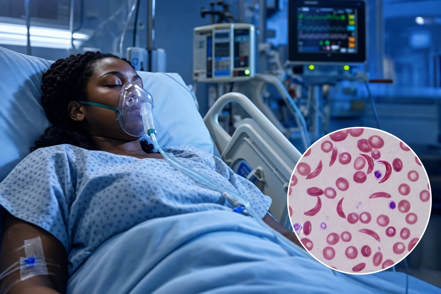
Pathophysiology — the molecular basis
From a single base change to multi-organ vasculopathy
1. The mutation (HBB c.20A>T; p.Glu6Val)
A single-nucleotide substitution (GAG→GTG) in codon 6 of the β-globin gene (HBB on chromosome 11p15.4) replaces the polar, negatively charged **glutamic acid with the hydrophobic valine at position 6** of the β-chain. This is autosomal recessive: homozygous HbSS = sickle cell anaemia (disease); heterozygous HbAS = sickle cell trait (usually asymptomatic). HbS proportion determines severity (HbSS > HbSβ⁰-thal > HbSC > HbSβ⁺-thal).
2. Deoxygenation → HbS polymerisation
Deoxygenated HbS (α₂β₂ where the β6 Val creates a hydrophobic patch) polymerises via longitudinal contact into **14-strand helical fibres** that distort the red cell. Polymerisation kinetics follow a **delay time** inversely related to HbS concentration²⁰–³⁰, so anything that increases deoxygenation, lowers pH, raises 2,3-DPG or concentrates HbS (dehydration) shortens delay time and accelerates sickling. **HbF inhibits polymerisation** (γ-chain lacks β6 Val) — the rationale for hydroxyurea and BCL11A-editing gene therapy.
3. Sickling → deformed, rigid, dehydrated erythrocyte
Repeated sickling–unsickling damages the membrane: activation of Gardos and K-Cl cotransport channels → **K⁺ and water loss → intracellular dehydration** (raises MCHC, the single most powerful driver of polymerisation). The cell becomes irreversibly sickled (ISC), rigid and adhesive. This reduces deformability → microvascular obstruction.
4. Haemolysis — the haemolytic phenotype
Intravascular and extravascular haemolysis releases free haemoglobin and arginase → **scavenges nitric oxide (NO)** and depletes arginine → endothelial dysfunction, pulmonary hypertension, priapism, leg ulcers, stroke. Markers of haemolysis: low/normal Hb, high reticulocytes, high LDH, indirect bilirubin, low haptoglobin, high plasma free Hb. A low haemoglobin phenotype (co-existence of α-thalassaemia trait, high HbF) shifts the picture towards a milder "viscosity" phenotype.
5. Endothelial activation + leucocyte/platelet adhesion
Sickled reticulocytes adhere (via VLA-4/α4β1, CD36, LW) to endothelium upregulated by cytokines/hypoxia (VCAM-1, ICAM-1, selectins). **P-selectin** on endothelium and platelets drives heterotypic aggregates — the target of crizanlizumab. Neutrophil trapping, platelet activation and complement activation generate the vaso-occlusive plug.
6. Vaso-occlusion → ischaemia–reperfusion
Microvascular occlusion in bone marrow, spleen, lung, brain and renal medulla produces **ischaemia–reperfusion injury**, oxidative stress and a sterile inflammatory response (IL-1β, IL-6, TNF-α). This is the final common pathway of the painful crisis, acute chest syndrome, splenic infarction, papillary necrosis and avascular necrosis.
7. Chronic vasculopathy
Cumulative injury produces functional asplenia (infarction by age 5–7 in HbSS), pulmonary hypertension (TRV >2.5 m/s, risk of death), sickle nephropathy (hyperfiltration → albuminuria → CKD), retinopathy (HbSC > HbSS), moyamoya (collaterals after cerebral infarction), and a shortened lifespan — median ~45–55 years, improving with comprehensive care.
Types of crisis
Vaso-occlusive (VOC)
Pain crisis — most common
- Sickled RBCs occlude microvasculature → ischaemia → pain
- Common sites: long bones (femur, humerus), spine, chest, abdomen
- Triggers: infection, dehydration, cold, hypoxia, stress, altitude
- Treatment: oxygen, hydration, opioid analgesia (morphine PCA), warmth
Acute chest syndrome (ACS)
#1 cause of death
- New pulmonary infiltrate + respiratory symptoms (fever, cough, dyspnoea, chest pain) in SCD patient
- Causes: infection (pneumonia), fat embolism (from bone marrow infarction), pulmonary infarction
- Treatment: antibiotics (ceftriaxone + azithromycin), oxygen, hydration, incentive spirometry, transfusion, bronchodilators
- Severe: exchange transfusion (reduce HbS <30%)
Other crises
Organ-specific
- Stroke: cerebral infarction (especially children). Urgent exchange transfusion.
- Splenic sequestration: sudden splenic enlargement + Hb drop (children). Transfuse.
- Aplastic crisis: parvovirus B19 → temporary RBC aplasia. Hb drops rapidly. Transfuse.
- Priapism: painful persistent erection. Aspiration + phenylephrine. Exchange transfusion if severe.
Crisis types in detail
Acute painful (VOC) crisis
~90% of admissions; #1 reason for ICU
- Mechanism: vaso-occlusion in bone marrow/skeletal muscle → ischaemic nociceptive pain + secondary inflammatory pain
- Pain often diffuse; classic dactylitis ("hand-foot syndrome") in infants. Back, chest, long bones, abdomen.
- Severe VOC driving ACS: new infiltrate within 24–72 h of admission — 50% of "ACS" events are triggered by rib infarction + hypoventilation from opioid analgesia
- Differentials to actively exclude: osteomyelitis, septic arthritis, intra-abdominal pathology, ACS, PE, acute abdomen
Acute chest syndrome (ACS)
#1 cause of death; ~10% of episodes
- Definition: **new pulmonary infiltrate on CXR + ≥1 of fever, chest pain, tachypnoea, wheeze, cough, hypoxia (SpO2 drop >2% from baseline), increased work of breathing**
- Aetiology (Vichinsky NASCSSG): infection ~30% (Chlamydia, Mycoplasma, RSV, S. pneumoniae), fat embolism ~10% (bone marrow infarction → fat emboli → POI + neurologic symptoms), infarction/occlusion, hypoventilation/atelectasis (rib infarction, opioid analgesia)
- Risk factors: HbSS, low HbF, high steady-state Hb, recent VOC, postoperative, young adult, winter
- Mortality 1–10% adults; recurrence ~50% → chronic lung disease, pulmonary hypertension
Stroke
Ischaemic in children; haemorrhagic in adults
- Ischaemic stroke: ~11% of HbSS by age 20 — one of the highest childhood stroke rates of any disease. Driven by large-vessel vasculopathy of the distal ICA/MCA/ACA → moyamoya.
- Primary screening: **annual transcranial Doppler (TCD)** ages 2–16. Abnormal (≥200 cm/s) → chronic transfusion (STOP trial).
- Acute management: **urgent exchange transfusion** (target HbS <30%, Hb ≤110 g/L) — do NOT delay for MRI if clearly lateralising deficit. Standard thrombolysis is NOT indicated for the vaso-occlusive mechanism.
- Secondary prevention: chronic transfusion for ≥1 year (STOP II / SWiTCH), transition to hydroxyurea after ~3 years if low-risk (TWiTCH).
Splenic sequestration
Children <5; functional asplenia later
- Acute pooling of blood in an enlarged spleen → sudden Hb drop ≥20 g/L + reticulocytosis + palpable spleen >2 cm. Can progress to hypovolaemic shock within hours.
- Major type: Hb <60 g/L + reticulocyte response; minor type: less severe. Triggered by viral infection.
- Treatment: **isovolaemic transfusion** (avoid over-transfusion — splenic release causes rebound Hb rise). Fluid resuscitation for shock.
- Recurrence rate high (~50%) → elective splenectomy after recovery. Chronic transfusion in younger children to delay splenectomy.
Aplastic crisis
Parvovirus B19 — self-limiting
- Parvovirus B19 infects erythroid progenitor cells (P-antigen/globoside) → transient red-cell aplasia (~7–10 days). With a RBC lifespan of only ~10–20 days in SCD, Hb falls precipitously.
- Hallmark: **very low reticulocytes** (<1% — distinguishes from sequestration/haemolysis), Hb 30–60 g/L, no rise in bilirubin/LDH. Serology: parvovirus B19 IgM+/PCR+.
- Management: **transfusion** until marrow recovers; isolate (parvovirus is contagious to pregnant staff/contacts — hydrops fetalis). IVIG for immunocompromised / persistent infection.
Priapism
Ischaemic emergency — salvage window 24 h
- Stuttering (recurrent, <4 h, often nocturnal) vs prolonged (>4 h, ischaemic). Sickling in corpora cavernosa → venous occlusion.
- Prolonged priapism = **urological emergency**: risk of permanent ED >50% if >24 h.
- Management: corporal aspiration + intracavernosal **phenylephrine** (α-agonist), ice, analgesia; **exchange transfusion** if refractory or recurrent; shunt surgery last resort. Pseudoephedrine/etafenone for stuttering prophylaxis.
Other organ-specific
Multi-organ & misc
- Hepatic sequestration / intrahepatic cholestasis: RUQ pain, jaundice, transaminitis, extreme bilirubin. Exchange transfusion.
- Renal: papillary necrosis (painless gross haematuria), sickle nephropathy (albuminuria, hyperkalaemia, type IV RTA).
- Dactylitis (hand–foot) in infants; avascular necrosis of femoral head; retinal artery occlusion; fat embolism syndrome.
- Acute multi-organ failure syndrome: sudden organ failure (lung, liver, kidney) + fever + falling platelets; **urgent exchange transfusion** — mimics sepsis/TTP.
Management
Sickle cell crisis ICU management
Oxygen + hydration
Supplemental oxygen (target SpO2 >95% — prevents further sickling). IV hydration: normal saline or Hartmann at 1-1.5x maintenance rate (promotes haemodilution, reduces blood viscosity, prevents further sickling). Do NOT over-hydrate (risk of pulmonary oedema, especially in ACS).
Analgesia — treat pain aggressively
Severe pain is the hallmark of VOC. Do NOT undertreat. Morphine PCA (1 mg bolus, 6 min lockout) or continuous infusion (1-5 mg/h). Add paracetamol + NSAIDs (if no renal impairment). Assess pain with VAS/NRS every 2h. Patient-controlled analgesia preferred — patients know their pain.
Investigate and treat triggers
Infection: blood/urine/sputum cultures, start empiric antibiotics (ACS: ceftriaxone + azithromycin). Dehydration: correct electrolytes, assess volume status. Check: FBC (Hb trend, reticulocyte count), LDH (haemolysis), bilirubin, lactate. CXR for ACS.
Transfusion — SIMPLE or EXCHANGE
Simple transfusion: if Hb <70 (or drop >20 from baseline). Target Hb 100-110 (do NOT exceed — hyperviscosity worsens sickling above Hb 110). Exchange transfusion: for severe complications (stroke, severe ACS, multi-organ failure, severe refractory pain). Goal: reduce HbS to <30%, maintain Hb 100-110. Automated erythrocytapheresis preferred (removes HbS-rich cells and replaces with normal HbA cells).
Acute chest syndrome specific management
Antibiotics (ceftriaxone + azithromycin — cover community pathogens + atypicals). Incentive spirometry (prevent atelectasis — major contributor to ACS). Bronchodilators (if wheeze). Simple transfusion if Hb <90 or PaO2 <70 despite oxygen. Exchange transfusion if deteriorating (worsening hypoxia, increasing infiltrates, respiratory failure). Consider ECMO if refractory.
Prevention of recurrence
Hydroxyurea (increases HbF — reduces sickling). Prophylactic penicillin (children — functional asplenia). Vaccination (pneumococcal, Haemophilus, meningococcal). Stem cell transplant (curative — for severe cases). CRISPR gene therapy (Casgevy — FDA approved Dec 2023 — edits BCL11A to increase HbF).
ICU bundle for the acute admission
First 6 hours of any sickle cell admission
0. Recognise the crisis & stratify severity
ABCDE. Is this ACS, stroke, sequestration, aplastic, priapism, or "simple" VOC? Sepsis screen for every febrile patient (functional asplenia). Early warning scores (NEWS2/MEWS) — SCD patients decompensate fast. Two large-bore cannulae; group & save + crossmatch; extended RBC phenotype (C, c, E, e, Kell) to reduce alloimmunisation.
1. Oxygen + monitoring
Target SpO2 ≥94% (or within 2% of patient baseline). Continuous SpO2, serial ABG if any respiratory signs. Escalate SpO2 fall >2% from baseline as evolving ACS.
2. Hydration — isotonic, controlled
Crystalloid (0.9% saline or Hartmann) at **1–1.5× maintenance** (~3–4 L/day adult; ~5 mL/kg/h). Avoid hypotonic dextrose-only (worsens hyponatraemia). Caution in CKD, cardiac disease, ACS (over-hydration precipitates pulmonary oedema). Reassess volume every 4 h.
3. Analgesia — rapid, titrated, opioid-first
Severe pain: morphine 0.1 mg/kg IV q10–20 min until controlled, then PCA/infusion. Avoid pethidine (meperidine) — norpethidine neurotoxicity, seizures. Adjuncts: paracetamol, NSAID (if eGFR ok), gabapentin for neuropathic component. Pain reassessment q15 min during titration, then q2 h. Naloxone available; antiemetic + laxative co-prescribed.
4. Investigate triggers
FBC + reticulocytes, U&E, LFTs, LDH, bilirubin, haptoglobin, CRP, lactate, group & screen, coagulation. Cultures (blood ×2, urine, sputum, throat). CXR (every febrile patient and any respiratory sign). Parvovirus PCR if reticulocytes low. Lipase, urinalysis, peripheral film. Venous Doppler if limb pain/swelling.
5. Empiric antibiotics
Fever in SCD = medical emergency. Ceftriaxone 2 g IV (cover encapsulated organisms — pneumococcus, meningococcus, H. influenzae). Add macrolide (azithromycin) if ACS suspected. Add antipseudomonal cover if line infection/known colonisation. Add oseltamivir in influenza season.
6. Incentive spirometry + VTE prophylaxis
Incentive spirometry q2 h while awake — halves ACS in patients with chest/back pain (Bellet trial principle). VTE prophylaxis (LMWH) for immobilised patients — SCD is prothrombotic.
Acute chest syndrome — the killer complication
ACS recognition, monitoring & escalation
Recognise early (any one is enough)
New infiltrate on CXR PLUS any of: T >38.5°C, RR increased, SpO2 drop >2% from baseline or <92%, chest pain, cough, wheeze, increased work of breathing. Beware: CXR may lag symptoms by 12–24 h — trend clinically and repeat CXR daily.
Bundle: antibiotics + respiratory support
Ceftriaxone 2 g IV + azithromycin 500 mg IV (cover S. pneumoniae, Chlamydia, Mycoplasma, H. influenzae, viruses). Oxygen to SpO2 ≥94%. Incentive spirometry q2 h. Bronchodilators if wheeze. Consider steroids if asthma overlap (short course; rebound VOC risk).
Simple transfusion — early
If Hb <90 g/L or falls >10 g/L from baseline, or PaO2/FiO2 <300 → simple/top-up transfusion to Hb ~100–110 g/L. Reduces HbS fraction by dilution.
Exchange transfusion — deteriorating
Indications: rapid progression (new multilobar infiltrate, rising O2 requirement, PaO2 <60 mmHg on FiO2 ≥0.5), fall in Hb >20 g/L, platelets falling, multi-organ failure. Target **HbS <30%, Hb ≤110 g/L**. Automated erythrocytapheresis preferred (isovolaemic, removes HbS-rich cells); manual exchange if not available.
Advanced respiratory support
HFNC/bipap to avoid intubation if possible (positive pressure reduces venous return, can worsen). If intubated: lung-protective ventilation (Vt 6 mL/kg, Pplat <30), permissive hypercapnia, PEEP titrated. **Inhaled nitric oxide** or epoprostenol for severe hypoxaemia. ECMO (V-V) for refractory hypoxaemia — centre-specific.
Disposition & follow-on
ICU for: rising FiO2, organ failure, exchange transfusion, unstable physiology. After recovery: initiate/optimise hydroxyurea, transfusion programme, plan vaccination, iron studies if transfused.
Transfusion strategies
Simple (top-up) transfusion
Raising Hb
- Indication: symptomatic anaemia (Hb <70, or <90 with ACS, or drop >20 from baseline). Aplastic crisis, sequestration (cautiously), pre-op in selected.
- Goal: Hb 100–110 g/L. NEVER exceed 110–120 (hyperviscosity worsens sickling, stroke risk).
- Phenotype-matched units (C, c, E, e, Kell) to reduce alloimmunisation (~30% of multi-transfused SCD patients form antibodies — anti-C, anti-E, anti-Kell common).
Exchange transfusion
Lowering HbS
- Indications: **acute stroke, severe/progressive ACS, multi-organ failure syndrome, severe refractory VOC, severe priapism, hepatic sequestration, fat embolism, pre-op major surgery**.
- Goal: HbS <30% (acute stroke, severe ACS) or <40% (elective), Hb ≤110 g/L. One volume exchange removes ~60% of HbS.
- Automated erythrocytapheresis isovolaemic (preferred — removes HbS-rich cells precisely); manual 2-volume exchange if apheresis unavailable. Central venous access often required; large FFP/blood demand.
Chronic transfusion programme
Stroke prevention
- Indications: secondary stroke prevention, primary prevention if abnormal TCD (STOP), severe/recurrent ACS, pregnancy in selected.
- Goal: keep HbS <30% every 3–4 weeks. Iron overload monitoring (ferritin, MRI T2* liver/heart). Chelation (deferasirox first-line).
- Transition to hydroxyurea after ~3 years if low risk (TWiTCH) — non-inferior with less iron overload.
Disease-modifying & preventive therapy
Hydroxyurea (hydroxycarbamide)
First-line; ↑HbF
- Mechanism: ribonucleotide reductase inhibition → myelosuppression + **raises HbF** (γ-chain lacks β6 Val → no polymerisation); also ↓reticulocytes, ↓WBC, ↑NO.
- Indications: ≥3 VOC/year, recurrent ACS, HbSS/HbSβ⁰-thal, from age 9 months (BABY HUG showed safety).
- Dose: start 15–20 mg/kg/day (adult max 35 mg/kg/day), titrate to max tolerated dose (neutrophils ≥2.0, platelets ≥80). Monitor FBC q4–6 weeks. Cytopenia is dose-limiting and reversible.
Crizanlizumab (anti-P-selectin)
Adjuvant VOC prevention
- Humanised monoclonal anti-P-selectin → blocks leucocyte/platelet–endothelium adhesion (SUSTAIN trial).
- Indication: ≥2 VOC/year despite hydroxyurea (or contraindication). IV infusion 5 mg/kg q4 weeks.
- Reduces crisis rate by ~45% (median 1.6 vs 3.0 crises/year). Not for acute treatment. Thrombocytopenia, infusion reactions.
Voxelotor (HbS polymerisation inhibitor)
Raises Hb, ↓haemolysis
- Allosteric modifier of HbS — increases oxygen affinity → stabilises oxy-state → **inhibits polymerisation** (HOPE trial).
- Indication: haemolytic anaemia phenotype; raises Hb by ~10 g/L. Oral 1500 mg daily.
- Note: voluntary withdrawal from US market 2023 (PH-adjusted safety review; vaso-occlusive events signal) — check local availability. Avoid in pregnancy.
L-glutamine (endorcel)
Adjunct
- Reduces oxidative stress (NAD⁺/NADH). Approved
; modest reduction in crisis/hospitalisation. Oral powder BID. Limited by GI side-effects and adherence.
Curative: HSCT
Myeloablative / RIC
- Matched-sibling allogeneic HSCT: >90% overall / ~85% event-free survival; only ~15% have a matched sibling donor. Haploidentimal & unrelated donor protocols expanding.
- Indications: severe phenotype (stroke, recurrent ACS, refractory VOC) especially children.
Curative: gene therapy (Casgevy)
FDA/MHRA-approved Dec 2023
- CRISPR/Cas9 ex-vivo edit of the **BCL11A erythroid enhancer** in autologous CD34⁺ cells → reactivates γ-globin → high HbF → no polymerisation.
- CLIMB-121: ~94% voxel-free from severe VOC for ≥12 months after exagamglogene autotemcel.
- Other platforms: lovo-cel (lentiviral βA-T87Q) — Bluebird; beti-cel (Lyfgenia, FDA 2023). Cost (~US$2M), myeloablative busulfan conditioning, fertility/secondary malignancy considerations.
Landmark trials & evidence
MSH (Charache 1995) — hydroxyurea in sickle cell anaemia
PMID 7715639
RCT, double-blind, 299 adults with ≥3 painful crises/year; hydroxyurea vs placebo
Key finding
Hydroxyurea reduced median crisis rate from 4.5 → 2.5/year, halved ACS episodes (25 vs 51), and reduced transfusions. Stopped early for efficacy. Foundation of hydroxyurea therapy.
STOP (Adams 1998–2000) — primary stroke prevention by transfusion
PMID 10830201
RCT, 130 children with abnormal TCD (≥200 cm/s); chronic transfusion vs observation
Key finding
Chronic transfusion reduced first stroke by ~90% (from 10%/year to <1%/year). Established TCD screening + transfusion for primary stroke prevention. Stopped early for overwhelming benefit.
TWiTCH (Ware 2016) — hydroxyurea non-inferior to transfusion
PMID 26670617
RCT non-inferiority, 121 children with abnormal TCD normalised after ≥1 year transfusion; switched to hydroxyurea vs continued transfusion
Key finding
Hydroxyurea non-inferior for maintaining normal TCD over 3 years. Allowed discontinuation of chronic transfusion and avoidance of iron overload in selected children.
SWiTCH (Ware 2012) — transfusion vs hydroxyurea for secondary stroke prevention
PMID 22318199
RCT, 133 children with prior stroke + iron overload; hydroxyurea + phlebotomy vs chronic transfusion + chelation
Key finding
Stopped early for futility (stroke recurrence 7% transfusion vs 10% hydroxyurea, NS, but failed non-inferiority). Demonstrated chronic transfusion remains standard for secondary stroke prevention; led to TWiTCH.
SUSTAIN (Ataga 2017) — crizanlizumab for VOC prevention
PMID 27959701
RCT, 198 patients ≥2 VOC/year; crizanlizumab high-dose vs low-dose vs placebo
Key finding
High-dose crizanlizumab increased crisis-free survival (median 4.07 vs 1.38 months) and reduced annual crisis rate (1.63 vs 2.98). First targeted anti-adhesion therapy.
HOPE (Vichinsky 2019) — voxelotor in SCD
PMID 31199090
RCT, 274 patients; voxelotor 1500 mg, 900 mg vs placebo
Key finding
Voxelotor 1500 mg raised Hb >10 g/L in 51% vs 6% placebo, reduced haemolysis markers. Approved for haemolytic anaemia (later withdrawn in US 2023 pending safety review).
Vichinsky 2000 — NASCSSG ACS aetiology study
PMID 10861320
Prospective, 671 episodes / 538 patients; comprehensive microbiology + imaging
Key finding
Identified infection (Chlamydia, Mycoplasma, viruses, bacteria), fat embolism and infarction as causes; ~50% had no pathogen isolated. Defined diagnostic criteria, severity predictors and outcomes (mortality 3% adults).
CLIMB-121 / Casgevy (Frangoul 2024) — CRISPR gene therapy
PMID 38661449
Phase 1–3, single-arm, 44 patients with severe SCD; exagamglogene autotemcel (BCL11A enhancer edit)
Key finding
~94% were voxel-free (no severe VOC) for ≥12 consecutive months during 24-month follow-up. First CRISPR therapy approved (FDA/MHRA Dec 2023). Curative intent; long-term follow-up ongoing.
Acute stroke in SCD
Stroke in SCD — recognise and act in minutes
Recognise
Sudden focal deficit, altered mental status, seizure, headache. Children: BE-FAST / paediatric stroke signs (hemiparesis, aphasia, visual change). High index of suspicion — SCD has one of the highest childhood stroke rates of any disease.
Image immediately
Non-contrast CT to exclude haemorrhage. MRI/MRA for infarct territory and large-vessel vasculopathy (distal ICA/MCA). **Do NOT delay exchange transfusion** for MRI if clearly lateralising ischaemic stroke.
Urgent exchange transfusion
Goal: HbS <30%, Hb ≤110 g/L (avoid hyperviscosity). Automated erythrocytapheresis preferred. Standard IV thrombolysis/thrombectomy NOT routinely indicated (mechanism is vaso-occlusive + vasculopathy, not cardio/atheroembolic) unless clear additional indication.
Secondary prevention
Chronic transfusion programme (every 3–4 weeks to HbS <30%) for ≥1 year. After ~3 years, transition to hydroxyurea (max tolerated dose) if low-risk (TWiTCH); lifelong in some. Control BP, hydration, fever.
Long-term
Repeat TCD/MRA surveillance (moyamoya risk). Consider revascularisation surgery (EDAS/pial synangiosis) for progressive moyamoya. Antiplatelet (aspirin) per stroke guidelines.
Splenic sequestration & aplastic crisis — paediatric emergencies
Splenic sequestration
Hypovolaemic shock risk
- Sudden pooling of blood in enlarging spleen → Hb drop ≥20 g/L (often <60) + reticulocytosis + palpable spleen >2 cm. Mostly children <5 (HbSS), but adults with HbSC retain splenic function longer.
- Treatment: **isovolaemic transfusion** (cautious — spleen releases trapped blood → rebound polycythaemia; target Hb ~80, recheck). IV fluids for shock.
- Recurrence ~50% → splenectomy (after age ~2) or chronic transfusion to delay surgery. Parent education in palpating the spleen.
Aplastic crisis
Parvovirus B19
- Parvovirus B19 infects erythroid precursors → transient RBC aplasia (~7–10 days). With shortened RBC lifespan, Hb falls to 30–60 g/L.
- Hallmark: **reticulocytes <1%** (vs high in sequestration), no rise in bilirubin/LDH. Parvovirus IgM+/PCR+.
- Treatment: transfuse until marrow recovery. **Isolate** (contagious — risk to pregnant contacts: hydrops fetalis). IVIG if persistent infection.
Priapism — urological emergency
Prolonged priapism (>4 h) management
Recognise
Prolonged, painful erection not resolved by ejaculation/urination. Stuttering (<4 h, often nocturnal) vs prolonged (>4 h, ischaemic). Corpora cavernosa rigid, glans spongy.
Analgesia + urology referral
Opioid analgesia (pain is severe). Urgent urology review — salvage window ~24 h before irreversible ED (>50% risk).
Corporal aspiration + phenylephrine
Aspirate 30–60 mL from corpora (send for gas — acidic, low glucose confirms ischaemic). Instil **intracavernosal phenylephrine** 100–500 µg q5–10 min (dilute 1:1,000,000; cardiac monitoring — α-agonist, hypertension/arrhythmia risk).
Exchange transfusion if refractory
If no detumescence after aspiration/phenylephrine or recurrent within 24 h → exchange transfusion (HbS <30%). Beware ASPEN syndrome (afibrinogenaemia, sudden severe thrombocytopenia) after exchange for priapism — rare but fatal.
Surgical shunt
Last resort — Winter/T-shunt / Ebbehoj. Distal glans–cavernosal shunt. High ED rate.
Prevent recurrence
Stuttering prophylaxis: pseudoephedrine 30–60 mg PO q6 h or etafenone; self-tamponade (squatting/exercise); hydroxyurea; chronic transfusion; beta-agonists (terbutaline). Consider oestrogen in adolescents.
Differential diagnosis of acute pain in SCD
VOC vs infection
Painful crisis vs osteomyelitis/septic arthritis
- VOC: usually multifocal, symmetric, no systemic toxicity (initially). Often no fever or low-grade.
- Osteomyelitis: unifocal, persistent, localised warmth/swelling, high fever, raised inflammatory markers, positive blood culture. Salmonella & S. aureus classic. MRI distinguishes.
- Septic arthritis: hot swollen joint, restricted movement; aspirate joint fluid.
Abdominal pain
Mesenteric vs surgical
- VOC of mesentery/abdominal organs: common, usually no peritonism. Often improves with hydration/analgesia.
- Exclude: appendicitis, cholecystitis (pigment stones — almost universal by adulthood), splenic/ hepatic sequestration, pancreatitis, mesenteric adenitis, ectopic, AAA in older adults.
- Worrying signs: peritonism, guarding, rising lactate, progressive organ dysfunction → surgical review.
Chest pain / dyspnoea
ACS vs PE/pneumonia
- ACS: new infiltrate on CXR. PE: SCD is prothrombotic; D-dimer unhelpful (always raised). CTPA if suspicion.
- Myocardial ischaemia (rare, young, but coronary vasculopathy + anaemia possible). Fat embolism (bone marrow infarction → triad of hypoxia, neuro signs, thrombocytopenia, petechiae).
- Aortic dissection, pneumothorax (especially with pneumonia/ventilation) — bedside ultrasound.
Anaesthesia & perioperative considerations
Perioperative management of SCD
Pre-op
Stratify phenotype (HbSS highest risk). Optimise Hb, hydrate, ensure transfusion history (alloantibodies). Pre-op transfusion for moderate/high-risk surgery (target Hb 90–100). Aggressive prophylaxis not needed for low-risk procedures (TAPS trial).
Intra-op
Avoid hypoxia, hypothermia, dehydration, acidosis, hypotension, venous stasis/tourniquets. Warm fluids, humidified gases, monitor SpO2 + end-tidal CO2. Adequate depth of anaesthesia. Regional anaesthesia excellent (epidural for thoracic/abdominal).
Post-op
Oxygen to SpO2 ≥94%, generous hydration, multimodal analgesia (regional where possible), **incentive spirometry q2 h** (prevents ACS), early mobilisation, VTE prophylaxis. Highest ACS risk 24–72 h post-op — monitor closely.
High-risk surgery
Major (cardiac, neuro, prolonged): pre-op exchange transfusion to HbS <30%, Hb 100–110.
Complications of transfusion
Alloimmunisation
~30% multi-transfused
- Anti-C, anti-E, anti-Kell most common. Mitigate with **extended phenotype-matched units** (Rh + Kell). Risk of delayed haemolytic transfusion reaction (DHTR) days–weeks later.
Iron overload
Chronic transfusion
- Each unit ~200 mg iron; no physiological excretion. Monitor ferritin + MRI T2* (liver, heart). Chelation: deferasirox (first-line), deferiprone, deferoxamine. Cardiac iron → cardiomyopathy/arrhythmia.
Acute transfusion reactions
AHTR, TACO, TRALI, allergic
- Acute haemolytic (ABO mismatch, alloantibody) — stop transfusion, supportive. TACO (circulatory overload). TRALI (within 6 h, hypoxia + bilateral infiltrates, no raised LA pressure).
Infection
Historical & residual
- HBV, HCV, HIV (now very low risk). Bacterial contamination (rare but high mortality). Parvovirus B19 (pure red cell aplasia). Prion (theoretical).
Exam practice
SAQ — Acute chest syndrome and exchange transfusion
10 minutes · 10 marks
A 26-year-old woman with HbSS sickle cell anaemia is admitted with a painful vaso-occlusive crisis. On day 2 she develops a fever (38.9°C), pleuritic chest pain, tachypnoea (RR 30) and new oxygen requirement (SpO2 91% on air). Chest X-ray shows a new right lower-lobe infiltrate. Hb has fallen from 82 to 64 g/L.
SAQ — Splenic sequestration and the acute anaemia crises
10 minutes · 10 marks
A 3-year-old boy with HbSS disease presents with sudden-onset pallor, lethargy, tachycardia and abdominal fullness. Hb 42 g/L (baseline 78), reticulocytes 18%, platelets 60 × 10⁹/L, and a palpable, tender spleen 6 cm below the costal margin. He is tachypnoeic but normotensive.
Clinical pearls
Red flags
Comparison of crisis types at a glance
Feature
- Pain (VOC)
- Acute chest
- Stroke
- Splenic sequestration
- Aplastic
- Priapism
Frequency
- Most common (~90%)
- ~10% of admissions
- ~11% by age 20 (HbSS)
- Children <5
- Any age (epidemic)
- Adult males ~35%
Key feature
- Severe ischaemic pain
- New infiltrate + resp symptoms
- Focal deficit / TCD ≥200
- Spleen + Hb drop + retics↑
- Reticulocytes <1%
- Painful erection >4 h
First treatment
- O2 + fluids + opioid PCA
- Abx + O2 + IS + transfusion
- Urgent EXCHANGE transfusion
- Cautious transfusion + fluids
- Transfuse + isolate
- Aspiration + phenylephrine
Definitive / specific
- Hydroxyurea prevention
- Exchange if worsening
- Chronic transfusion → HU
- Splenectomy (recurrence)
- Self-limiting (7–10 d)
- Exchange if refractory
Key points to take to the exam
References
- [1]Rees DC, Williams TN, Gladwin MT. Sickle-cell disease Lancet, 2010.PMID 21131035
- [2]Yawn BP, Buchanan GR, Afenyi-Annan AN, et al. Management of sickle cell disease: summary of the 2014 evidence-based report by expert panel members JAMA, 2014.PMID 25203083
- [3]Kato GJ, Piel FB, Reid CD, et al. Sickle cell disease Nat Rev Dis Primers, 2018.PMID 29542687
- [4]Kavanagh PL, Fasipe TA, Wun T. Sickle Cell Disease: A Review JAMA, 2022.PMID 35788790
- [5]Adams RJ. Lessons from the Stroke Prevention Trial in Sickle Cell Anemia (STOP) study J Child Neurol, 2000.PMID 10830201
- [6]Frangoul H, Locatelli F, Sharma A, et al. Exagamglogene Autotemcel for Severe Sickle Cell Disease N Engl J Med, 2024.PMID 38661449
- [7]Kato GJ. Priapism in sickle-cell disease: a hematologist's perspective J Sex Med, 2012.PMID 21554552
- [8]Charache S, Terrin ML, Moore RD, et al. Effect of hydroxyurea on the frequency of painful crises in sickle cell anemia. Investigators of the Multicenter Study of Hydroxyurea in Sickle Cell Anemia N Engl J Med, 1995.PMID 7715639
- [9]Ware RE, Davis BR, Schultz WH, et al. Hydroxycarbamide versus chronic transfusion for maintenance of transcranial doppler flow velocities in children with sickle cell anaemia-TCD With Transfusions Changing to Hydroxyurea (TWiTCH): a multicentre, open-label, phase 3, non-inferiority trial Lancet, 2016.PMID 26670617
- [10]Ware RE, Helms RW, SWiTCH Investigators. Stroke With Transfusions Changing to Hydroxyurea (SWiTCH) Blood, 2012.PMID 22318199
- [11]Piel FB, Tewari S, Brousse V, et al. Defining global strategies to improve outcomes in sickle cell disease: a Lancet Haematology Commission Lancet Haematol, 2023.PMID 37451304
- [12]Bhasin N, Sarode R. Acute Chest Syndrome in Sickle Cell Disease Transfus Med Rev, 2023.PMID 37741793
- [13]Kato GJ, Steinberg MH, Gladwin MT. Intravascular hemolysis and the pathophysiology of sickle cell disease J Clin Invest, 2017.PMID 28248201
- [14]Serjeant GR, Mason K, Topley JM, et al. Outbreak of aplastic crises in sickle cell anaemia associated with parvovirus-like agent Lancet, 1981.PMID 6116082
- [15]Vichinsky EP, Neumayr LD, Earles AN, et al. Causes and outcomes of the acute chest syndrome in sickle cell disease. National Acute Chest Syndrome Study Group N Engl J Med, 2000.PMID 10861320
- [16]Ataga KI, Kutlar A, Kanter J, et al. Crizanlizumab for the Prevention of Pain Crises in Sickle Cell Disease N Engl J Med, 2017.PMID 27959701
- [17]Vichinsky E, Hoppe CC, Ataga KI, et al. A Phase 3 Randomized Trial of Voxelotor in Sickle Cell Disease N Engl J Med, 2019.PMID 31199090