ICU · Respiratory
Acute severe pneumonia: aspiration risk and prevention in ICU
Also known as Aspiration prevention in ICU · ETT cuff management · Subglottic secretion drainage · Head of bed elevation · Post-extubation dysphagia · Cuff leak test · Water swallow test (3-ounce) · FEES (fibreoptic endoscopic evaluation of swallowing) · Enteral feeding aspiration prevention · Aspiration risk factors
Aspiration of oropharyngeal/gastric secretions around the ETT cuff is the primary mechanism for VAP. Prevention strategies target reducing microaspiration and bacterial colonisation. Evidence-based bundle: (1) Head of bed elevation 30-45 degrees. (2) Daily sedation interruption + spontaneous breathing trial. (3) Oral care with chlorhexidine. (4) Subglottic secretion drainage (ETT with subglottic suction port). (5) ETT cuff pressure monitoring (20-30 cmH2O). (6) Avoid unnecessary intubation (use NIV when possible). (7) Hand hygiene. Additional: early mobilisation, peptic ulcer prophylaxis, DVT prophylaxis. Each measure independently reduces VAP — combined bundle has greatest effect.
On this page & tools
Your progress
Saved locally on this device.
Target exams
Red flags
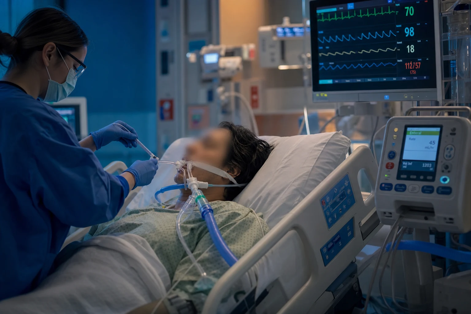
Pathophysiology: mechanisms of aspiration in the ICU [1]
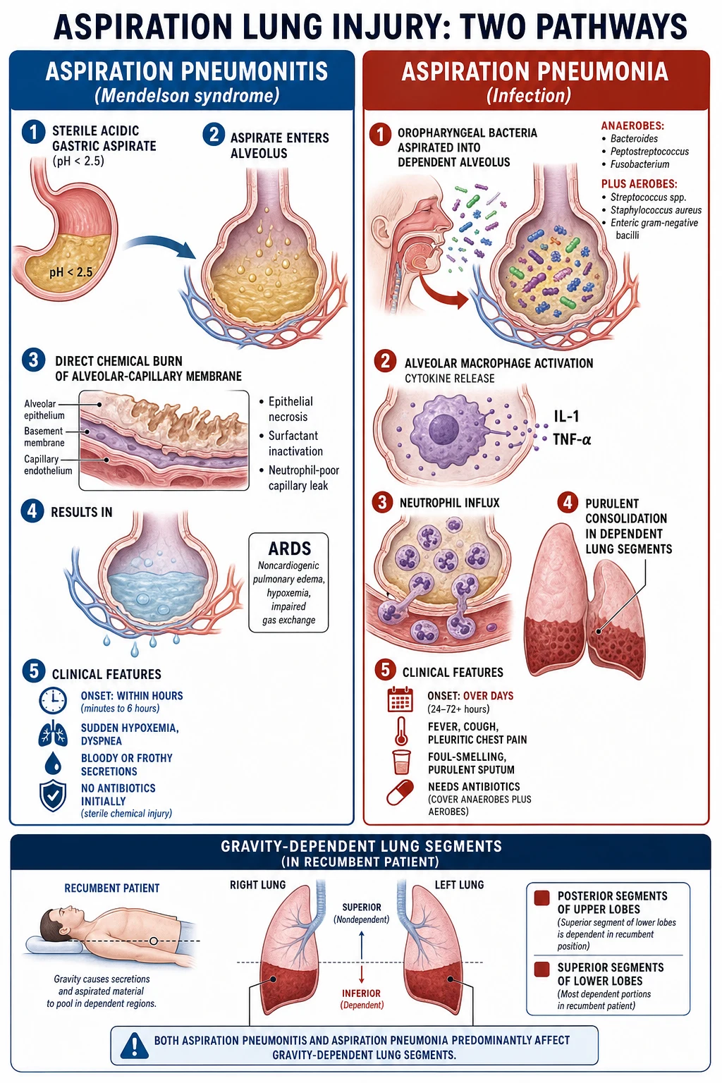
Aspiration in the critically ill patient occurs by one of three mechanisms, and each has different implications for prevention.[1]
- Microaspiration around the ETT cuff — the dominant mechanism of ventilator-associated pneumonia (VAP). Even an apparently well-sealed high-volume low-pressure cuff forms longitudinal folds (channels) through which liquid tracks. Contaminated subglottic secretions pool above the cuff (the subglottic mucous pool) and seep past into the lower airway. This process is silent: there is no cough, because the cuff stents the glottis open and the tube abolishes the laryngeal sensory reflex.
- Macro-aspiration of gastric or oropharyngeal contents — the non-intubated or recently extubated patient with depressed consciousness, an impaired swallow, or active gastro-oesophageal reflux aspirates a larger volume. The clinical picture may be aspiration pneumonitis (chemical) or aspiration pneumonia (infective).
- Silent aspiration without a protective cough — common in the elderly, after stroke, after extubation, and in any neurologically impaired patient. Bedside clinical examination misses it; instrumental assessment (FEES or VFSS) is required to detect it confidently.[6]
Risk factors for aspiration
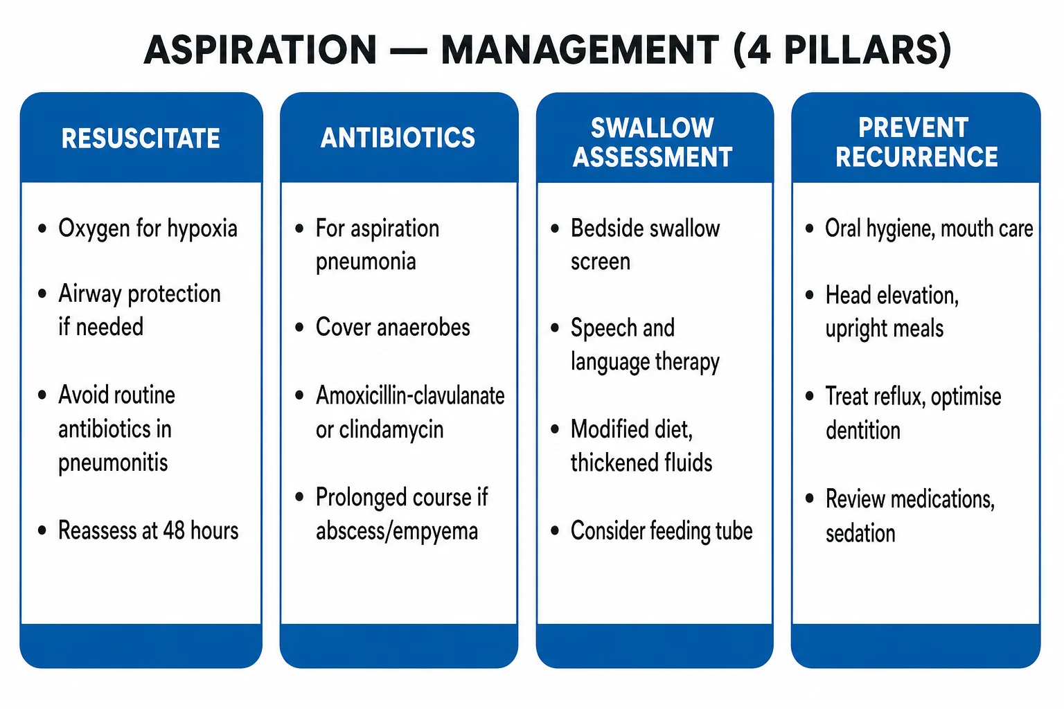
The risk factors cluster into four domains. The more domains that overlap in a single patient, the higher the aspiration risk — and the more aggressive the prevention bundle must be. The classic independent predictors were quantified in the Drakulovic randomised trial (below).[3]
Depressed consciousness
Airway reflexes lost
- GCS < 8 — loss of glottic closure and cough
- General anaesthesia and procedural sedation
- Alcohol intoxication and recreational drug overdose
- Post-ictal state after a seizure
- Opioid and benzodiazepine excess; hepatic encephalopathy
Neurological deficit
Swallow disordered
- Stroke — brainstem, lacunar, and large bilateral cortical infarcts
- Neuromuscular disease — myasthenia gravis, Guillain-Barre, ALS
- Dementia, Parkinson disease, advanced multiple sclerosis
- Critical illness polyneuromyopathy
- Recent extubation — laryngeal injury, vocal cord palsy
Airway device & ventilation
Defences bypassed
- Endotracheal tube — bypasses upper airway, stents glottis open
- Mechanical ventilation > 7 days
- Tracheostomy — cuff and altered laryngeal sensation
- Prolonged or repeated intubation; traumatic / difficult intubation
- Excessive sedation preventing daily awakening
Gastrointestinal
Reflux & stasis
- Nasogastric tube — splints the gastro-oesophageal junction open
- Gastroparesis, ileus, and GI obstruction
- Large gastric residual volumes; overfeeding
- Active vomiting; bulbar palsy with pooling
- Post-operative state; recent upper GI surgery
Patient factors
Background risk
- Age > 70 years
- COPD, diabetes mellitus, current smoker
- Malnutrition and low albumin
- Head and neck surgery or radiotherapy
- Poor oral hygiene and heavy oropharyngeal colonisation
The aspiration prevention bundle
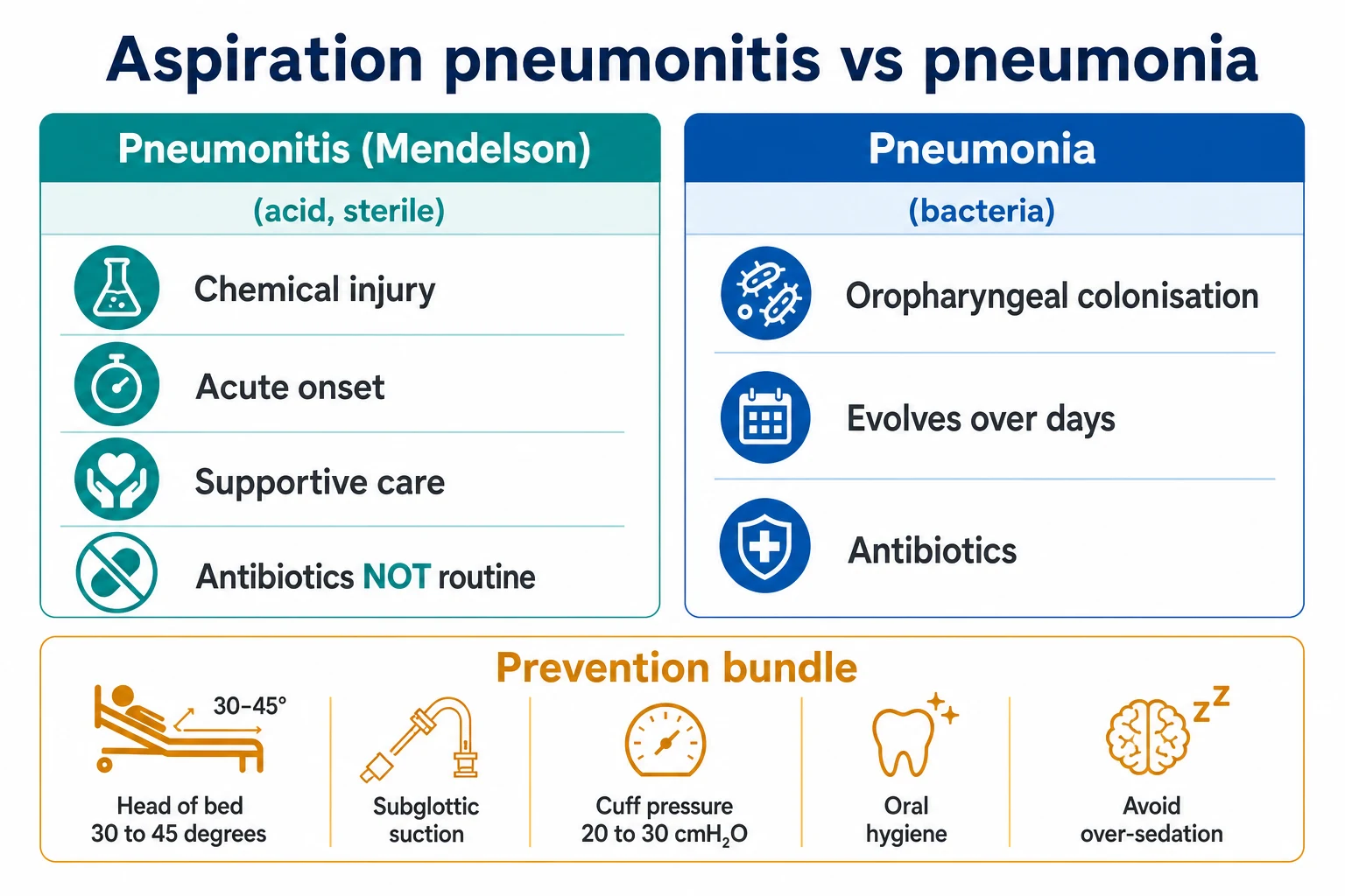
No single measure eliminates aspiration — the bundle works because each element targets a different point in the pathogenesis (reduce colonisation, reduce reflux, reduce microaspiration, reduce duration of exposure). Bundle all measures and audit compliance.[1][2]
Comprehensive aspiration prevention bundle
Avoid unnecessary intubation
Use NIV or high-flow nasal cannula for COPD exacerbation, cardiogenic pulmonary oedema, and immunocompromised respiratory failure. Every extra day of intubation raises VAP risk by about 1-3%. The most effective prevention is to never intubate, or to extubate as soon as safe.
Head of bed elevation 30-45 degrees
The simplest and most consistently effective single measure. Gravity opposes retrograde flow of gastric contents. Drakulovic 1999: semirecumbent vs supine reduced microbiologically-confirmed pneumonia from 23% to 5%. Audit compliance with a bed-angle indicator — it is the measure most often forgotten at night. Exceptions: severe hypotension, spinal precautions, prone positioning, pelvic traction.
Subglottic secretion drainage (SSD)
Use an ETT with a subglottic suction port (e.g. HI-LO Evac) for every patient expected to be ventilated > 48-72 h. Continuous or intermittent suction removes the contaminated mucous pool above the cuff. Lacherade 2010: intermittent SSD reduced VAP from 25.6% to 14.8% (42% relative reduction), with benefit in both early- and late-onset VAP.
Maintain cuff pressure 20-30 cmH2O
Check at least once per shift with a manometer — never estimate by palpation. Too low (< 20) allows channel leak and microaspiration. Too high (> 30) risks mucosal ischaemia, tracheal stenosis, and tracheo-oesophageal fistula. Cuff pressure drifts with position change, coughing, and changes in PEEP — re-check after turning the patient.
Oral care with chlorhexidine plus toothbrushing
0.12% or 0.2% chlorhexidine mouthwash every 8-12 h, plus mechanical toothbrushing every 12 h, suction pooled secretions before turning. Reduces oropharyngeal bacterial load. Note: universal chlorhexidine is controversial — some meta-analyses signal increased mortality in non-cardiac patients, and several guidelines have narrowed the recommendation. Apply as per local protocol.
Minimise sedation — daily SAT plus SBT
Daily Sedation Awakening Trial paired with a Spontaneous Breathing Trial reduces ventilator-days (the ABC trial, Girard). Fewer intubated days means fewer aspiration days. Pair with a delirium screen (CAM-ICU or ICDSC) and an early-mobilisation pathway.
Early mobilisation
Patients who sit out of bed or march on the spot within 72 h have shorter ventilation and fewer respiratory infections. Mobilisation also aids secretion clearance and gut motility.
Hand hygiene and infection control
WHO Five Moments for hand hygiene, alcohol rub before and after every patient contact, and sterile suction technique. Hand hygiene is the single most effective cross-infection control measure and prevents patient-to-patient spread of resistant organisms.
Structured enteral feeding protocol
Start enteral (not parenteral) feeding early. Use a prokinetic (metoclopramide or erythromycin) if gastric residuals are high, check gastric residual volumes, avoid overfeeding, elevate the head of bed, and consider continuous rather than bolus feeding in high-risk patients. Switch to post-pyloric (NJ) feeding if residuals remain high or reflux is severe.
Selective stress-ulcer prophylaxis
Give a PPI or H2 blocker only to genuinely high-risk patients (mechanical ventilation > 48 h, coagulopathy, shock, major burns, history of GI bleed). Universal prophylaxis raises gastric pH, increases gastric bacterial colonisation, and may increase VAP and C. difficile risk. The SUP-ICU trial found no mortality benefit from routine pantoprazole.
Minimising aspiration from enteral feeding
Enteral feeding is essential, but it is also an independent aspiration risk factor. The goal is to feed the gut, not the lung. [1]
Continuous infusion
Steady gastric delivery
- Lower peak gastric distension and residual volumes
- Probably less reflux and microaspiration in high-risk patients
- Easier to titrate to tolerance; preferred in severe critical illness
- Requires a feeding pump; less physiologic
- Residuals easier to interpret and trend
Bolus feeding
Intermittent gravity boluses
- More physiologic; frees the patient from a pump for mobilisation
- Higher transient gastric distension may trigger reflux
- Useful for stable, longer-term patients and rehabilitation
- Avoid in gastroparesis, high residuals, or severe reflux
- Smaller, more frequent boluses are better tolerated
Gastric (NG / OG)
First-line route
- Easier and faster to place at the bedside
- More physiologic; preserves stomach reservoir function
- Higher risk of reflux and microaspiration
- Residual volume monitoring guides tolerance
- Add a prokinetic if residuals persistently high
Post-pyloric (NJ)
For high-risk / intolerance
- Reduces reflux of gastric contents
- Preferred in severe reflux, gastroparesis, pancreatitis, high residuals
- Does NOT eliminate aspiration — oropharyngeal secretions are still aspirated
- Placement harder; often needs endoscopy or fluoroscopy
- Lower residual-volume monitoring needed
Enteral feeding protocol to reduce aspiration
Confirm tube position before every feed
Verify NG/NJ position with pH <= 5.5 aspirate or X-ray. Never feed through a malpositioned tube — pulmonary placement causes direct aspiration of formula.
Keep head of bed at 30-45 degrees during and after feeding
Never feed a patient flat unless absolutely contraindicated. Maintain elevation for at least 30-60 min after a bolus feed.
Check gastric residual volumes
Check every 4-6 h initially. Residuals < 250-500 mL are usually tolerated; hold or reduce the rate for higher volumes. Excessive residuals signal gastroparesis and predict reflux and aspiration.
Add a prokinetic for high residuals
Metoclopramide 10 mg IV QDS or erythromycin 250 mg IV QDS reduces residuals and aspiration. Combine for refractory intolerance. Reassess daily.
Prefer continuous infusion in the high-risk patient
Switch from bolus to continuous infusion for severe critical illness, gastroparesis, or known reflux. Reduce the rate rather than stopping feeds if intolerance develops.
Escalate to post-pyloric feeding if intolerance persists
Place an NJ tube if high residuals or reflux continue despite a prokinetic, or in severe pancreatitis, severe reflux, or documented aspiration of gastric feeds.
Avoid overfeeding
Do not exceed target energy delivery. Overfeeding increases gastric distension, reflux, hypercapnia, and hepatic dysfunction — and raises aspiration risk.
Swallow assessment
Any patient who has been intubated, has had a stroke, or has depressed consciousness must remain nil-by-mouth until swallow safety has been assessed. Up to 41% of critically ill adults have post-extubation dysphagia, and around a third aspirate silently — so a normal-looking patient can still be aspirating.[6]
Swallow screen and assessment pathway
Rest the patient before assessing
Wait at least 4 hours after extubation (and ideally 12-24 h) before a swallow assessment. Residual sedation, laryngeal oedema, and vocal-cord dysfunction all distort the result in the first hours after extubation.
Bedside pre-screen
Before any fluid: is the patient alert and able to sit upright at 30-90 degrees? Can they control saliva, produce a strong voluntary cough, and a clear voice? Excessive secretions, a wet or hoarse voice, or a weak cough = fail the pre-screen — keep nil-by-mouth and refer to speech and language therapy (SALT).
3-ounce (90 mL) water swallow test
If the pre-screen is passed, ask the patient to drink 90 mL of water continuously without interruption. FAIL if coughing or choking during or for 1 minute after, a wet-hoarse voice, or inability to finish the full volume. A fail mandates nil-by-mouth and formal SALT assessment.
Formal SALT bedside assessment
A speech and language therapist performs a structured bedside examination of oral control, laryngeal elevation, and trial textures (puriss, thickened and thin fluids, solids). Identifies most overt aspiration but still misses silent aspiration.
FEES — fibreoptic endoscopic evaluation of swallowing
A thin flexible endoscope passed trans-nasally gives a direct view of the larynx and pharynx during swallowing of dyed food and fluid. Performed at the bedside, portable, and the test of choice to detect silent aspiration and to guide diet modification.
VFSS — videofluoroscopic swallow study
A modified barium swallow under fluoroscopy in radiology. Gold-standard dynamic imaging of the oral, pharyngeal, and oesophageal phases, and quantifies residue and aspiration. Reserved for complex cases and to plan rehabilitation when FEES is inconclusive.
Reintroduce diet in stages
If the screen is passed, start on the safest texture (thickened fluids, soft puriss) and advance as tolerated. A failed screen means keep nil-by-mouth, feed via NG/NJ, and arrange SALT-led texture progression.
Bedside water swallow test
Quick screen
- Fast, cheap, no equipment — a good triage tool
- Detects overt aspiration (cough, wet voice)
- Misses silent aspiration
- 3-ounce / 90 mL continuous-sip version is the standard
- A pass does not guarantee safety — observe the first meals
FEES
Bedside instrumental
- Direct laryngeal and pharyngeal visualisation
- Detects silent aspiration and pooling
- Portable — performed at the ICU bedside
- Guides immediate texture and strategy modification
- Requires trained endoscopist and equipment
VFSS
Radiology gold standard
- Dynamic fluoroscopy of all swallow phases
- Best for complex anatomy and rehabilitation planning
- Requires transfer to radiology and radiation exposure
- Detects silent aspiration with high sensitivity
- Reserved for selected, complex cases
Post-extubation aspiration risk
Extubation removes a sealed airway and exposes a newly injured larynx — the perfect setup for aspiration. Post-extubation dysphagia is common, frequently silent, and independently associated with longer stay, pneumonia, and reintubation. [1]
Why extubation causes dysphagia
- Laryngeal injury from the tube — ulceration, granuloma, and oedema impair glottic closure.
- Vocal-cord dysfunction — palsy, arytenoid dislocation, or reduced mobility.
- Reduced laryngeal sensation from nerve compression and critical illness.
- Generalised weakness — critical illness polyneuromyopathy weakens the swallow and cough.
- Residual sedation and delirium in the first hours after extubation. [1]
Post-extubation dysphagia — the numbers
The cuff leak test
The cuff leak test predicts post-extubation upper-airway obstruction from laryngeal oedema, which in turn drives stridor, reintubation, and swallow difficulty. [1]
Pre-extubation airway and aspiration risk assessment
Assess the cuff leak before planned extubation
Perform a cuff leak test on every high-risk patient (prolonged intubation, traumatic intubation, female, large tube, head/neck surgery, known laryngeal pathology). A low or absent leak signals laryngeal oedema.
Give steroids if the leak is low
Methylprednisolone 20 mg IV or dexamethasone 4-8 mg, starting at least 4 hours (ideally 12 hours) before extubation. Jaber 2009 and Kuriyama 2017: in cuff-leak-selected high-risk patients this roughly halved stridor and reintubation, with a number-needed-to-treat around 5-9. There is no benefit in unselected patients.
Extubate to appropriate respiratory support
Plan the post-extubation airway: high-flow nasal cannula or NIV for patients at risk of respiratory failure. Have reintubation equipment and a skilled operator immediately available.
Keep nil-by-mouth and rest the larynx
No oral intake for at least 4 hours, and ideally 12-24 hours, after extubation. Let residual sedation clear and oedema settle before challenging swallow.
Perform a swallow screen before feeding
Bedside pre-screen, then a 3-ounce water swallow test. A failed screen keeps the patient nil-by-mouth and triggers formal SALT assessment and FEES.
Reintroduce diet in stages under SALT guidance
Start with the safest texture (thickened fluids, soft puriss) and advance as tolerated. Continue NG or NJ feeding until safe oral intake is established.
Watch for stridor
Inspiratory stridor within minutes to hours of extubation signals laryngeal oedema — sit the patient up, give nebulised adrenaline and helium-oxygen if available, and prepare for reintubation.
Tracheostomy and decannulation
For the tracheostomied patient, aspiration risk persists until decannulation and beyond. [1]
Tracheostomy swallow and decannulation pathway
Downsize and deflate the cuff
Progress to a cuffless or fenestrated tube once the patient tolerates ventilation off the ventilator. Cuff deflation restores trans-laryngeal airflow and glottic function.
Use a one-way speaking valve (Passy-Muir)
A speaking valve restores upper-airway airflow, subglottic pressure, and sensation, which improves swallow and cough and helps assess readiness for decannulation.
Assess swallow with the cuff down
With a speaking valve in place, perform a swallow screen and SALT assessment. A patient who cannot manage secretions with the cuff down is not ready to decannulate.
Capping and resting trials
Cap the tube for progressively longer periods (12-24 h continuous) while the patient breathes normally and manages secretions. Successful capping plus effective cough and safe swallow is a precondition for decannulation.
Decannulate once safe
Decannulate when the patient tolerates continuous capping, has an effective cough, manages secretions, and has passed a swallow assessment. Cover the stoma and observe for 24-48 h.
Landmark trials
Drakulovic 1999 — semirecumbent vs supine body position (Lancet; PMID 10584721)
Design
Single-centre randomised trial stopped at planned interim analysis; 86 intubated, mechanically ventilated patients assigned to semirecumbent (n=39) or supine (n=47) body position
Primary outcome
Nosocomial pneumonia (clinical plus quantitative microbiological criteria). Microbiologically-confirmed pneumonia: 5% semirecumbent vs 23% supine; clinically suspected 8% vs 34%
Independent risk factors
Supine position OR 6.8; enteral nutrition OR 5.7; mechanical ventilation >= 7 days OR 10.9; GCS < 9. Worst combination — enteral nutrition plus supine position: 50% developed pneumonia
Clinical bottom line
Head-of-bed elevation is the simplest, cheapest, and most consistently effective VAP prevention measure. This is the trial that made 30-45 degrees a universal standard and quantified enteral nutrition, long ventilation, and depressed consciousness as independent aspiration risk factors.
Lacherade 2010 — intermittent subglottic secretion drainage (AJRCCM; PMID 20522796)
Design
Multicentre (4 French ICUs) randomised controlled trial; 333 intubated patients expected to require > 48 h ventilation, randomised to intermittent subglottic secretion drainage (n=169) vs no drainage (n=164)
Primary outcome
Overall incidence of microbiologically confirmed VAP: 14.8% (SSD) vs 25.6% (control); relative risk reduction 42.2% (95% CI 10.4-63.1)
Subgroups
Significant reduction in BOTH early-onset VAP (1.2% vs 6.1%) and late-onset VAP (18.6% vs 33.0%). No significant difference in ventilation duration or hospital mortality
Clinical bottom line
Intermittent subglottic secretion drainage significantly reduces VAP, including late-onset VAP, in patients ventilated > 48 h. Use a subglottic-suction ETT for every patient expected to be ventilated beyond 48-72 h.
Jaber 2009 — steroids for post-extubation stridor and reintubation (Critical Care; PMID 19344515)
Design
Meta-analysis of 7 prospective, double-blind randomised trials; 1846 mechanically ventilated adults, 949 receiving steroids before extubation
Key findings
Overall: steroids reduced reintubation (RR 0.58, NNT 28) and stridor (RR 0.48, NNT 11). The effect was concentrated in high-risk patients selected by a low cuff leak: reintubation RR 0.38 (NNT 9) and stridor RR 0.40 (NNT 5)
Timing
Benefit required administration at least 4 hours before extubation. No clear benefit when high-risk patients were not selected
Clinical bottom line
Steroids prevent post-extubation stridor and reintubation — but ONLY in patients selected by the cuff leak test and ONLY if given at least 4 hours before extubation. Do not give steroids routinely to every patient before extubation.
Kuriyama 2017 — prophylactic corticosteroids before extubation (Chest; PMID 28232056)
Design
Updated systematic review and meta-analysis of 11 randomised trials; 2472 mechanically ventilated adults, through February 2016
Key findings
Prophylactic steroids reduced post-extubation airway events (RR 0.43) and reintubation (RR 0.42). In cuff-leak-selected high-risk patients the effect was larger: airway events RR 0.34 and reintubation RR 0.35. No benefit in unselected patients
Safety
Adverse events were rare
Clinical bottom line
Confirms and extends Jaber 2009: select patients for steroids using the cuff leak test. Targeted prophylaxis before elective extubation halves airway complications in genuinely high-risk patients.
McIntyre 2021 — incidence of post-extubation dysphagia (Australian Critical Care; PMID 32739246)
Design
Systematic review and meta-analysis of 38 studies (peer-reviewed and grey literature) through October 2019; 5798 patient events, 1957 dysphagic episodes; random-effects model
Primary outcome
Pooled weighted incidence of post-extubation dysphagia 41% (95% CI 33-50%). Reported range across studies 3-62% owing to heterogeneity in assessment methods and timing
Silent aspiration
Of the patients with dysphagia, about 36% aspirated silently (95% CI 22-50%)
Clinical bottom line
Post-extubation dysphagia is common (about 4 in 10 extubated adults) and a third aspirate silently. Screen every newly extubated patient before feeding, and use FEES or VFSS when bedside screening is equivocal or concern persists.
Aspiration prevention — effect sizes at a glance
What each intervention delivers
Additional exam pearls
Exam practice
SAQ — Post-extubation dysphagia and aspiration risk
10 minutes · 10 marks
A 72-year-old woman is admitted to ICU with severe community-acquired pneumonia requiring 6 days of mechanical ventilation via a 7.5 mm endotracheal tube. Intubation was difficult (two attempts). She is now alert and triggering well on pressure support. The cuff leak test returns a leak of 80 mL. You plan extubation for tomorrow morning.
Additional red flags
Prevention measures
Evidence-based aspiration prevention
Head of bed elevation 30-45°
Simplest and most consistently effective measure. Reduces microaspiration by gravity. Audit compliance (often suboptimal). Exception: severe hypotension, spinal precautions, prone positioning. Use a bed angle indicator. Nurse in semi-recumbent position at all times.
Daily SAT + SBT
Sedation Awakening Trial + Spontaneous Breathing Trial. Reduces days of mechanical ventilation → fewer days intubated → lower VAP risk. ABC trial (Girard): SAT+SBT pairing reduced mortality and ventilation days. Stop/reduce sedation daily → assess readiness → trial off ventilator → extubate when ready.
Oral care with chlorhexidine
0.12% or 0.2% chlorhexidine mouthwash every 8-12 hours. Reduces oropharyngeal bacterial load → fewer bacteria aspirated. Cochrane: reduces VAP incidence. Brush teeth every 12h. Suction oropharyngeal secretions before turning. Moisturise lips. Note: routine chlorhexidine may increase mortality in some populations (controversial — some guidelines have removed universal recommendation).
Subglottic secretion drainage
Use ETT with subglottic suction port (HI-LO Evac tube). Continuously or intermittently drains contaminated secretions above the cuff. Reduces microaspiration. Overview of systematic reviews: significant VAP reduction. Both continuous and intermittent effective. Particularly beneficial for patients expected to be ventilated >48-72h.
ETT cuff pressure monitoring
Maintain cuff pressure 20-30 cmH2O. Too low (<20): microaspiration through channels ("channel leak"). Too high (>30): mucosal ischaemia → tracheal stenosis, tracheo-oesophageal fistula. Check cuff pressure at least once per shift using manometer (not by palpation — unreliable). Adjust as needed. Be aware: cuff pressure varies with changes in body position, PEEP, intrathoracic pressure.
Clinical pearls
Red flags
References
- [1]Torres A, et al. Ventilator-associated pneumonia in adults: a narrative review Intensive Care Med, 2020.PMID 32157357
- [2]Klompas M, et al. Notum palmitoleoyl-protein carboxylesterase regulates Fas cell surface death receptor-mediated apoptosis via the Wnt signaling pathway in colon adenocarcinoma Bioengineered, 2021.PMID 34402722
- [3]Drakulovic MB, Torres A, Bauer TT, Nicolas JM, Nogue S, Ferrer M Supine body position as a risk factor for nosocomial pneumonia in mechanically ventilated patients: a randomised trial Lancet, 1999.PMID 10584721
- [4]Lacherade JC, De Jonghe B, Guezennec P, et al. Intermittent subglottic secretion drainage and ventilator-associated pneumonia: a multicenter trial Am J Respir Crit Care Med, 2010.PMID 20522796
- [5]McClave SA, Taylor BE, Martindale RG, et al. Guidelines for the Provision and Assessment of Nutrition Support Therapy in the Adult Critically Ill Patient: Society of Critical Care Medicine (SCCM) and American Society for Parenteral and Enteral Nutrition (A.S.P.E.N.) JPEN J Parenter Enteral Nutr, 2016.PMID 26773077
- [6]McIntyre M, Doeltgen S, Dalton N, Koppa M, Chimunda T Post-extubation dysphagia incidence in critically ill patients: A systematic review and meta-analysis Aust Crit Care, 2021.PMID 32739246
- [7]Jaber S, Jung B, Chanques G, Bonnet F, Marret E Effects of steroids on reintubation and post-extubation stridor in adults: meta-analysis of randomised controlled trials Crit Care, 2009.PMID 19344515
- [8]Kuriyama A, Umakoshi N, Sun R Prophylactic Corticosteroids for Prevention of Postextubation Stridor and Reintubation in Adults: A Systematic Review and Meta-analysis Chest, 2017.PMID 28232056