ICU · Antimicrobial Stewardship
Acute severe community-acquired pneumonia: nosocomial complications and prevention
Also known as Nosocomial infections in CAP patients · Superinfection prevention · ICU-acquired infections in pneumonia
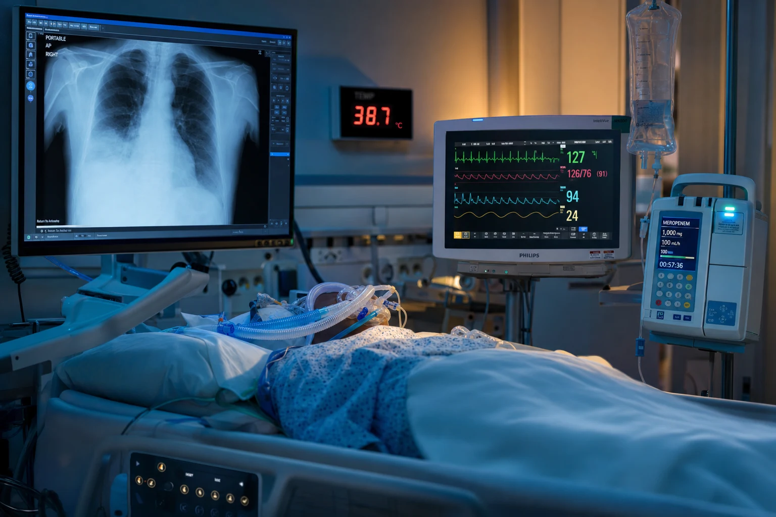
CAP patients admitted to ICU are at high risk for nosocomial (hospital-acquired) superinfections during their stay. Common: VAP (ventilator-associated pneumonia — 1, develops 48h after intubation), CRBSI (catheter-related bloodstream infection), C. difficile colitis, UTI (catheter-associated), surgical site infection (if surgery performed). Risk factors: prolonged ventilation, broad-spectrum antibiotics, immunosuppression, severity of illness. Prevention: VAP bundle (head elevation, daily SAT+SBT, oral chlorhexidine, subglottic suction, cuff pressure), CRBSI bundle (full barrier precautions, chlorhexidine skin prep, daily review of line necessity), antibiotic stewardship (minimise duration, de-escalate), early mobilisation, hand hygiene.
On this page & tools
Your progress
Saved locally on this device.
Target exams
Red flags
Clinical pearls
Red flags
Nosocomial pneumonia: definitions
Pneumonia acquired in hospital is classified by where and how the patient acquired it. The terminology matters because the likely pathogens and empiric antibiotic choices differ from community-acquired pneumonia (CAP). The 2005 ATS/IDSA guidelines introduced the term HCAP (healthcare-associated pneumonia) for patients with recent hospitalisation, residence in a nursing home, home wound care, or recent IV antibiotics/chemotherapy — on the assumption that these patients harboured resistant organisms.[3]
HAP
Hospital-acquired pneumonia
- New pneumonia >48 hours after hospital admission
- NOT intubated at onset (distinguishes from VAP)
- Incidence ~3-5 per 1,000 admissions, ~5-10 per 1,000 patient-days
- Pathogens: enteric Gram-negatives (E. coli, Klebsiella, Enterobacter), S. aureus (incl. MRSA)
- Mortality 15-25% (higher than CAP)
- Empiric therapy guided by local antibiogram and MDR risk factors
VAP
Ventilator-associated pneumonia
- New pneumonia >48 hours AFTER endotracheal intubation
- The #1 nosocomial infection in the ICU
- Incidence ~1-3 per 1,000 ventilator-days (was higher before prevention bundles)
- Attributable mortality ~13%, attributable cost and length-of-stay substantial
- Early-onset (<4 days): MSSA, pneumococcus, H. influenzae, antibiotic-sensitive GNs
- Late-onset (≥4 days): MDR Pseudomonas, Acinetobacter, ESBL Klebsiella, MRSA
HCAP
Healthcare-associated — ABANDONED
- Defined in 2005 ATS/IDSA guidelines — nursing-home residents, recent hospitalisation, home IV therapy/wound care, dialysis
- Premised on assumption of high MDR risk → broad empiric antibiotics
- REMOVED in 2016 IDSA/ATS guidelines — risk factors were poor predictors of MDR
- Routine broad MDR coverage caused overtreatment and harm
- Do NOT use the HCAP label — base empiric therapy on local antibiogram + patient risk factors instead
Why CAP patients are uniquely vulnerable
A patient admitted to the ICU for severe CAP is the prototype high-risk host for nosocomial superinfection because several risk factors converge simultaneously:[1]
- Prolonged mechanical ventilation (the dominant risk — each extra ventilator day raises VAP risk ~1-3%)
- Broad-spectrum antibiotics disrupt normal oropharyngeal and gut flora → colonisation with resistant organisms
- Impaired consciousness / sedation → loss of the gag and cough reflexes → microaspiration
- ETT cuff + supine position → pooled secretions bypass the cuff
- Severity of illness and immune paralysis (critical-illness-associated immunosuppression)
- Invasive devices: central lines (CRBSI), urinary catheters (CAUTI), NG tubes (sinusitis/aspiration)
Epidemiology and attributable burden
Nosocomial infection burden in ICU
VAP develops in roughly 8-28% of mechanically ventilated patients depending on population and prevention-bundle adherence, with a median onset around day 5 of ventilation. The daily hazard is highest in the first week and accrues at roughly 1% per ventilator-day in the early period — the rationale that "the fastest way to prevent VAP is to extubate." Multiple observational studies estimate attributable mortality of ~13% (the excess death beyond the underlying critical illness), with markedly worse outcomes when the causative organism is MDR or when initial empiric therapy is inappropriate.[7][2]
Pathogenesis of VAP
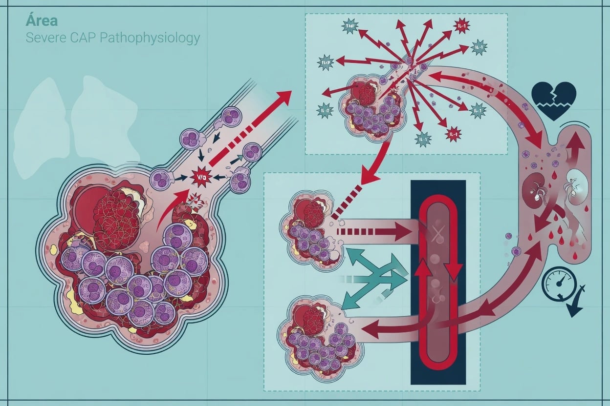
VAP is fundamentally a disease of microaspiration of contaminated oropharyngeal secretions around the endotracheal tube (ETT) cuff. The ETT does not seal the airway — it is a foreign body that facilitates bacterial entry while paralysing the airway's mechanical defences.[2]
The four-step pathogenic cascade
How VAP develops — the pathogenic sequence
1. Colonisation of the oropharynx
Within hours of intubation, the oropharynx is colonised by enteric Gram-negatives and S. aureus. Broad-spectrum antibiotics and critical illness eradicate normal commensals (viridans streptococci), allowing pathogenic overgrowth. Gastric alkalinisation (PPIs, enteral feeds) allows gastric bacterial overgrowth that refluxes upward.
2. Formation of the ETT biofilm
Bacteria adhere to the polyvinyl-chloride ETT surface and secrete an exopolysaccharide matrix (biofilm) within 24h. Biofilm is resistant to both antibiotics and host immunity (bacteria 10-1000x more resistant inside). Fragments of biofilm embolise distally with each breath, inoculating the lower airway. Biofilm persists despite appropriate antibiotics.
3. Microaspiration around the cuff
The ETT cuff cannot form a perfect seal. Secretions pool in the subglottic space ("silent aspiration") and leak past folds in the cuff (longitudinal folds, or channels formed when cuff pressure fluctuates). Supine position, low cuff pressure, and patient transport increase leakage. Each intubated patient aspirates up to ~50% of pooled secretions.
4. Overwhelm host defences → pneumonia
Impaired mucociliary clearance (ETT bypasses, sedation suppresses cough), reduced neutrophil function (critical-illness immunoparalysis), and bacterial virulence overwhelm residual host defences. Inoculum reaches the alveoli → alveolar macrophage activation → neutrophil influx → consolidation = VAP. Each intubated day repeats the cycle; risk rises >1% per day.
Risk factors for VAP
Modifiable
Target the prevention bundle here
- Supine positioning (Drakulovic: supine > doubles VAP risk)
- Low cuff pressure (<20 cmH2O) → leakage around cuff
- Prolonged ventilation (each day adds ~1-3% risk)
- Over-sedation — abolishes cough and delays weaning
- Re-intubation (disturbs cuff seal, aspirates gastric contents)
- Enteral feeding via large-bore NG (reflux, gastric overdistension)
- Inadequate hand hygiene by staff
Non-modifiable
Identify high-risk patients
- Age >60, chronic lung disease, ARDS
- Immunosuppression, burns, trauma (especially head injury)
- Severity of illness (APACHE II), coma
- Emergency intubation / intubation outside ICU
- Pre-existing oropharyngeal/gastric colonisation with MDR organisms
- Season (winter — more viral co-pathogens)
Microbiology: early vs late VAP

The single most important microbiological principle: the likely pathogens depend on the time since intubation and on prior antibiotic exposure. Early VAP (within ~4 days) reflects aspiration of the patient's own community/commensal flora; late VAP reflects the MDR organisms that have colonised the ICU environment and the patient's airway under antibiotic pressure.[3]
Early-onset VAP
< 4 days of ventilation
- Usually aspiration of endogenous community-acquired flora
- Methicillin-sensitive S. aureus (MSSA)
- Streptococcus pneumoniae
- Haemophilus influenzae
- Antibiotic-sensitive enteric Gram-negatives (E. coli, Klebsiella, Proteus)
- Anaerobes (if frank aspiration)
- Usually NOT MDR → narrower empiric therapy (e.g. ceftriaxone, ampicillin-sulbactam, or respiratory fluoroquinolone)
- Better prognosis than late-onset VAP
Late-onset VAP
≥ 4 days of ventilation
- Reflects ICU-acquired, antibiotic-selected MDR organisms
- Pseudomonas aeruginosa (most common late pathogen)
- Acinetobacter baumannii (highly MDR; carbapenem-resistant strains emerging)
- Klebsiella / Enterobacter / Serratia (ESBL- and carbapenemase-producers)
- MRSA (methicillin-resistant Staphylococcus aureus)
- Stenotrophomonas maltophilia (often dually resistant)
- Requires broad empiric MDR cover: anti-Pseudomonal beta-lactam + MRSA agent ± aminoglycoside/colistin; de-escalate on culture results
- Higher mortality, longer ICU stay
The VAP prevention bundle
No single intervention eliminates VAP; the evidence base favours a multimodal "bundle" of cheap, low-risk measures applied together. The original Institute for Healthcare Improvement (IHI) VAP bundle and subsequent society guidelines converge on a core set of elements.[7][2]
The five core prevention elements
VAP prevention bundle — apply every element, every day
1. Semirecumbent position 30-45°
Elevate the head of the bed 30-45° unless contraindicated (spinal precautions, hypotension, prone ventilation). Drakulovic (Lancet 1999): supine vs semirecumbent — VAP 34% vs 8% (p=0.003). Supine position is an independent risk factor; the effect is largest during enteral feeding. Document HOB angle at every turn.
2. Daily sedation interruption (SAT) + spontaneous breathing trial (SBT)
Wake the patient daily (if safe) and assess readiness to wean. Coordinated SAT+SBT reduces duration of ventilation and VAP. The fastest way to prevent VAP is to extubate. Avoid over-sedation (abolishes cough, promotes pooling).
3. Oral care with chlorhexidine
0.12-2% chlorhexidine oral rinse/gel every 8-12h reduces oropharyngeal colonisation. Most benefit in cardiac surgery; benefit in general ICU is debated and recent meta-analyses question mortality signal, but it remains widely recommended. Mechanical tooth-brushing is an additive effect.
4. Subglottic secretion drainage (SSD) ETT
Use an ETT with a separate dorsal lumen above the cuff to continuously aspirate pooled subglottic secretions. Reduces early VAP (~50% RRR) especially in patients ventilated >48-72h. Cost-prevents at high VAP incidence. Not suitable for all patients ( cuff seal issues, suction trauma).
5. Maintain cuff pressure 20-30 cmH2O
Check cuff pressure at least every 8h. <20 → microaspiration; >30 → tracheal mucosal ischaemia and stenosis. Use a cuff-pressure manometer, not pilot-balloon palpation. Polyurethane/thin cuffs and cuff-shape (tapered) designs further reduce leakage.
6. Hand hygiene + gloves
WHO '5 Moments for Hand Hygiene'. The single most effective, lowest-cost infection-control measure. Staff hand contamination is the main route of cross-colonisation between patients. Monitor and feedback compliance.
7. DVT and stress ulcer prophylaxis
These are part of the original "ventilator bundle" not because they prevent VAP directly, but because the bundled approach reduces all-cause ICU morbidity. Stress ulcer prophylaxis: give only for proven indications (mechanical ventilation >48h OR coagulopathy) — unnecessary PPIs increase C. diff and may increase VAP.
8. Early mobilisation and minimising sedation
Mobilise as soon as haemodynamically stable. Reduces ICU-acquired weakness, delirium, duration of ventilation — and therefore VAP. Pair with SAT/SBT and a light sedation target (RASS 0 to -1).
Bundle element
Effect / evidence
- Semirecumbent 30-45° — RCT-level, large effect (Drakulovic 1999)
- Daily SAT + SBT — reduces ventilation duration (ABC, Awakening and Breathing Controlled trial)
- Subglottic secretion drainage — ~50% RRR in early VAP, meta-analysis supported
- Cuff pressure 20-30 cmH2O — mechanistic + observational
- Oral chlorhexidine — reduces oropharyngeal colonisation; mortality signal debated
- Hand hygiene (5 Moments) — reduces cross-transmission of MDR organisms
Adjunctive / controversial
Selective use
- Selective digestive decontamination (SDD) / oropharyngeal decontamination (SOD) — reduces VAP and ICU mortality in some settings, but resistance-selection and C. diff concerns limit adoption outside low-resistance regions
- Silver-coated ETT (North American Silver-Coated Endotracheal Tube trial) — delayed VAP onset but no mortality benefit, costly
- Probiotics — reduce VAP in some meta-analyses; quality of evidence low
- Routine stress ulcer prophylaxis — only for ventilation >48h or coagulopathy
- Kinetic beds / continuous lateral rotation — modest VAP reduction, mainly neuro/trauma
- Early tracheostomy — does NOT reduce VAP (TracMan, S&P trials)
Does NOT prevent VAP
Discard these
- Routine sucralfate over PPI — no VAP advantage
- Gastric overfeeding / large boluses — may increase reflux-aspiration
- Scheduled ETT changes — biofilm reforms; changing tubes does not reduce VAP
- Aerosolised antibiotics for prophylaxis — resistance, no routine role
- Strict glucose control 81-108 (NICE-SUGAR) — hypoglycaemia harm, no VAP benefit
Diagnosis of VAP
VAP is a clinical diagnosis supported by microbiology — there is no single gold-standard test. The 2016 guideline does not mandate any one strategy; both invasive (bronchoscopic) and non-invasive (tracheal aspirate) sampling are acceptable, provided results are interpreted with the clinical picture and used to de-escalate antibiotics.[3]
Clinical criteria
The diagnosis requires a new or progressive infiltrate on imaging plus at least two of: fever >38°C, leucopenia (<4) or leucocytosis (>12), and purulent respiratory secretions. Sensitivity is high but specificity is modest — atelectasis, pulmonary oedema, PE, ARDS and drug reactions all mimic VAP. [1]
Clinical Pulmonary Infection Score (CPIS)
The CPIS (Pugin, 1991) combines six domains — fever, leucocyte count, secretions, oxygenation (PaO2/FiO2), radiography, and semi-quantitative tracheal-aspirate culture — into a score from 0 to 12.[6] A CPIS >6 supports VAP.
| CPIS domain | 0 points | 1 point | 2 points |
|---|---|---|---|
| Temperature (°C) | 36.5-38.4 | 38.5-38.9 | ≤36 or ≥39 |
| Leucocytes (x10&sup9;/L) | 4-11 | <4 or >11 | <4 or >11 + bands ≥50% |
| Tracheal secretions | None | Non-purulent | Purulent |
| PaO2/FiO2 (mmHg) | >240 or ARDS | — | ≤240 (no ARDS) |
| Chest radiograph | No infiltrate | Diffuse/patchy | Localised |
| Semi-quantitative culture | 0 / pathogenic | 1+ / pathogenic | 2+ / pathogenic |
Qualitative vs quantitative sampling
Non-invasive: endotracheal aspirate (qualitative/semi-quant)
- Suction catheter through the ETT; sample sent for Gram stain + culture
- Simple, cheap, no bronchoscopy, minimal risk
- High sensitivity (~75-90%) — a NEGATIVE aspirate usefully EXCLUDES VAP
- Low specificity (~70%) — oropharyngeal contamination overestimates pathogens
- Best used as a "rule-out" and to guide de-escalation
Invasive: bronchoalveolar lavage (BAL) / protected specimen brush (PSB)
- Bronchoscopically-directed or "blind" mini-BAL into the affected segment
- Quantitative culture thresholds: BAL >10&sup4; CFU/mL (PSB >10³)
- Higher specificity (~80-85%) — better discrimination of true pathogens
- More expensive, needs expertise, transient hypoxaemia/bleeding risk
- No consistent mortality benefit vs non-invasive strategy (meta-analyses)
Choosing a strategy
- Either strategy is acceptable (2016 guideline); standardise within your unit
- Obtain a lower-airway sample BEFORE starting or changing antibiotics
- Use results to DE-ESCALATE therapy, not to decide whether to treat
- Rapid molecular tests (e.g. BioFire, MRSA PCR) can guide early narrowing
- Biomarkers (procalcitonin, CRP, sTREM-1) are adjuncts, not sole decision-makers
Antibiotic therapy and duration
Empiric therapy principles
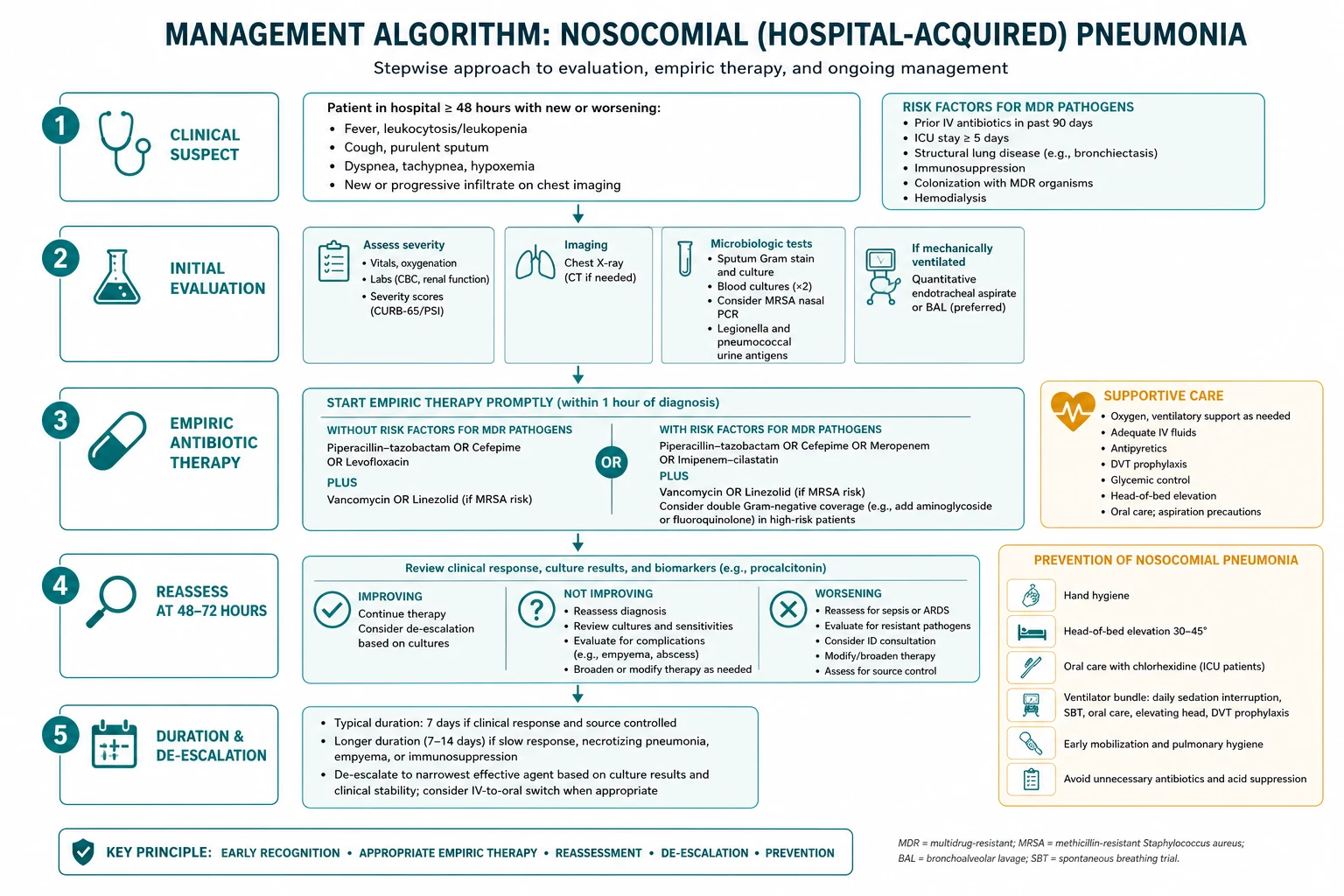
Empiric therapy must cover the likely MDR pathogens based on time since intubation, prior antibiotics, local antibiogram, and unit resistance patterns.[3]
- Early VAP (<4 days), no MDR risk factors: ceftriaxone, ampicillin-sulbactam, or a respiratory fluoroquinolone (cover MSSA, pneumococcus, sensitive enteric GNs).
- Late VAP (≥4 days) or any MDR risk factor: two anti-Pseudomonal agents of different classes (e.g. piperacillin-tazobactam or cefepime or meropenem, PLUS an aminoglycoside or fluoroquinolone) PLUS MRSA cover (vancomycin or linezolid) if MRSA risk factors exist.
- Carbapenem-resistant organisms: colistin or polymyxin B ± a second agent; consider high-dose meropenem extended-infusion if MIC allows.
De-escalate to the narrowest effective agent once culture and sensitivity results return (typically 48-72h). Inappropriate initial therapy (failure to cover the causative organism) doubles VAP mortality — the most important reason to err broad initially, then narrow.[3]
How long to treat: the PNEUMA trial
The PNEUMA trial (Chastre et al., JAMA 2003) is the landmark study establishing short-course therapy for VAP. It randomised 401 patients with microbiologically-confirmed VAP to 8 vs 15 days of appropriate antibiotics.[4]
PNEUMA trial — 8 vs 15 days of antibiotics for VAP
Interpretation: Among patients with VAP caused by non-lactose-fermenting Gram-negatives (Pseudomonas, Acinetobacter, Stenotrophomonas), recurrence was higher with the 8-day course, prompting some experts to extend treatment to 10-14 days for these pathogens. Otherwise, an 8-day course is non-inferior to 15 days and reduces antibiotic exposure — the foundation of the 2016 guideline's recommendation for a 7-day course for both HAP and VAP (clinical response permitting).[3][4]
Beyond pneumonia: other ICU-acquired infections
VAP is the most visible nosocomial infection in a ventilated CAP patient, but the same risk factors — invasive devices, broad antibiotics, immunoparalysis — drive a cluster of related infections that must be screened for whenever a CAP patient develops new fever or inflammatory markers. [1]
| Infection | Source | Key diagnostic step | Prevention bundle |
|---|---|---|---|
| CRBSI / CLABSI | Central venous catheter | Paired blood cultures (peripheral + from line); dwell time >5 d | Maximal barrier insertion, chlorhexidine prep, daily line necessity review, remove when not essential |
| Catheter-associated UTI (CAUTI) | Urinary catheter | Urine culture >10³ CFU/mL + symptoms; avoid treating asymptomatic bacteriuria | Avoid catheter; remove ASAP; closed drainage; sterile insertion |
| C. difficile infection | Antibiotic disruption of gut flora | Stool toxin EIA/NAAT (GDH + toxin); send for ANY new diarrhoea | Antimicrobial stewardship; avoid unnecessary PPIs; isolate promptly |
| Surgical site infection | Operative wound (if surgery) | Wound inspection, culture of discharge | Prophylactic antibiotic timing, normoglycaemia, normothermia |
| Sinusitis (nosocomial) | NG/ETT obstructing sinus drainage | CT sinuses + sinus aspiration cultures | Use orogastric over nasogastric tubes; semirecumbent position |
| Decubitus ulcer infection | Pressure injury | Wound swab/biopsy; deep tissue in sepsis | Regular turning, pressure-relieving mattress, skin care, nutrition |
Management algorithm summary
Approach to suspected nosocomial infection in a ventilated CAP patient
1. Recognise the trigger
New fever >38°C, rising inflammatory markers (CRP/procalcitonin), purulent secretions, new/worsening oxygenation, new infiltrate on CXR, or haemodynamic instability. Do NOT assume it is infection — consider PE, drug fever, ARDS, atelectasis, pancreatitis.
2. Localise the source
Examine the patient: chest (auscultate, review CXR/CT), central line site and duration, urine output and catheter, abdomen (distension, diarrhoea → C. diff), wounds, sinuses. Send paired blood cultures (peripheral + each lumen of lines in situ >48h).
3. Sample before antibiotics
For suspected VAP: endotracheal aspirate or BAL for Gram stain and culture BEFORE starting/changing antibiotics. Send C. diff toxin for any new diarrhoea. Urine culture only if symptomatic. Consider rapid molecular panels.
4. Start empiric broad therapy
Cover likely MDR organisms based on time since intubation, prior antibiotics and local antibiogram. Two anti-Pseudomonals + MRSA cover if late VAP or MDR risk. Inappropriate initial therapy doubles mortality — go broad first.
5. Reassess at 48-72 hours
Review cultures, sensitivities and clinical course. De-escalate to the narrowest effective agent. Stop MRSA cover if no MRSA. If no infection confirmed and patient improving, consider stopping antibiotics entirely (antibiotic time-out).
6. Treat for the shortest effective duration
7-8 days for most VAP/HAP (PNEUMA trial). Extend to 10-14 days only for non-lactose-fermenting Gram-negatives, slow clinical response, undrained foci, or bacteraemia. Procalcitonin can support stopping decisions.
7. Reinforce the prevention bundle
Confirm head-of-bed 30-45°, daily SAT/SBT, oral care, cuff pressure 20-30, subglottic suction if available, line removal review, hand hygiene. Address modifiable risk factors to prevent recurrence and the next infection.
8. Source control and de-escalation
Remove infected lines (replace at new site if still needed), drain collections, relieve obstruction. Continue stewardship rounds to keep antibiotic pressure — and thus resistance and C. diff risk — as low as possible.
Prognosis
Outcomes after nosocomial infection in ICU
Prognostic modifiers: causative organism (Pseudomonas/Acinetobacter/MRSA worse than MSSA/pneumococcus), appropriateness of initial empiric therapy, host immunity, severity of underlying illness, and the speed of source control. Inappropriate initial empiric therapy is the single most modifiable mortality risk — this is why guidelines recommend erring broad and de-escalating.[3]
Evidence and landmark trials
PNEUMA
JAMA 2003
401 pts with VAP — 8 vs 15 days antibiotics
Key finding
No difference in mortality (18.8% vs 17.6%) or recurrence. 8-day group had more antibiotic-free days (13.1 vs 8.4). Higher recurrence with non-lactose-fermenting GNs at 8 days.
Practice change
Short-course (7-8 day) therapy became standard for uncomplicated VAP
Drakulovic
Lancet 1999
86 intubated pts — semirecumbent 45° vs supine
Key finding
VAP 8% semirecumbent vs 34% supine (p=0.003). Supine position and enteral feeding independent risk factors.
Practice change
Semirecumbent 30-45° became a core VAP-bundle element
IDSA/ATS 2016
Clin Infect Dis
Evidence-based guideline update for HAP/VAP
Key finding
Removed HCAP. Recommends 7-day therapy, local antibiogram-guided empiric therapy, either invasive or non-invasive diagnostics, against CPIS/biomarkers alone.
Practice change
Restructured empiric therapy around local antibiogram + patient risk factors, not HCAP label
ABC (SAT+SBT)
Lancet 2008
Coordinated daily sedation interruption + spontaneous breathing trial vs usual care
Key finding
Reduced duration of mechanical ventilation (median 9.7 vs 12.9 days) and ICU stay without excess adverse events.
Practice change
Paired SAT+SBT became standard weaning practice
TracMan
NEJM 2009
909 intubated pts — early (day 1-4) vs late (day 10+) tracheostomy
Key finding
No difference in mortality, ventilator-free days or VAP rate. Early tracheostomy did not help.
Practice change
Early routine tracheostomy not recommended; avoid for VAP prevention
NICE-SUGAR
NEJM 2009
6104 ICU pts — intensive glucose 81-108 vs conventional <180 mg/dL
Key finding
Intensive control increased mortality (27.5% vs 24.9%) and severe hypoglycaemia. No VAP benefit.
Practice change
Tight glucose control abandoned; moderate targets preferred
Exam practice
SAQ — Ventilator-associated pneumonia in a CAP patient
10 minutes · 10 marks
A 68-year-old man is admitted to ICU with severe CAP (Streptococcus pneumoniae bacteraemia) requiring intubation. He is on day 6 of ventilation, having received ceftriaxone + azithromycin, then escalated to piperacillin-tazobactam on day 4 for ongoing sepsis. Today he spikes a fever of 38.9°C, has thick purulent secretions, and a new right lower lobe infiltrate on CXR. WBC 16.2, PaO2/FiO2 210 on FiO2 0.5/PEEP 8.
Clinical pearls — deep dive
Red flags — critical pitfalls
References
- [1]Martin-Loeches I, Torres A. Severe community-acquired pneumonia Eur Respir Rev, 2022.PMID 36517046
- [2]Klompas M, et al. Notum palmitoleoyl-protein carboxylesterase regulates Fas cell surface death receptor-mediated apoptosis via the Wnt signaling pathway in colon adenocarcinoma Bioengineered, 2021.PMID 34402722
- [3]Kalil AC, Metersky ML, Klompas M, et al. Management of Adults With Hospital-acquired and Ventilator-associated Pneumonia: 2016 Clinical Practice Guidelines by the Infectious Diseases Society of America and the American Thoracic Society Clin Infect Dis, 2016.PMID 27418577
- [4]Chastre J, Wolff M, Fagon JY, et al. Comparison of 8 vs 15 days of antibiotic therapy for ventilator-associated pneumonia in adults: a randomized trial JAMA, 2003.PMID 14625336
- [5]Drakulovic MB, Torres A, Bauer TT, et al. Supine body position as a risk factor for nosocomial pneumonia in mechanically ventilated patients: a randomised trial Lancet, 1999.PMID 10584721
- [6]Pugin J, Auckenthaler R, Mili N, et al. [The diagnosis of pneumonia in the ventilated patient] Schweiz Med Wochenschr, 1990.PMID 2251483
- [7]Muscedere J, Dodek P, Keenan S, et al. Comprehensive evidence-based clinical practice guidelines for ventilator-associated pneumonia: prevention J Crit Care, 2008.PMID 18359430