ICU · Antimicrobial Stewardship
Catheter-related bloodstream infection (CRBSI)
Also known as Central line-associated bloodstream infection (CLABSI) · CRBSI · Catheter-related sepsis · Line sepsis
CRBSI is bloodstream infection originating from an intravascular catheter. The 1 preventable ICU-acquired infection. Common organisms: coagulase-negative staphylococci (1 — Staph epidermidis), S. aureus, Enterococcus, Gram-negative bacilli, Candida. Diagnosis: positive blood cultures from peripheral vein AND catheter (same organism, differential time to positivity 2h, or semi-quantitative culture 15 CFU). Management: REMOVE the catheter + targeted antibiotics (7-14 days). Prevention: full barrier precautions during insertion, chlorhexidine skin antisepsis, optimal site selection (subclavian jugular femoral), daily review of line necessity, removal when no longer needed. Bundle approach can reduce CRBSI to near-zero.
On this page & tools
Your progress
Saved locally on this device.
Target exams
Red flags
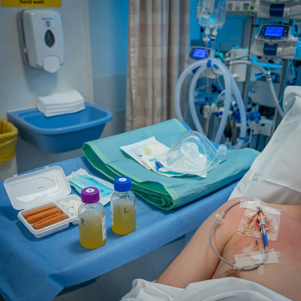
Diagnosis
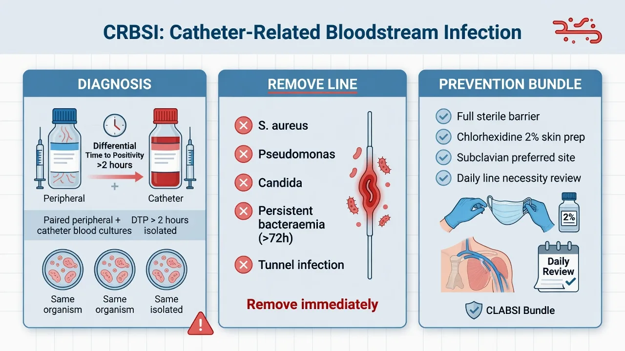
Management
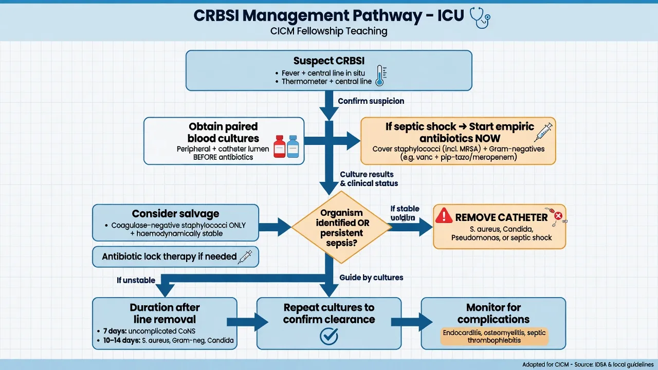
CRBSI management
REMOVE the catheter
Remove the infected catheter immediately (unless it is a tunneled catheter and the organism is coagulase-negative staph — in which case antibiotic lock therapy may be attempted). Source control. If continued central access needed: insert new line at DIFFERENT site (do NOT guidewire exchange an infected line).
Empiric antibiotics
Vancomycin 1g IV BD (covers staph including MRSA — most common CRBSI organisms). Add piperacillin-tazobactam if Gram-negative risk (severe sepsis, immunocompromised, prolonged hospitalisation). Add echinocandin if Candida suspected (multiple sites colonised, TPN, prolonged antibiotics).
Targeted therapy when organism identified
Coagulase-negative staph: vancomycin 5-7 days (or line lock if catheter retained). MSSA: flucloxacillin 2g IV Q4H (switch from vancomycin once susceptibilities known). MRSA: vancomycin 14 days. Enterococcus: ampicillin (if susceptible) or vancomycin. Gram-negative: organism-specific. Candida: echinocandin → fluconazole step-down.
Duration
Coagulase-negative staph: 5-7 days (after catheter removal). S. aureus: minimum 14 days (longer if endocarditis, metastatic infection). Enterococcus: 7-14 days. Gram-negative: 7-14 days. Candida: 14 days after first negative culture + ophthalmology review.
Search for complications
S. aureus CRBSI: echocardiogram to exclude endocarditis (mandatory — 25% have valvular involvement). Persistent bacteraemia >72h: search for metastatic foci (vertebral osteomyelitis, epidural abscess, septic pulmonary emboli). Repeat blood cultures every 48-72h until negative.
Prevention bundle
At insertion
Prevent contamination
- Full maximal sterile barrier precautions (cap, mask, sterile gown, gloves, large sterile drape)
- Chlorhexidine 2% alcoholic skin antisepsis (let it dry before puncture)
- Site selection: subclavian (lowest infection) > internal jugular > femoral (highest)
- Ultrasound guidance (reduces attempts, complications)
- Aseptic technique throughout
Daily maintenance
Prevent colonisation
- Daily review: is this line still needed? Remove if not.
- Hand hygiene before accessing line (WHO 5 moments)
- Chlorhexidine-impregnated sponge dressing at exit site (reduces colonisation)
- Scrub the hub: chlorhexidine/alcohol wipe before each access
- Avoid routine replacement of CVCs (not evidence-based for infection prevention)
- Do NOT routinely apply antibiotic ointment to exit site (resistance risk)
Site selection
CVC site infection rates
Clinical pearls
Red flags
Detailed CRBSI prevention — the evidence-based bundle
CRBSI prevention bundle — expanded with evidence
| Component | Evidence | Implementation |
|---|---|---|
| Maximal sterile barriers | Pronovost Keystone: reduced CRBSI by 66% in 103 ICUs. Full body drape + cap + mask + sterile gown + sterile gloves for ALL CVC insertions — even emergencies | Mandate checklist before insertion. "Stop if any item missing" |
| Chlorhexidine 2% skin antisepsis | Superior to povidone-iodine (RR 0.49). Allow to dry ≥30 seconds. Chlorhexidine is alcohol-based → rapid onset + persistent activity for >48h | Apply in concentric circles from centre outward. Do NOT wipe off |
| Site selection | Subclavian = LOWEST infection (clean skin, easy dressing). IJV = moderate (more colonised skin, harder to keep dressing clean). Femoral = HIGHEST infection (3-8x higher — groin flora). AVOID femoral unless emergency | Preferred: subclavian (if no coagulopathy/resp failure). Second: IJV (US-guided). Last resort: femoral (remove within 24-48h) |
| Ultrasound guidance | Reduces mechanical complications (pneumothorax, arterial puncture, haematoma) by 70%. Reduces attempts. Does NOT directly reduce infection but indirectly by reducing time/trauma | Real-time US for ALL CVC insertions (NICE 2002 recommendation) |
| Daily review of necessity | Each day CVC remains → 3-5% increased infection risk. The STRONGEST predictor of CRBSI is DURATION of catheterisation. The "Get the Line OUT" campaign | Ward round question EVERY DAY: 'Does this patient need this line TODAY?' Remove if: antibiotics complete, no vasopressors, no TPN, no inaccessible venous access |
| Chlorhexidine-impregnated dressing | Tegaderm CHG sponge reduces CRBSI by 40% in high-risk patients (lines >5 days). Study: 1,636 patients, CRBSI 1.3 vs 0.4 per 1000 catheter days | Apply Biopatch at insertion site under transparent dressing for ALL CVCs expected to remain >5 days. Change every 7 days |
| Antimicrobial-impregnated catheters | Minocycline-rifampicin or chlorhexidine-silver sulfadiazine CVCs reduce colonisation and CRBSI. Use for patients at HIGH risk (burns, immunocompromised, prolonged need) | Not for routine use (cost, potential resistance) |
| Hub disinfection | Hub is the #1 entry point for CRBSI (catheter manipulation). Scrub the hub with chlorhexidine/alcohol for ≥15 seconds before EVERY access. Allow to dry | "Scrub the hub" education campaign for all staff |
| Dressing care | Transparent dressing (allows visual inspection). Change every 7 days OR if soiled/damp/loosened. Gauze dressing if patient diaphoretic or site oozing (change every 48h) | Document dressing date. Inspect daily for redness, swelling, discharge |
CRBSI treatment — by organism
CRBSI management protocol — the step-by-step
- SUSPECT CRBSI: Fever ± rigors ± elevated WCC ± local signs (erythema, discharge at exit site) in patient with CVC. NEW onset sepsis without other source = consider CRBSI
- DRAW CULTURES: (a) 2 sets blood cultures from PERIPHERAL vein (not from the catheter). (b) 1 set from EACH lumen of the catheter. (c) Culture the catheter tip if removing (semi-quantitative Maki roll-plate method — >15 CFU = significant colonisation)
- DIFFERENTIAL TIME TO POSITIVITY: If catheter culture positive >2h BEFORE peripheral → the catheter is the source. If <2h difference → source is elsewhere (catheter colonised but not source)
- START EMPIRIC ANTIBIOTICS: Vancomycin 1g IV q12h (covers MRSA + coagulase-negative staph — the #1 CRBSI organisms) ± Gram-negative cover if septic or immunocompromised (piperacillin-tazobactam 4.5g q6h)
- REMOVE THE CATHETER (in most cases): ALWAYS remove for S. aureus, Candida, Pseudomonas, Enterococcus. CONSIDER removing for coagulase-negative staph (if tunneled catheter — may try antibiotic lock therapy). Do NOT exchange over a guidewire (new catheter contaminated from same tract)
- INSERT NEW CATHETER at DIFFERENT site if ongoing central access needed
- DURATION BY ORGANISM: Coagulase-negative Staph → 5-7 days (if line removed + improving). S. aureus → 14 days MINIMUM (screen for endocarditis/osteomyelitis — echo mandatory). Candida → 14 days after FIRST negative culture. Gram-negative → 7-14 days. Pseudomonas → 14 days
- ANTIBIOTIC LOCK THERAPY (for salvage): For tunneled catheters (Hickman, Portacath) with coagulase-negative Staph and NO tunnel infection: instil vancomycin 5mg/mL into lumen for 12h/day x 14 days. Success 60-80% for CNS, <50% S. aureus, <30% Candida
Additional clinical pearls
[1]Definition — CRBSI vs CLABSI
Pathogens — the typical ICU CRBSI microbiology
Gram-positive (≈60-70%)
Skin flora — dominant
- Coagulase-negative staphylococci (CoNS — Staph epidermidis): #1 organism (~30-40% of CRBSI). Slow-growing, biofilm-forming. Often catheter-salvageable.
- Staphylococcus aureus (MSSA + MRSA): ~10-20%. Aggressive — 25% endocarditis, mandatory metastatic workup and minimum 14 days therapy.
- Enterococci (E. faecalis, E. faecium — VRE): ~8-12%. Often in patients on broad-spectrum antibiotics or with intra-abdominal pathology.
Gram-negative (≈20-25%)
Hospital flora / gut
- Enterobacterales: E. coli, Klebsiella, Enterobacter, Serratia. Common with hub contamination or contaminated infusate.
- Pseudomonas aeruginosa: associated with moisture, contaminated flushes, water-borne exposure. ALWAYS remove catheter; 14 days therapy.
- Stenotrophomonas maltophilia, Acinetobacter baumannii: opportunists in long-stay, heavily antibiotic-exposed patients.
Candida (≈8-12%)
Fungal
- Candida albicans (~50%) and non-albicans (C. glabrata, C. parapsilosis, C. krusei, C. tropicalis).
- Risk factors: TPN, broad-spectrum antibiotics, haematologic malignancy, prolonged ICU stay, femoral catheter, diabetes.
- ALWAYS remove catheter. ALWAYS request ophthalmology review (chorioretinitis ~16%). Echinocandin first-line; fluconazole step-down if susceptible.
Other / atypical
Less common
- Corynebacterium jeikeium, Bacillus spp., Micrococcus — consider only if ≥2 positive sets and clinical signs.
- Mycobacteria (M. chelonae, M. fortuitum, M. mucogenicum): consider in dialysis catheters, immunosuppressed, or outbreak setting.
- Malassezia furfur: lipophilic yeast — classically associated with lipid emulsion / TPN.
Pathogenesis — biofilm and the two routes of colonisation
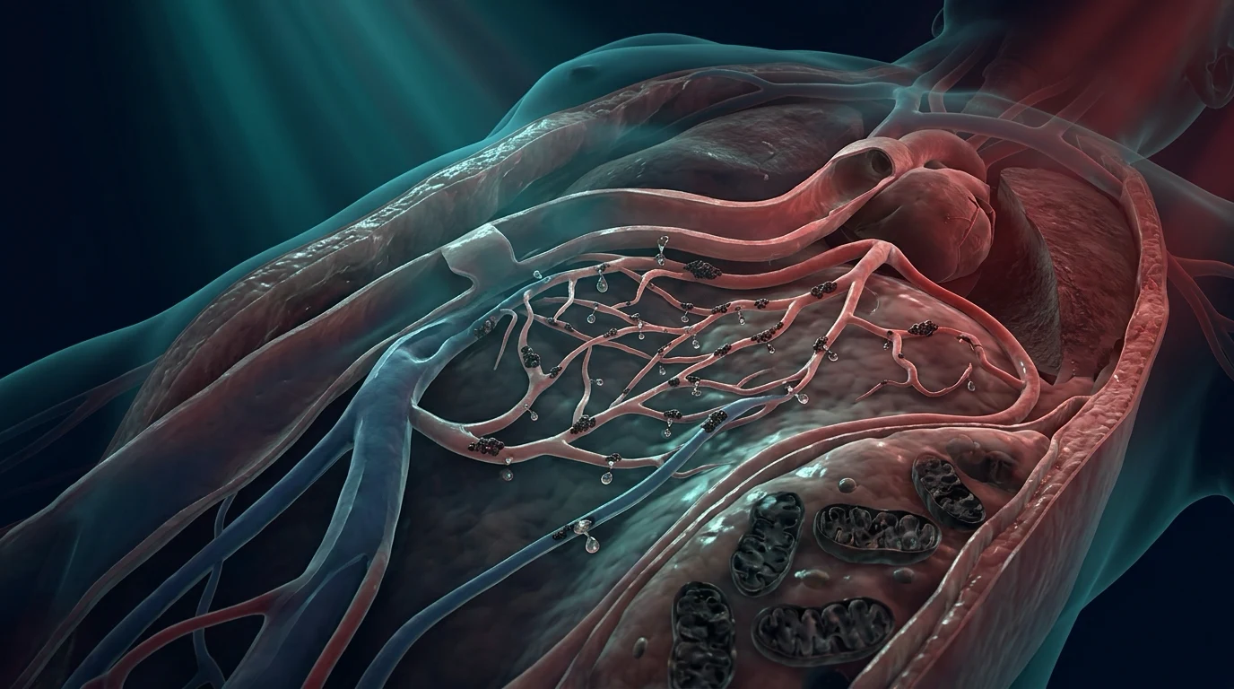
Extraluminal route
Migration along external catheter surface
- Skin micro-organisms at the insertion site migrate DOWN the external surface of the catheter → colonise the distal intravascular segment → bloodstream.
- DOMINANT mechanism for SHORT-term catheters (<10-14 days).
- Targeted by: chlorhexidine skin antisepsis, chlorhexidine-impregnated dressings, antimicrobial-impregnated catheters, optimal site selection.
Intraluminal route
Hub contamination
- Micro-organisms from staff hands or skin contaminate the catheter HUB during manipulation → migrate DOWN the internal lumen → bloodstream.
- DOMINANT mechanism for LONG-term catheters (>14 days) and tunneled devices.
- Targeted by: "scrub the hub" (CHG/alcohol ≥15 s before every access), needleless connector care, aseptic technique for line draws/administration.
Biofilm
The common pathogenic denominator
- Organisms (especially CoNS and S. aureus) adhere to the catheter and secrete an extracellular polymeric substance (EPS) slime layer.
- Biofilm renders organisms 10-1000× more resistant to antibiotics and host immunity than planktonic bacteria — explains why systemically active antibiotics ALONE often fail to clear catheter-based infection.
- Removal of the catheter is the definitive source-control measure — physical removal of biofilm-bearing foreign body.
- Antibiotic-lock therapy works because it achieves 100-1000× the MIC within the lumen to penetrate biofilm.
Less common routes
Infusate / haematogenous
- Contaminated infusate (rare but dramatic — outbreaks of Burkholderia cepacia, Serratia, Citrobacter from contaminated fluids).
- Haematogenous seeding from a distant focus (e.g., intra-abdominal abscess) onto an indwelling catheter.
Risk factors for CRBSI
Catheter-related
Modifiable at insertion
- Type: non-tunnelled CVC = HIGHEST risk. Tunnelled cuffed (Hickman) and implanted ports = LOWEST risk (~1 per 1000 catheter-days). PICCs intermediate; arterial catheters surprisingly low (~1-2 per 1000 catheter-days).
- Number of lumens: each additional lumen increases infection risk — multi-lumen catheters have higher CRBSI than single-lumen. Use the minimum number of lumens needed.
- Site: femoral > internal jugular > subclavian (see Site selection).
- Dwelling time: STRONGEST independent risk factor — each extra day at risk adds 3-5% cumulative risk.
- Insertion under EMERGENT conditions (e.g., code, trauma) — breaks in aseptic technique.
Patient-related
Often non-modifiable
- Severe underlying illness (high APACHE), immunosuppression, neutropenia, burns.
- Skin disruption: psoriasis, burns, dermatitis at insertion site.
- Extreme age (neonate or elderly), diabetes mellitus, malnutrition.
Care-related
Modifiable at maintenance
- Total parenteral nutrition (TPN) — lipids and high glucose promote microbial growth and biofilm (especially Candida and CoNS).
- Frequent catheter access (drug administration, blood sampling) — each manipulation is a contamination opportunity.
- Tracheostomy in situ (proximity to IJV line — secretions contaminate dressing).
- Dressing disruption / soiling / moisture.
- Non-aseptic hub care (failure to scrub the hub).
Diagnostic methods — practical detail
[4]Diagnostic methods compared
| Method | Specimen | Threshold | When useful | Catheter removed? |
|---|---|---|---|---|
| DTP | Paired peripheral + catheter blood (same organism) | Catheter positive ≥2 h before peripheral | Catheter retained / suspected CRBSI | No |
| Quantitative paired blood cultures | Paired peripheral + catheter blood | Catheter:peripheral ratio ≥3:1 | DTP equivocal, lab supports it | No |
| Maki roll-plate (semi-quant) | Distal catheter tip (5 cm) | >15 CFU/plate | Catheter already removed | Yes |
| Brun-Buisson (quantitative) | Sonicated intravascular segment | >10³ CFU/mL | Suspicion of intraluminal source | Yes |
| Hub-blood / paired sets | 2 sets from hub + 2 peripheral | Match organism + DTP ≥2 h | Routine bedside practice | No |
Mimics and differential diagnosis
Other ICU-acquired bacteraemia
Same organisms, different source
- Ventilator-associated pneumonia (VAP) — same organisms (S. aureus, Gram-negatives).
- Intra-abdominal source (biliary, post-operative collections).
- Urinary tract infection, wound infection, sinusitis, cholecystitis.
- Use paired DTP >2 h (catheter first) to confirm the catheter is the source — if not, search elsewhere.
Non-infectious causes of fever
Mimics the septic picture
- Drug fever (β-lactams, anticonvulsants), transfusion reaction, PE/DVT, MI, pancreatitis, alcohol withdrawal, malignant hyperthermia, neuroleptic malignant syndrome, adrenal crisis.
- Always send cultures BEFORE starting empirical antibiotics — a single set of "positive cultures" treated as CRBSI may simply reflect contamination (especially single-set CoNS).
Contamination vs true bacteraemia
The CoNS trap
- CoNS are common skin contaminants. Treat as TRUE bacteraemia only if: ≥2 positive sets drawn from different sites at different times, with consistent clinical picture and organism susceptibility.
- A SINGLE positive CoNS set → likely contamination; do NOT treat without clinical signs.
Empiric and targeted therapy by organism
Empiric (before organism known)
Active against MRSA + Gram-negatives ± Candida
- Vancomycin 15-20 mg/kg IV q8-12h (or daptomycin 6-10 mg/kg q24h if vancomycin-allergic / VRE suspected) — covers CoNS and MRSA.
- Add piperacillin-tazobactam 4.5 g IV q6-8h (or cefepime / meropenem) if severe sepsis, neutropenia, prolonged hospital stay, or known Gram-negative colonisation.
- Add an echinocandin (caspofungin 70 mg load then 50 mg/day, micafungin 100 mg/day, anidulafungin 200 mg load then 100 mg/day) if Candida suspected (multiple colonised sites, TPN, broad-spectrum antibiotics, prolonged ICU stay).
- De-escalate as soon as organism + susceptibilities are known (24-48 h).
Coagulase-negative staph
Catheter often salvageable
- Vancomycin 1 g IV q12h (or daptomycin if VRE / vancomycin-intolerant).
- If line removed + uncomplicated: 5-7 days. If line retained (tunneled): systemic + antibiotic-lock × 10-14 days; salvage ~60-80%.
Staphylococcus aureus
ALWAYS remove line — high metastatic risk
- MSSA: switch to flucloxacillin / nafcillin / cefazolin (β-lactam superior to vancomycin for MSSA).
- MRSA: vancomycin (target AUC/MIC 400-600) or daptomycin (cannot use if pneumonia — inactivated by surfactant).
- Minimum 14 days from first NEGATIVE blood culture. Add gentamicin + rifampicin for the first 3-5 days ONLY if endocarditis / prosthetic hardware.
- Mandatory echocardiography (TTE; TOE if TTE negative and any suspicion).
Enterococcus
Consider VRE
- Ampicillin (if susceptible) ± gentamicin synergy; vancomycin if ampicillin-resistant.
- Linezolid or daptomycin for VRE (E. faecium).
- Always remove catheter; 7-14 days.
Gram-negative bacilli
Organism-specific
- Escalate to organism-directed therapy: ceftriaxone, cefepime, piperacillin-tazobactam, meropenem for ESBL/AmpC; colistin / polymyxin for carbapenem-resistant.
- Pseudomonas: ALWAYS remove catheter; 14 days with two active agents initially.
- 7-14 days; reassess after first negative culture.
Candida
ALWAYS remove line; ophthalmology
- Echinocandin first-line (caspofungin, micafungin, anidulafungin). Step-down to fluconazole once stable and susceptible (C. albicans, C. parapsilosis, C. tropicalis).
- Treat for 14 days AFTER first negative blood culture.
- Day 1: ophthalmology review (chorioretinitis in ~16%). If present, extend therapy and prefer fluconazole (vitreous penetration).
- Investigate for intra-abdominal source if recent abdominal surgery.
Antibiotic duration by organism — quick reference
Recommended therapy duration from first negative culture
| Organism | Remove catheter? | Antibiotic duration | Special considerations |
|---|---|---|---|
| Coagulase-negative staph | Yes (preferred); lock if salvaged | 5-7 days (line removed) | Lock × 10-14 d if tunneled line salvaged |
| MSSA | YES — always | ≥14 days | Echocardiography mandatory; screen for metastatic foci |
| MRSA | YES — always | ≥14 days (AUC-guided vancomycin) | TOE if any suspicion of endocarditis |
| Enterococcus | YES | 7-14 days | Treat VRE with linezolid or daptomycin |
| Gram-negative (Enterobacterales) | Yes | 7-14 days | Escalate based on susceptibility |
| Pseudomonas aeruginosa | YES — always | 14 days | Remove promptly; high metastatic risk |
| Candida | YES — always | 14 days after first negative culture | Ophthalmology review day 1 |
| Mycobacteria (NTM) | YES | Prolonged, organism-specific | Public-health notification; outbreak investigation |
Persistent bacteraemia — the metastatic search
Persistent bacteraemia >72 h despite line removal and appropriate antibiotics
Reassess source
Confirm line removed. Repeat blood cultures every 48-72 h to document clearance. Confirm organism and susceptibility — re-confirm vancomycin MIC, ESBL/Carba-R status. Consider superimpressed infection (VAP, intra-abdominal).
Echocardiography
Transthoracic echo (TTE) first. Transoesophageal echo (TOE) if TTE negative AND S. aureus, persistent bacteraemia, prosthetic valve, intracardiac device (pacemaker/ICD), or injection drug use. Sensitivity of TOE for vegetations ~90%.
Septic thrombophlebitis
Doppler ultrasound of the catheterised vein — suppurative thrombosis of the IJV (Lemierre-like), subclavian, or femoral vein. CT venography if suspicion persists.
Distant / metastatic foci
Vertebral osteomyelitis and epidural abscess (spinal MRI if back pain or persistent S. aureus bacteraemia). Septic pulmonary emboli (CT chest — wedge-shaped peripheral opacities). Splenic / hepatic abscess, psoas abscess (CT abdomen).
Prosthetic material
Prosthetic joints (hip / knee), prosthetic vascular grafts, pacemaker / IVD / LVAD leads, neurosurgical shunts — all potential seeded foci requiring imaging and often surgical source control.
Prolong therapy
Once metastatic focus identified, extend therapy (4-6 weeks for uncomplicated vertebral osteomyelitis, ≥6 weeks for endocarditis). Engage infectious diseases + relevant surgical specialty early.
Prevention bundle — the expanded evidence base
CRBSI prevention bundle — every element with the evidence
| Element | Key evidence | Practical implementation |
|---|---|---|
| Maximal sterile barriers at insertion | Pronovost Keystone (NEJM 2006) — bundle implementation reduced median CRBSI from 2.7 to 0 per 1000 catheter-days across 103 ICUs (66% relative reduction), sustained at 18 months.[3] | Mandate checklist; "stop the line" if any item missing. Cap + mask + sterile gown + sterile gloves + full-body drape for EVERY CVC, including emergent insertions. |
| Chlorhexidine 2% alcoholic skin antisepsis | Superior to povidone-iodine (RR ~0.49 for CRBSI). Allow to air-dry ≥30 s before puncture.[5] | Apply in concentric circles outward from insertion point; do NOT blot or wipe. Re-apply if puncture attempt fails. |
| Optimal site selection | Subclavian = lowest CRBSI; femoral = highest. 3SITES trial (NEJM 2015): subclavian had lowest bloodstream infection and DVT but higher pneumothorax; femoral highest infection + thrombosis.[9] | First-line subclavian (if no coagulopathy / no respiratory compromise / no chest-wall deformity). Second IJV (US-guided). Last resort femoral — remove within 24-48 h. |
| Ultrasound guidance | Reduces mechanical complications (pneumothorax, arterial puncture) by ~70%; reduces attempts and time. | Real-time 2-D US for ALL elective CVC insertions; out-of-plane / in-plane according to operator skill. |
| Daily review of necessity | Each extra day at risk adds 3-5% cumulative infection probability; duration is the strongest independent predictor.[4] | Daily ward-round question: "Does this patient need this line TODAY?" Remove if antibiotics complete, no vasopressors, no TPN, no inaccessible access. |
| CHG-impregnated sponge dressing | Timsit JAMA 2009 (1,636 patients): CRBSI 1.3 vs 0.4 per 1000 catheter-days (HR 0.24); significant for lines expected to remain >5 days.[6] | Apply Biopatch under transparent dressing at insertion; change every 7 d or if soiled. |
| CHG bathing | Climo NEJM 2013 (7高危 wards): 23% reduction in MRSA / VRE acquisition and CLABSI.[10] | 2% CHG bathing/wipes daily in high-acuity ICU patients. Not for routine ward use. |
| Antimicrobial-impregnated catheters | Darouiche NEJM 1999: minocycline-rifampin superior to chlorhexidine-silver sulfadiazine for CRBSI prevention; Hanna JCO 2004 confirms in cancer patients.[7][8] | Reserve for high-risk patients: expected dwelling >5 days, burns, immunosuppressed, limited access, high baseline CRBSI unit. |
| Scrub the hub | Hub is the dominant intraluminal portal of entry. | CHG/alcohol wipe × ≥15 s before EVERY access; allow to dry. "Scrub the hub" staff education + audit. |
| Dressing care | Transparent dressings allow visual inspection; gauze for diaphoretic / oozing sites. | Change every 7 d OR if soiled / damp / loosened. Gauze every 48 h. Inspect daily. |
| Avoid routine replacement | Routine guidewire exchange of uninfected lines does NOT prevent CRBSI and increases mechanical complications.[5] | Replace only for clinical indication, not on a schedule. Replace lines inserted emergently (broken aseptic technique) within 48 h at a new site. |
| Avoid antibiotic ointment at exit site | Promotes fungal colonisation and antimicrobial resistance.[5] | Do NOT routinely apply mupirocin / polymyxin / bacitracin / neomycin at exit site. |
Antimicrobial-impregnated catheters — when to use them
[1]Trial cards — landmark CRBSI evidence
Pronovost 2006 (NEJM) — Keystone ICU CRBSI bundle (the landmark)
Design
Prospective cohort with sequential implementation across 103 ICUs in Michigan, USA (~375,757 catheter-days). CVC insertion bundle: hand hygiene, full barrier precautions, chlorhexidine skin antisepsis, optimal site selection (avoid femoral), daily review of necessity with removal of unnecessary lines.
Intervention
Multifaceted quality-improvement intervention: education, central-line cart, checklist, daily goals, physician feedback, plus measurement.
Primary outcome
Median CRBSI rate fell from 2.7 to 0 per 1000 catheter-days at 3 months (rate ratio 0.62), then 0.0 sustained at 18 months — a 66% relative reduction. Effect sustained long-term.
Safety
No increase in mechanical complications or inappropriate line removals; net safety benefit strongly favourable.
Clinical bottom line
A simple, cheap, reproducible 5-element bundle reduced CRBSI dramatically across diverse ICUs — established the standard of care for CVC insertion. The single most influential CRBSI study ever published.
Timsit 2009 (JAMA) — chlorhexidine-impregnated sponge dressing
Design
Multicentre randomised trial, 1,636 ICU patients, 2,829 CVCs; CHG-impregnated sponge (Biopatch) under transparent dressing vs standard dressing.
Intervention
CHG-impregnated gel disc at exit site vs identical-looking non-impregnated dressing. Less-frequent (7-day) dressing changes vs standard (3-day) in 2 × 2 factorial.
Primary outcome
Major CRBSI: 1.3 vs 0.4 per 1000 catheter-days (hazard ratio 0.24, 95% CI 0.09-0.64). Benefit concentrated in CVCs in situ >7 days and arterial catheters. No effect on shorter-dwell lines.
Safety
Local contact dermatitis in ~5% (mostly mild, more common with prolonged use). No systemic CHG toxicity.
Clinical bottom line
CHG-impregnated sponge dressings reduce CRBSI in lines expected to dwell >5-7 days — now a standard component of the prevention bundle in long-dwell ICU patients.
Darouiche 1999 (NEJM) — minocycline-rifampin vs chlorhexidine-silver sulfadiazine CVCs
Design
Prospective, randomised, multicentre clinical trial; 738 CVCs in adults. Both arms had antimicrobial-impregnated catheters (one internal+external, one external only).
Intervention
Minocycline-rifampin-impregnated CVC vs chlorhexidine-silver sulfadiazine-impregnated CVC (first generation, external surface only).
Primary outcome
Catheter colonisation 36/356 (10%) minocycline-rifampin vs 57/382 (15%) CHG-SS; CRBSI 1/356 (0.3%) vs 13/382 (3.4%) — minocycline-rifampin superior. RR for CRBSI 0.09 (95% CI 0.01-0.67).
Safety
No increase in adverse events or antimicrobial resistance detected at short-term follow-up.
Clinical bottom line
Established minocycline-rifampin-impregnated catheters as the most effective antimicrobial CVC for high-risk patients. The benchmark comparison for antimicrobial-impregnated lines.
Parienti 2015 (NEJM) — 3SITES: intravascular complications by site
Design
Multicentre randomised controlled trial, 3,471 adults requiring a first CVC at 3 sites (subclavian, jugular, femoral) in 8 French ICUs. 2 × 2 design (site ± catheter-type).
Intervention
CVC insertion at subclavian vs internal jugular vs femoral vein (randomised when feasible).
Primary outcome
Composite of catheter-related bloodstream infection AND symptomatic deep-vein thrombosis: subclavian 0.4% vs jugular 1.5% vs femoral 3.5% (femoral highest, p<0.001).
Safety
Mechanical complications: pneumothorax 1.1% subclavian vs ~0% jugular/femoral. Arterial puncture higher in femoral. Net balance favours subclavian unless respiratory/coagulation risk.
Clinical bottom line
Subclavian site has lowest combined infectious + thrombotic risk; femoral highest. Subclavian remains preferred when feasible (no coagulopathy / no respiratory compromise). The definitive head-to-head site comparison.
Climo 2013 (NEJM) — daily chlorhexidine bathing (HABIT)
Design
Cluster-randomised, crossover trial in 9 ICUs and bone-marrow transplant units; 7,727 patients. 2% CHG-impregnated washcloths vs non-antimicrobial washcloths for daily bathing.
Intervention
Daily bathing with 2% CHG washcloths (Hibiclens-equivalent) vs non-antimicrobial washcloths for 6-month periods, then crossover.
Primary outcome
Composite primary bloodstream-infection rate (MRSA + VRE acquisition) reduced by 28%. CLABSI specifically reduced from 6.1 to 4.8 per 1000 patient-days.
Safety
Mild skin irritation in ~2%; no anaphylaxis. Easy to implement; no additional staffing.
Clinical bottom line
Daily CHG bathing is a low-cost adjunct that reduces multidrug-resistant organism acquisition and CLABSI in high-acuity units. Part of the bundle in many ICUs but not universal — skin irritation, meticillin exposure concerns, and patient choice require local policy.
Marra 2015 (Infection) — attributable mortality of CLABSI (meta-analysis)
Design
Systematic review and meta-analysis of studies reporting adjusted mortality for CLABSI (matched / multivariable-adjusted).
Outcome
Pooled attributable mortality from CLABSI: odds ratio 1.27 (95% CI 1.04-1.56) — i.e., ~25-30% relative excess mortality attributable to CLABSI after adjustment. Prolonged ICU stay (mean +5-7 days) and excess cost (~USD 20,000-45,000 per case) consistently reported.
Heterogeneity
Wide 95% CI reflecting varied case-mix and adjustment quality; some studies showed no excess mortality. Effect largest for S. aureus and Candida; smallest for CoNS (often contaminant / less virulent).
Clinical bottom line
CLABSI is associated with substantial excess mortality, length-of-stay and cost — strong economic and ethical justification for sustained investment in prevention bundles and zero-tolerance reporting.
Additional clinical pearls — extended (21-40)
Antimicrobial-impregnated catheter comparison
Antimicrobial-impregnated CVC types — comparative properties
| Property | Minocycline-rifampin | Chlorhexidine-silver sulfadiazine (1st gen) | CHG-silver sulfadiazine (2nd gen) |
|---|---|---|---|
| Surface coated | Internal + external | External only | Internal + external |
| Spectrum | Gram-positive (incl. MRSA), Gram-negative, Candida | Gram-positive, some Gram-negative, Candida | Broader than 1st-gen |
| Duration of activity | Prolonged (≥30 days) | Shorter (≤7-14 days) | Improved vs 1st gen |
| CRBSI reduction vs standard | Most effective in head-to-head RCTs | Effective in meta-analysis | Improved vs 1st gen |
| Resistance concern | Rifampin resistance reported (rare with short-term use) | Less resistance signal | Less resistance signal |
| Cost | Higher | Moderate | Higher |
| Recommended use | High-risk, long-dwell >5-7 days, haematology-oncology, burns | Lower-risk, intermediate dwell; or when minocycline-rifampin unavailable | High-risk, prolonged dwell |
Red flags — extended
Putting it together — the one-minute CRBSI mental model
Rapid CRBSI mental model
Diagnose
Paired blood cultures from peripheral + each lumen. Same organism + catheter positive ≥2 h before peripheral (DTP >2h) → catheter is the source. If line being removed → send Maki tip (>15 CFU = colonisation).
Decide on the line
REMOVE for S. aureus, Pseudomonas, Candida, Enterococcus, severe sepsis, tunnel-tract infection, prosthetic material, persistent bacteraemia. SALVAGE ATTEMPT (with antibiotic lock) only for CoNS in tunneled catheters with no tunnel infection.
Start empiric therapy
Vancomycin ± anti-pseudomonal β-lactam ± echinocandin — de-escalate at 24-48 h once organism known.
Stage therapy by organism
CoNS 5-7 d; S. aureus ≥14 d + echo + metastatic workup; Enterococcus 7-14 d; Gram-negatives 7-14 d; Candida 14 d after first negative + ophthalmology.
Reassess at 72 h
Persistent bacteraemia → search for metastatic foci (TOE, MRI spine, CT chest/abdomen, Doppler of catheterised vein). Extend therapy to 4-6 weeks once deep focus identified.
Prevent the next one
Bundle: maximal barrier precautions, CHG skin prep, subclavian preferred, US guidance, daily review of necessity, CHG dressing in long-dwell lines, scrub the hub. Sustained near-zero is achievable.
SAQs
SAQ — CRBSI diagnosis and prevention bundle
10 minutes · 10 marks
A 64-year-old man, day 9 of an unplanned ICU admission for severe pancreatitis, has a triple-lumen right internal jugular CVC (inserted on the day of admission) and a femoral arterial line. He develops a new fever (39.1°C), rigors, and a rising lactate (1.2 → 3.4 mmol/L) with no obvious alternate source. Blood cultures are sent and broad-spectrum antibiotics are started.
SAQ — Catheter removal decisions in suspected/confirmed CRBSI
10 minutes · 10 marks
A 52-year-old woman with a long-term tunneled Hickman catheter for haemodialysis is admitted to the ICU with rigors and hypotension 30 minutes after her dialysis session. Blood cultures from the catheter and a peripheral vein both grow Staphylococcus aureus (MSSA) within 6 hours of each other. She has no exit-site or tunnel-tract inflammation. She urgently needs ongoing dialysis.
References
- [1]Ling ML, Apisarnthanarak A, Madriaga G, et al. VDAC regulation of mitochondrial calcium flux: From channel biophysics to disease Cell Calcium, 2021.PMID 33529977
- [2]Chopra V, Flanders SA, Saint S, et al. (MAGIC criteria). Notum palmitoleoyl-protein carboxylesterase regulates Fas cell surface death receptor-mediated apoptosis via the Wnt signaling pathway in colon adenocarcinoma Bioengineered, 2021.PMID 34402722
- [3]Pronovost P, Needham D, Berenholtz S, et al. An intervention to decrease catheter-related bloodstream infections in the ICU N Engl J Med, 2006.PMID 17192537
- [4]Mermel LA, Allon M, Bouza E, et al. Clinical practice guidelines for the diagnosis and management of intravascular catheter-related infection: 2009 Update by the Infectious Diseases Society of America Clin Infect Dis, 2009.PMID 19489710
- [5]O'Grady NP, Alexander M, Burns LA, et al. (HICPAC/CDC). Guidelines for the prevention of intravascular catheter-related infections Am J Infect Control, 2011.PMID 21511081
- [6]Timsit JF, Schwebel C, Bouadma L, et al. Chlorhexidine-impregnated sponges and less frequent dressing changes for prevention of catheter-related infections in critically ill adults: a randomized controlled trial JAMA, 2009.PMID 19318651
- [7]Darouiche RO, Raad II, Heard SO, et al. A comparison of two antimicrobial-impregnated central venous catheters. Catheter Study Group N Engl J Med, 1999.PMID 9878638
- [8]Hanna H, Benjamin R, Chatzinikolaou I, et al. Long-term silicone central venous catheters impregnated with minocycline and rifampin decrease rates of catheter-related bloodstream infection in cancer patients: a prospective randomized clinical trial J Clin Oncol, 2004.PMID 15284269
- [9]Parienti JJ, Mongardon N, Megarbane B, et al. (3SITES study). Intravascular Complications of Central Venous Catheterization by Insertion Site N Engl J Med, 2015.PMID 26398070
- [10]Climo MW, Yokoe DS, Warren DK, et al. Effect of daily chlorhexidine bathing on hospital-acquired infection N Engl J Med, 2013.PMID 23388005
- [11]Marra AR, Opilla M, Edmond MB, et al. Attributable mortality of central line associated bloodstream infection: systematic review and meta-analysis Infection, 2015.PMID 25331552
- [12]Minter DJ, Appa A, Chambers HF, Doernberg SB. Contemporary Management of Staphylococcus aureus Bacteremia-Controversies in Clinical Practice Clin Infect Dis, 2023.PMID 37950887