ICU · Antimicrobial Stewardship
Fungal infections in the ICU: candidaemia and aspergillosis
Also known as Candidaemia · Invasive candidiasis · Invasive pulmonary aspergillosis · Echinocandin · Voriconazole · Candida score · Candida auris · Galactomannan · Beta-D-glucan
Fungal infections in ICU: candidaemia (most common fungal infection — Candida albicans ~50%, non-albicans species rising — C. glabrata 25% with increasing fluconazole resistance, C. auris emerging multidrug-resistant) and invasive pulmonary aspergillosis (immunocompromised and severe COPD/cirrhosis/influenza/COVID-19 patients). Candidaemia risk factors: central venous catheter (1), broad-spectrum antibiotics, TPN, abdominal surgery, diabetes, immunosuppression, prolonged ICU stay. Diagnosis: blood cultures (gold standard but slow 48-72 h), beta-D-glucan (sensitive non-specific), Candida score (≥3 = high risk). Treatment: echinocandin (caspofungin/micafungin/anidulafungin) first-line for candidaemia, step down to fluconazole when stable/susceptible, liposomal amphotericin B for resistant. Remove central line (source control). Ophthalmology review (endophthalmitis 5-10%). Aspergillosis: voriconazole first-line; diagnosis by galactomannan (BAL preferred), CT halo sign, and AspICU criteria.
On this page & tools
Your progress
Saved locally on this device.
Target exams
Red flags
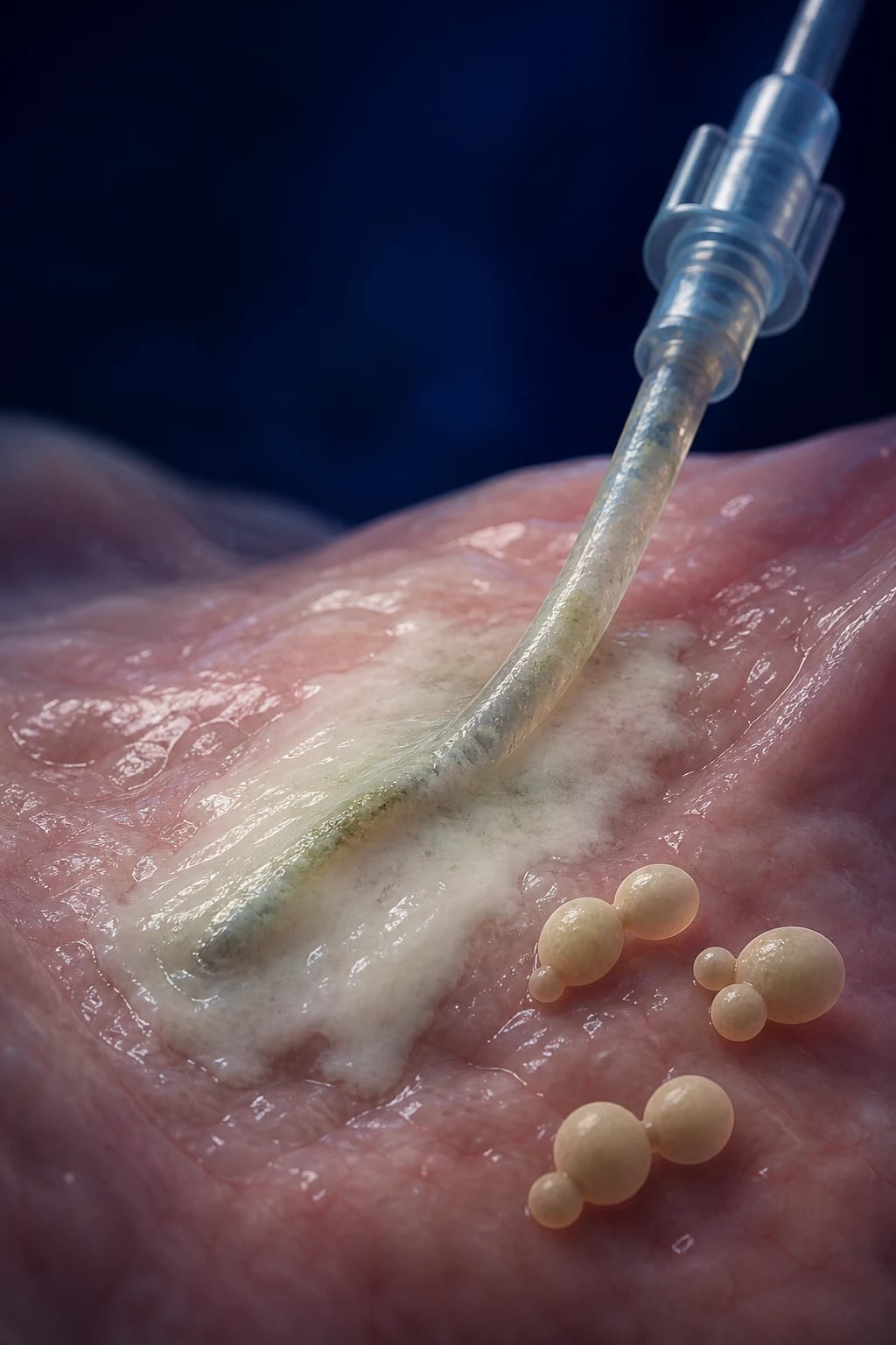
Epidemiology and Candida species distribution
Candida species are the third to fourth most common cause of nosocomial bloodstream infection in the ICU and the most common fungal bloodstream pathogen. Roughly 1 in 10 ICU patients colonised with Candida in multiple sites will progress to invasive candidiasis. Mortality attributable to candidaemia is 15-49%, and the attributable excess length of stay is significant. [1]
C. albicans
~50% of candidaemia
- Most common species; typically fluconazole-susceptible
- Forms true hyphae → tissue invasion
- Higher virulence than non-albicans species
- Predominant in surgical and non-malignancy ICU patients
C. glabrata
~25% of candidaemia
- Increasing in frequency, especially in older patients and those with prior azole exposure
- Reduced susceptibility or resistance to FLUCONAZOLE (dose-dependent susceptible, often requires echinocandin)
- Does not form hyphae → less tissue invasion but persistent bloodstream infection
- Step down from echinocandin only if azole-susceptible confirmed
C. parapsilosis
~10-15%
- Common in neonates, TPN, and line-associated infection
- Forms biofilm on catheters — strongly linked to central line infection
- Intrinsically has REDUCED susceptibility to echinocandins (paradoxical) — fluconazole usually preferred once identified
- Affects the choice: if C. parapsilosis isolated, consider fluconazole (if susceptible) over echinocandin
C. tropicalis
~5-10%
- High virulence — often associated with metastatic infection (endophthalmitis, skin lesions)
- Common in neutropenic and haematological malignancy patients
- Usually fluconazole-susceptible but resistance rising in some regions
- High mortality — aggressive management required
C. krusei
~2-5%
- INTRINSICALLY RESISTANT to fluconazole — never use fluconazole
- Reduced susceptibility to voriconazole in some isolates
- Echinocandin is first-line; liposomal amphotericin B is alternative
- Associated with haematological malignancy and prior fluconazole prophylaxis
C. auris
Emerging MDR
- Multidrug-resistant: most strains resistant to fluconazole, many to amphotericin B, some to echinocandins
- Persists in the environment and on skin — survives on surfaces for weeks
- Causes outbreaks in ICU — mandatory CONTACT ISOLATION, dedicated equipment, environmental cleaning
- Difficult to identify with conventional biochemistry — may be misidentified as C. haemuloni or other species
- High mortality (30-60%) and high colonisation rate → screen contacts
Candidaemia
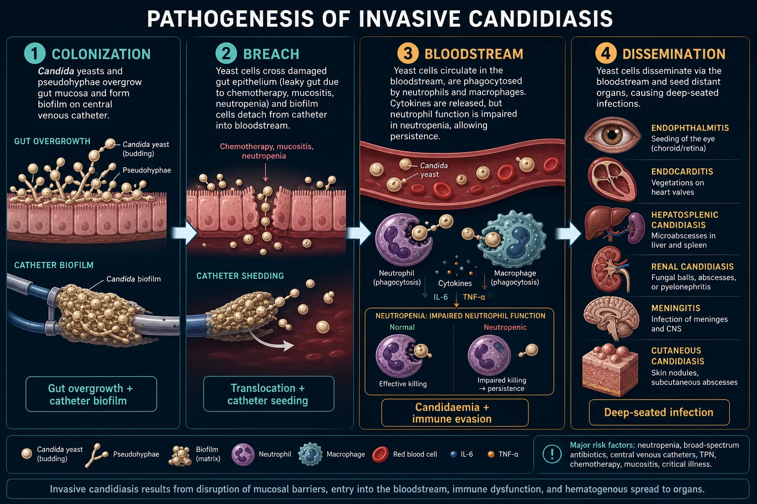
Risk factors and Candida score
Invasive candidiasis in the ICU is fundamentally a disease of disrupted host defences and iatrogenic exposures. The central venous catheter is the #1 risk factor and the source in up to 70% of ICU-acquired cases — Candida species readily form biofilms on intravascular material. Heavy colonisation (especially at multiple non-contiguous sites) precedes invasion: the more sites colonised, the higher the risk of bacteraemia. [1]
Risk factors
For invasive candidiasis
- Central venous catheter (the #1 risk factor — biofilm formation; source in up to 70%)
- Total parenteral nutrition (TPN) — promotes GI translocation and line colonisation
- Broad-spectrum antibiotics (prolonged course) — disrupts gut flora, permits Candida overgrowth
- Abdominal surgery (especially GI perforation, anastomotic leak, repeat laparotomy)
- Acute severe pancreatitis — pancreatic necrosis is a nidus for Candida
- Prolonged ICU stay (>7 days; risk rises sharply after 10-14 days)
- Immunosuppression (neutropenia, high-dose corticosteroids, solid organ or stem cell transplant)
- Haemodialysis (vascular access + immunosuppression of uraemia)
- Diabetes mellitus (hyperglycaemia promotes Candida growth and impaired neutrophil function)
- Malignancy, especially haematological (leukaemia, lymphoma, post-chemotherapy neutropenia)
- Severe burns (loss of skin barrier + colonisation)
- Prior Candida colonisation at multiple sites (the strongest single predictor)
Candida score
Leon 2006 — bedside screening tool
- Score ≥3 = HIGH RISK → consider empirical/preemptive antifungal (start therapy without waiting for cultures)
- Components (1 point each): severe sepsis OR septic shock (+1.0); total parenteral nutrition (+1.0); broad-spectrum antibiotics (+1.0); surgery (+0.5 weighting); Candida colonisation at multiple sites is implicit in the screening population
- Simplified bedside version: 1 point each for — severe sepsis, TPN, broad-spectrum antibiotics, candiduria (positive urine culture)
- Limitation: LOW SPECIFICITY (most patients with score ≥3 do NOT have invasive candidiasis) — use as a trigger for investigation/empirical therapy, not standalone diagnosis
- Validated for NON-NEUTROPENIC critically ill patients; do NOT apply in neutropenia (use a different paradigm)
- A score ≥3 carries ~3x higher likelihood of invasive candidiasis vs lower scores
Colonisation index
Alternative risk measure
- Colonisation index = number of non-blood sites positive for Candida / total number of sites cultured
- Index ≥0.5 = high risk of progression to invasive candidiasis (in neutropenic/haematology patients)
- More labour-intensive than Candida score; used in haemato-oncology rather than general ICU
- Both the score and the colonisation index are PROXY measures — confirmation requires positive blood culture or sterile-site culture
Diagnosis
Diagnosis of invasive candidiasis is challenging — blood cultures are the gold standard but lack sensitivity (only 50% sensitivity in deep-seated candidiasis) and take 48-72 hours to turn positive. Adjunctive biomarkers help bridge the gap. Two negative blood cultures do NOT exclude invasive candidiasis — deep-seated infection (hepatosplenic, endocarditis) may be culture-negative. [1]
Blood cultures
Gold standard
- The gold standard — positive culture from a sterile site confirms invasive candidiasis
- Limitation: SLOW (48-72 h to positivity) and only ~50% sensitive for deep-seated disease
- Draw BEFORE starting antifungals where possible; use aerobic AND anaerobic (BACTEC/MycoF) bottles
- At least 2 sets from different sites; if central line present, draw paired peripheral + line cultures
- Persistent candidaemia >72 h despite appropriate therapy → search for deep focus (echo, ophthalmology, imaging)
- Species identification AND susceptibility testing guides step-down therapy
Beta-D-glucan
Cell wall marker
- Detects (1→3)-β-D-glucan — a component of MOST fungal cell walls (Candida, Aspergillus, Pneumocystis; NOT Mucorales or Cryptococcus which lack it)
- SENSITIVE (~75-80%) but NON-SPECIFIC (~70-80%) for invasive candidiasis
- Two consecutive positive results (>80 pg/mL) in a high-risk patient supports empirical therapy
- FALSE POSITIVES: IV immunoglobulin, albumin, haemodialysis with cellulose membranes, cotton gauze swabs, recent surgery (sponges), certain antibiotics (piperacillin-tazobactam historically, amoxicillin-clavulanate)
- FALSE NEGATIVES: C. krusei and C. parapsilosis produce less β-D-glucan; localised infection without fungaemia
- Serial values useful to monitor response — falling titres suggest response
T2Candida panel
Rapid molecular
- T2 magnetic resonance — detects Candida DNA directly in whole blood within 3-5 hours
- Identifies the 5 common species (C. albicans, C. tropicalis, C. parapsilosis, C. krusei, C. glabrata)
- High sensitivity (>90%) and specificity (>90%) vs blood culture
- Limitation: expensive, not widely available, detects DNA (may include dead organisms — does not distinguish active infection)
- Best used as a rapid rule-OUT in high-risk patients with negative conventional cultures
Mannan / anti-mannan
Antigen/antibody
- Mannan (cell wall antigen) + anti-mannan antibody — combined assay
- Sensitivity moderate (~60-80%), specificity higher (~80-90%)
- More useful for chronic/deep-seated candidiasis than acute candidaemia
- Not routinely used in ICU in most centres; serial testing improves yield
Treatment
Candidaemia management protocol — IDSA 2016
1. Echinocandin FIRST-LINE
Caspofungin 70 mg loading then 50 mg daily. OR micafungin 100 mg daily. OR anidulafungin 200 mg loading then 100 mg daily. Echinocandins are preferred because: (a) candidacidal (not just static) — they inhibit β-1,3-glucan synthase in the cell wall; (b) broad Candida coverage including fluconazole-resistant C. glabrata and intrinsically resistant C. krusei; (c) few drug interactions vs azoles; (d) proven superior to amphotericin B and at least equivalent to fluconazole in outcomes. Exception: if C. parapsilosis suspected or identified (reduced echinocandin susceptibility — paradoxical), prefer fluconazole if susceptible.
2. Source control — REMOVE central line
Remove the central venous catheter as soon as possible — it is the source in up to 70% of ICU cases. The IDSA recommends catheter removal in ALL patients with candidaemia where the catheter is plausible source. Studies show catheter removal reduces duration of bacteraemia and improves mortality. If no alternative access exists, change over guidewire AND add antibiotic/antifungal lock therapy. Remove any other infected foreign material (urinary catheter, surgical hardware) if implicated.
3. Ophthalmology review
ALL patients with candidaemia require dilated fundoscopy by an ophthalmologist — ideally within the first week. Endophthalmitis occurs in 5-10% and may be asymptomatic initially. Lesions are typically yellow-white chorioretinal infiltrates with vitritis. If present: intravitreal amphotericin B ± voriconazole PLUS systemic fluconazole (excellent ocular penetration) — echinocandins do NOT achieve adequate intraocular levels. Delayed treatment → permanent visual loss. Repeat exam during therapy as lesions can develop or progress.
4. Switch fluconazole only if C. parapsilosis identified
If blood cultures grow C. parapsilosis, switch to fluconazole (if susceptible) because C. parapsilosis has reduced echinocandin susceptibility due to FKS mutation. Otherwise continue echinocandin until species and susceptibilities are known.
5. Step down to fluconazole when stable
Once clinically stable (usually after 5-7 days of echinocandin), blood cultures negative, AND species identified as fluconazole-susceptible (C. albicans, C. parapsilosis, C. tropicalis): step down to oral or IV fluconazole 400 mg daily (after a 800 mg loading dose if naive). If C. glabrata: step down only if fluconazole-susceptible or voriconazole-susceptible; otherwise continue echinocandin. C. krusei: never use fluconazole (intrinsic resistance) — continue echinocandin or switch to voriconazole.
6. Duration — 2 weeks after first negative culture
Treat for 14 days AFTER the first negative blood culture AND clinical resolution of symptoms/signs. Longer if: metastatic infection (endophthalmitis, endocarditis, osteomyelitis, hepatosplenic), persistent bacteraemia, or unstable patient. Perform ECHOCARDIOGRAPHY if persistent bacteraemia beyond 72 h despite appropriate therapy — to exclude Candida endocarditis (which needs 6+ weeks and surgical evaluation).
7. Investigate for deep-seated focus
Persistent candidaemia despite appropriate therapy (>72 h) or relapse after completion: search for occult focus with echocardiography (endocarditis), ophthalmology (endophthalmitis), abdominal CT (hepatosplenic candidiasis, pancreatic abscess), and consider infected thrombus (remove or anticoagulate). Persistent infection usually means SOURCE CONTROL failure, not drug failure.
Antifungal agents — mechanisms, spectrum, and key toxicities
Echinocandins
Caspofungin, micafungin, anidulafungin
- MECHANISM: inhibit β-1,3-glucan synthase → disrupt fungal cell wall → CANDIDACIDAL
- Spectrum: excellent for MOST Candida species (including azole-resistant C. glabrata, C. krusei); limited activity vs C. parapsilosis (paradoxical); limited activity vs Aspergillus (only growth inhibition, not killing)
- First-line for candidaemia and empirical therapy in ICU
- Toxicity: well tolerated — histamine release with rapid infusion (flushing), mild transaminitis; rare hepatic dysfunction
- Caspofungin: dose increase to 70 mg daily if patient on rifampicin or >80 kg (some guidance)
- Micafungin: does not require dose adjustment for hepatic/renal impairment
- Anidulafungin: no dose adjustment needed for any organ dysfunction
Fluconazole
Triazole
- MECHANISM: inhibits fungal lanosterol 14α-demethylase (CYP51) → depletes ergosterol → static
- Spectrum: C. albicans, C. parapsilosis, C. tropicalis (usually susceptible); REDUCED activity vs C. glabrata; INTRINSIC RESISTANCE in C. krusei
- Step-down therapy when species susceptible and patient stable; prophylaxis in selected high-risk
- Excellent bioavailability (oral = IV); good CSF, urine, ocular penetration
- Toxicity: QT prolongation, hepatitis, drug interactions (inhibits CYP2C9/3A4 — interacts with warfarin, phenytoin, tacrolimus, statins)
- Dose adjustment required for renal function (eGFR <50)
Voriconazole
Triazole — first-line for aspergillosis
- MECHANISM: as fluconazole but broader fungal spectrum
- Spectrum: Aspergillus (first-line), most Candida (including C. krusei), Scedosporium, Fusarium; NOT Mucorales
- First-line for invasive aspergillosis; alternative for C. krusei or fluconazole-resistant candidiasis
- Excellent bioavailability; variable metabolism (CYP2C19 polymorphism) → THERAPEUTIC DRUG MONITORING mandatory
- Toxicity: hepatotoxicity, visual disturbances (photopsia — benign but alarming), skin reactions (photosensitivity, SCC with chronic use), QT prolongation, periostitis, hallucinations
- Multiple drug interactions (CYP2C19/2C9/3A4 substrate and inhibitor)
Liposomal amphotericin B
Polyene — broad spectrum
- MECHANISM: binds ergosterol → forms pores in fungal membrane → cell death; CANDIDACIDAL
- Broadest antifungal spectrum — Candida, Aspergillus, Cryptococcus, Mucorales, dimorphic fungi (Histoplasma)
- Indicated for: resistant Candida (C. auris, C. glabrata not responding to echinocandin), Mucorales (first-line with surgical debridement), Cryptococcus (with flucytosine)
- Liposomal formulation (AmBisome) preferred — much less nephrotoxicity than conventional deoxycholate form
- Toxicity: nephrotoxicity (dose-related — pre-hydrate, monitor creatinine/electrolytes; loss of K+, Mg2+), infusion reactions (chills, fever — premedicate), anaemia (bone marrow suppression)
- Dose: 3-5 mg/kg/day liposomal (vs 0.6-1 mg/kg/day conventional deoxycholate)
Isavuconazole
Triazole — newer
- MECHANISM: as voriconazole; broader spectrum including Mucorales
- Spectrum: Aspergillus, Mucorales (first-line alternative), most Candida
- Advantages: fewer drug interactions, better tolerated, no visual disturbances, longer half-life (once daily)
- SECURE trial: non-inferior to voriconazole for aspergillosis with fewer adverse events; VITAL trial established activity against Mucorales
- Toxicity: hepatotoxicity, gastrointestinal; QT SHORTENING (unique — opposite to other azoles)
Flucytosine (5-FC)
Pyrimidine analogue
- MECHANISM: converted to 5-fluorouracil → inhibits fungal DNA/RNA synthesis
- Spectrum: Cryptococcus, some Candida — NEVER used as monotherapy (rapid resistance)
- Always combined: with amphotericin B for Cryptococcus meningitis; with amphotericin for selected Candida (endocarditis, meningitis)
- Toxicity: bone marrow suppression (anaemia, leukopenia, thrombocytopenia), hepatotoxicity, colitis — dose adjust for renal function; monitor levels
Candida auris — the emerging multidrug-resistant threat
Candida auris was first described in 2009 (isolated from the ear canal of a Japanese patient) and has since emerged globally as a cause of healthcare-associated outbreaks with high mortality. It is the first globally spreading fungal pathogen with true multidrug resistance and has been declared an urgent threat by the CDC and a priority pathogen by the WHO. [1]
Microbiology
Why it is dangerous
- Often MISIDENTIFIED by conventional biochemical systems (VITEK 2, API) — may be reported as C. haemuloni, C. famata, or Rhodotorula
- Requires MALDI-TOF mass spectrometry or molecular methods (PCR, sequencing of D1/D2 or ITS regions) for accurate identification
- Resistant to most disinfectants — survives on surfaces and equipment for weeks
- Grows at 42°C and in high-salt conditions — persistent in hospital environments
Resistance
Antifungal profile
- Nearly all strains RESISTANT to fluconazole (>90%)
- Variable resistance to voriconazole and other azoles (30-50%)
- Reduced susceptibility to amphotericin B in a substantial proportion (some isolates MIC ≥2)
- Most strains still SUSCEPTIBLE to echinocandins (the first-line), but echinocandin resistance has emerged — must test
- Some isolates are pan-resistant (resistant to all three classes)
- Antifungal susceptibility testing is MANDATORY for every C. auris isolate
Infection control
Containment
- CONTACT ISOLATION in a single room with dedicated equipment (stethoscope, BP cuff, thermometer)
- Gown and gloves for ALL entries — strict hand hygiene (alcohol gel works, but wash if hands visibly soiled)
- Dedicated nursing staff where possible (cohorting) — limit staff movement between cases
- Environmental cleaning with sporicidal agent (bleach / hydrogen peroxide vapour)
- Screening of close contacts (axilla/groin swabs, nares) to detect colonisation
- Notify public health; report to local surveillance system
- Decolonisation generally NOT effective — focus on preventing transmission
Treatment
IDSA interim guidance
- First-line: ECHINOCANDIN (caspofungin/micafungin/anidulafungin) — while awaiting susceptibilities
- If echinocandin-resistant or pan-resistant: combination therapy or high-dose liposomal amphotericin B (if susceptible)
- Source control paramount — REMOVE infected lines, drain abscesses, debride infected tissue
- Infectious diseases consultation MANDATORY for all C. auris infections
- Monitor for persistent bacteraemia — high rate of deep-seated infection
Suspected C. auris in your ICU — what to do
1. Recognise the red flags
Candida species with atypical susceptibility (fluconazole-resistant from a non-haematology patient), outbreak of yeast infections in the ICU, or patient recently admitted from a facility with known C. auris. International transfer, especially from South Asia, Middle East, Africa, or South America, raises index of suspicion.
2. Confirm identification
Send isolate for MALDI-TOF or molecular confirmation (PCR). Do not accept a biochemical identification as final. Notify the laboratory that C. auris is suspected so they use the correct database.
3. Isolate immediately
Place the patient in a single room under CONTACT precautions with dedicated equipment. Do not wait for confirmation — act on suspicion. Screen close contacts (axilla/groin swabs).
4. Treat with echinocandin + source control
Start echinocandin immediately. Send for full susceptibility testing (fluconazole, voriconazole, amphotericin B, echinocandin). Remove any infected central line. Escalate based on susceptibilities — consider combination therapy or amphotericin B if echinocandin-resistant.
5. Notify and screen
Notify infection control, microbiology, public health. Screen all room contacts and recent ward contacts. Audit environmental cleaning. Consider ward closure to new admissions if outbreak declared.
Invasive pulmonary aspergillosis
Invasive pulmonary aspergillosis (IPA) was traditionally a disease of severely immunocompromised patients (prolonged neutropenia, stem cell transplant). It is now increasingly recognised in the non-neutropenic ICU population — COPD exacerbations treated with prolonged steroids, cirrhosis, severe influenza, COVID-19, and after liver transplantation. Mortality in ICU-acquired IPA is 50-80%, driven by delayed diagnosis and underlying disease severity. [1]
The diagnostic framework for the non-neutropenic ICU patient differs from the classical EORTC/MSG criteria (which apply to haematology patients). The AspICU criteria (Blot 2012) provide a clinically applicable algorithm using host factors, clinical features, and mycological evidence. [1]
Risk groups
Who gets aspergillosis
- Classical high-risk: prolonged neutropenia (>10 days, post-chemotherapy), haematological malignancy (AML, MDS), allogeneic stem cell transplant (especially GVHD), solid organ transplant (lung, liver)
- Severe inherited immunodeficiency (chronic granulomatous disease, hyper-IgE syndrome)
- Increasingly recognised in ICU: COPD exacerbation on prolonged/high-dose steroids (≥0.8 mg/kg/day prednisolone equivalent for >2 weeks), decompensated cirrhosis, severe influenza/COVID-19 (CAPA — COVID-19-associated pulmonary aspergillosis), severe ARDS, burns
- Chronic pulmonary aspergillosis: structural lung disease (cavitary TB, bronchiectasis, COPD with apical bullae), sarcoidosis
- Post-influenza aspergillosis (IAPA): newly described entity — severe influenza (ICU) + Aspergillus within days of admission; high mortality even in immunocompetent hosts
Diagnosis — AspICU criteria
Blot 2012
- AspICU algorithm (for non-neutropenic ICU): does NOT require classical host factors
- Requires: Aspergillus in respiratory specimen (BAL or sputum culture/PCR/cytology) PLUS one of: (a) compatible clinical/radiological picture (new infiltrate, pleuritic chest pain, haemoptysis), (b) galactomannan positive (BAL OD index ≥0.5 or serum ≥0.5), (c) b-D-glucan positive
- CT chest: HALO SIGN (nodule with ground-glass halo = early haemorrhagic infarction, ~day 0-5); AIR CRESCENT SIGN (cavity forming as necrotic tissue separates, ~day 10-14); wedge-shaped infarct
- Galactomannan: BAL fluid preferred (sensitivity ~80-90%, specificity ~85-95%) over serum (sensitivity lower ~40-50% in non-neutropenic)
- Beta-D-glucan: NON-SPECIFIC — positive in Candida, Aspergillus, Pneumocystis; negative in Mucorales and Cryptococcus
- Culture/histology: definitive (hyphae invading tissue on biopsy) but rarely feasible in unstable ICU patients
- Aspergillus PCR: emerging — high sensitivity, rapid; useful as rule-out; not standardised
- FALSE POSITIVE galactomannan: piperacillin-tazobactam (historically; now less common), amoxicillin-clavulanate, Plasmalyte, GI translocation of dietary galactomannan, gluconate-containing fluids, twin gestation in neonates
Differential of halo sign
Not always aspergillus
- Invasive aspergillosis (most common in immunocompromised)
- Mucormycosis (need different therapy — amphotericin B + surgery; voriconazole does NOT work)
- Bacterial pneumonia (Pseudomonas, Staphylococcus, Nocardia)
- Viral pneumonitis (CMV, influenza, HSV)
- Pulmonary haemorrhage, granulomatosis with polyangiitis, neoplasm
- The REVERSE halo sign (atoll sign) is more suggestive of MUCORMYCOSIS or organising pneumonia
Aspergillosis treatment
[1]Suspected invasive aspergillosis in the non-neutropenic ICU patient
1. Identify the at-risk patient
COPD on prolonged steroids, cirrhosis, severe influenza/COVID-19, post-liver transplant, prolonged neutropenia, GVHD. New or worsening infiltrate, pleuritic chest pain, or haemoptysis in these patients → consider IPA. Do not wait for definitive signs — mortality rises with delay.
2. Image and sample
CT chest (look for halo sign, air crescent sign, wedge infarct, reverse halo). Perform bronchoscopy with BAL: send for Aspergillus culture, galactomannan (BAL OD index ≥0.5 positive), PCR, and cytology. Send serum galactomannan and beta-D-glucan. Do not delay empirical therapy if high suspicion.
3. Start empirical voriconazole immediately
Voriconazole 6 mg/kg IV BD x 2 doses, then 4 mg/kg IV BD. Send trough level after 5-7 days (target 1-5.5 mg/L). If CYP2C19 poor metaboliser (or toxicity): switch to isavuconazole. If Mucorales cannot be excluded (reverse halo sign, diabetic ketoacidosis, voriconazole prophylaxis breakthrough): start liposomal amphotericin B until Mucorales excluded.
4. Reassess and adjust
Repeat CT and galactomannan at 1-2 weeks. Falling galactomannan and clinical/radiological improvement = response. Persistent/rising galactomannan or progressive infiltrate = treatment failure → reconsider diagnosis (Mucorales? resistant Aspergillus?), switch agent (amphotericin B, posaconazole, combination), check source (persistent immunosuppression, cavitation).
5. Reduce immunosuppression if possible
Wean corticosteroids to the lowest effective dose. In neutropenia, use G-CSF. Reduce calcineurin inhibitors if feasible (balance with graft risk). Treat underlying driver (influenza with oseltamivir, COVID-19 with antivirals).
6. Duration and follow-up
Minimum 6-12 weeks, longer if immunosuppression persists. Step down to oral voriconazole/isavuconazole when stable and absorbing. Continue until clinical, radiological, and mycological resolution. Lifelong secondary prophylaxis may be needed if immunosuppression cannot be reversed.
Brief note on other ICU fungal infections
While candidaemia and aspergillosis dominate, several other fungal infections appear in specific ICU populations. Distinguishing them matters because the treatment is entirely different. [1]
Mucormycosis (Mucorales)
Rhinocerebral / pulmonary
- Risk: DIABETIC KETOACIDOSIS (#1 — iron acquisition in acidosis), neutropenia, deferoxamine therapy, voriconazole prophylaxis breakthrough
- Angioinvasive — rapid necrosis, black eschar (rhinocerebral), pulmonary infarcts
- RESISTANT to voriconazole and echinocandins — first-line is LIPOSOMAL AMPHOTERICIN B (high dose 5-10 mg/kg/day) plus URGENT SURGICAL DEBRIDEMENT
- Reverse halo sign on CT more suggestive of mucormycosis than aspergillosis
- Mortality 50-90% — depends on speed of debridement and reversal of underlying cause
Cryptococcus
Meningitis / disseminated
- Risk: HIV/AIDS (CD4 <100), solid organ transplant, idiopathic CD4 lymphocytopenia
- Presents as subacute meningitis (headache, fever, altered mental state); can cause ARDS
- Diagnosis: serum and CSF cryptococcal antigen (CrAg — sensitive); India ink CSF; culture
- Treatment: liposomal amphotericin B + flucytosine for induction (2 weeks) → fluconazole consolidation (8 weeks) → fluconazole maintenance (6-12 months)
- Watch for IRIS (immune reconstitution inflammatory syndrome) when starting antiretrovirals
- Manage raised intracranial pressure with serial lumbar punctures / shunt
Pneumocystis jirovecii pneumonia
PCP
- Risk: HIV/AIDS (CD4 <200), transplant, prolonged steroids, malignancy on therapy
- Hypoxic pneumonitis with relatively clear chest X-ray early; classic ground-glass on CT
- Diagnosis: elevated beta-D-glucan (NOT galactomannan), raised LDH, silver stain/MGG stain or PCR on induced sputum/BAL
- Treatment: high-dose co-trimoxazole (trimethoprim-sulfamethoxazole) 15-20 mg/kg/day TMP; ADD STEROIDS if PaO2 <70 mmHg or A-a gradient >35
- Prophylaxis: co-trimoxazole in patients on >20 mg prednisolone for >4 weeks (or equivalent)
Endemic mycoses
Histoplasma, Blastomyces, Coccidioides
- Geographic exposure (Ohio/Mississippi river valleys for histoplasmosis; southwest USA for coccidioidomycesis)
- Disseminated disease in immunosuppressed — can mimic TB or bacterial sepsis
- Treatment: liposomal amphotericin B (severe) → itraconazole step down
- Consider in returning travellers or in regions of endemicity
Antifungal prophylaxis and empirical therapy strategies
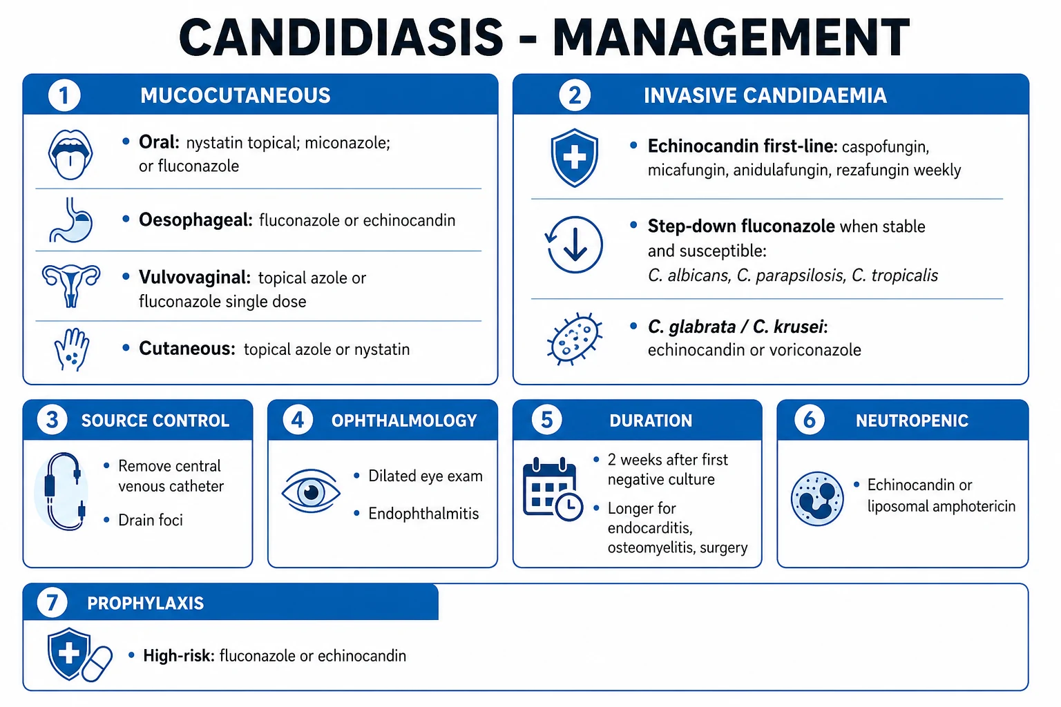
Prophylaxis
Preventing infection in high-risk
- Fluconazole 400 mg/day: liver transplant (first 4 weeks, longer if high-risk), prolonged neutropenia (haematology), stem cell transplant with GVHD
- Posaconazole: neutropenia with AML/MDS post-chemotherapy, GVHD (covers Aspergillus — fluconazole does not)
- Micafungin: alternative in neutropenia if azoles not tolerated
- Routine prophylaxis in GENERAL ICU patients is NOT recommended — does not improve mortality, drives resistance, high cost
- Targeted prophylaxis may be considered in units with high incidence of invasive candidiasis (>10%)
Empirical therapy
Treat on suspicion
- Start antifungal in HIGH-RISK patient with CLINICAL SUSPICION (sepsis, new fever, signs of infection) without microbiological confirmation — most commonly in neutropenic sepsis with persistent fever beyond 4-7 days
- Agent: echinocandin or liposomal amphotericin B (covers Aspergillus too)
- Candida score ≥3 + clinical signs: empirical echinocandin reasonable
- Empirical therapy should NOT be routine in all ICU patients with fever — drives resistance and exposes patients to toxicity
Preemptive therapy
Treat on biomarker
- Start antifungal based on BIOMARKER positivity (β-D-glucan, galactomannan, T2 Candida) in a high-risk patient even without clinical signs
- Most stewardship-friendly approach — avoids over-treatment
- Requires serial biomarker testing infrastructure
- Risk: false positives lead to unnecessary exposure
TrialCard — landmark evidence
Herbrecht 2002 (NEJM) — voriconazole vs amphotericin B for primary therapy of invasive aspergillosis
Design
Multicentre, randomised, open-label; 277 patients with definite or probable invasive aspergillosis (mostly haematology/neutropenia)
Intervention
Voriconazole vs amphotericin B deoxycholate (as primary therapy)
Primary outcome
Successful response at 12 weeks: 52.8% voriconazole vs 31.6% amphotericin B; voriconazole also improved SURVIVAL (70.8% vs 57.9% at 12 weeks)
Safety
Voriconazole better tolerated; amphotericin B had more nephrotoxicity and infusion reactions
Clinical bottom line
Established VORICONAZOLE as first-line therapy for invasive aspergillosis — replaced amphotericin B as the standard. The landmark study for aspergillosis treatment.
Mora-Duarte 2002 (NEJM) — caspofungin vs amphotericin B for invasive candidiasis
Design
Multicentre, randomised, double-blind, non-inferiority; 224 patients with invasive candidiasis (mostly non-neutropenic)
Intervention
Caspofungin 70 mg load then 50 mg/day vs amphotericin B deoxycholate
Primary outcome
Modified intention-to-treat: caspofungin 73.4% vs amphotericin B 61.7% — caspofungin NON-INFERIOR and showed superior efficacy
Safety
Caspofungin significantly fewer drug-related adverse events; amphotericin B had nephrotoxicity (rising creatinine, hypokalaemia), infusion reactions
Clinical bottom line
Established echinocandin as equivalent or superior to amphotericin B with markedly better tolerability. Paved the way for echinocandin as first-line for candidaemia.
Reboli 2007 — REVIVE trial: anidulafungin vs fluconazole for invasive candidiasis (NEJM)
Design
Multicentre, randomised, double-blind, non-inferiority; 245 patients with candidaemia/invasive candidiasis
Intervention
Anidulafungin 200 mg load then 100 mg/day vs fluconazole 800 mg load then 400 mg/day
Primary outcome
Global response at end of IV therapy: anidulafungin 75.6% vs fluconazole 60.2% — anidulafungin NON-INFERIOR (and superior in modified analysis)
Safety
Similar adverse event rates; both well tolerated
Clinical bottom line
Echinocandins are at least equivalent to fluconazole for invasive candidiasis, with better activity against fluconazole-resistant species (C. glabrata, C. krusei) — supports echinocandin as first-line.
Leon 2006 — derivation of the Candida score (Crit Care Med)
Design
Multicentre, prospective, observational cohort; 1,699 non-neutropenic ICU patients with Candida colonisation
Aim
Derive a bedside score to predict invasive candidiasis (positive blood or sterile-site culture)
Result
Four independent predictors (1 point each): severe sepsis/septic shock, total parenteral nutrition, broad-spectrum antibiotics, surgery. Score ≥3 = high risk of invasive candidiasis (sensitivity ~75%, specificity ~60%)
Validation
Subsequently validated in independent cohorts — score ≥3 carries ~3x likelihood of invasive candidiasis
Clinical bottom line
The standard bedside screening tool in non-neutropenic ICU patients — use a score ≥3 to trigger investigation/empirical therapy, but recognise the LOW SPECIFICITY. Do not apply in neutropenia.
Ostrosky-Zeichner 2005 — performance of (1→3)-β-D-glucan assay for invasive fungal infection (Clin Infect Dis)
Design
Multicentre, prospective, case-control evaluation; 163 patients with proven invasive fungal infection vs controls
Test
Fungitell assay — serum (1→3)-β-D-glucan
Performance
For invasive candidiasis: sensitivity 80%, specificity 83% (single positive); two consecutive positives improved specificity. For aspergillosis: sensitivity ~70-80%
Key teaching
β-D-glucan is SENSITIVE but NON-SPECIFIC — positive in Candida, Aspergillus, Pneumocystis, Fusarium; NEGATIVE in Cryptococcus and Mucorales (they do not produce β-D-glucan). Many false positives (IVIG, albumin, dialysis, certain antibiotics, gauze).
Clinical bottom line
Best used as a RULE-OUT in high-risk patients (two negatives makes invasive fungal infection unlikely) and to support empirical therapy when serially positive in a high-risk patient. Do not interpret in isolation.
Blot 2012 — AspICU study: a clinical algorithm to diagnose invasive pulmonary aspergillosis in critically ill patients (AJRCCM)
Design
Multicentre, retrospective cohort; 224 ICU patients with Aspergillus-positive respiratory specimens
Problem
Classical EORTC/MSG criteria are designed for haematology patients with neutropenia — most ICU cases have NO classical host factors
Algorithm (AspICU)
Aspergillus in BAL/sputum PLUS one of: compatible clinical/radiological picture; positive galactomannan (BAL OD ≥0.5 or serum ≥0.5); positive beta-D-glucan
Outcome
Mortality in proven/probable IPA was ~60-70% in ICU. AspICU algorithm correctly identified disease without requiring neutropenia or classical radiology.
Clinical bottom line
Established the operational framework for diagnosing IPA in the non-neutropenic ICU patient — now standard alongside galactomannan and CT. Always pair with culture and imaging.
Lockhart 2017 — simultaneous emergence of multidrug-resistant Candida auris on 3 continents (Clin Infect Dis)
Design
Multicentre molecular epidemiology; 54 C. auris isolates from Pakistan, India, South Africa, Venezuela
Key findings
Four distinct clades emerged independently on three continents (South Asia, South Africa, South America) — each clonal within region. Nearly all resistant to fluconazole; 35% to voriconazole; variable to amphotericin B; most susceptible to echinocandins
Significance
C. auris is the first globally spreading fungal pathogen with true MDR; identification challenges (misidentified by biochemical systems) and environmental persistence drive outbreaks
Clinical bottom line
Defines C. auris as a unique infection control and therapeutic challenge — requires accurate identification (MALDI-TOF/molecular), contact isolation, screening of contacts, and echinocandin first-line with susceptibility testing.
SAQs
SAQ — Candidaemia in a non-neutropenic ICU patient
10 minutes · 10 marks
A 58-year-old man, day 12 of an unplanned ICU admission for severe necrotising pancreatitis, has a triple-lumen right internal jugular CVC, is on total parenteral nutrition and broad-spectrum meropenem, and has Candida colonisation at multiple sites. He develops a new fever (39.2°C) with hypotension and a rising lactate. Blood cultures drawn from a peripheral vein and from the catheter both grow Candida albicans at 36 hours.
SAQ — Invasive pulmonary aspergillosis in a non-neutropenic ICU patient
10 minutes · 10 marks
A 67-year-old woman with COPD on long-term prednisolone 30 mg/day, admitted with an acute exacerbation treated with a further 5-day course of IV hydrocortisone, develops worsening hypoxia and new pleuritic chest pain on ICU day 3. CT chest shows multiple nodules with surrounding ground-glass halos. Bronchoscopy grows Aspergillus fumigatus; BAL galactomannan OD index is 5.2. She is not neutropenic.
Clinical pearls
Red flags
References
- [1]Kullberg BJ, Arendrup MC. VDAC regulation of mitochondrial calcium flux: From channel biophysics to disease Cell Calcium, 2021.PMID 33529977
- [2]Lortholary O, et al. Notum palmitoleoyl-protein carboxylesterase regulates Fas cell surface death receptor-mediated apoptosis via the Wnt signaling pathway in colon adenocarcinoma Bioengineered, 2021.PMID 34402722
- [3]Pappas PG, Kauffman CA, Andes DR, et al. Clinical Practice Guideline for the Management of Candidiasis: 2016 Update by the Infectious Diseases Society of America Clin Infect Dis, 2016.PMID 26679628
- [4]Herbrecht R, Denning DW, Patterson TF, et al. Allergic skin and systemic reactions in a patient with pure red cell aplasia and anti-erythropoietin antibodies challenged with different epoetins J Am Soc Nephrol, 2002.PMID 12191983
- [5]Mora-Duarte J, Betts R, Rotstein C, et al. Characterization of freezing tolerance and vernalization in Vern-, a spring-type Brassica napus line derived from a winter cross Planta, 2002.PMID 12447535
- [6]Reboli AC, Rotstein C, Pappas PG, et al. MRI patterns of atrophy associated with progression to AD in amnestic mild cognitive impairment Neurology, 2008.PMID 17898323
- [7]Leon C, Ruiz-Santana S, Saavedra P, et al. Evaluation of nutritional status in children with refractory epilepsy Nutr J, 2006.PMID 16640779
- [8]Ostrosky-Zeichner L, Alexander BD, Kett DH, et al. [gamma ]-Hydroxybutyrate poisoning: Poisoning from toy beads BMJ, 2008.PMID 18202042
- [9]Ullmann AJ, Cornely OA, Burchardt A, et al. Spatial compartmentalization of tumor necrosis factor (TNF) receptor 1-dependent signaling pathways in human airway smooth muscle cells. Lipid rafts are essential for TNF-alpha-mediated activation of RhoA but dispensable for the activation of the NF-kappaB and MAPK pathways J Biol Chem, 2006.PMID 16982613
- [10]Patterson TF, Thompson GR 3rd, Denning DW, et al. Long-term outcome of thalamic deep brain stimulation in two patients with Tourette syndrome J Neurol Neurosurg Psychiatry, 2010.PMID 20660922
- [11]Lockhart SR, Etienne KA, Vallabhaneni S, et al. Drug-Eluting vs Bare-Metal Stents in Patients With Chronic Kidney Disease and Coronary Artery Disease: Insights From a Systematic Review and Meta-Analysis J Invasive Cardiol, 2018.PMID 28915510
- [12]Blot SI, Taccone FS, Van den Abeele AM, et al. Tomato RNA polymerase II interacts with the rod-like conformation of the left terminal domain of the potato spindle tuber viroid positive RNA genome J Gen Virol, 2012.PMID 22422064
- [13]Playford EG, Eggimann P, Calandra T. Can wear explain the histological variation around metal-on-metal total hips? Clin Orthop Relat Res, 2015.PMID 25141844
- [14]Chowdhary A, Sharma C, Meis JF. A Potentially Volatile Situation Clin Chem, 2019.PMID 30923064