ICU · infectious-diseases
Acute Neutropenic Sepsis — Comprehensive ICU Management
Also known as Neutropenic sepsis · Febrile neutropenia · Neutropenic fever · Chemotherapy-induced neutropenia · MASCC score · Anti-pseudomonal coverage · Neutropenic enterocolitis
Neutropenic sepsis (febrile neutropenia) — a life-threatening infection in patients with neutrophil count <0.5 × 10^9/L (or <1.0 with expected decline) AND fever (38.3C single or 38.0C sustained over 1h). Occurs in cancer patients receiving cytotoxic chemotherapy (nadir 7-14 days post-chemotherapy), haematological malignancy, post-stem-cell transplant, or congenital neutropenia. Mortality: 5-10% overall (up to 30-50% with septic shock). The 1 rule: EMPIRICAL BROAD-SPECTRUM ANTIBIOTICS WITHIN 1 HOUR ('door-to-needle' time — just like sepsis). Do NOT wait for cultures or source identification. Pathogens: gram-negative bacilli (1 — Pseudomonas aeruginosa is the MOST DANGEROUS — has high mortality in neutropenic patients — must cover), gram-positive cocci (Staphylococcus, Streptococcus viridans — from mucositis), fungal (Candida, Aspergillus — if persistent fever 4-7 days despite antibiotics). Management: (1) empirical anti-pseudomonal beta-lactam (piperacillin-tazobactam 4.5 g IV q6h OR cefepime 2 g IV q8h OR meropenem 1 g IV q8h) WITHIN 1 HOUR, (2) blood cultures (2 sets from different sites + from any central line) BEFORE antibiotics if possible (but DO NOT delay 1 hour), (3) MASCC risk stratification (high-risk → IV antibiotics + ICU; low-risk → oral antibiotics + possible outpatient), (4) add vancomycin if line infection suspected, mucositis, or haemodynamic instability, (5) add antifungal (caspofungin or liposomal amphotericin) if fever persists 4-7 days despite antibiotics, (6) G-CSF (controversial — not routine — may benefit high-risk patients with expected prolonged neutropenia 7 days).
On this page & tools
Your progress
Saved locally on this device.
Target exams
Red flags
Overview
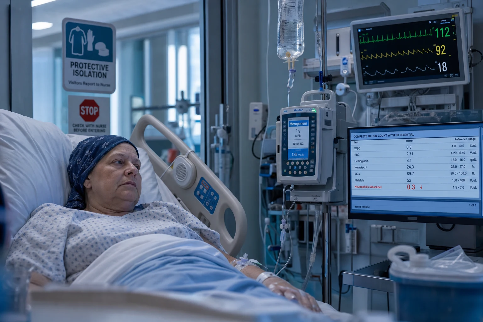
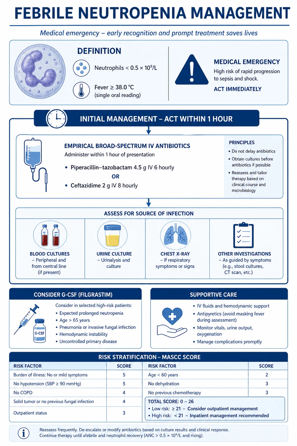
Neutropenic sepsis is one of the MOST TIME-CRITICAL infections in ICU. The neutropenic patient has NO localising signs — no abscess formation (no neutrophils), no purulent sputum, no localised erythema — the infection disseminates RAPIDLY → bacteraemia → septic shock → death within hours if untreated. The intensivist must have a VERY LOW threshold to give empirical broad-spectrum antibiotics in ANY cancer patient with fever.[1][2]
Pathogen biology — the immune defect dictates the pathogen
The single most useful framework for the exam: map the type of immune defect to the expected pathogen. Cytotoxic chemotherapy damages multiple arms simultaneously (it is myelosuppressive AND lymphocyte-toxic AND breaches mucosal barriers), so a real patient often has overlapping defects — but the dominant defect predicts the dominant threat. Know this table cold.[2][4]
Immune defect → expected pathogen (the exam table)
| Immune defect | Typical clinical setting | Bacteria | Fungi | Viruses | Key principle |
|---|---|---|---|---|---|
| Neutropenia (<0.5 × 10^9/L) | Cytotoxic chemo 7–14 days post, AML induction, post-HSCT pre-engraftment | Pseudomonas aeruginosa, E. coli, Klebsiella, Enterobacter, Serratia, Staph aureus, Strep viridans (via mucositis) | Aspergillus, Candida (hepatosplenic), Mucorales (rare) | — | Gram-negative bacteraemia is the immediate killer; anti-Pseudomonal cover is non-negotiable |
| T-cell defect / lymphopenia | HIV, transplant on calcineurin inhibitors, fludarabine, alemtuzumab, steroids, purine analogues | Listeria monocytogenes, Nocardia, Salmonella, Mycobacterium (TB/MAI), Legionella | Pneumocystis jirovecii, Cryptococcus, Histoplasma, Aspergillus (if steroid), Coccidioides | CMV, HSV, VZV, EBV, JC virus (PML) | CD4 count predicts the pathogen; co-trimoxazole prophylaxis covers PCP + Nocardia + Toxoplasma |
| B-cell defect / hypogammaglobulinaemia | CLL, multiple myeloma, post-rituximab, post-splenectomy, ibrutinib | Streptococcus pneumoniae, Haemophilus influenzae, Neisseria meningitidis (encapsulated organisms) | — | Enterovirus, Parvovirus B19 | Encapsulated organisms — conjugate + polysaccharide vaccination is critical; IVIG if profound |
| High-dose corticosteroids (≥0.3 mg/kg prednisone ≥3 weeks) | ALL induction, GVHD, lymphoma, autoimmune | Nocardia, Listeria, Staph, gram-negatives | Aspergillus (the classic steroid–fungus link), Pneumocystis, Candida, Mucorales | HSV, VZV, CMV reactivation | "Steroid + new pulmonary infiltrate = think Nocardia or Aspergillus until proven otherwise"; risk persists for weeks AFTER steroids stopped |
| Mucositis (breach of mucocutaneous barrier) | High-dose cytarabine, methotrexate, HSCT conditioning, anthracyclines | Streptococcus viridans, oral anaerobes, Enterococcus, coag-neg staph | Candida (oral/oesophageal) | HSV (reactivation in mucosal breaks) | Strep viridans bacteraemia can cause septic shock + ARDS ("viridans shock syndrome"); add vancomycin |
| Asplenia / hyposplenism | Post-splenectomy, sickle cell, radiation | Encapsulated: S. pneumoniae, H. influenzae b, N. meningitidis, Capnocytophaga (dog bites), Bordetella holmesii | Babesiosis (parasite) | — | "Overwhelming post-splenectomy infection" (OPSI) — minutes-to-hours sepsis; standing daily penicillin + vaccines |
| Complement blockade / deficiency | Eculizumab (anti-C5), congenital | Neisseria, S. pneumoniae | — | — | Eculizumab patients need meningococcal vaccine + standing penicillin prophylaxis |
Why neutropenia is uniquely dangerous
Neutrophils perform three jobs that nothing else replaces: (1) phagocytosis of extracellular bacteria and fungi, (2) abscess formation (walls off infection locally), and (3) oxidative killing via the myeloperoxidase–NADPH oxidase burst. Without neutrophils:[5]
- Bacteria are NOT contained locally → they enter the bloodstream within hours → bacteraemia → septic shock.
- No pus, no abscess, no localising signs. A neutropenic patient with pneumonia may have a normal chest X-ray (no infiltrate because there are no neutrophils to cause alveolar consolidation) — the infiltrate appears only at neutrophil recovery ("neutrophil recovery infiltrate").
- Cultures may be negative in 30–50% — the absence of neutrophils means less inflammatory amplification and a lower bacterial load detectable in a standard 20 mL blood-culture volume. [1]
This is why the 1-hour antibiotic rule exists: the neutropenic host has no "buffer". In a normal host, a few hours of untreated bacteraemia may be tolerated; in the neutropenic host, those same hours can be fatal. [1]
The Pseudomonas aeruginosa problem
Pseudomonas deserves its own exam emphasis because it is the single most lethal organism in neutropenic sepsis (mortality 20–40% in bacteraemia, up to 50% with shock). Reasons:[5]
- Intrinsic virulence — produces elastase, proteases, exotoxin A, and a Type III secretion system (ExoS, ExoU) that cause tissue destruction and immune evasion.
- Biofilm formation — colonises central venous catheters, urinary catheters and devitalised tissue (typhlitis, burns); biofilm resists both neutrophils and antibiotics.
- Natural resistance — low outer-membrane permeability, AmpC beta-lactamase, efflux pumps → inherently resistant to many agents (no ceftriaxone, no ampicillin, no tetracycline, no erythromycin).
- Rapid resistance acquisition — under antibiotic pressure it upregulates efflux pumps and acquires carbapenemases. [1]
Clinical sources: respiratory (esp. ventilated), central line, urinary tract, typhlitis/enterocolitis, skin. Two classic Pseudomonas skin signs every fellow should know:
- Ecthyma gangrenosum — Pseudomonas vasculitis → haemorrhagic/necrotic ulcer with an erythematous halo (septicaemic spread); classically axilla, groin, or perianal.
- Green nail syndrome — pyocyanin pigment staining nails (chronic, less acute). [1]
The moulds — Aspergillus and Mucorales
- Aspergillus fumigatus — angioinvasive in prolonged neutropenia (>10–14 days). Invades pulmonary vessels → haemoptysis (can be fatal), vessel thrombosis → pulmonary infarction. CT: halo sign (nodule with ground-glass halo = haemorrhage around an infarct) early; later air-crescent sign (air crescent as neutrophils return and necrotic tissue separates). Diagnosis: serum/BAL galactomannan, culture, biopsy. Treatment: voriconazole first-line (improved survival vs amphotericin in Herbrecht 2002 NEJM), isavuconazole, liposomal amphotericin B.[8]
- Mucorales (Rhizopus, Mucor, Absidia) — rare but devastating. Risk: prolonged neutropenia + high-dose steroids + diabetic ketoacidosis + iron overload/deferoxamine. Angioinvasive → rhinocerebral mucormycosis (black eschar in nasal cavity, facial pain, cranial nerve palsies) or pulmonary. Does NOT have a positive galactomannan (distinguishes from Aspergillus). Treatment: surgical debridement + liposomal amphotericin B + reverse immunosuppression. Mortality 50–80%.[8]
The viral reactivations
- HSV/VZV — reactivation is near-universal in HSV-seropositive HSCT recipients during conditioning + engraftment; oesophagitis, oral/genital ulcers, cutaneous dissemination, hepatitis, pneumonitis. Diagnosis: PCR. Treatment: IV aciclovir 5 mg/kg q8h (severe) or oral valaciclovir (mild). Prophylaxis: aciclovir 400 mg PO BD.
- CMV — T-cell defect driven; reactivation in HSCT (day 30–100) and solid-organ transplant. Monitor with quantitative PCR weekly; pre-emptive valganciclovir 900 mg BD when threshold reached (avoids universal-prophylaxis toxicity). Disease: pneumonitis, colitis, retinitis, hepatitis, CMV syndrome (fever + marrow suppression). Treatment: valganciclovir/ganciclovir ± foscarnet for resistance; letermovir for prophylaxis only.[4]
- EBV — drives post-transplant lymphoproliferative disorder (PTLD) after solid-organ or HSCT; reduction of immunosuppression + rituximab ± chemotherapy.
- Adenovirus — disseminated disease in HSCT (hepatitis, pneumonitis, haemorrhagic colitis); treat cidofovir (nephrotoxic) or brincidofovir.
The encapsulated-organism / B-cell defect
In hypogammaglobulinaemia (CLL, myeloma, post-rituximab) and asplenia, the threat is encapsulated bacteria — Streptococcus pneumoniae, Haemophilus influenzae type b, Neisseria meningitidis — and Capnocytophaga after dog bites. These cause overwhelming post-splenectomy infection (OPSI): Waterhouse-Friderichsen adrenal haemorrhage, DIC, death within hours. Empiric therapy: ceftriaxone 2 g (meningitis dose) ± vancomycin + azithromycin, add dexamethasone if pneumococcal meningitis. Prevention: conjugate + polysaccharide vaccination, daily phenoxymethylpenicillin, patient alert card/medic-alert bracelet.[4]
Management protocol — the 1-hour rule
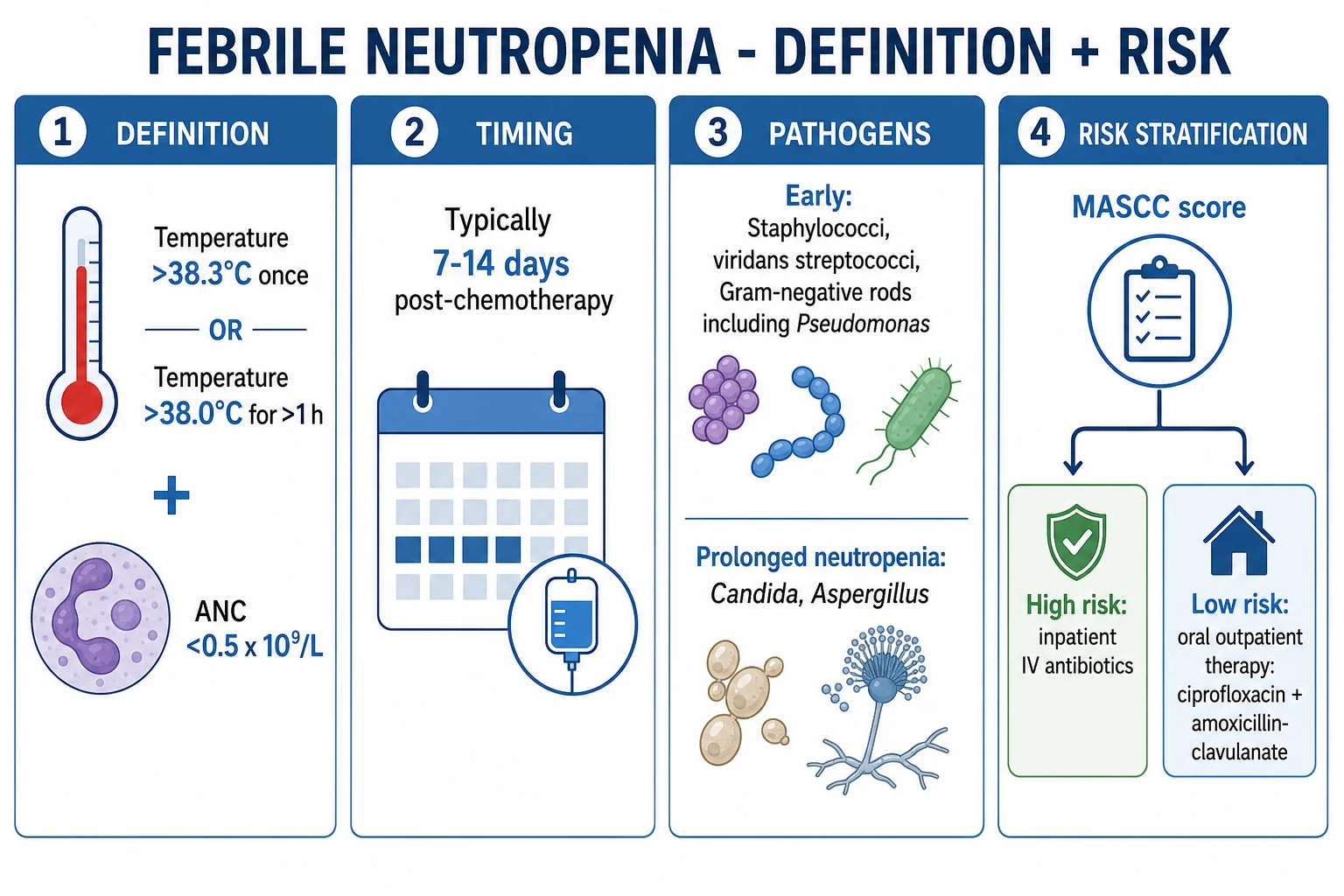
Neutropenic sepsis management — the first 6 hours
- RECOGNISE — any cancer patient on chemotherapy (or within 3-4 weeks of chemotherapy) with fever (>38.3C single OR >38.0C sustained for 1h) + neutrophils <0.5 (or <1.0 with expected decline) = NEUTROPENIC SEPSIS. Do NOT attribute fever to "tumour fever" or "drug fever" until infection is excluded
- RESUSCITATE (if septic): oxygen, IV fluids (20 mL/kg crystalloid if hypotensive), vasopressors (noradrenaline) if MAP <65. Follow SSC 2021 hour-1 bundle
- BLOOD CULTURES — 2 sets from PERIPHERAL sites + 1 set from EACH lumen of any central venous catheter. If possible, obtain BEFORE antibiotics. BUT: if the patient is septic/unstable, DO NOT delay antibiotics >1 hour for cultures
- GIVE EMPIRICAL ANTI-PSEUDOMONAL BETA-LACTAM WITHIN 1 HOUR:
- Piperacillin-tazobactam 4.5 g IV q6h (first-line — broad coverage including Pseudomonas, anaerobes, many gram-positives)
- OR Cefepime 2 g IV q8h (anti-pseudomonal cephalosporin — good gram-negative, reasonable gram-positive)
- OR Meropenem 1 g IV q8h (reserved for known ESBL/CRE colonisation or severe sepsis — broadest coverage but promotes resistance)
- DO NOT use: ceftriaxone (NO Pseudomonas coverage), vancomycin alone (NO gram-negative coverage), fluoroquinolone alone (resistance)
- ADD VANCOMYCIN if: (a) central line infection suspected (tenderness, erythema, exit site pus), (b) severe mucositis (Strep viridans risk), (c) haemodynamic instability (broaden coverage), (d) known MRSA colonisation, (e) catheter has been in >7 days. Dose: 15-20 mg/kg IV q8-12h (target trough 15-20)
- INVESTIGATE THE SOURCE — while antibiotics are running:
- Physical examination (skin, oral cavity for mucositis, perianal area for perirectal abscess, central line site)
- Urine culture (UTI — but may be culture-negative in neutropenia)
- Chest X-ray (pneumonia — but may be absent in early neutropenic pneumonia)
- CT chest/abdomen if persistent fever or localising symptoms (looking for: pneumonia, sinusitis, typhlitis, perirectal abscess, liver/splenic lesions = fungal)
- Stool culture + C. difficile toxin (if diarrhoea)
- Viral PCR (HSV, VZV, CMV, EBV — if appropriate)
- MONITOR — continuous: ECG, SpO2, BP (arterial line if septic). 4-hourly: temperature, FBC (neutrophil trend), U&E, LFTs, CRP, lactate (clearance = response to therapy)
- ASSESS AT 48-72 HOURS:
- FEVER RESOLVED + cultures negative → continue antibiotics until neutrophil recovery (>0.5) or 72h afebrile (whichever is longer)
- FEVER RESOLVED + cultures positive → targeted antibiotics for 7-14 days (organism-specific)
- FEVER PERSISTENT (>72h) + cultures negative → ADD EMPIRICAL ANTIFUNGAL (caspofungin 70 mg day 1 then 50 mg daily — covers Candida; or liposomal amphotericin B 3 mg/kg/day — broader but more toxic). Re-image (CT chest for Aspergillus — halo sign, reversed halo). Check galactomannan (Aspergillus cell wall antigen) + beta-D-glucan (broad fungal marker)
MASCC risk index — high vs low risk stratification
MASCC Risk Index Score (Multinational Association for Supportive Care in Cancer)
| Factor | Points | Notes |
|---|---|---|
| Burden of illness: no/mild symptoms | 5 | If severe symptoms (bedridden) → 0 points → HIGH RISK |
| No hypotension (SBP >90) | 5 | Hypotension → HIGH RISK |
| No COPD | 4 | COPD → HIGH RISK |
| Solid tumour or no prior fungal infection | 4 | Haematological malignancy → 0 points → HIGH RISK |
| No dehydration | 3 | Dehydration → HIGH RISK |
| Outpatient at onset of fever | 3 | Inpatient onset → 0 points |
| Age <60 | 2 | Age ≥60 → 0 points |
| Score ≥21 | LOW RISK | May consider oral antibiotics (ciprofloxacin + amoxicillin-clavulanate) + outpatient management |
| Score <21 | HIGH RISK | IV antibiotics + hospital admission ± ICU monitoring |
MASCC risk index — detailed component-by-component scoring
The MASCC (Multinational Association of Supportive Care in Cancer) risk-index score was validated prospectively by Klastersky et al. in >1,000 neutropenic fever episodes to predict low-risk status (defined as fever resolution without serious medical complications). It uses seven weighted clinical variables available at the bedside. Maximum score = 26; a score ≥21 identifies a low-risk patient with sensitivity ~91% and specificity ~68% for the absence of serious complications (positive predictive value for low-risk ~91%).[3]
MASCC components in detail — what each one measures and why
| Component (points if present) | What you are scoring | Rationale |
|---|---|---|
| Burden of illness: no or mild symptoms (5), moderate (3), or severe/moribund (0) | The physician's global clinical impression of how sick the patient looks AT PRESENTATION | The single most heavily weighted item. A patient who "looks well" is overwhelmingly likely to have an uncomplicated course. "Severe" (moribund/bedridden) → 0 → automatically high-risk. Score the BURDEN, not the fever |
| No hypotension, SBP >90 mmHg (5) | Systolic BP at presentation | Hypotension signals early septic shock — the strongest predictor of complications and death. Even pre-shock (SBP 90–100 with tachycardia) is concerning |
| No chronic obstructive pulmonary disease (4) | History of COPD | COPD is a proxy for frailty and for a lung that cannot tolerate a pneumonia — lung involvement is the commonest complication site |
| Solid tumour OR no previous fungal infection (4) | Underlying malignancy type and fungal history | Haematological malignancy (AML, lymphoma, post-HSCT) carries deeper, longer neutropenia and higher fungal/bacterial risk than a solid tumour on adjuvant chemo. Prior invasive fungal infection = a reservoir of disease |
| Outpatient status at onset of fever (3) | Was the fever first noted as an outpatient? | Inpatient-acquired fever implies hospital pathogens (resistant gram-negatives, VRE), line exposure, and a sicker baseline |
| Age <60 years (2) | Chronological age | Age ≥60 independently increases mortality in neutropenic sepsis (less physiological reserve). Paediatric patients are scored on the separate MASCC paediatric index |
How to use it: add the points. ≥21 = low risk (consider oral antibiotics ± outpatient). <21 = high risk (IV antibiotics, inpatient, monitor for shock). In practice EVERY patient sick enough to be in ICU is high-risk regardless of the score — the MASCC score's real exam use is to justify safely stepping a stable ward patient DOWN to oral therapy, not to justify ICU admission. [1]
Caveats: (1) Do NOT let the calculation delay antibiotics — score it AFTER the 1-hour antibiotic dose. (2) MASCC is validated for adults with solid tumours or haematological cancer, not for neutropenia from non-malignant causes (drug-induced agranulocytosis, aplastic anaemia) where it is unvalidated. (3) The Clinical Index of Stable Febrile Neutropenia (CISNE) is an alternative validated in solid tumours (uses ECOG, COPD, chronic disease, mucositis, monocytes <200, bilirubin/AST elevation); a CISNE score ≥1 predicts complications.[3]
Empirical antifungal strategy for persistent fever (day 4–7)
If fever persists beyond 72–96 hours of appropriate broad-spectrum antibacterial therapy in a high-risk neutropenic patient, the working diagnosis shifts to invasive fungal infection (IFI). The probability of IFI rises steeply the longer the neutropenia lasts: by day 14 of profound neutropenia, >50% of persistently febrile patients have evidence of mould or yeast disease.[6][8]
Persistent-fever antifungal decision pathway (days 3–7+)
- DAY 3–4 of persistent fever — re-examine, repeat cultures (blood, urine, any line), send galactomannan (serum; also BAL if bronchoscopy done) and (1→3)-β-D-glucan (pan-fungal). Perform high-resolution CT chest + CT sinuses/abdomen as guided by symptoms. Look for: halo sign, nodules, reversed-halo (mould); hepatosplenic lesions (chronic disseminated candidiasis); sinus opacification (mucormycosis/Aspergillus).
- DAY 4–7 — add empirical antifungal if still febrile and neutropenic. Two equally valid strategies depending on local epidemiology and prior prophylaxis:
- Mould-active strategy (liposomal amphotericin B 3 mg/kg/day IV) — default if the patient was already on a mould-active azole (posaconazole/isavuconazole) and breakthrough is suspected, or if mucormycosis is in the differential. Broader (covers Aspergillus + Mucorales + Candida) but nephrotoxic (monitor K+, Mg2+, creatinine) and causes infusion reactions.
- Echinocandin strategy (caspofungin 70 mg day 1 → 50 mg/day; OR micafungin 100 mg/day; OR anidulafungin 200 mg → 100 mg/day) — first-line if the concern is primarily Candida, if the patient has renal impairment, or as a diagnostic-driven holding measure. Excellent against Candida (including fluconazole-resistant glabrata/krusei), limited mould activity, low toxicity. NOT adequate if galactomannan is positive (Aspergillus proven) → switch to a mould-active azole.[9]
- Mould-active azole (voriconazole 6 mg/kg BD ×2 then 4 mg/kg BD; or isavuconazole 200 mg q8h ×6 then 200 mg/day) — preferred if Aspergillus is microbiologically documented (positive galactomannan/BAL) — voriconazole improved survival vs amphotericin in Herbrecht 2002. Requires therapeutic drug monitoring (voriconazole trough 1–5 mg/L) and checks for CYP2C19 polymorphism/interactions.[8]
- DAY 7+ — pre-emptive / diagnostic-driven (preferred modern approach) — rather than blanket empirical therapy, escalate antifungal therapy ONLY if there is microbiological or radiological evidence of IFI (galactomannan positive, β-D-glucan positive, new CT lesion). Broncho-alveolar lavage for galactomannan + culture if a new pulmonary infiltrate appears. This reduces unnecessary antifungal exposure and resistance.
- Continue antifungal until: neutrophil recovery (>0.5 × 10^9/L), afebrile ≥48 h, AND any documented fungal infection treated to a complete response. If an IFI is confirmed, therapy continues for weeks–months (e.g., voriconazole for ≥6–12 weeks for invasive aspergillosis) often long after discharge.
Galactomannan vs beta-D-glucan — interpretation
| Test | What it detects | Sensitivity | Specificity | False positives | False negatives | Cut-off |
|---|---|---|---|---|---|---|
| Galactomannan (serum) | Aspergillus cell-wall polysaccharide (galactofuranose) — specific to Aspergillus and some Penicillium | ~70–80% (higher in BAL) | ~85–90% | Piperacillin-tazobactam (historical contamination; modern formulations cleaner but still possible), amoxicillin-clavulanate, gut translocation, Bifidobacterium (paediatrics), fermented foods | Mucorales (no galactomannan — a useful distinguishing feature), prior effective antifungal | Serum ODI ≥0.5 (single or two consecutive); BAL ODI ≥1.0 |
| (1→3)-β-D-glucan | Pan-fungal cell-wall component (Aspergillus, Candida, Pneumocystis, Fusarium, Trichosporon) | ~75–80% | ~80% | Dialysis membranes/cellulose filters, IV immunoglobulin, albumin, gauze, some historical antibiotic batches | Mucorales and Cryptococcus (do NOT make β-D-glucan — useful negative); zygomycetes; blastomycosis | ≥80 pg/mL (Fungitell assay) |
| Combination | Serial GM + BDG as screening, with BAL GM for diagnosis | Higher with serial screening (twice weekly) | — | — | — | Two consecutive positives more specific than one |
Key exam point: a positive galactomannan + a compatible CT (halo/nodule) = treat for invasive pulmonary aspergillosis with voriconazole/isavuconazole. A positive β-D-glucan with negative galactomannan suggests Candida or Pneumocystis. Mucormycosis is BOTH negative (no galactomannan, no β-D-glucan) — the diagnosis is tissue biopsy showing broad, ribbon-like, non-septate hyphae with right-angle branching; treat with surgical debridement + liposomal amphotericin B and consider isavuconazole.[8]
Echinocandin vs liposomal amphotericin B — the empirical choice
Echinocandin vs liposomal amphotericin B (L-AmB) for empirical / persistent-fever antifungal therapy
| Feature | Echinocandin (caspofungin, micafungin, anidulafungin) | Liposomal amphotericin B |
|---|---|---|
| Spectrum | Excellent Candida (incl. resistant species); weak/no mould activity | Broad: most Candida, Aspergillus, Mucorales, endemic fungi |
| Mechanism | Inhibits β-1,3-glucan synthase (cell wall) | Binds ergosterol → pore in cell membrane |
| Toxicity | Low: mild LFT rise, headache, histamine infusion reaction | Infusion reactions (chills, fever), hypokalaemia, hypomagnesaemia, nephrotoxicity, anaemia |
| Drug interactions | Minimal (not a major CYP substrate) | Additive nephrotoxicity with calcineurin inhibitors, contrast, aminoglycosides |
| Renal failure | Safe — no dose adjustment | Lipid formulation safer than deoxycholate but still monitor; minimise concurrent nephrotoxins |
| Dosing | Caspofungin 70 mg → 50 mg/day; micafungin 100 mg/day; anidulafungin 200 mg → 100 mg/day | L-AmB 3 mg/kg/day (5–10 mg/kg/day for documented mucormycosis) |
| When to choose | Persistent fever + low mould suspicion; renal impairment; diagnostic-driven holding therapy; on azole prophylaxis | Suspected mould including Mucorales; breakthrough on azole; galactomannan negative but progressive sinus/lung disease |
Clinical pearls
G-CSF (granulocyte colony-stimulating factor) — the controversy
G-CSF (filgrastim 5 mcg/kg/day SC; pegfilgrastim 6 mg SC once per chemo cycle; lenograstim) stimulates myeloid progenitor proliferation, shortening the neutropenic nadir. The evidence and the ASCO/IDSA position:[7]
G-CSF — the case for and against
| Argument FOR (use it) | Argument AGAINST (don't use it routinely) |
|---|---|
| Prophylactic use (given WITH chemotherapy) in high-risk regimens reduces the incidence of febrile neutropenia by ~50%; recommended when the expected febrile-neutropenia rate is ≥20% | Does NOT reduce infection-related mortality in pooled RCT/meta-analysis (Bohlius, Lancet 2007) |
| Shortens hospital stay by ~1–2 days and IV antibiotic duration | Cost (~USD 100–300/day; pegfilgrastim ~USD 3,000/dose) |
| Therapeutic trial evidence (in pneumonia/typhlitis/organ failure) suggests possible benefit in sicker patients | Risk of Sweet's syndrome, vasculitis, interstitial pneumonitis, splenic rupture, flare of sickle cell disease; theoretical stimulation of myeloid leukaemia clones |
| May enable maintaining chemotherapy dose-intensity in curative-intent therapy | Does not improve complete remission rate or overall survival of the underlying malignancy |
| Useful to mobilise stem cells for harvest pre-HSCT | Filgrastim can transiently worsen respiratory failure (capillary leak) in patients with pre-existing pneumonia/ARDS |
ASCO 2015 guidance on WHEN to use G-CSF:[7]
- Primary prophylaxis (with the first cycle) — when expected febrile-neutropenia risk ≥20% (high-risk regimens, age >65 + extensive disease, prior chemo/radiation, poor performance status, pre-existing neutropenia, renal/liver dysfunction, HIV).
- Secondary prophylaxis (after a prior cycle with a neutropenic complication) — to maintain dose-intensity in curative-intent therapy.
- Therapeutic use (during established febrile neutropenia) — NOT routine. Consider selectively in: pneumonia, hypotension/sepsis, invasive fungal infection, multi-organ dysfunction, expected neutropenia >7–10 days (e.g., AML induction, allogeneic HSCT). Filgrastim 5 mcg/kg/day SC, continue until ANC >1.0 after the nadir.
Bottom line for the exam: "G-CSF is NOT routine in established neutropenic sepsis — it reduces hospital days and febrile episodes but does NOT reduce mortality; reserve it for high-risk patients with expected prolonged neutropenia, documented fungal infection, or organ failure."[7]
Specific clinical scenarios
1. Stem cell transplant (HSCT)
The HSCT timeline dictates the dominant infectious threat, and the exam expects you to know this sequence:[4]
Infectious threats after haematopoietic stem cell transplant — by phase
| Phase | Time after HSCT | Dominant immune defect | Dominant pathogens | Prophylaxis |
|---|---|---|---|---|
| Pre-engraftment | Days 0–30 | Profound neutropenia + mucositis + ablative conditioning | Gram-negatives (Pseudomonas), gram-positives (Strep viridans, coag-neg staph), Candida, HSV reactivation (if seropositive), respiratory viruses (RSV, influenza, parainfluenza) | Anti-pseudomonal cover, fluconazole or posaconazole, aciclovir (HSV+), PJP prophylaxis started at engraftment |
| Early post-engraftment | Days 30–100 | Delayed T-cell reconstitution, acute GVHD (steroids!) | CMV reactivation (pre-emptive monitoring with PCR), adenovirus, PCP (PJP), Aspergillus (esp. with GVHD/steroids), BK virus (haemorrhagic cystitis) | Co-trimoxazole (PCP), weekly CMV PCR monitoring (pre-emptive valganciclovir/ganciclovir if positive), posaconazole (GVHD), mould-active surveillance |
| Late post-engraftment | >Day 100 | Chronic GVHD, prolonged T- and B-cell defects, hypogammaglobulinaemia | Encapsulated bacteria (S. pneumoniae, H. influenzae), VZV, EBV (PTLD), CMV (late), PCP (if still immunosuppressed), hepatitis B/C reactivation | Co-trimoxazole (long-term if immunosuppressed), VZV vaccine (live vaccines deferred ≥2 yr), IVIG if hypogammaglobulinaemia, re-vaccination (pneumococcal, Hib, meningococcal — immunity is "reset") |
Key ICU points: CMV in HSCT = reactivation driven by T-cell depletion; monitor with quantitative PCR weekly day 30–100; pre-emptive valganciclovir 900 mg BD when the PCR threshold is reached (avoids the toxicity of universal prophylaxis). Adenovirus can cause fulminant hepatitis, pneumonitis, and haemorrhagic colitis — treat with cidofovir (or brincidofovir). BK virus causes haemorrhagic cystitis (supportive; cidofovir/fluoroquinolones sometimes used).[4]
2. CAR-T cell therapy and cytokine release syndrome (CRS)
Chimeric antigen receptor T-cell therapy (tisagenlecleucel, axicabtagene ciloleucel, lisocabtagene maraleucel, idecabtagene vicleucel, ciltacabtagene autoleucel) for B-cell malignancies and multiple myeloma causes a unique constellation of toxicities that mimic and coexist with neutropenic sepsis. A patient with CAR-T CRS in ICU has prolonged cytopenias (weeks–months) PLUS a cytokine-driven inflammatory syndrome.[10]
CRS grading (ASTCT 2019 consensus):
- Grade 1 — fever + no hypotension + no hypoxia (looks like simple febrile neutropenia).
- Grade 2 — fever + hypotension responding to fluids OR hypoxia responding to <40% O2.
- Grade 3 — fever + hypotension on ONE vasopressor OR hypoxia needing ≥40% O2.
- Grade 4 — fever + multiple vasopressors (excluding vasopressin) OR positive-pressure ventilation. [1]
The overlap that traps you: CRS Grade 1 = fever ± tachycardia, indistinguishable from early neutropenic sepsis at presentation. Therefore: ALL CAR-T patients with fever get the full neutropenic-sepsis workup + empirical anti-pseudomonal antibiotics within 1 hour — THEN assess for CRS. Do not attribute fever to "expected CRS" until infection is excluded or concomitantly treated.[10]
Management of CRS:
- Grade 1 — supportive + antibiotics; antipyretics.
- Grade ≥2 — tocilizumab (anti-IL-6 receptor) 8 mg/kg IV (max 800 mg), may repeat q8h (max 3 doses/24 h, 4 total). FDA-approved for CRS. Corticosteroids (dexamethasone 10 mg q6h or methylprednisolone 1 g/day) for refractory CRS or where neurotoxicity (ICANS) coexists.
- Neurotoxicity (ICANS — immune effector cell-associated neurotoxicity syndrome): confusion, aphasia, tremor, seizures, cerebral oedema. Assess with the ICE score hourly; treat with dexamethasone ± tocilizumab; consider anti-IL-6 (siltuximab) if refractory. [1]
Pearl: tocilizumab does NOT mask bacterial infection (it blocks IL-6 but does not suppress fever from sepsis reliably), so it can be given alongside antibiotics. But steroids DO mask infection — give concomitantly with antibiotics, never as monotherapy in this setting.[10]
3. Post-chemotherapy (solid tumour vs haematological)
- Solid tumour adjuvant therapy (breast, lung, colorectal, sarcoma): neutropenia typically lasts 5–7 days, nadir day 7–14. Empirical anti-pseudomonal cover, antifungal if persistent fever, but lower overall fungal risk than haematology. MASCC scoring more likely to identify low-risk patients suitable for oral therapy/outpatient.
- Acute leukaemia induction (AML 7+3 cytarabine–daunorubicin; ALL induction): neutropenia lasts 3–4 weeks, deep (often ANC 0), high fungal risk → mould-active prophylaxis (posaconazole) from day 1, fluoroquinolone prophylaxis, aciclovir if HSV+. Antifungal threshold for empirical therapy is lower (add by day 4 of fever).[4]
- High-dose cytarabine — severe mucositis + Strep viridans bacteraemia (shock syndrome) + keratitis + cerebellar toxicity. Add vancomycin empirically; consider penicillin prophylaxis (viridans strep is often penicillin-susceptible).
- Rituximab/fludarabine/alemtuzumab (anti-CD20, purine analogue, anti-CD52) — profound, prolonged T- and B-cell defects; high risk PCP (co-trimoxazole prophylaxis mandatory), CMV reactivation (alemtuzumab — monitor CMV PCR), HBV reactivation (rituximab — screen HBsAg/anti-HBc, give entecavir/tenofovir prophylaxis if positive).[4]
4. The "missing neutrophils" mimics — beware non-chemotherapy neutropenia
Not all neutropenic fever is chemotherapy-related. In ICU also consider:
- Drug-induced agranulocytosis — clozapine, carbimazole/thiamazole, sulfasalazine, anti-epileptics, ticlopidine. Treat identically (1-hour antibiotics + G-CSF — the G-CSF evidence is BETTER here than in chemo neutropenia + stop the offending drug).
- Aplastic anaemia — often prolonged neutropenia; supportive + immunosuppression (ATG/ciclosporin) or HSCT.
- Congenital neutropenias — severe congenital neutropenia (ELANE), cyclic neutropenia, Shwachman-Diamond syndrome; on chronic G-CSF.
- Severe sepsis itself — cytokine-mediated margination/apoptosis can cause transient neutropenia (the "septic neutropenia" of overwhelming infection, e.g., typhoid, OPSI). [1]
Antimicrobial prophylaxis protocols
Prophylaxis is the highest-yield "preventive ICU" topic in neutropenic-sepsis exams. The regimen is dictated by the expected depth and duration of neutropenia and the specific immune defect.[4][7]
Prophylaxis by pathogen — drug, dose, indication, caveat
| Pathogen | Drug & dose | Indication | Caveat |
|---|---|---|---|
| Pneumocystis jirovecii (PCP/PJP) | Co-trimoxazole 480 mg (SS) or 960 mg (DS) PO daily (or DS three times weekly); alternatives: dapsone 100 mg daily, atovaquone 1500 mg daily, inhaled pentamidine monthly | Steroids ≥20 mg prednisone for ≥4 weeks; HSCT; ALL; alemtuzumab; purine analogues (fludarabine/cladribine); calcineurin inhibitors; HIV with CD4 <200 | Stop during IV high-dose methotrexate (marrow toxicity, antagonises rescue). Co-trimoxazole also covers Toxoplasma, Nocardia, many pneumococci — broad protective effect |
| Aspergillus (mould) | Posaconazole 300 mg IV/PO day 1 BD then daily (suspension 200 mg TDS); isavuconazole alternative; voriconazole also active | AML/MDS induction, GVHD on steroids, prolonged neutropenia >10–14 days, allogeneic HSCT with GVHD | Fluconazole does NOT cover moulds. Posaconazole improved survival vs fluconazole in GVHD prophylaxis (Cornely/Ullmann 2007 NEJM). Monitor LFTs, QT |
| Candida | Fluconazole 400 mg daily (or posaconazole/micafungin in high-risk) | AML induction, HSCT pre-engraftment, prolonged neutropenia | Fluconazole covers most Candida but NOT C. glabrata/krusei reliably; echinocandin preferred if colonised with resistant species or prior fluconazole exposure |
| CMV | Valganciclovir 900 mg daily (pre-emptive when PCR positive; universal prophylaxis in D+/R– transplant); IV ganciclovir for active disease; letermovir 480 mg daily for prophylaxis (HSCT) | HSCT (D+/R– mismatch), alemtuzumab, heavily immunosuppressed | Letermovir is prophylaxis only (not treatment); valganciclovir/ganciclovir treat active CMV but cause marrow toxicity (worsen neutropenia) |
| HSV/VZV | Aciclovir 400 mg PO BD (HSV) / 800 mg daily (VZV); valaciclovir 500 mg BD | HSCT if HSV+ serology; heavily immunosuppressed; post-fludarabine/alemtuzumab | Almost universal in HSV-seropositive HSCT recipients during conditioning + engraftment |
| HBV reactivation | Entecavir 0.5 mg daily or tenofovir 300 mg daily | Rituximab, ofatumumab, chemo in HBsAg+ or anti-HBc+ | Screen ALL patients pre-rituximab with HBsAg + anti-HBc; antiviral prophylaxis (better than monitoring) for ≥6–12 months post-rituximab |
| Pneumococcus / encapsulated | Pneumococcal conjugate (PCV15/PCV20) + polysaccharide PPSV23; Hib; MenACWY + MenB; influenza annually | Asplenia, post-HSCT (re-vaccinate from 6 months), CLL, myeloma, hypogammaglobulinaemia | Vaccinate BEFORE splenectomy/chemo where possible; avoid live vaccines during chemo; conjugate before polysaccharide (immunological priming) |
| Bacterial (febrile-neutropenia prophylaxis) | Levofloxacin 500 mg daily or ciprofloxacin 500 mg BD | AML induction, HSCT pre-engraftment, regimens with expected FN rate ≥20% | Drives fluoroquinolone resistance; balance against reduction in febrile-neutropenia episodes. Local antibiogram essential |
Vaccination timing after HSCT (the exam detail): re-vaccination starts ~6 months post-transplant (3 months if no GVHD/immunosuppression) because the graft's immune memory is "reset". Sequence: PCV first (conjugate primes T-cell memory), then PPSV23 eight weeks later to broaden serotypes; inactivated influenza from 6 months annually; live vaccines (MMR, VZV) deferred ≥24 months if no GVHD/immunosuppression.[4]
De-escalation and stopping antibiotics
- Documented bacteraemia — de-escalate to the narrowest effective agent once sensitivities are known; treat organism-appropriate duration (S. aureus 4–6 wk; Enterobacterales 7–14 d from first negative culture; Candida 2 wk after first negative culture + source control).[9]
- Unexplained fever, cultures negative, now afebrile — stop antibiotics once ANC >0.5 for 2 consecutive days OR afebrile ≥72 h and clinically stable (whichever is LATER). The ASCO/IDSA position allows stopping in the stable afebrile low-risk patient.[1]
- NEVER stop empirical therapy in a persistently neutropenic patient who is still febrile without adding/reviewing antifungal cover.
- Empirical antifungal started for persistent fever should continue until neutrophil recovery + afebrile + any documented IFI treated to response; if the workup is fully negative and the patient recovers, stop.
Red flags
Prognosis
Neutropenic sepsis outcomes
| Factor | Mortality | Notes |
|---|---|---|
| Overall (all neutropenic fever) | 5-10% | Has improved with early antibiotics + better supportive care |
| With septic shock | 30-50% | The strongest predictor of mortality |
| Pseudomonas aeruginosa bacteraemia | 20-40% | The most dangerous pathogen |
| MASCC low-risk | <5% | Can be managed as outpatient |
| MASCC high-risk | 10-20% | Requires IV antibiotics ± ICU |
| Duration of neutropenia >14 days | 20-30% | Prolonged neutropenia → higher fungal risk + mortality |
Key trials and evidence
ASCO/IDSA 2018 Guidelines — neutropenic sepsis management (PMID 32219360)
Source
American Society of Clinical Oncology / Infectious Diseases Society of America joint guideline update
Key principle 1
Empirical anti-pseudomonal beta-lactam within 1 hour — piperacillin-tazobactam first-line
Key principle 2
MASCC risk index stratifies: high-risk → IV + admit; low-risk (score ≥21) → oral + possible outpatient
Key principle 3
Add vancomycin only for specific indications (line infection, mucositis, shock, MRSA)
Key principle 4
Empirical antifungal for persistent fever >4-7 days
Clinical bottom line
The definitive guideline for neutropenic sepsis management — 1-hour antibiotics + MASCC stratification + targeted additions
Herbrecht 2002 NEJM — voriconazole vs amphotericin B for invasive aspergillosis
Source
New England Journal of Medicine 2002;347:408–415 — definitive randomised trial in primary invasive aspergillosis
Design
Randomised open-label: voriconazole vs amphotericin B deoxycholate, then other licensed antifungal
Population
277 patients with definite/probable invasive aspergillosis (largely haematological malignancy, neutropenia, HSCT)
Key result
Successful outcome: voriconazole 52.8% vs amphotericin 31.6% (p<0.001); survival at 12 weeks 70.8% vs 57.9%
Clinical bottom line
Voriconazole became the FIRST-LINE therapy for invasive aspergillosis — the trial that established the mould-active azole era
Walsh 2004 NEJM — caspofungin vs liposomal amphotericin B for empirical antifungal therapy (PMID 15385695)
Source
New England Journal of Medicine 2004;351:1391–1402
Design
Randomised double-blind non-inferiority: caspofungin vs liposomal amphotericin B in persistent febrile neutropenia
Population
1,095 patients with febrile neutropenia persisting >96 h on broad-spectrum antibiotics
Key result
Caspofungin non-inferior overall, with FEWER breakthrough fungal infections and less nephrotoxicity/infusion toxicity than liposomal amphotericin B
Clinical bottom line
Validated the echinocandin as a low-toxicity empirical antifungal for persistent febrile neutropenia — the basis for the echinocandin arm of the day 4–7 strategy
Bohlius 2007 Lancet — IPD meta-analysis of CSF prophylaxis in cancer chemotherapy
Source
Lancet 2007;370:113–122 — individual-patient-data meta-analysis of 14,939 patients across 17 RCTs
Key result
CSF prophylaxis reduced febrile neutropenia (RR ~0.7) and infection-related mortality during chemotherapy, but the infection-related mortality reduction was small and driven by high-risk subgroups; no clear all-cause mortality benefit
Clinical bottom line
Underpins ASCO guidance: G-CSF for primary prophylaxis in high-risk regimens (expected FN ≥20%) but NOT routine therapeutic use in established neutropenic sepsis
Cornely 2007 / Ullmann 2007 NEJM — posaconazole prophylaxis in neutropenia and GVHD
Source
Two landmark NEJM 2007 trials — posaconazole vs fluconazole/itraconazole prophylaxis
Cornely (AML/MDS induction)
Posaconazole reduced proven/probable invasive fungal infections (2% vs 8%) and improved overall survival vs fluconazole/itraconazole in prolonged neutropenia
Ullmann (GVHD)
Posaconazole non-inferior to fluconazole for fungal infections overall but superior for invasive aspergillosis and trended to a survival benefit
Clinical bottom line
Established posaconazole as the preferred mould-active prophylaxis in AML/MDS induction and GVHD — the standard of care
Lee 2014 Blood — cytokine release syndrome consensus grading (PMID 25253717)
Source
Blood 2014;124:188–195 — consensus diagnostic and management framework for CRS
Key contribution
Defined CRS grading and the tocilizumab/corticosteroid escalation pathway adopted worldwide
Clinical bottom line
The reference for managing CAR-T CRS in ICU — tocilizumab for grade ≥2, steroids for refractory CRS/ICANS
Viva / SAQ — worked example
SAQ — Neutropenic sepsis with invasive pulmonary aspergillosis
10 minutes · 10 marks
A 58-year-old man with newly diagnosed AML is day 12 after induction chemotherapy (7+3 cytarabine + daunorubicin). He presents with fever 39.2°C and rigors. Neutrophils 0.1 × 10^9/L (expected to fall further), platelets 22, MAP 78 mmHg, HR 112, RR 24, SpO2 95% on room air, lactate 2.6 mmol/L. He has a tunnelled central venous catheter. He has been on posaconazole and levofloxacin prophylaxis. Blood cultures are being drawn.
Summary — the exam one-liners
- Neutrophils under 0.5 plus fever over 38.3 = neutropenic sepsis → empirical anti-pseudomonal beta-lactam within one hour (piperacillin-tazobactam first-line; meropenem if prophylaxis/resistance). Never ceftriaxone.
- Pseudomonas aeruginosa is the #1 killer — anti-Pseudomonal cover is mandatory.
- No localising signs in neutropenia — cultures are negative in up to 50%; treat on clinical grounds.
- Persistent fever beyond 4-7 days despite antibiotics = think invasive fungal infection — add empirical antifungal (echinocandin for Candida, liposomal amphotericin B if mould/Mucor suspected, mould-active azole if Aspergillus is documented).
- Map the immune defect to the pathogen: neutropenia → Pseudomonas/Aspergillus; T-cell → PCP/CMV/Nocardia/Listeria; B-cell/asplenia → encapsulated organisms; steroids → Nocardia/Aspergillus/PCP/Strongyloides.
- MASCC score 21 or more = low risk (oral, maybe outpatient); below 21 = high risk (IV, admit). Every ICU patient is high-risk.
- G-CSF reduces febrile episodes but not mortality — NOT routine; reserve for prolonged neutropenia, documented fungal infection, or organ failure.
- CAR-T fever = neutropenic sepsis until proven otherwise — full 1-hour pathway then assess CRS; tocilizumab with antibiotics, never steroids alone. [1]
References
- [1]Taplitz RA, et al. The Importance of Addressing Advance Care Planning and Decisions About Do-Not-Resuscitate Orders During Novel Coronavirus 2019 (COVID-19) JAMA, 2020.PMID 32219360
- [2]Freifeld AG, et al. What younger forensic pathologists need for their training: valuable comments from our interviews Leg Med (Tokyo), 2013.PMID 22981177
- [3]Klastersky J, et al. Developing an internet-based support system for adolescents with depression JMIR Res Protoc, 2012.PMID 23612485
- [4]Flowers CR, et al. The tumor suppressor Rb critically regulates starvation-induced stress response in C. elegans Curr Biol, 2013.PMID 23664972
- [5]Marrs R, et al. The Neurologic Assessment in Neuro-Oncology (NANO) scale: a tool to assess neurologic function for integration into the Response Assessment in Neuro-Oncology (RANO) criteria Neuro Oncol, 2017.PMID 28453751
- [6]Koehler P, et al. Reply to the Editor- Anticoagulation in atrial fibrillation after intracranial hemorrhage: could the hemorrhage location influence the outcome? Heart Rhythm, 2017.PMID 27871984
- [7]Smith TJ, et al. Dietary Advice for Melanoma: Not Ready for Prime Time J Clin Oncol, 2015.PMID 26124491
- [8]Patterson TF, et al. Practice Guidelines for the Diagnosis and Management of Aspergillosis: 2016 Update by the Infectious Diseases Society of America Clin Infect Dis, 2016.PMID 27365388
- [9]Pappas PG, et al. Clinical Practice Guideline for the Management of Candidiasis: 2016 Update by the Infectious Diseases Society of America Clin Infect Dis, 2016.PMID 26679628
- [10]Lee DW, et al. An agent-based model for mRNA export through the nuclear pore complex Mol Biol Cell, 2014.PMID 25253717