ICU · Infectious Diseases
Acute severe community-acquired pneumonia: Pseudomonas pneumonia
Also known as Pseudomonas aeruginosa pneumonia · Pseudomonas CAP · Antipseudomonal therapy · Nosocomial Pseudomonas pneumonia · MDR Pseudomonas pneumonia
Pseudomonas aeruginosa is an aerobic Gram-negative bacillus — an opportunistic, water-borne pathogen that forms biofilms and thrives in moist environments and on indwelling devices. It is a rare cause of community-acquired pneumonia (CAP, <1% of uncomplicated CAP) but a major cause of hospital-acquired pneumonia (HAP) and ventilator-associated pneumonia (VAP), and a feared pathogen in patients with structural lung disease (bronchiectasis, cystic fibrosis), immunocompromise/neutropenia, and prior broad-spectrum antibiotic exposure. Clinical course is severe and rapidly progressive, with ICU mortality of 20-40%. Diagnosis rests on sputum or bronchoalveolar-lavage (BAL) culture with Gram stain showing Gram-negative bacilli. Management mandates TWO active agents from different classes for severe disease — an anti-pseudomonal beta-lactam (piperacillin-tazobactam, cefepime, ceftazidime, or meropenem) PLUS an aminoglycoside (tobramycin, amikacin, gentamicin) OR an antipseudomonal fluoroquinolone (ciprofloxacin, levofloxacin) — with de-escalation once susceptibilities return and a standard duration of 7 days. Pseudomonas is a resistance virtuoso: AmpC beta-lactamase, ESBLs, carbapenemases (especially metallo-beta-lactamases), efflux pumps, porin loss and target modification all drive multidrug-resistant (MDR), extensively drug-resistant (XDR) and pandrug-resistant (PDR) phenotypes.
On this page & tools
Your progress
Saved locally on this device.
Target exams
Red flags
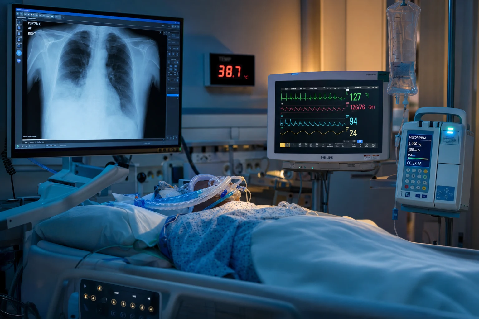
Microbiology and pathogenesis
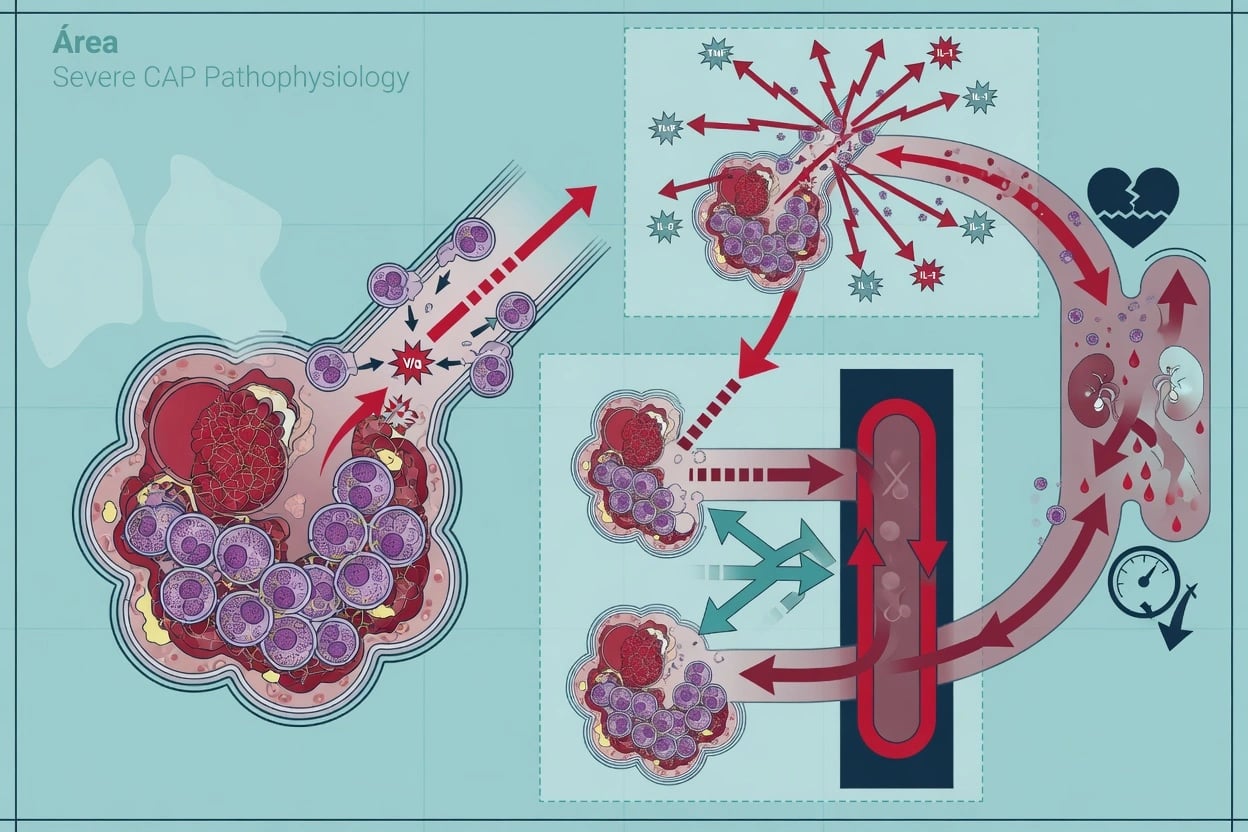
Virulence factors — how Pseudomonas damages the lung
Adhesion and biofilm
Type IV pili and polar flagellum mediate adhesion to mucosal surfaces and the endotracheal tube. Once attached, bacteria secrete exopolysaccharide (alginate in mucoid CF strains) and assemble into a biofilm — a sessile community shielded from antibiotics, antibody and neutrophils. Biofilm bacteria are phenotypically tolerant (persister cells), explaining why eradication is so difficult in chronic infection.<Cite id="5" />
Exotoxin A
Exotoxin A is an ADP-ribosyltransferase that inactivates elongation factor 2 (EF-2) — identical mechanism to diphtheria toxin — halting host-cell protein synthesis. Causes tissue necrosis and is a major virulence determinant in invasive disease.
Type III secretion system (T3SS) toxins
A molecular syringe injects four effector toxins directly into host cells: **ExoS** and **ExoU** (most virulent — a phospholipase causing rapid necrosis and haemorrhage), **ExoT** (inhibits phagocytosis and epithelial wound healing), **ExoY** (adenylyl cyclase). T3SS-positive strains (especially ExoU) are independently associated with severe disease and high mortality in Pseudomonas pneumonia and bacteraemia.<Cite id="6" />
Proteases, elastase, phospholipase
Elastase (LasB) degrades elastin, IgG, IgA, complement and surfactant proteins — destroys alveolar architecture and disables opsonisation. Alkaline protease and protease IV degrade complement and fibrin. Phospholipase C (haemolysin) destroys surfactant and lung epithelium, contributing to ARDS. Mucoid exopolysaccharide (alginate) impairs phagocytosis.
Pigments
Pyocyanin (blue-green pigment — characteristic pus/sputum colour) generates reactive oxygen species, depletes glutathione, impairs ciliary function and induces neutrophil apoptosis. Pyoverdine (fluorescent green-yellow) is a high-affinity siderophore that scavenges iron. Pyocyanin + pyoverdine together account for the classic blue-green "pus" of Pseudomonas infection.
Quorum sensing
Cell-to-cell signalling (las, rhl, pqs systems) coordinates population-wide expression of virulence genes and biofilm maturation once a threshold density (quorum) is reached. Quorum sensing is a therapeutic target (experimental quorum-sensing inhibitors).<Cite id="5" />
Risk factors
[1] [3]Clinical features
Pseudomonas pneumonia — clinical syndromes
Community-acquired Pseudomonas pneumonia (rare)
Occurs almost exclusively in patients with risk factors (bronchiectasis/CF, severe COPD, immunocompromise, recent antibiotics). Presents as a severe, rapidly progressive CAP — high fever, rigors, productive cough with purulent (sometimes green-tinged) sputum, dyspnoea, pleuritic chest pain, and early hypoxaemia. CXR typically shows bilateral lower-zone bronchopneumonia; may be multilobar. Bacteraemia in 10-20%. Mortality 20-40% — among the highest of any CAP pathogen.<Cite id="4" />
Hospital-acquired pneumonia (HAP) — non-ventilated
Onset ≥48 h after admission. Risk = prolonged hospitalisation, prior antibiotics, reduced consciousness (aspiration), ICU. Presentation may be subtle: new fever, purulent respiratory secretions, worsening oxygenation, rising inflammatory markers. CXR new infiltrate. Often multidrug-resistant from the outset — empiric therapy must reflect local antibiogram.
Ventilator-associated pneumonia (VAP)
Pneumonia arising ≥48 h after intubation. *P. aeruginosa* is the single most common VAP pathogen (~20% of cases) and the leading cause of late-onset VAP (≥5 days after admission). Diagnosis via quantitative/semi-quantitative BAL or endotracheal aspirate cultures. Purulent, often green, secretions; fever, rising WBC, worsening oxygenation, new/worsening infiltrate. High attributable mortality, especially with inappropriate initial therapy.<Cite id="2" />
Bacteraemic / neutropenic Pseudomonas pneumonia
In neutropenic/immunocompromised hosts Pseudomonas frequently causes primary bacteraemia with secondary pulmonary seeding (or pneumonia with secondary bacteraemia). Look for **ecthyma gangrenosum**, septic shock, rapidly progressive multilobar infiltrates. Mortality 30-50%. This is a true medical emergency — empiric antipseudomonal therapy from the first hour of febrile neutropenia.<Cite id="12" />
Chronic colonisation (CF / bronchiectasis)
Not pneumonia per se, but chronic Pseudomonas biofilm infection of structurally abnormal airways. Mucoid (alginate-producing) phenotypes dominate. Acute exacerbations = increased sputum volume/purulence, dyspnoea, reduced FEV1 — treat with antipseudomonal agents guided by prior sputum culture, often combining IV + inhaled (tobramycin, colistin, aztreonam) therapy. Differentiate colonisation from acute infection using clinical + inflammatory markers.<Cite id="7" />
Diagnosis
Diagnostic workup — samples and what they tell you
Lower-respiratory samples (the diagnostic cornerstone)
Obtain BEFORE (or as soon as possible after) antibiotic doses. **Sputum Gram stain + culture** (good-quality sample: <10 squamous epithelial cells / low-power field, >25 neutrophils). Gram stain shows Gram-negative bacilli; culture grows oxidase-positive, lactose-non-fermenting, pigmented colonies on MacConkey agar. For intubated patients: **endotracheal aspirate** (sensitivity high, specificity lower) or **bronchoalveolar lavage (BAL) / protected specimen brush (PSB)** with quantitative cultures (diagnostic thresholds: BAL ≥10^4 CFU/mL, PSB ≥10^3 CFU/mL). BAL is preferred for VAP — better specificity, less contamination.<Cite id="2" />
Blood cultures
Two sets before antibiotics. Bacteraemia complicates 10-20% of CAP and up to 25% of HAP/VAP due to Pseudomonas, and is an independent predictor of mortality. Persistent bacteraemia on appropriate therapy (≥48-72 h) mandates search for endocarditis, metastatic abscess, infected catheter or inadequate source control.
Antibiotic susceptibility testing
CRITICAL — Pseudomonas develops resistance on therapy, so ALWAYS check susceptibilities. Test panel: piperacillin-tazobactam, ceftazidime, cefepime, meropenem/imipenem, amikacin, gentamicin, tobramycin, ciprofloxacin, levofloxacin, colistin, and newer agents (ceftolozane-tazobactam, ceftazidime-avibactam, ceftazidime-avibactam-aztreonam, imipenem-relebactam). Request **MBL/carbapenemase detection** (phenotypic + molecular) in resistant strains — guides escalation therapy. Repeat cultures if clinical failure.<Cite id="6" />
Biomarkers
**Procalcitonin** (PCT) supports a bacterial (vs viral) process and guides duration — stop when PCT falls >80% from peak or <0.25 ng/mL in stable patients. **CRP** trends with response but is nonspecific. **WBC** — leucopenia in severe Pseudomonas sepsis is ominous. None is specific to Pseudomonas; use trends, not single values. Falling PCT supports successful de-escalation.
Imaging
**Chest X-ray**: bilateral, often lower-zone predominant bronchopneumonia; may show multilobar consolidation, cavitation, pleural effusion, lobar expansion. **CT chest**: defines cavitation, abscess, empyema, necrotising pneumonia; excludes alternative diagnoses. **Point-of-care ultrasound**: B-lines, subpleural consolidation, pleural effusion at the bedside. Radiographic progression often outpaces clinical deterioration — repeat imaging.<Cite id="3" />
Severity assessment
Calculate **CURB-65** or **SMART-COP/SMRT-CO** (the latter preferred for ICU triage in ANZ). Apply **qSOFA/SOFA**, serum lactate, organ-failure assessment. Identify who needs ICU (mechanical ventilation, vasopressors) vs ward. Pseudomonas CAP with septic shock or ARDS physiology demands ICU from the outset.
Management

Pseudomonas pneumonia management — step by step
Risk assessment and empiric escalation
Identify any Pseudomonas risk factor: structural lung disease (bronchiectasis/CF, severe COPD with frequent exacerbations), recent (<90 d) broad-spectrum IV antibiotics, healthcare exposure (recent hospital, nursing home, dialysis), immunosuppression (neutropenia, high-dose steroids, transplant), prior Pseudomonas isolation, or intubation (VAP). If ANY is present, ADD anti-pseudomonal cover to empiric CAP therapy from the first hour.<Cite id="1" />
Empiric anti-pseudomonal beta-lactam (class 1)
**Piperacillin-tazobactam** 4.5 g IV q6-8h (extended infusion preferred) **OR cefepime** 2 g IV q8h **OR ceftazidime** 2 g IV q8h **OR meropenem** 1 g IV q8h (imipenem alternative). Choose based on local antibiogram, prior isolate susceptibilities, and allergy. Extended/prolonged infusion (over 3-4 h) maximises time above MIC for these time-dependent agents and improves outcomes in severe Gram-negative infection.
Empiric second agent (class 2) — for severe/high-risk
Add **aminoglycoside**: tobramycin 5-7 mg/kg IV once daily (often more active than gentamicin vs Pseudomonas) OR amikacin 15-20 mg/kg OD OR gentamicin 5-7 mg/kg OD. **OR** add **antipseudomonal fluoroquinolone**: ciprofloxacin 400 mg IV q8h (most active vs Pseudomonas) OR levofloxacin 750 mg IV daily. Fluoroquinolone preferred when aminoglycoside toxicity is a concern (renal failure, elderly); aminoglycoside preferred when high likelihood of fluoroquinolone resistance (recent FQ exposure).<Cite id="2" />
De-escalation (48-72 h)
Once susceptibilities return, **de-escalate to the single most-active beta-lactam** (or most-active single agent) the isolate is susceptible to. Continue dual therapy only if: (1) neutropenia or bacteraemia with septic shock, (2) MDR/XDR isolate where combination may be required, (3) high-risk of treatment failure. Do NOT maintain combination therapy routinely beyond de-escalation — it adds nephrotoxicity (aminoglycoside) without mortality benefit.
Duration
**7 days** is the standard for Pseudomonas HAP/VAP/pneumonia responding to therapy (shorter courses reduce resistance selection and toxicity without increasing recurrence). **Prolong to 10-14 days** for: bacteraemia, metastatic infection, slow clinical response, immunocompromise, necrotising/cavitating pneumonia, or undrained abscess. **Procalcitonin-guided stopping** (stop when PCT >80% fall or <0.25 ng/mL) safely shortens duration in stable patients.<Cite id="2" />
Source control and monitoring
Obtain repeat sputum/blood cultures if treatment failure. **Renal function daily** (aminoglycoside/vancomycin nephrotoxicity). **Aminoglycoside levels**: trough <1 mg/L (gentamicin/tobramycin), peak 3-5 mg/L after 2-3 doses — once-daily extended-interval dosing for concentration-dependent killing. **Clinical monitoring**: fever, WBC, oxygenation, inflammatory markers, vasopressor wean. Drain empyema; remove/change infected catheters; reconsider endotracheal tube.
Supportive ICU care
**Lung-protective ventilation** (Vt 6 mL/kg PBW, plateau <30 cmH2O) — ARDS physiology common. **Vasopressors** for septic shock (noradrenaline first-line, ± vasopressin, ± hydrocortisone 200 mg/day for refractory shock). **Conservative fluid strategy** (capillary leak worsens pulmonary oedema). **Venous thromboembolism and stress-ulcer prophylaxis** per ICU protocols. **Glycaemic control**. Nutritional support. Early mobilisation.
Duration of therapy in VAP — the PNEUMA trial
Multicentre, randomised, unblinded, non-inferiority trial
Population: 401 ICU patients with microbiologically confirmed VAP (including a substantial proportion due to P. aeruginosa and other non-fermenting Gram-negative bacilli)
Key finding
Primary endpoint — 28-day all-cause mortality and pulmonary infection recurrence — were equivalent between groups. The 8-day arm had significantly more antibiotic-free days. **Exception**: patients with non-fermenting Gram-negative bacilli (predominantly Pseudomonas and Acinetobacter) had higher recurrence rates with shorter therapy (though no mortality difference).
Antibiotic resistance mechanisms
Resistance mechanisms — intrinsic and acquired
1. AmpC beta-lactamase (intrinsic, chromosomal)
*P. aeruginosa* constitutively expresses a chromosomal AmpC (class C) beta-lactamase that hydrolyses penicillins and 1st/2nd-gen cephalosporins. **Derepression** (induction by beta-lactam exposure or mutation of the regulatory gene *ampD*) massively upregulates AmpC → resistance to 3rd-gen cephalosporins (ceftazidime, ceftriaxone) and piperacillin-tazobactam. This is why ceftriaxone is NOT used for Pseudomonas, and why inducer beta-lactams (ceftazidime, imipenem) can select resistant subpopulations during therapy. Cefepime and carbapenems are relatively stable to AmpC.<Cite id="5" />
2. Extended-spectrum beta-lactamases (ESBLs) and PSE/oxacillinases
Acquired (often plasmid-encoded) ESBLs (PER, VEB, GES types) and Pseudomonas-specific enzymes (PSE, OXA-type) hydrolyse 3rd-gen cephalosporins and aztreonam. Often co-transfer with aminoglycoside- and fluoroquinolone-resistance genes, producing MDR phenotypes. Carbapenems and cefepime generally retain activity (cefepime is a poor AmpC/ESBL inducer).<Cite id="5" />
3. Carbapenemases — especially metallo-beta-lactamases (MBLs)
Carbapenemases hydrolyse carbapenems and nearly all beta-lactams. The most clinically important in Pseudomonas are the **metallo-beta-lactamases (MBLs)** — VIM, IMP, NDM — which require zinc for activity and are NOT inhibited by avibactam or classical beta-lactamase inhibitors (they hydrolyse every beta-lactam **except aztreonam**). Other carbapenemases: KPC (inhibited by avibactam/relebactam), GES-type, OXA-type. MBL-producing Pseudomonas are rising globally and carry very high mortality.<Cite id="8" />
4. Efflux pumps
Multi-drug efflux systems pump antibiotics out of the cell. The major Pseudomonas pumps: **MexAB-OprM** (effluxes beta-lactams, quinolones, tetracyclines, chloramphenicol, macrolides, trimethoprim), **MexXY-OprM** (aminoglycosides, cefepime), **MexCD-OprJ** (fluoroquinolones, 4th-gen cephalosporins), **MexEF-OprN** (fluoroquinolones, carbapenems). Upregulation by mutation produces broad multidrug resistance — a key driver of MDR/XDR. Efflux inhibitors are in development but not yet clinical.
5. Porin loss (OprD)
The outer-membrane porin **OprD** is the uptake channel for carbapenems (imipenem especially). Mutational loss of OprD (under carbapenem selective pressure) confers **imipenem/meropenem resistance** without carbapenemase production. OprD loss is a common and clinically important mechanism — often combined with efflux upregulation to produce carbapenem-resistant strains.
6. Target modification
Mutations in **DNA gyrase (gyrA) and topoisomerase IV (parC)** confer fluoroquinolone resistance. Methylation of the aminoglycoside-binding site (16S rRNA methyltransferases such as RmtA-H) confers high-level resistance to ALL aminoglycosides. Mutations in **PBP3 (ftsI, ceftazidime target)** and **PmrAB/PhoPQ two-component systems** (lipopolysaccharide modification → polymyxin resistance) round out the resistance arsenal.
7. Biofilm-mediated tolerance
Biofilm-embedded bacteria are **phenotypically tolerant** — slow growth, low metabolic rate, persister cells and extracellular matrix physically impede antibiotic penetration. This is NOT classical genetic resistance (susceptibility testing on planktonic cells looks normal) but is the chief reason chronic Pseudomonas infections (CF, bronchiectasis, VAP with biofilm-laden ETT) are so refractory. Inhaled antibiotics achieve high local concentrations to overcome biofilm tolerance.
Carbapenem-resistant Pseudomonas aeruginosa bacteraemia — mortality burden
Systematic review and meta-analysis of cohort studies
Population: 13 cohort studies, 5,272 episodes of *P. aeruginosa* bacteraemia (carbapenem-resistant vs carbapenem-susceptible)
Key finding
Pooled **all-cause mortality was significantly higher in carbapenem-resistant P. aeruginosa bacteraemia** (attributable increase in mortality vs susceptible strains). Excess mortality was driven by (1) inappropriate initial empiric therapy (CRPA rarely covered by standard empiric regimens) and (2) virulence. MBL-producing strains carried the highest mortality.
Mortality and prognosis
[1] [8] [12]Complications
[2] [6]Prevention
Preventing Pseudomonas pneumonia
Infection control in ICU
Hand hygiene (the single most effective intervention), contact precautions for MDR/XDR cases, environmental cleaning of water systems/sinks/taps, dedicated equipment (stethoscopes, thermometers), surveillance cultures in high-risk units, antimicrobial stewardship to limit selection pressure. Avert cross-transmission by cohorting and isolating colonised patients.<Cite id="2" />
Ventilator bundle (VAP prevention)
Evidence-based VAP bundle reduces Pseudomonas VAP incidence: **head-of-bed elevation 30-45°**, **daily sedation interruption + spontaneous breathing trials**, **subglottic secretion drainage**, **oral care with chlorhexidine** (debated), **DVT and stress-ulcer prophylaxis**, minimising pooling of condensate in the circuit. Early extubation and non-invasive ventilation where feasible reduces intubation days.<Cite id="2" />
Antimicrobial stewardship
Antibiotic restriction (esp. carbapenems, fluoroquinolones, broad-spectrum cephalosporins), cycling/rotation in some ICUs, de-escalation at 48-72 h, PK/PD-optimised dosing (extended infusions), and short-course therapy all reduce Pseudomonas resistance selection. Stewardship is a structural prerequisite for MDR control.<Cite id="6" />
Device management
Remove unnecessary central/urinary catheters; prefer peripheral access when safe; use antimicrobial-impregnated catheters in high-risk; manage endotracheal tube biofilm (subglottic suction, early tracheostomy decisions); early enteral nutrition to reduce bacterial translocation.
CF / bronchiectasis-specific suppression
Chronic **inhaled tobramycin, aztreonam, or colistin** suppresses Pseudomonas load and reduces exacerbations in chronic colonisation. Macrolide maintenance (azithromycin) in non-CF bronchiectasis reduces exacerbation frequency (immunomodulatory, not antimicrobial). Vaccines against Pseudomonas remain experimental.
Special populations
Special situations and modifications
Cystic fibrosis / bronchiectasis
Often chronically colonised with mucoid, multi-resistant Pseudomonas. Acute exacerbations: IV anti-pseudomonal therapy guided by **prior sputum susceptibilities** (often 2 agents), frequently combining a beta-lactam + aminoglycoside (e.g. ceftazidime + tobramycin), 14 days, PLUS continue inhaled suppressive therapy. Differentiate colonisation from infection using clinical + inflammatory markers. Consult the CF/breath team early.<Cite id="7" />
Neutropenic / immunocompromised
Febrile neutropenia: empiric **antipseudomonal beta-lactam** (cefepime, piperacillin-tazobactam, or meropenem) is first-line monotherapy; add aminoglycoside ± vancomycin in shock or suspected catheter/pneumonia source. Look for **ecthyma gangrenosum**. Reverse neutropenia (G-CSF) where appropriate. Early infectious-diseases consult.<Cite id="12" />
Burns
Burn wounds are rapidly colonised by Pseudomonas; septicaemia is a leading cause of death. Topical silver sulfadiazine/mafenide; surgical debridement; systemic antipseudomonal therapy for invasive infection. Strict isolation.
Penicillin allergy
Non-anaphylactic: cefepime/ceftazidime (low cross-reactivity with penicillins). Anaphylaxis (true type I): aztreonam (monobactam — minimal cross-reactivity) ± aminoglycoside or fluoroquinolone. Avoid meropenem/imipenem if type I penicillin allergy (cross-reactivity low but real).<Cite id="1" />
Renal impairment
Adjust all anti-pseudomonal agents (beta-lactams, aminoglycosides, colistin, fluoroquinolones). Prefer **extended-infusion beta-lactam** dosing adjusted to renal function. Aminoglycoside level monitoring is mandatory; consider ciprofloxacin if aminoglycoside risk is prohibitive. Renal replacement therapy affects dosing — consult drug-specific RRT dosing references.
Short-answer questions
SAQ — Late-onset ventilator-associated pneumonia with green secretions
10 minutes · 10 marks
A 68-year-old man is intubated on day 7 of an ICU admission for aspiration pneumonia complicating a STEMI. He has been on piperacillin-tazobactam for 5 days. Today he spikes 39.2 °C, his ventilator circuit fills with thick green secretions, P/F ratio falls from 320 to 165, and vasopressor requirement rises. CXR shows a new right lower-zone consolidation. BAL Gram stain shows Gram-negative bacilli.
SAQ — Choosing the antipseudomonal agent in a neutropenic patient with ecthyma gangrenosum
10 minutes · 10 marks
A 34-year-old man with acute myeloid leukaemia on induction chemotherapy (day 9, neutrophils 0.1 ×10^9/L) is admitted with rigors and hypotension. Examination reveals a necrotic black eschar with surrounding erythema over the right axilla. Blood cultures are pending. The unit antibiogram shows 30 % piperacillin-tazobactam resistance and 18 % meropenem resistance in Pseudomonas aeruginosa.
Clinical pearls
Red flags
References
- [1]Metlay JP, Waters GW, Long AC, et al. Diagnosis and Treatment of Adults with Community-acquired Pneumonia. An Official Clinical Practice Guideline of the American Thoracic Society and Infectious Diseases Society of America Am J Respir Crit Care Med, 2019.PMID 31573350
- [2]Torres A, Niederman MS, Chastre J, et al. International ERS/ESICM/ESCMID/ALAT guidelines for the management of hospital-acquired pneumonia and ventilator-associated pneumonia: Guidelines for the management of hospital-acquired pneumonia (HAP)/ventilator-associated pneumonia (VAP) of the European Respiratory Society (ERS), European Society of Intensive Care Medicine (ESICM), European Society of Clinical Microbiology and Infectious Diseases (ESCMID) and Asociación Latinoamericana del Tórax (ALAT) Eur Respir J, 2017.PMID 28890434
- [3]Martin-Loeches I, Torres A, Nagavci B, et al. ERS/ESICM/ESCMID/ALAT guidelines for the management of severe community-acquired pneumonia Intensive Care Med, 2023.PMID 37012484
- [4]Mandell LA, Wunderink RG, Anzueto A, et al. Infectious Diseases Society of America/American Thoracic Society consensus guidelines on the management of community-acquired pneumonia in adults Clin Infect Dis, 2007.PMID 17278083
- [5]Qin S, Xiao W, Zhou C, et al. Pseudomonas aeruginosa: pathogenesis, virulence factors, antibiotic resistance, interaction with host, technology advances and emerging therapeutics Signal Transduct Target Ther, 2022.PMID 35752612
- [6]Reynolds D, Kollef M. The Epidemiology and Pathogenesis and Treatment of Pseudomonas aeruginosa Infections: An Update Drugs, 2021.PMID 34743315
- [7]Garau J, Gomez L. Pseudomonas aeruginosa pneumonia Curr Opin Infect Dis, 2003.PMID 12734446
- [8]Zhang Y, Chen XL, Huang AW, Liu YH. Mortality attributable to carbapenem-resistant Pseudomonas aeruginosa bacteremia: a meta-analysis of cohort studies Emerg Microbes Infect, 2016.PMID 27004762
- [9]Maraolo AE, Nacca D, Morello S, et al. Ceftolozane/tazobactam for difficult-to-treat Pseudomonas aeruginosa infections: A systematic review of its efficacy and safety for off-label indications Int J Antimicrob Agents, 2020.PMID 31923569
- [10]Vaiman M, Lazary A, Raveh D, et al. Ecthyma gangrenosum and ecthyma-like lesions: review article Eur J Clin Microbiol Infect Dis, 2015.PMID 25407372
- [11]Spernovasilis N, Iliaki P, Ilia A, et al. Skin manifestations of Pseudomonas aeruginosa infections Curr Opin Infect Dis, 2021.PMID 33492004
- [12]Bodey GP, Bolivar R, Fainstein V, Jadeja L. Pseudomonas bacteremia. Retrospective analysis of 410 episodes Arch Intern Med, 1985.PMID 3927867