ICU · Infectious Diseases
Acute pyelonephritis and urosepsis
Also known as Pyelonephritis · Urosepsis · Emphysematous pyelonephritis · Pyonephrosis · Complicated urinary tract infection
Acute pyelonephritis is upper urinary tract infection (renal parenchyma + renal pelvis). Urosepsis = sepsis from a urinary source — one of the most common causes of sepsis in ICU. Pathogenesis: ascending infection from bladder (E. coli 1, ~80%; Proteus, Klebsiella, Enterococcus, Pseudomonas); haematogenous spread in immunocompromised. Risk factors: urinary obstruction (stones, BPH, tumour), diabetes, pregnancy, catheterisation, immunocompromise, female gender, vesicoureteric reflux. Presentation: fever, rigors, flank pain, costovertebral angle tenderness, dysuria, frequency, nausea/vomiting, septic shock (urosepsis). Diagnosis: urinalysis (nitrites, leucocyte esterase, WBC), urine culture greater than 10^5 CFU/mL, blood cultures, imaging (CT — obstruction, abscess, emphysematous pyelonephritis in diabetics). Treatment: antibiotics (ceftriaxone ± gentamicin for severe; oral ciprofloxacin/co-amoxiclav for uncomplicated), source control (relieve obstruction, drain abscess, remove/replace catheter). Emphysematous pyelonephritis (diabetics + gas): surgical emergency.
On this page & tools
Your progress
Saved locally on this device.
Target exams
Red flags
Background and definitions
The urinary tract is a continuum, and infection is classified by anatomical level and by complexity: [1]
- Lower UTI — urethritis, cystitis (bladder). Confined below the bladder neck.
- Upper UTI — pyelonephritis (renal parenchyma + collecting system), and prostatitis/epididymitis in men.
- Uncomplicated — infection in a structurally and neurologically normal urinary tract in a non-pregnant, immunocompetent adult woman.
- Complicated — anything that worsens prognosis: male sex, pregnancy, diabetes, immunocompromise, urinary obstruction, indwelling catheter, recent instrumentation, renal failure, stones, or anatomic abnormality. All upper UTI in men, pregnancy, and the immunocompromised is treated as complicated. [1]
Urosepsis is sepsis (Sepsis-3 definition — life-threatening organ dysfunction caused by a dysregulated host response) arising from a urinary source.[6] It is among the commonest causes of sepsis in ICU patients and accounts for 20-30% of all severe sepsis/septic shock episodes. Urosepsis is notable for a relatively favourable prognosis compared with pulmonary or abdominal sepsis provided source control is achieved early — untreated obstruction, pyonephrosis or emphysematous pyelonephritis carry mortality of 20-40%.[2][3]
Microbiology
Escherichia coli
#1 cause (~80%)
- Gram-negative rod; normal bowel flora; uropathogenic clones express virulence factors — P-fimbriae (bind renal epithelium), haemolysin, aerobactin, capsule
- Cause >80% of community-acquired uncomplicated pyelonephritis; ~50% of catheter/healthcare-associated
- Ascending infection: periurethral colonisation → bladder → ureteric reflux/ascent → renal pelvis → parenchyma
- Increasing fluoroquinolone and trimethoprim resistance worldwide — check local antibiogram
Proteus mirabilis
Stone-former
- Urease-producing — splits urea to ammonia → alkaline urine → struvite (magnesium ammonium phosphate) and apatite stones
- Associated with staghorn calculi and recurrent infection — the "stone is the nidus"
- More common in men (BPH), catheterised patients, and those with urinary diversion
- Hard to eradicate while the stone remains — source control = stone removal
Klebsiella spp.
Enterobacterales
- Gram-negative rod; common in healthcare-associated infection, diabetes, and obstruction
- K. pneumoniae increasingly ESBL- and carbapenemase-producing (KPC, NDM) — a major ICU threat
- Can be intrinsically resistant to ampicillin; carbapenem or ceftazidime-avibactam may be needed
Enterococcus
Gram-positive
- E. faecalis > E. faecium; cause ~5-10% of pyelonephritis, higher after instrumentation/catheterisation
- Intrinsically resistant to cephalosporins (low-affinity PBP) — must use ampicillin, vancomycin, or nitrofurantoin
- VRE (vancomycin-resistant enterococcus) — linezolid, daptomycin, tigecycline
- Often polymicrobial with Gram-negatives in complicated/catheter UTI
Pseudomonas aeruginosa
Catheter / HCA
- Classic healthcare-associated pathogen — catheter, instrumentation, stones, obstruction
- Intrinsically resistant to many agents; needs anti-pseudomonal beta-lactam (piperacillin-tazobactam, ceftazidime, cefepime, meropenem) ± aminoglycoside
- Biofilm formation on catheters — must REMOVE/CHANGE the catheter as source control
- Also consider in structurally abnormal tracts and after broad-spectrum antibiotics
Other / special
Less common
- Staphylococcus saprophyticus — second commonest cause of UTI in young sexually-active women (usually cystitis)
- Staphylococcus aureus in urine — suspect haematogenous seeding / endocarditis unless clearly a contaminant; look for a distant source
- Candida spp. — common in catheterised, diabetic, ICU, antibiotic-exposed, TPN patients; treat only if symptomatic, neutropenic, or pre-procedure
- Mycobacterium tuberculosis — sterile pyuria with pyuria; consider in endemic areas / immunocompromised
Pathophysiology
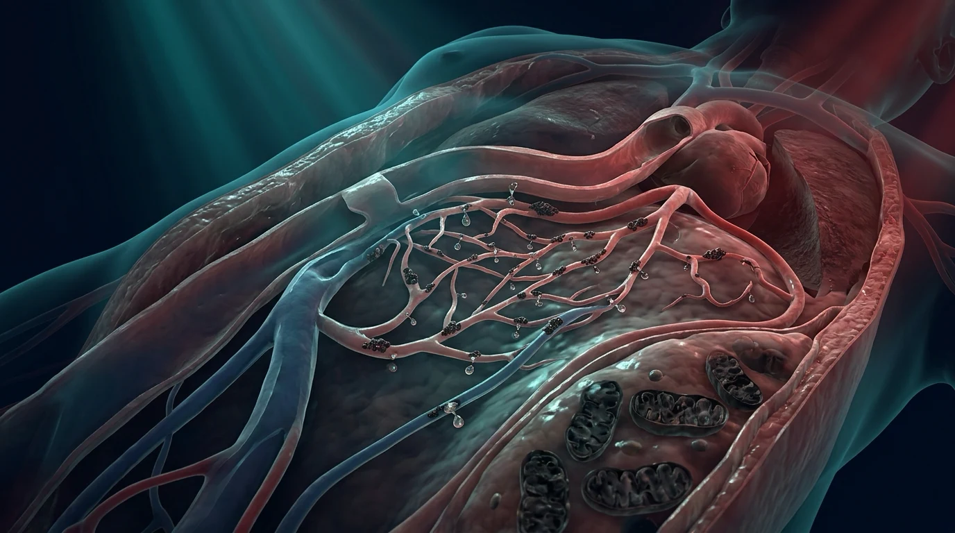
The kidney is normally sterile. Infection reaches it by two routes: [1]
1. Ascending infection (dominant route)
Organisms colonise the periurethral area and distal urethra → ascend the urethra to the bladder (cystitis) → travel up the ureters to the renal pelvis (pyelitis) → invade the renal parenchyma (pyelonephritis) via the collecting ducts. Facilitating factors: [1]
- Short urethra in women (hence female predominance).
- Sexual activity — mechanical introduction of periurethral bacteria.
- Vesicoureteric reflux (VUR) — incompetent ureterovesical junction allows bladder urine (and organisms) to reflux up the ureters during micturition.
- Urinary stasis / incomplete emptying — from obstruction (BPH, stones, tumour, neurogenic bladder, pregnancy), reducing the mechanical washout that normally clears organisms.
- Bacterial virulence factors — uropathogenic E. coli expresses P-fimbriae (Gal-Gal binding) that adhere to urothelium and resist washout; flagella aid ascent; haemolysin and siderophores (aerobactin) damage tissue and evade innate immunity. [1]
2. Haematogenous spread
Bacteraemia seeds the kidney via the cortical arteries — uncommon in the normal host, but a recognised route in the immunocompromised (endocarditis, IV drug use, disseminated infection) and with Staphylococcus aureus, Candida, Salmonella, and Mycobacterium tuberculosis. S. aureus in the urine with no lower-tract symptoms should prompt a search for an extra-urinary source (endocarditis, osteomyelitis, IV line infection). [1]
Inflammatory cascade
Once in the parenchyma, organisms elicit a neutrophil-predominant interstitial nephritis with tubular oedema, microabscess formation and (in severe disease) papillary necrosis, perinephric abscess extension, or gas production (emphysematous pyelonephritis — fermenting organisms in poorly-controlled diabetics). The inflammatory cytokine response (IL-1, IL-6, TNF-α) drives the systemic features — fever, rigors, and, when dysregulated, septic shock.[6][2]
Risk factors
Female sex
Anatomy
- Short urethra + proximity to anus/periurethral flora → ascending colonisation
- Lifetime risk of UTI ~50% in women; recurrent in 20-30%
- Sexual activity and spermicide use (alters vaginal flora) increase risk
Obstruction
Stasis
- Stones (calcium, struvite, uric acid) — act as a nidus and impair washout
- BPH in men; urethral stricture; pelvic tumour; retroperitoneal fibrosis
- Functional obstruction: neurogenic bladder (spinal cord injury, diabetes, MS), anticholinergics
- Pregnancy — progesterone relaxes ureteric smooth muscle + mechanical compression by gravid uterus
- Vesicoureteric reflux (VUR) — congenital, may persist into adulthood; causes recurrent pyelonephritis and renal scarring
Diabetes mellitus
Immune + tissue
- Impaired neutrophil function (chemotaxis, phagocytosis) and glycosuria (bacterial growth substrate)
- Predisposes to severe complications: emphysematous pyelonephritis, papillary necrosis, perinephric abscess, fungal UTI
- Higher rates of bacteraemia and antimicrobial-resistant organisms
Pregnancy
Physiological
- Physiological ureteric dilation (progesterone) + mechanical compression → stasis; asymptomatic bacteriuria in 2-10%
- Untreated ASB progresses to pyelonephritis in 20-30% (vs <1% if treated) — universal screening + treatment
- Pyelonephritis in pregnancy: high risk of premature labour, ARDS, septic shock — admit, IV antibiotics
Catheter / instrumentation
Healthcare-associated
- Indwelling urinary catheter — biofilm formation; risk ~5% per catheter-day
- Catheter-associated UTI (CAUTI) is the commonest healthcare-associated infection
- Recent urological procedure (TURP, cystoscopy, stent change, biopsy)
- Predisposes to Pseudomonas, Proteus, Enterococcus, Candida, polymicrobial infection
Immunocompromise
Host defence
- Transplant (renal, on calcineurin inhibitors + mycophenolate + steroids)
- Chemotherapy / neutropenia, haematological malignancy, advanced HIV
- High-dose corticosteroids, anti-TNF biologics
- Higher risk of atypical organisms (Candida, CMV, BK virus, mycobacteria) and haematogenous spread
Clinical features
The clinical hallmark is the triad of fever ± rigors, flank pain, and urinary symptoms, with costovertebral angle (CVA) tenderness on examination. [1]
Upper UTI (pyelonephritis)
Kidney
- Fever (often >38.5°C) ± rigors — indicates bacteraemia/systemic involvement
- Flank / loin pain — unilateral or bilateral; CVA tenderness (Murphy's punch sign)
- Nausea, vomiting, anorexia — frequently present; may mimic intra-abdominal pathology
- Lower-tract symptoms usually coexist (dysuria, frequency, urgency, suprapubic pain) but may be ABSENT in the elderly, immunocompromised, or catheterised
- Ileus, abdominal distension; occasionally a palpable ballotable mass (severe hydronephrosis, abscess)
- Septic shock: hypotension, tachycardia, mottled skin, altered mental state, oliguria (urosepsis)
Lower UTI (cystitis)
Bladder
- Dysuria, frequency, urgency, suprapubic pain/discomfort
- Cloudy or bloody urine, offensive odour
- Usually AFEBRILE or low-grade fever (<38°C); no flank pain; no rigors
- Presence of fever, rigors, or flank pain suggests UPPER UTI — different management pathway
- Haematuria (gross) — think haemorrhagic cystitis, stones, or malignancy if recurrent
Atypical / special
Beware
- Elderly: atypical presentation — confusion, falls, incontinence, anorexia without fever (delirium as the only sign)
- Pregnancy: may present as preterm labour; respiratory symptoms (ARDS) in severe cases
- Diabetic: may be emphysematous — look for gas, crepitus, severe metabolic derangement
- Spinal cord injury / neurogenic bladder: absence of pain/dysuria — fever and autonomic dysreflexia may be the only clues
- Immunocompromised: blunted inflammatory response — cultures positive with minimal symptoms
Upper vs lower UTI — exam discriminator
The clinical distinction matters: upper UTI requires longer parenteral therapy, imaging, and source control; lower UTI is usually oral therapy. Discriminators: [1]
- Fever >38°C, rigors, flank pain, CVA tenderness, nausea/vomiting → upper UTI.
- Afebrile, isolated dysuria/frequency/suprapubic pain → lower UTI (cystitis).
- A useful exam point: the presence of systemic features (rigors, high fever, haemodynamic instability) mandates blood cultures, imaging, and admission. [1]
Diagnosis
Diagnostic approach to suspected pyelonephritis / urosepsis
Urinalysis (dipstick + microscopy)
BEDSIDE first test. Positive nitrites (Enterobacterales convert nitrate → nitrite; FALSE NEGATIVE if bladder dwell <4 h, or non-nitrate-formers like Enterococcus, Pseudomonas, Staphylococcus). Leucocyte esterase (neutrophil enzyme — detects pyuria). Microscopy: >10 WBC/high-power field. Haematuria (microscopic) common. Sterile pyuria (pyuria with negative routine culture) → think Mycobacterium tuberculosis, Chlamydia, Ureaplasma, fungal, partially-treated infection, or interstitial nephritis.
Urine culture & susceptibility
Clean-catch midstream urine (MSU) — gold standard. SIGNIFICANT GROWTH: >10^5 CFU/mL in a symptomatic woman (classic Kass criterion); >10^3 in a symptomatic man or on suprapubic aspirate; ANY growth from suprapubic aspirate (normally sterile). Catheter specimen: >10^3 with symptoms. Always collect BEFORE antibiotics if possible. Send Gram stain — rapid clue to Gram-negative rod (E. coli) vs Gram-positive (Enterococcus).
Blood cultures × 2
Send in ALL patients with fever/rigors, sepsis, or immunocompromise BEFORE antibiotics. Positive in ~20-30% of pyelonephritis (higher with obstruction, diabetes, stones). Blood-culture-positive urosepsis: same organism in urine and blood confirms the urinary source. Antibiotics within 1 h of recognition in septic shock (Surviving Sepsis).
Bloods — FBC, CRP, U&E, LFTs, lactate, venous/arterial gas
Leucocytosis with neutrophilia; raised CRP (mirrors severity). U&E: AKI common in sepsis or with bilateral obstruction (post-renal). Lactate: marker of tissue hypoperfusion / severity (Sepsis-3). Venous gas: metabolic acidosis, AKI. Glucose/HbA1c: undiagnosed/poorly-controlled diabetes. Procalcitonin: rising — may help distinguish bacterial infection and guide antibiotic duration.
Imaging — CT abdomen/pelvis with contrast (THE key modality)
INDICATED in: failure to improve within 48-72 h of antibiotics; diabetes; immunocompromise; suspected obstruction/stones; recurrent infection; male; severe sepsis/septic shock; palpable mass. CT with IV contrast is gold standard — identifies obstruction, stones (non-contrast better for tiny calculi), abscess, pyonephrosis, emphysematous change, perinephric extension. Bedside ultrasound (POCUS) is a rapid ICU bedside screen for hydronephrosis (obstruction) but is INSUFFICIENT to exclude parenchymal disease — a normal US does not exclude pyelonephritis.
Exclude obstruction EARLY
Pyonephrosis (pus under pressure in an obstructed kidney) is a urological emergency — every uroseptic patient needs the question "is there obstruction?" answered within hours. Bedside POCUS for hydronephrosis is a rapid triage; CT confirms. Relieve obstruction (JJ stent or percutaneous nephrostomy) — drainage of infected urine often produces dramatic haemodynamic improvement.
Special tests as indicated
Prostatic involvement (tender prostate on PR — avoid vigorous massage to avoid bacteraemia) suggests prostatitis → 4-week course. Renal abscess >3-5 cm → drainage. Atypical organisms (TB: sterile pyuria + AFB/NAAT on morning urines ×3; fungal: Candida with pseudohyphae). Post-treatment culture not routine unless recurrent/complicated.
Imaging — what to look for
CT with IV contrast
Gold standard
- Wedge-shaped areas of decreased enhancement / striated nephrogram — acute pyelonephritis
- Round non-enhancing lesion with rim enhancement = abscess
- Gas within renal parenchyma/collecting system = emphysematous pyelonephritis (classify class I-IV)
- Dilated collecting system with fluid/debris level = pyonephrosis
- Perinephric stranding/fluid collection = extension beyond capsule
- Non-contrast series identifies radiopaque stones (calcium, struvite)
Ultrasound (incl. POCUS)
Bedside / safe
- First-line in pregnancy (no radiation) and the unstable ICU patient at bedside
- Detects hydronephrosis (obstruction), large abscesses, gross emphysematous change, stones
- NORMAL US does NOT exclude pyelonephritis — parenchymal inflammation often sonographically occult
- Use to guide percutaneous nephrostomy and abscess drainage
Plain CT (stone protocol)
Calculus
- Highest sensitivity for radiopaque and radiolucent (uric acid) stones
- Use when the clinical question is "is there an obstructing stone?"
- Low radiation; can be combined with contrast series
MRI / nuclear medicine
Rarely needed
- MRI — gadolinium — alternative when iodinated contrast contraindicated; pregnancy (after US)
- DMSA / DTPA renal scans — assess renal scarring and differential function (mainly paediatric / recurrent VUR work-up)
Management
Initial resuscitation (urosepsis / septic shock)
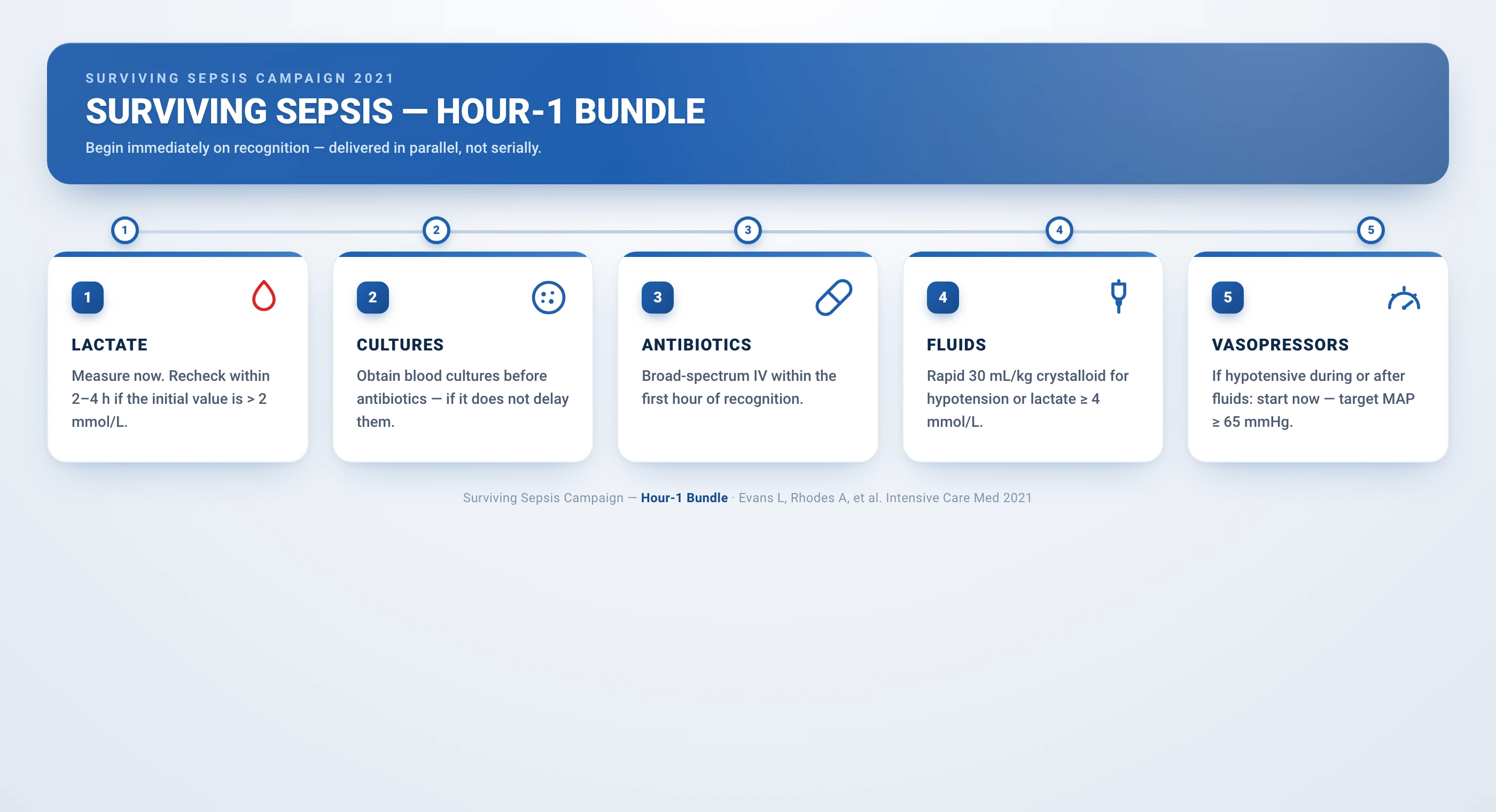
First hour bundle (Surviving Sepsis adapted to urosepsis)
Measure lactate; obtain blood cultures ×2 BEFORE antibiotics
Lactate >2 mmol/L = tissue hypoperfusion; >4 = severe sepsis. Two sets of blood cultures (aerobic + anaerobic) from separate sites, plus urine culture, all before the first antibiotic dose. Do NOT delay antibiotics beyond 45 min to obtain cultures in shock.
Broad-spectrum antibiotics within 1 hour
Empiric IV therapy covering common Gram-negatives + Enterococcus, with anti-pseudomonal cover if healthcare-associated/cathetered/immunocompromised. Options: ceftriaxone 2 g IV daily ± gentamicin (community); piperacillin-tazobactam 4.5 g IV TDS or ceftazidime (HCA/anti-pseudomonal); add ampicillin if Enterococcus risk; meropenem for known ESBL/carbapenemase. De-escalate to culture sensitivities at 48-72 h.
Crystalloid 30 mL/kg for hypotension or lactate ≥4
Balanced crystalloid (Hartmann's / Plasma-Lyte) preferred over 0.9% saline (hyperchloraemic acidosis, AKI). Reassess fluid responsiveness (passive leg raise, IVC POCUS, dynamic indices) before further boluses — uroseptic patients are often elderly with limited cardiac reserve.
Vasopressors for MAP <65 mmHg
Norepinephrine first-line (alpha + modest beta-1 agonist). Target MAP 65 mmHg. Add vasopressin 0.03 U/min if escalating norepinephrine. Do not withhold vasopressors to "fill more" — early vasopressors reduce fluid overload.
Source control — EARLY and definitive
Relieve obstruction (JJ stent or percutaneous nephrostomy — the latter in septic/unstable); drain abscess >3 cm (percutaneous radiologically-guided); remove/replace urinary catheter; nephrectomy for refractory emphysematous pyelonephritis. Aim for source control within 6-12 h of recognition. Drainage of an obstructed infected system often produces dramatic improvement.
Supportive ICU care
Lung-protective ventilation if ARDS; renal replacement therapy for refractory AKI/electrolyte/acid-base; VTE prophylaxis; stress-ulcer prophylaxis if intubated/coagulopathic; glycaemic control 140-180 mg/dL; early enteral nutrition; monitor lactate trend.
Antimicrobial therapy
Uncomplicated pyelonephritis
Adult non-pregnant woman, mild-moderate
- Oral therapy acceptable if able to tolerate PO and no sepsis
- Ciprofloxacin 500 mg PO BD × 7 days (check local resistance <10%)
- Levofloxacin 750 mg PO daily × 5 days
- Co-amoxiclav (amoxicillin-clavulanate) 875/125 mg PO BD × 7-10 days (if organism susceptible)
- Trimethoprim-sulfamethoxazole 160/800 mg PO BD × 14 days (if known susceptible & local resistance <20%)
- Nitrofurantoin and fosfomycin are NOT reliable for tissue/upper-tract levels — reserve for cystitis
Severe / complicated / urosepsis
IV therapy
- Ceftriaxone 2 g IV daily ± gentamicin 5-7 mg/kg IV once daily (synergy + broad Gram-negative cover)
- Piperacillin-tazobactam 4.5 g IV TDS (anti-pseudomonal; good for catheter/HCA)
- Ceftazidime 2 g IV TDS or cefepime 2 g IV TDS (anti-pseudomonal cephalosporin)
- Meropenem 1 g IV TDS — known ESBL, carbapenemase, or critically ill with resistant organism risk
- Add ampicillin 2 g IV QID if Enterococcus suspected (cephalosporins do not cover it)
- Duration: 7-14 days (7 days for rapid responders with source control; 14 days for slow response, abscess, bacteraemia)
Pregnancy
Special
- Ceftriaxone 1-2 g IV daily (or cefuroxime) — cephalosporin first-line
- AVOID fluoroquinolones (cartilage/arthropathy), tetracyclines (teeth/bone), trimethoprim 1st trimester (folate antagonist) & 3rd trimester (kernicterus — displaces bilirubin)
- AVOID aminoglycosides if possible (fetal ototoxicity) — use only if severe & short course
- Always admit; IV therapy; 10-14 day course; test-of-cure culture 1-2 weeks after
Prostatitis / epididymitis
Longer course
- Ciprofloxacin or levofloxacin × 4-6 weeks (penetrate prostatic tissue — fluoroquinolones concentrate in prostate)
- Acute bacterial prostatitis can cause urosepsis — IV therapy then oral step-down
- Consider STI screen (Chlamydia, gonorrhoea) in young sexually-active men
Catheter-associated UTI
CAUTI
- REMOVE or replace the catheter FIRST — biofilm-laden catheter is the source
- Do NOT treat asymptomatic catheter bacteriuria (does not reduce infection, selects resistance)
- Treat only symptomatic infection: fever, rigors, flank pain, new confusion, haemodynamic instability
- Catheter specimen culture guides therapy; organisms often polymicrobial (Pseudomonas, Proteus, Enterococcus, Candida)
Source control — the cornerstone
Source control in urosepsis
Relieve obstruction
Pyonephrosis = pus under pressure in an obstructed kidney = urological emergency. Decompression: percutaneous nephrostomy (radiology-guided — preferred in the unstable/septic patient at bedside or IR suite) OR retrograde JJ ureteric stent (cystoscopy under anaesthetic — requires theatre, may seed infection retrograde). Drainage of thick pus under pressure often produces immediate haemodynamic improvement. Send drained fluid for culture.
Drain abscess
Renal/perinephric abscess >3 cm rarely resolves with antibiotics alone — needs percutaneous catheter drainage (radiology). <3 cm may respond to antibiotics. Surgical drainage reserved for multiloculated/failed percutaneous drainage. Send pus for culture.
Remove/replace urinary catheter
The catheter is often the source in CAUTI/urosepsis. Remove if no longer needed; replace if still required (fresh catheter). Change suprapubic catheters and urological stents that are due. Sample urine from a NEW catheter, not a long-standing one (biofilm).
Treat stones (definitive)
Struvite (infection) stones must be completely removed — residual fragments harbour urease-producing organisms and the stone recurs. Options: ESWL, PCNL, ureteroscopy. Eradicate the nidus to cure the infection. Do NOT leave staghorn calculi in situ.
Nephrectomy for refractory emphysematous pyelonephritis
When medical + percutaneous management fails, or there is extensive destruction (Huang-Tsai class III/IV — extensive gas or gas through kidney + perinephric), emergency nephrectomy is life-saving. Mortality 20-40% overall; highest for extensive disease and delayed intervention.
Emphysematous pyelonephritis — the surgical emergency
Emphysematous pyelonephritis (EPN) is a necrotising, gas-forming infection of the kidney, almost always in diabetics (90%+), caused by gas-forming organisms (E. coli most common, also Klebsiella, Proteus, Pseudomonas). The gas (CO₂, H₂) results from mixed-acid fermentation of glucose by the organism in tissues with high glucose and impaired perfusion.[8]
Huang-Tsai classification (CT-based)
Class I
Gas in collecting system only
- Bubbly/linear gas in the renal parenchyma or collecting system without extension
- Best prognosis — often responds to antibiotics + relief of obstruction
- Mortality ~20%
Class II
Gas in renal parenchyma
- Gas in renal parenchyma without extrarenal extension
- Requires antibiotics + percutaneous drainage ± nephrectomy if not improving
- Mortality ~35%
Class IIIa
Extension of gas into perinephric space
- Gas extends to perinephric space
- High risk of abscess, septic shock; aggressive source control required
Class IIIb
Extension of gas into pararenal space
- Gas extends beyond Gerota's fascia to pararenal space
- Extensive disease — high mortality; early nephrectomy often required
Class IVa/b
Bilateral or solitary kidney
- Bilateral EPN or EPN in a solitary kidney
- Nephrectomy not possible (would render anuric) — bilateral percutaneous drainage, antibiotics, dialysis if needed
- Worst prognosis — mortality approaches 50%+
Management of EPN
Emphysematous pyelonephritis management
Resuscitate + broad-spectrum antibiotics
ABC; IV fluids (balanced crystalloid); vasopressors for shock; broad-spectrum Gram-negative cover (piperacillin-tazobactam or meropenem) + source control. Aggressive glucose control. These patients are often profoundly septic and acidotic.
CT with contrast — classify
Confirm gas in kidney; classify by Huang-Tsai (extent); look for obstruction, abscess, bilateral involvement. This drives the decision between medical/percutaneous and surgical management.
Percutaneous drainage + medical therapy first-line
Class I-II and many class III: drainage of gas/pus/obstruction via percutaneous nephrostomy or catheter drains (often multiple, repeated) + antibiotics + glycaemic control. Systematic review evidence favours percutaneous drainage over upfront nephrectomy with lower mortality.
Nephrectomy if failure or extensive disease
Indications: clinical deterioration despite 48-72 h of drainage + antibiotics; extensive class IIIb/IV destruction; lack of access to repeated drainage. Emergency nephrectomy is life-saving in refractory disease. Bilateral/solitary kidney: bilateral drainage, avoid nephrectomy.
Correct glycaemia and predisposing factors
Insulin infusion for hyperglycaemia; relieve obstruction; treat the underlying diabetes. Address the reason gas formed — without glycaemic control, recurrence on the contralateral side is a risk.
Percutaneous drainage vs nephrectomy for emphysematous pyelonephritis (Somani, systematic review, J Urol 2008)
Systematic review of retrospective case series (no RCT possible — rare disease)
Population: Adults with radiologically confirmed emphysematous pyelonephritis across reported series
Key finding
Medical therapy alone had the highest mortality (~50%); addition of percutaneous drainage reduced mortality to ~13.5%, better than emergency nephrectomy (~25%). Combined percutaneous drainage + medical therapy became the new standard first-line; nephrectomy reserved for failure.
Practice change
In emphysematous pyelonephritis, treat with broad-spectrum antibiotics PLUS percutaneous drainage first; reserve nephrectomy for extensive or refractory disease.
Sepsis-3 definitions (Singer, JAMA 2016)
International consensus task force redefining sepsis and septic shock
Population: Adult patients with suspected infection — derivation and validation cohorts
Key finding
SOFA-based sepsis definition and the septic shock criteria identified a population with mortality ~40% (vs ~10% for sepsis without shock). qSOFA (RR ≥22, altered mentation, SBP ≤100) — a bedside screen — predicted poor outcome outside ICU.
Practice change
Apply Sepsis-3 definitions to urosepsis: organ dysfunction (SOFA ≥2) defines sepsis; vasopressor-dependent hypotension + lactate >2 despite fluids defines septic shock and mandates aggressive resuscitation + early source control.
Complications
Pyonephrosis
Obstructed infected kidney
- Pus under pressure in an obstructed collecting system — urological emergency
- Risk: septic shock, irreversible renal destruction, kidney loss
- Management: urgent decompression (nephrostomy or JJ stent) + antibiotics
- Drainage often produces dramatic haemodynamic improvement
Renal/perinephric abscess
Walled-off infection
- Collection of pus within the kidney (renal/cortical) or between kidney and Gerota's fascia (perinephric)
- Risk factors: diabetes, obstruction, stones, staphylococcal bacteraemia (haematogenous)
- <3 cm may resolve with antibiotics; >3 cm → percutaneous catheter drainage
- Surgical drainage for multiloculated/failed percutaneous drainage
Emphysematous pyelonephritis
Gas-forming
- Necrotising gas-forming infection — diabetics; mortality 20-40%
- Percutaneous drainage + antibiotics first-line; nephrectomy for refractory/extensive
- See dedicated section above
Papillary necrosis
Sloughed papillae
- Ischaemic necrosis of renal papillae — diabetes, obstruction, analgesic abuse, sickle cell
- Sloughed papilla may obstruct ureter (acute obstruction); ring-shadow on imaging
- May be asymptomatic or cause haematuria, flank pain, AKI
Septic shock & AKI
Systemic
- Vasodilatory shock, multi-organ failure; AKI from sepsis ± post-renal obstruction
- Mortality 10-20% in urosepsis with shock (better than pulmonary/abdominal sepsis with source control)
- May require vasopressors, RRT, lung-protective ventilation
Renal scarring / CKD / recurrence
Long-term
- Recurrent childhood pyelonephritis + VUR → renal scarring, hypertension, CKD (reflux nephropathy)
- Adults: residual scarring after severe infection; address predisposing factors to prevent recurrence
- Emphysematous/obstructive disease may leave a non-functioning kidney
Special situations
Pregnancy
High-risk
- Universal screening for asymptomatic bacteriuria (treat — prevents pyelonephritis in 20-30%)
- Pyelonephritis in pregnancy: ADMIT, IV cephalosporin; risk of premature labour, ARDS, septic shock
- Avoid fluoroquinolones, tetracyclines, trimethoprim (1st & 3rd trimester), aminoglycosides
- Ultrasound first-line imaging (avoid CT radiation); MRI if needed
- Right-sided predilection (right ureter more compressed by gravid uterus, dextro-rotation)
Diabetes mellitus
Severe complications
- Higher risk of bacteraemia, emphysematous pyelonephritis, papillary necrosis, perinephric abscess, fungal UTI
- Aggressive imaging (CT) — do not miss gas
- Tight(ish) glucose control; check feet for source; may have neuropathic bladder (stasis)
- Higher incidence of resistant organisms
Catheterised / catheter-associated
HCA
- Asymptomatic bacteriuria is universal in long-term catheters — DO NOT treat
- Treat only symptomatic infection; remove/replace catheter as source control
- Polymicrobial, resistant organisms (Pseudomonas, Proteus, Enterococcus, Candida)
- Consider alternative to long-term catheter (suprapubic, condom, intermittent self-catheterisation)
Renal transplant
Immunocompromised
- Immunosuppression blunts presentation — cultures positive with few symptoms
- Atypical organisms: CMV, BK virus, Candida, mycobacteria; opportunistic infection
- Anastomotic stricture / obstruction common; surgical complications (urinoma, leak)
- Coordinate with transplant team; avoid drug interactions (calcineurin inhibitors + macrolides/azoles)
Elderly / delirious
Atypical
- May present without fever or dysuria — confusion, falls, incontinence, anorexia
- High threshold to image; consider obstruction (BPH, cervical/prostate tumour)
- Beware over-diagnosis of UTI from asymptomatic bacteriuria — not every positive culture in a confused elderly patient is infection
Prognosis
Uncomplicated pyelonephritis
Good
- Mortality <1% with appropriate therapy
- Most respond within 48-72 h; oral step-down after clinical improvement
- Test-of-cure culture if recurrent, complicated, or pregnant
Complicated / urosepsis
Moderate
- Mortality 10-20% in septic shock — better than pulmonary/abdominal sepsis IF source control achieved
- Favourable prognosis reflects the accessibility of the source for control (catheter drainage, stent)
- Delayed source control worsens outcome dramatically
Emphysematous pyelonephritis
Poor
- Mortality 20-40% overall; higher with extensive (class IIIb/IV) disease, thrombocytopenia, acute renal failure, altered sensorium, shock
- Scoring systems (Huang-Tsai class; EPIC score — emphysematous pyelonephritis) guide prognosis and surgical decision-making
- Nephrectomy mortality ~25%; medical-only mortality ~50%
Prognostic factors
What matters
- Speed of source control; adequacy of initial antibiotics; host factors (age, diabetes, immunocompromise)
- Severity of sepsis (lactate, SOFA, vasopressor requirement); presence of bacteraemia
- Presence of obstruction, abscess, or gas — all worsen prognosis
SAQ — Urosepsis with septic shock and obstructive uropathy
SAQ — Urosepsis with septic shock in a post-TURP diabetic patient
10 minutes · 10 marks
A 68-year-old man with type 2 diabetes and benign prostatic hyperplasia is readmitted to ICU on day 4 after a transurethral resection of prostate (TURP) with an indwelling Foley catheter. He is confused, febrile (39.2 °C) and shivering. BP 78/45, HR 128, RR 28, SpO2 94% on room air, capillary refill 5 s. Lactate 4.8 mmol/L, WBC 24.6 x10^9/L, creatinine 220 umol/L (baseline 90), pH 7.28, bicarbonate 16. Urine is cloudy; Gram stain shows Gram-negative rods. Bedside POCUS demonstrates right-sided hydronephrosis with a 1.5 cm obstructing ureteric stone.
SAQ — Obstructive uropathy with pyonephrosis and post-obstructive diuresis
10 minutes · 10 marks
A 72-year-old man with bladder-outlet obstruction from advanced prostate cancer presents with a 3-day history of right flank pain, anuria, fever 38.9 °C and new confusion. BP 96/58, HR 112, RR 24. Creatinine 340 umol/L (baseline 110), K+ 5.9 mmol/L, pH 7.22, lactate 3.2. WBC 22 x10^9/L. Urine Gram stain shows Gram-negative rods. Bedside ultrasound shows bilateral hydronephrosis with a markedly distended bladder; in-and-out catheterisation drains 1.4 L of cloudy urine. A subsequent CT urogram confirms bilateral hydroureteronephrosis with a large obstructing mass at the bladder neck and a right perinephric fluid collection suggestive of pyonephrosis.
Clinical pearls
Red flags
Exam practice — viva-style questions
[1]Summary answer (for the oral exam)
"Acute pyelonephritis is infection of the renal parenchyma and collecting system, most commonly by ascending E. coli, and urosepsis is sepsis arising from a urinary source — one of the commonest causes of sepsis in ICU. I would diagnose it at the bedside with a history of fever, rigors, flank pain and urinary symptoms plus costovertebral angle tenderness, confirm it with urinalysis showing nitrites, leucocyte esterase and pyuria, a urine culture with significant growth, blood cultures that are positive in 20-30%, and a contrast CT of the abdomen and pelvis to exclude obstruction, abscess, gas and stones — remembering that a normal bedside ultrasound does not exclude parenchymal disease. My management follows the first-hour sepsis bundle: cultures before antibiotics, broad-spectrum intravenous therapy covering common Gram-negatives and Enterococcus — ceftriaxone with or without gentamicin for community disease, piperacillin-tazobactam or meropenem for healthcare-associated or anti-pseudomonal cover, with ampicillin added if Enterococcus is suspected — balanced crystalloid for hypotension or lactate above four, norepinephrine for a MAP below 65, and above all EARLY SOURCE CONTROL: relief of obstruction by percutaneous nephrostomy or stent for pyonephrosis, drainage of any abscess greater than three centimetres, removal or replacement of an indwelling catheter, and definitive treatment of any struvite stone. I would treat for seven to fourteen days, de-escalating to oral therapy as the patient improves and cultures return. In a diabetic with gas in the kidney I would diagnose emphysematous pyelonephritis — a surgical emergency — classify it by the Huang-Tsai system, treat with antibiotics and percutaneous drainage first-line, and reserve emergency nephrectomy for extensive or refractory disease, knowing the mortality is twenty to forty per cent. In pregnancy I would admit, give an intravenous cephalosporin and avoid fluoroquinolones, tetracyclines, trimethoprim and aminoglycosides. Throughout, I would reassess at 48 to 72 hours and re-image if the patient is not improving, because the prognosis of urosepsis — mortality under one per cent when uncomplicated, ten to twenty per cent in septic shock, and twenty to forty per cent in emphysematous disease — hinges on rapid antibiotics and definitive source control." [1]
Related conditions
- Sepsis and septic shock — apply Sepsis-3 definitions and the Surviving Sepsis one-hour bundle to urosepsis.[6]
- Acute kidney injury — post-renal AKI from obstruction; septic AKI from hypoperfusion; may require RRT.
- Asymptomatic bacteriuria — screen and treat in pregnancy and before urological instrumentation; otherwise do NOT treat (catheter, elderly).
- Vesicoureteric reflux / reflux nephropathy — congenital; recurrent childhood pyelonephritis → renal scarring, hypertension, CKD.
- Renal/perinephric abscess — complication needing percutaneous or surgical drainage.
- Prostatitis — acute bacterial (urosepsis source) and chronic; longer antibiotic course (4-6 weeks).
References
- [1]Johnson JR, Russo TA Acute Pyelonephritis in Adults N Engl J Med, 2018.PMID 29298155
- [2]Wagenlehner FM, Lichtenstern C, Rolfes C, et al. Urosepsis--from the view of the urologist Int J Antimicrob Agents, 2011.PMID 21993485
- [3]Wagenlehner FM, Tandogdu Z, Bjerklund Johansen TE An update on classification and management of urosepsis Curr Opin Urol, 2017.PMID 27846034
- [4]Somani BK, Nabi G, Thorpe P, et al. Is percutaneous drainage the new gold standard in the management of emphysematous pyelonephritis? Evidence from a systematic review J Urol, 2008.PMID 18353396
- [5]Gupta K, Hooton TM, Naber KG, et al. International clinical practice guidelines for the treatment of acute uncomplicated cystitis and pyelonephritis in women: A 2010 update by the Infectious Diseases Society of America and the European Society for Microbiology and Infectious Diseases Clin Infect Dis, 2011.PMID 21292654
- [6]Singer M, Deutschman CS, Seymour CW, et al. The Third International Consensus Definitions for Sepsis and Septic Shock (Sepsis-3) JAMA, 2016.PMID 26903338
- [7]Chen CC, Lin SH, Huang HC, et al. Performance of Scoring Systems in Predicting Clinical Outcomes of Patients with Emphysematous Pyelonephritis: A 14-Year Hospital-Based Study J Clin Med, 2022.PMID 36555916
- [8]Tsu JH, Ma WK, Chan WC, et al. Emphysematous pyelonephritis: an 8-year retrospective review across four acute hospitals Asian J Surg, 2013.PMID 23810162