ICU · Infectious Diseases
Severe dengue in the ICU
Also known as Dengue haemorrhagic fever · Dengue shock syndrome · Severe dengue · DENV infection
Severe dengue is a tropical flaviviral infection transmitted by Aedes aegypti mosquitoes, caused by one of four serotypes (DENV 1-4) and defined by plasma leakage, haemorrhage, and organ impairment. The disease runs three phases: febrile (2-7 days of high fever, retro-orbital pain, myalgia, rash, leucopenia), critical (begins at DEFERVESCENCE — plasma leak, haemoconcentration, dengue shock syndrome, bleeding, 24-48h window of deterioration), and recovery (reabsorption of extravascular fluid, bradycardia, diuresis). Pathophysiology centres on antibody-dependent enhancement (ADE) on secondary infection with a heterologous serotype driving a cytokine storm (TNF, IL-6, IL-8), endothelial/ glycocalyx dysfunction, capillary leak, and consumptive thrombocytopenia. WHO warning signs: abdominal pain, persistent vomiting, fluid accumulation, mucosal bleeding, lethargy, hepatomegaly, rising haematocrit with falling platelets. Severe dengue criteria (2009 WHO): dengue shock syndrome (DSS — compensated then decompensated shock from plasma leak), severe bleeding, and organ impairment (AST/ALT >1000, impaired consciousness, myocarditis, AKI). Management: careful isotonic crystalloid resuscitation titrated to haematocrit and perfusion (10-20 mL/kg bolus in compensated shock), transfusion for major bleeding, NO prophylactic platelets, and avoidance of NSAIDs/aspirin. ICU care addresses shock, organ support, and NIV for pulmonary oedema from fluid leak / over-resuscitation. No specific antiviral exists; supportive care is definitive.
On this page & tools
Your progress
Saved locally on this device.
Target exams
Red flags
Virology and epidemiology
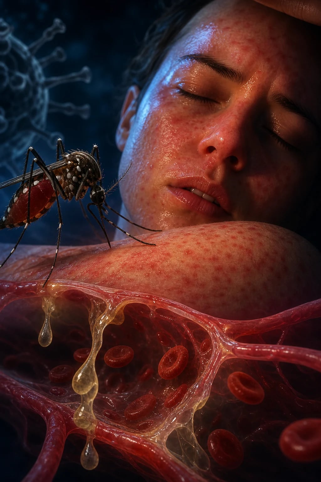
Dengue is caused by four antigenically distinct serotypes of dengue virus (DENV 1, 2, 3, 4), all members of the genus Flavivirus (family Flaviviridae). Each serotype is an independent virus — infection with one confers lifelong homologous immunity but only transient cross-protection against the others. The genome is a single-stranded positive-sense RNA encoding three structural proteins (capsid C, premembrane prM, envelope E) and seven non-structural proteins (NS1-NS5). The E glycoprotein is the principal target of neutralising antibody and mediates cellular entry; NS1 is the secreted antigen used in rapid diagnostics and is itself implicated in endothelial injury and vascular leak. [1]
The principal vector is the female Aedes aegypti mosquito (a day-biting, peri-domestic breeder in stagnant water); Aedes albopictus is a secondary vector in temperate regions. Humans are the amplifying host — there is no animal reservoir in the urban transmission cycle. Dengue is the most important arboviral disease of humans, with an estimated 100-400 million infections and ~20,000 deaths annually across >100 endemic countries, and incidence has expanded 30-fold in 50 years owing to urbanisation, climate change, and travel. Both primary and secondary infections occur in adults in hyperendemic regions of Southeast Asia, the Pacific, and Latin America; in ANZ, cases are almost all imported returning travellers (Indonesia, Thailand, Vietnam, Philippines being the commonest exposures). [1]
DENV-1
Serotype 1
- Genotype diversity drives regional epidemics — strains vary in virulence
- Common cause of secondary infection in adults — associated with classic DSS
- Cosmopolitan distribution; periodic genotype replacement causes outbreaks
DENV-2
Serotype 2
- Asian genotypes (e.g. Cosmopolitan, Asian I) historically linked to severe disease
- Strongly associated with antibody-dependent enhancement in secondary infection
- Often the serotype recovered in fatal adult cases and DHF outbreaks
DENV-3
Serotype 3
- Associated with hepatitis and encephalitis phenotypes
- Re-emergence after years of absence causes large susceptible-population epidemics
DENV-4
Serotype 4
- Often clinically milder but sylvatic cycles and genotype shifts occur
- Secondary DENV-4 still causes severe disease via ADE
Clinical phases
Dengue phases (click each)
Reabsorption phase
Lasts 48-72h. Plasma reabsorbed from extravascular space into intravascular compartment. Haematocrit falls (may drop rapidly if over-resuscitated → haemodilution). Relative bradycardia, wide pulse pressure, diuresis, appetite returns, rash desquamates. AVOID fluid overload — pulmonary oedema from reabsorption is the iatrogenic danger of this phase.
The three phases are clinically distinct and dictate management. The defining and most examined concept is that deterioration coincides with defervescence, not with peak fever — the intuitive belief that "the fever is gone, the patient is better" is a classic trap. [1]
Febrile phase (days 1-3)
Onset — high fever
- Sudden-onset high fever (39-40°C) with chills
- Bifrontal/retro-orbital headache, severe myalgia and arthralgia ("breakbone fever")
- Nausea, vomiting, anorexia, flushed facies, conjunctival injection
- Early rash: transient blanching erythema; palate petechiae; positive tourniquet test
- Leucopenia, thrombocytopenia beginning, mild HCT rise
- Monitor for warning signs; oral/IV maintenance fluids; paracetamol only
Critical phase (days 3-7)
Defervescence — leak
- Fever settles but patient clinically WORSENS
- Plasma leak window: HCT rising, platelets falling, effusions/ascites
- Compensated → decompensated shock (DSS) within hours
- NS1 still detectable; IgM begins to rise from day 5
- Most dangerous phase — close monitoring (HCT q4-6h, urine output, perfusion)
- Isotonic crystalloid titrated to HCT; avoid both under- and over-resuscitation
Recovery phase (days 7-10)
Reabsorption
- Appetite returns, bradycardia, strong diuresis ("recovery polyuria")
- HCT falls to baseline; platelets climb back; rash desquamates (convalescent)
- Risk: iatrogenic pulmonary oedema if over-resuscitated during leak phase
- Taper IV fluids as extravascular fluid is reabsorbed
- Convalt icing confluent petechial/ischaemic rash pathognomonic
Pathophysiology
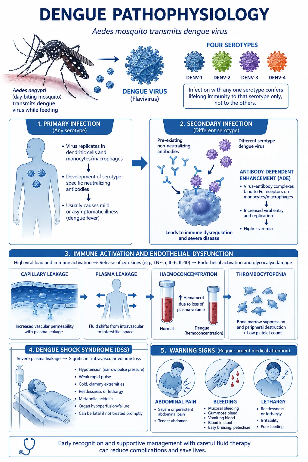
Understanding the mechanism of severe dengue is high-yield exam territory because it explains the single most important epidemiological fact: secondary infection with a heterologous serotype is the dominant risk factor for severe disease. This is antibody-dependent enhancement (ADE). [1]
After a primary DENV infection, the host develops serotype-specific neutralising antibodies plus a population of cross-reactive non-neutralising antibodies. On subsequent infection with a different serotype, these pre-existing antibodies bind but fail to neutralise the new virus; instead, the virus-antibody complex is taken up more efficiently by Fc-γ receptor-bearing monocytes, macrophages, and dendritic cells, producing a higher intracellular viral burden (a higher serum viral load). This is the core of ADE. [1]
The pathophysiological cascade from ADE to shock
1. Heterologous secondary infection + ADE
Non-neutralising anti-E/prM antibodies opsonise the new serotype; Fc-γ receptor-mediated entry into monocytes/macrophages yields a 10-100-fold increase in infected cells and serum viral load relative to primary infection.<Cite id="5" /> This is the strongest known risk factor for severe dengue. Katzelnick et al (Science 2017) quantified this in a Nicaraguan paediatric cohort: intermediate antibody titres conferred the highest risk of severe disease — a phenomenon analogous to the J-shaped risk curve seen with the Dengvaxia vaccine.
2. Massive cytokine storm
Heavily infected monocytes release a storm of TNF-α, IL-6, IL-8, IL-10, IFN-γ, MCP-1, and vasoactive mediators. Complement activation (especially C5a) and mast-cell degranulation amplify the response. The result is systemic endothelial activation and dysfunction.
3. Endothelial and glycocalyx injury → vascular permeability
NS1 antigen directly disrupts endothelial glycocalyx and tight junctions; cytokines up-regulate adhesion molecules and increase gap formation. The capillary leak is transient and selective — large molecules and albumin escape, producing haemoconcentration and hypoalbuminaemia, while the basement membrane remains intact (so leak is reversible). This is the structural basis of plasma leakage.
4. Plasma leakage → haemoconcentration → shock
Intravascular volume is lost into the interstitial space and serous cavities (pleura, peritoneum). Haematocrit rises (often >20% above baseline), pulse pressure narrows, and perfusion falls. The shock of DSS is therefore a hypovolaemic/distributive hybrid driven by leak, not by cardiogenic failure or primary sepsis — although secondary bacterial infection can supervene.
5. Thrombocytopenia and haemorrhage
Megakaryocyte suppression (direct viral effect), peripheral consumption, and immune-mediated platelet destruction (anti-NS1 antibodies cross-react with platelet surface proteins) combine to drive platelets often below 20 x10^9/L. Bleeding is worsened by coagulopathy (prolonged APTT, reduced fibrinogen) and capillary fragility, though true disseminated intravascular coagulation is uncommon.
6. Organ impairment
Direct viral cytopathic effect and immune injury produce hepatitis (AST/ALT elevation, occasionally >1000 with acute liver failure), myocarditis (conduction abnormalities, LV dysfunction, troponin rise), encephalopathy/encephalitis, and AKI. Comorbidity (diabetes, cardiac disease, asthma, chronic kidney disease, pregnancy) magnifies organ involvement.<Cite id="13" />
A clinical correlate: because the leak is reversible and the basement membrane is intact, patients who survive the critical phase recover rapidly once reabsorption begins — but this same reabsorption is the mechanism of iatrogenic pulmonary oedema when excess fluid was given during the leak phase. [1]
[5] [6]Warning signs
[1]A meta-analysis of febrile-phase predictors (Sangkaew et al, Lancet Infect Dis 2021) identified the most robust warning markers.[7]
Haematological
Platelet and HCT trajectory
- Platelets <100 x10^9/L — strongest single predictor
- Rising haematocrit with falling platelets (the divergent trend)
- Leucopenia with relative lymphocytosis and atypical lymphocytes
Clinical
Perfusion and bleeding
- Persistent vomiting, abdominal pain (third spacing), hepatomegaly
- Mucosal bleeding, menorrhagia, melena
- Restlessness or lethargy — early encephalopathy
- Cold extremities, delayed capillary refill, narrow pulse pressure
Laboratory
Biomarkers
- Elevated AST/ALT (hepatic involvement)
- Hypoalbuminaemia (leak marker)
- Metabolic acidosis (lactate >2 mmol/L, bicarbonate <18)
- Coagulopathy (prolonged APTT, low fibrinogen)
Host risk factors
Comorbidity
- Secondary infection (serostatus / prior dengue history)
- Diabetes, chronic cardiac or renal disease, asthma
- Pregnancy, extremes of age, obesity
Severe dengue criteria — 2009 WHO
The 2009 WHO classification (replacing the older DF/DHF/DSS scheme) divides dengue into dengue without warning signs, dengue with warning signs, and severe dengue. Severe dengue requires any ONE of three criteria. [1]
Severe plasma leakage
Dengue shock syndrome (DSS)
- Leading cause of dengue death
- Compensated shock: tachycardia, narrow pulse pressure (<20 mmHg), cold peripheries, delayed capillary refill (>2s), normal SBP
- Hypotensive (decompensated) shock: SBP <90 mmHg (or >40 mmHg fall from baseline), weak/impalpable pulse
- Driven by reversible capillary leak — HCT rising, albumin falling, effusions
- Grades I-IV in the older DHF scheme: I = positive tourniquet only; II = spontaneous bleeding; III = compensated shock; IV = profound shock
Severe bleeding
Haemorrhagic
- Major bleeding requiring transfusion: GI haemorrhage (haematemesis, melena), menorrhagia, intracranial, pulmonary, massive epistaxis
- May be masked by haemoconcentration — a falling HCT in the leak phase can signal bleeding, NOT improvement
- Risk highest with profound thrombocytopenia, prolonged shock, peptic ulcer disease, anticoagulants
- Distinct from trivial petechiae or gum oozing, which do NOT by themselves constitute severe bleeding
Severe organ impairment
End-organ failure
- Hepatitis: AST/ALT >1000 IU/L; acute liver failure with encephalopathy and coagulopathy possible
- CNS: impaired consciousness, seizures, encephalopathy/encephalitis (genuine CNS viral replication occurs)
- Cardiac: myocarditis, conduction block, LV dysfunction, cardiogenic shock, troponin elevation
- Renal: AKI (usually pre-renal or ATN from prolonged shock; rarely immune complex GN)
- ARDS and refractory metabolic acidosis
An important distinction: "expanded dengue syndrome" describes unusual organ manifestations (e.g. acute pancreatitis, encephalitis, myocarditis, Guillain-Barré, haemophagocytic lymphohistiocytosis, retinitis) that occur beyond the classical plasma-leak triad, increasingly recognised in adults. [1]
Diagnosis
Diagnostic strategy is phase-dependent. NS1 antigenaemia, viraemia, and IgM/IgG seroconversion follow a predictable timeline, so the right test depends on the day of illness.[1][11]
Diagnostic approach to suspected dengue
1. NS1 antigen — EARLY phase (days 1-5)
Detects the secreted viral NS1 glycoprotein in serum. Sensitivity ~70-90% in the first 5 days (highest in primary infection), falling after defervescence. Combined NS1 + IgM RDTs maximise diagnostic yield across the illness. False negatives in secondary infection and very early (<24h). Can also be detected in saliva (lower sensitivity).
2. IgM and IgG serology (from day 5)
IgM appears around day 5, peaks at 2 weeks, wanes by 3 months. IgG appears later and persists for life. **IgM positive + IgG positive = "secondary" pattern** (high IgG, low IgG avidity) — a clue to ADE risk. A four-fold rise in paired sera (acute vs convalescent) is the reference standard but is retrospective.
3. RT-PCR / viral isolation — EARLY and confirmatory
Detects viral RNA in serum during the first 5 days. Most sensitive early test; also allows serotyping (DENV 1-4) and genotyping for epidemiology. Not for acute decision-making in most settings due to cost and turnaround.
4. Supportive and severity markers
Full blood count (leucopenia, thrombocytopenia, atypical lymphocytes, rising HCT), albumin (low = leak), AST/ALT, coagulation (APTT), lactate and venous gas (acidosis), creatinine, troponin, glucose (hypoglycaemia). Chest X-ray/POCUS (right pleural effusion, ascites, thickened gallbladder wall). Blood cultures if secondary sepsis suspected (algid shock).
NS1 antigen
Early (days 1-5)
- Rapid, cheap, point-of-care; high early sensitivity
- Best single test in the febrile phase
- Lower sensitivity in secondary infection (immune complexing)
- Useful across all serotypes
IgM / IgG
Late (from day 5)
- Identifies recent and past infection
- IgG/IgM ratio distinguishes primary vs secondary
- Cross-reacts with other flaviviruses (Zika, yellow fever, JEV, West Nile)
- Confirms diagnosis retrospectively with paired sera
RT-PCR
Early + confirmatory
- Highest sensitivity in first 5 days
- Allows serotyping (DENV 1-4)
- Reference standard but costly, slow
- Used for outbreak investigation and vaccine evaluation
Fluid management — the critical skill
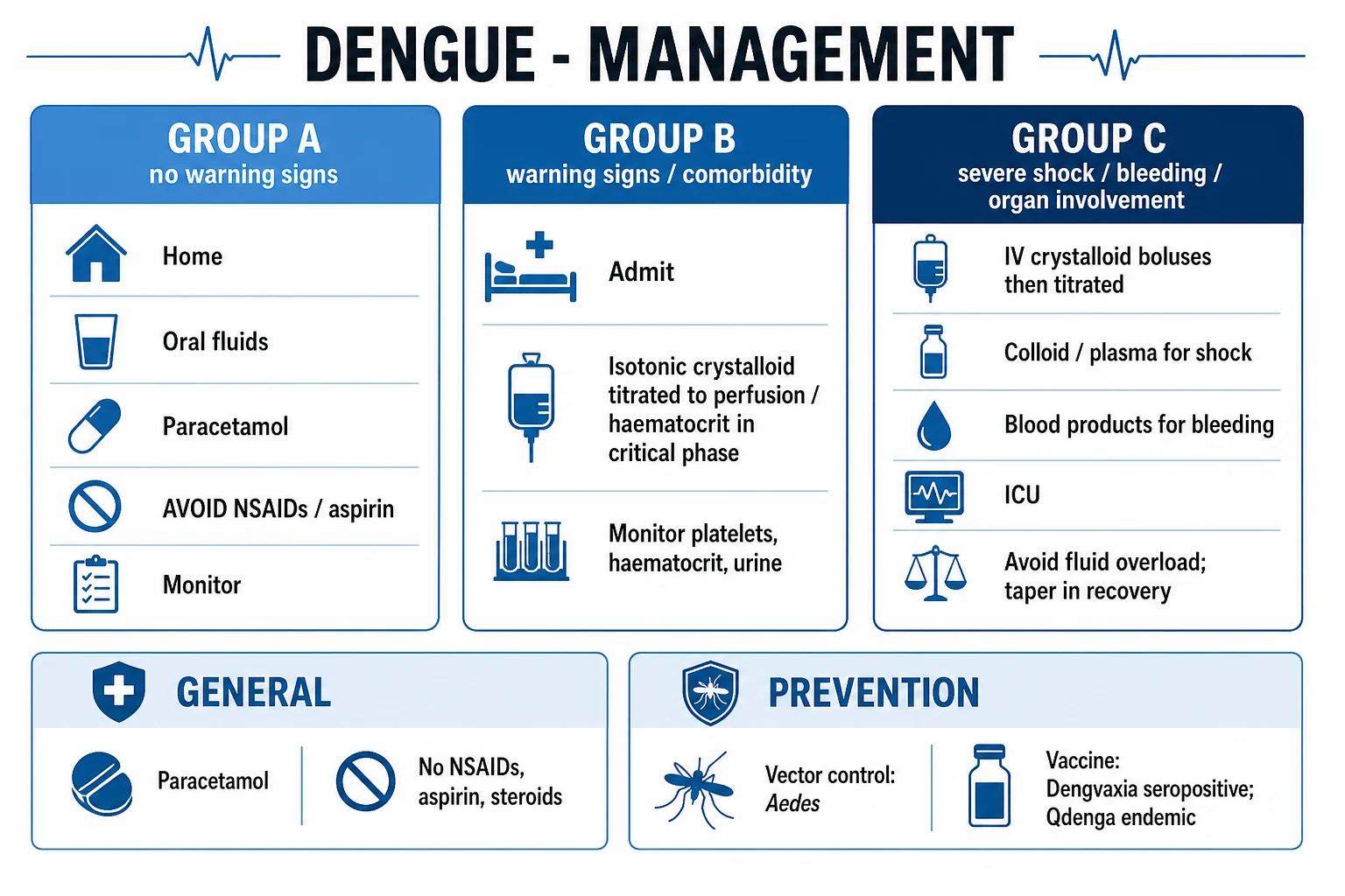
Fluid therapy is the cornerstone of dengue management: insufficient fluid permits shock and organ injury; excess fluid causes pulmonary oedema when reabsorption begins. There is no specific antiviral — fluid is the treatment. [1]
Dengue fluid management — the critical skill
Monitor closely during transition to critical phase
When fever defervesces: check haematocrit every 4-6h, platelets every 12-24h, BP and perfusion every 1-2h, urine output hourly. Rising haematocrit = plasma leakage. Falling platelets (<100,000) with rising haematocrit = progression to critical phase. Bedside POCUS for pleural effusion / ascites / IVC.
Isotonic crystalloid for plasma leak
When haematocrit rises >20% from baseline: start isotonic crystalloid (Hartmann or 0.9% saline). Rate: 5-7 mL/kg/h for 1-2h, then reduce to 3-5 mL/kg/h based on response. Goal: maintain perfusion (BP, urine output 0.5 mL/kg/h) without causing fluid overload.
TITRATE to haematocrit
The KEY to dengue fluid management: titrate crystalloid to haematocrit. If haematocrit rising: increase fluid rate. If haematocrit stable or falling: decrease rate. Do NOT give fluid blindly based on BP alone — haematocrit is the guide.
Colloid for refractory shock
If shock persists despite crystalloid (haematocrit still rising, BP low): add colloid (albumin 5%, starch). Colloid stays intravascular longer than crystalloid. Give boluses (10-20 mL/kg) and reassess. Wills et al (NEJM 2005) showed dextran and starch were superior to saline for profound DSS, while no fluid was clearly best for less severe shock.<Cite id="3" />
AVOID excessive fluid
The biggest danger in dengue: fluid overload. During the RECOVERY phase, extravascular fluid is reabsorbed into the intravascular space. If the patient has been over-resuscitated, this causes acute pulmonary oedema. Signs of overload: bilateral crackles, rising oxygen requirement, enlarged liver, falling HCT with haemodilution. Reduce/stop fluids. Give diuretics if needed.
Platelet transfusion
Do NOT routinely transfuse platelets. Dengue thrombocytopenia is from consumption and immune destruction, not production failure. Transfuse ONLY if: (1) active bleeding with low platelets, (2) platelets <10,000 with bleeding risk, (3) before invasive procedure. Prophylactic platelet transfusion does NOT prevent bleeding and carries transfusion risks.<Cite id="12" />
The landmark randomised evidence underpinning fluid choice in DSS is the Wills et al NEJM trial, which compared six crystalloid and colloid regimens in Vietnamese children. [1]
Wills 2005 (NEJM) — Comparison of three fluid solutions for resuscitation in dengue shock syndrome
Randomised, double-blind, controlled trial in Vietnamese children with DSS (moderate and severe shock arms)
Population: Children with dengue shock syndrome at the Hospital for Tropical Diseases, Ho Chi Minh City (n=230 moderate, n=51 severe)
Key finding
In moderate shock, Ringer's lactate alone gave the highest recurrence of shock; colloid (dextran) had a small early advantage. In SEVERE shock, the colloid dextran 70 was clearly superior to crystalloid — fewer children requiring a second resuscitation and a faster recovery of pulse pressure.
Crystalloid (Ringer / 0.9% saline)
First-line
- Cheap, available, safe — first-line for moderate shock
- Ringer/Hartmann preferred (less hyperchloraemic acidosis than 0.9% saline)
- 10-20 mL/kg bolus over 30 min; reassess; titrate to HCT and perfusion
- Risk: more of it leaves the intravascular space — watch for overload
Colloid (albumin 5%, dextran, starch)
Refractory / severe
- Stays intravascular longer — superior for profound or recurrent shock
- Preferred in SEVERE DSS and when crystalloid fails to stabilise HCT/perfusion
- Albumin preferred to synthetic colloids in many ICUs (starches carry AKI risk)
- No proven survival advantage over crystalloid in less severe disease
What to AVOID
Hypotonic solutions
- NO hypotonic solutions (5% dextrose, 0.45% saline) for resuscitation — leak out of vessel
- NO aggressive boluses in compensated shock without reassessment
- NO routine blood products unless bleeding
- NO prophylactic platelets
ICU care for severe dengue
ICU admission is required for DSS (especially decompensated), severe bleeding, organ impairment, or any patient requiring vasoactive support, mechanical ventilation, or close haemodynamic monitoring. [1]
ICU management of severe dengue
1. Recognise and grade shock
Compensated shock: SBP normal but pulse pressure <20 mmHg, tachycardia, cold peripheries, capillary refill >2s. Hypotensive (decompensated) shock: SBP <90 or >40 mmHg below baseline. Reassess every 15-30 min during resuscitation. Use POCUS (IVC collapsibility, cardiac function, pleural effusion, ascites) to guide therapy.
2. Judicious fluid resuscitation
10-20 mL/kg isotonic crystalloid bolus over 30 min for compensated shock; repeat once if needed, titrating to HCT (must not rise further), pulse pressure, and urine output. For refractory shock add colloid (albumin 5%, 10-20 mL/kg boluses). The minimum effective dose of fluid is the goal — over-resuscitation causes pulmonary oedema when reabsorption begins.
3. Vasopressors for fluid-refractory shock
If shock persists after 40-60 mL/kg crystalloid ± colloid, start a vasopressor (noradrenaline first-line; adrenaline if severe). Be alert to coexisting cardiogenic shock from myocarditis — early bedside echo is essential to distinguish distributive/hypovolaemic shock (normal/high cardiac output) from cardiogenic shock (low output, LV dysfunction).
4. Transfusion for major bleeding
Crossmatch early. Transfuse packed red cells for major haemorrhage or a falling HCT during the leak phase (which signals bleeding, not improvement). Fresh frozen plasma/cryoprecipitate for documented coagulopathy with bleeding. Platelet transfusion ONLY for active bleeding or peri-procedural need — NOT prophylactically.<Cite id="12" />
5. Organ support
NIV or invasive ventilation for pulmonary oedema (from fluid leak or over-resuscitation) or ARDS — lung-protective ventilation if intubated. Renal replacement therapy for AKI with refractory acidosis, hyperkalaemia, or fluid overload. Treat hypoglycaemia (5% or 10% dextrose infusion), electrolyte disturbances (hyponatraemia, hypokalaemia from third spacing and vomiting), and acidosis. Avoid hepatotoxic and nephrotoxic drugs.
6. Monitor and transition to recovery
Once reabsorption begins (HCT falls, diuresis, bradycardia), taper and then stop IV fluids to avoid overload. Treat iatrogenic pulmonary oedema with oxygen, diuretics, NIV. Watch for secondary bacterial infection (Gram-negative, *S. aureus*) — cultures and empiric antibiotics if shock fails to respond or new fever in recovery.
Key trials and evidence
Katzelnick 2017 (Science) — Antibody-dependent enhancement of severe dengue disease in humans
Prospective paediatric cohort study with longitudinal serology (Nicaragua)
Population: Children followed for up to 12 years with serial antibody titres and dengue surveillance
Key finding
Risk of severe dengue was J-shaped against antibody titre: very high titres were protective, but INTERMEDIATE titres (specifically 1:21 to 1:80) were associated with the highest risk of severe disease — a 7-fold increased risk compared with the protective high-titre group.
Sangkaew 2021 (Lancet Infect Dis) — Predictors of severe dengue: systematic review and meta-analysis
Systematic review and meta-analysis of febrile-phase predictors
Population: Children and adults in the febrile phase of dengue across endemic and non-endemic settings
Key finding
The strongest predictors were: abdominal pain, vomiting, clinical fluid accumulation, bleeding (especially mucosal), lethargy, hepatomegaly, and the haematological trend of rising haematocrit with falling platelets — validating the WHO warning-sign set. Platelet count and haematocrit trajectory had the highest predictive odds ratios.
Capeding 2014 (Lancet) — CYD14: tetravalent dengue vaccine in Asian children
Phase 3, randomised, observer-masked, placebo-controlled trial across five Asian countries
Population: Children aged 2-14 years (n=10,275)
Key finding
Vaccine efficacy against symptomatic virologically confirmed dengue was 56.5% over 25 months. Crucially, efficacy varied by serotype (high for DENV-4, lower for DENV-2) and by baseline serostatus — protection was much lower in seronegative recipients, with a signal of increased hospitalisation for dengue in the youngest (seronegative) recipients.
Ngo 2001 (Clin Infect Dis) — Four intravenous fluid regimens in the first hour of DSS
Randomised, double-blind comparison of four IV fluid regimens in the first hour
Population: Vietnamese children with dengue shock syndrome
Key finding
No single fluid was clearly superior for the initial resuscitation of DSS in the first hour; all four regimens achieved haemodynamic stabilisation in the majority. This trial established that crystalloid is a reasonable initial choice and informed the subsequent 2005 NEJM trial design.
Differential diagnosis
The returning traveller with fever and thrombocytopenia has a wide differential; in ANZ, dengue and malaria must both be actively excluded. [1]
Malaria
Plasmodium spp
- Fever pattern (cyclical in established infection), splenomegaly, hyperparasitaemia
- Thick/thin blood film and RDT; thrombocytopenia and anaemia common to both
- IV artesunate is the treatment — do not delay for dengue workup
- Travel to sub-Saharan Africa strongly favours malaria over dengue
Typhoid
Salmonella Typhi
- Stepwise rising fever, relative bradycardia, rose-spot rash, constipation then diarrhoea
- Blood/marrow culture; leucopenia and transaminitis overlap with dengue
- Treated with ceftriaxone or azithromycin
Leptospirosis
Leptospira
- Exposure to fresh water/animals; conjunctival suffusion, myalgia (calves), jaundice, AKI (Weil disease)
- Serology and PCR; can cause hepatorenal failure resembling severe dengue
- Treated with benzylpenicillin or doxycycline
Other arboviruses
Chikungunya, Zika, JEV
- Chikungunya: prominent symmetric polyarthralgia (often more than fever)
- Zika: mild fever, conjunctivitis, maculopapular rash; serology cross-reacts
- JEV: encephalitis picture; geographic overlap with dengue
Sepsis / bacterial
Gram-negative, rickettsial
- Sepsis with thrombocytopenia is indistinguishable from early severe dengue — blood cultures, lactate
- Scrub typhrus (Orientia) in rural Asia: eschar, lymphadenopathy, doxycycline
- Melioidosis (Burkholderia pseudomallei) in Southeast Asia/Australia: high mortality, ceftazidime
Prevention and vaccine
Vector control (removal of breeding sites, Aedes surveillance, indoor residual spraying, personal protection with repellents and long sleeves) remains the backbone of prevention, but two tetravalent live-attenuated vaccines now feature in national programmes: [1]
- CYD-TDV (Dengvaxia) — first licensed dengue vaccine; recommended ONLY for seropositive individuals (seronegative recipients have increased risk of severe dengue via ADE).[6]
- TAK-003 (Qdenga) — second-generation tetravalent vaccine with a DENV-2 backbone; broader protection including in seronegative recipients; licensed in several non-US jurisdictions.
- TV003/TV005 — NIH-developed tetravalent candidate with promising durability.
No antiviral agent is yet licensed for dengue; the target of supportive care remains correct fluid balance and organ support. [1]
SAQ — Dengue shock syndrome and recovery-phase complications
SAQ — Dengue shock syndrome in a returning traveller
10 minutes · 10 marks
A 29-year-old man who returned from Bali 6 days ago presents on day 5 of fever, which has now settled. He is alert but peripherally cold with a narrow pulse pressure (BP 96/78), HR 118, capillary refill 4 seconds. Haematocrit 0.52 (baseline 0.40), platelets 18 x10^9/L, albumin 28 g/L, AST 280 IU/L. A right pleural effusion and mild ascites are present on POCUS. NS1 antigen is positive. He has passed 15 mL of urine in the last 2 hours.
SAQ — Severe dengue with organ impairment and recovery-phase pulmonary oedema
10 minutes · 10 marks
A 45-year-old woman with dengue (DENV-2 confirmed, secondary infection) is in ICU on day 6 of illness. Her fever settled yesterday. She has received 4 L of crystalloid over 12 hours for compensated shock. Today her haematocrit has fallen from 0.55 to 0.34, she is tachypnoeic (RR 32) with bilateral crackles, SpO2 90% on 6 L, has a wide pulse pressure (130/60), and is passing 400 mL/hour of urine. Troponin is mildly elevated at 0.06 ng/mL with a normal echocardiogram.
Clinical pearls
Red flags
References
- [1]Simmons CP, Farrar JJ, Nguyen van Vinh Chau, Wills B. Dengue N Engl J Med, 2012.PMID 22494122
- [2]Wilder-Smith A, Ooi EE, Horstick O, Wills B. Dengue Lancet, 2019.PMID 30696575
- [3]Wills BA, Nguyen MD, Ha TL, et al. Comparison of three fluid solutions for resuscitation in dengue shock syndrome N Engl J Med, 2005.PMID 16135832
- [4]Ngo NT, Cao XT, Kneen R, et al. Acute management of dengue shock syndrome: a randomized double-blind comparison of 4 intravenous fluid regimens in the first hour Clin Infect Dis, 2001.PMID 11170909
- [5]Katzelnick LC, Gresh L, Halloran ME, et al. Antibody-dependent enhancement of severe dengue disease in humans Science, 2017.PMID 29097492
- [6]Capeding MR, Tran NH, Hadinegoro SR, et al. Clinical efficacy and safety of a novel tetravalent dengue vaccine in healthy children in Asia: a phase 3, randomised, observer-masked, placebo-controlled trial Lancet, 2014.PMID 25018116
- [7]Sangkaew S, Ming D, Boonyasiri A, et al. Risk predictors of progression to severe disease during the febrile phase of dengue: a systematic review and meta-analysis Lancet Infect Dis, 2021.PMID 33640077
- [8]Trung DT, Trieu HT, Wills BA. Microvascular Fluid Exchange: Implications of the Revised Starling Model for Resuscitation of Dengue Shock Syndrome Front Med (Lausanne), 2020.PMID 33415117
- [9]Pang J, Leo YS, Lye DC. Critical care for dengue in adult patients: an overview of current knowledge and future challenges Curr Opin Crit Care, 2016.PMID 27583589
- [10]Lee TH, Lee LK, Lye DC, Leo YS. Current management of severe dengue infection Expert Rev Anti Infect Ther, 2017.PMID 27786589
- [11]Rosenberger KD, Phung Khanh L, Tobian F, et al. Early diagnostic indicators of dengue versus other febrile illnesses in Asia and Latin America (IDAMS study): a multicentre, prospective, observational study Lancet Glob Health, 2023.PMID 36796983
- [12]Archuleta S, Chia PY, Wei Y, et al. Predictors and Clinical Outcomes of Poor Platelet Recovery in Adult Dengue With Thrombocytopenia: A Multicenter, Prospective Study Clin Infect Dis, 2020.PMID 31626692
- [13]Pang J, Hsu JP, Yeo TW, et al. Diabetes, cardiac disorders and asthma as risk factors for severe organ involvement among adult dengue patients: A matched case-control study Sci Rep, 2017.PMID 28045096
- [14]Waggoner JJ, Katzelnick LC, Burger-Calderon R, et al. Antibody-Dependent Enhancement of Severe Disease Is Mediated by Serum Viral Load in Pediatric Dengue Virus Infections J Infect Dis, 2020.PMID 32236481