ICU · Infectious Diseases
Toxic shock syndrome in the ICU
Also known as Toxic shock syndrome (TSS) · Staphylococcal TSS · Streptococcal TSS (STSS) · Superantigen-mediated disease
Toxic shock syndrome (TSS) is a life-threatening toxin-mediated illness caused by SUPERANTIGEN-producing bacteria. STAPHYLOCOCCAL TSS: Staphylococcus aureus TSST-1 toxin. STREPTOCOCCAL TSS: Streptococcus pyogenes (GAS) SpeA/SpeC toxins. Superantigens bypass normal antigen presentation → MASSIVE polyclonal T-cell activation → cytokine storm (IL-1, IL-2, TNF-alpha, IFN-gamma) → multi-organ failure. Clinical: HIGH fever, diffuse rash (desquamates later), hypotension, multi-organ involvement (GI vomiting/diarrhoea, renal, hepatic, CNS confusion, mucosal hyperaemia). Management: (1) Source control (remove tampon, drain abscess, debride wound). (2) Antibiotics (clindamycin — SUPPRESSES toxin production + standard anti-staph/strep cover). (3) IVIG (neutralises superantigen + anti-inflammatory). (4) Supportive (vasopressors, fluids, organ support). Mortality: staphylococcal ~3-5%, streptococcal ~30-60%.
On this page & tools
Your progress
Saved locally on this device.
Target exams
Red flags
Clinical pearls
Red flags
Pathophysiology — the superantigen mechanism in depth
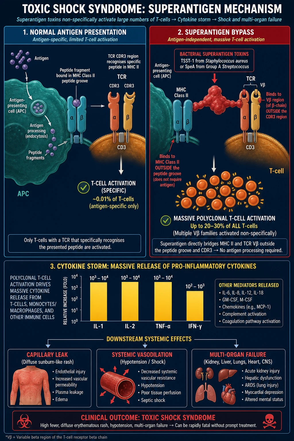
Toxic shock syndrome is the paradigmatic toxin-mediated, not infection-mediated illness. The host is destroyed not by invasive bacteria but by a single protein toxin that subverts the adaptive immune system so profoundly that up to a third of all circulating T-cells discharge their cytokines simultaneously.[1][11] Understanding this mechanism is the single highest-yield piece of pathophysiology for the CICM/FFICM viva, because every management step — clindamycin, source control, IVIG — is a direct consequence of it.
Normal antigen presentation versus superantigen activation
In conventional antigen presentation, a protein antigen is taken up by an antigen-presenting cell (APC), proteolytically cleaved into short peptides, and a single peptide is loaded into the groove of an MHC class II molecule. A naive T-cell whose T-cell receptor (TCR) happens to recognise that exact peptide-MHC complex binds, becomes activated, and clonally expands. Because only T-cells with the matching (cognate) receptor respond, roughly 1 in 10,000 to 1 in 100,000 T-cells (about 0.001-0.01%) are engaged.[11]
A superantigen bypasses every step of this. It is a stable secreted protein (22-29 kDa, resistant to boiling and proteolysis) that binds directly to two molecules: [1]
- The lateral surface of MHC class II on the APC — outside the peptide-binding groove, so it does not need any specific peptide to be present.
- The lateral surface of the T-cell receptor V-beta chain — outside the antigen-combining site, so it does not need TCR antigen specificity. [1]
By cross-bridging MHC II and the TCR, the superantigen physically juxtaposes the APC and the T-cell and delivers the activation signal to every T-cell bearing that particular V-beta family, regardless of what antigen that T-cell was "for". The result is massive polyclonal T-cell activation — up to 20-30% of all circulating T-cells fire at once (an amplification of roughly 10,000-fold over a normal response).[1][11]
Normal antigen presentation vs superantigen activation — the 10,000-fold amplification
| Feature | Conventional antigen | Superantigen |
|---|---|---|
| Antigen processing by APC | Required (proteolysis to peptide) | NOT required — intact toxin |
| Binding site on MHC II | Inside the peptide groove | Lateral surface, outside groove |
| Binding site on TCR | Antigen-combining site (CDR) | Lateral V-beta chain |
| T-cell specificity needed | Yes — cognate TCR only | No — any TCR of that V-beta family |
| Fraction of T-cells activated | ~0.001-0.01% | 20-30% (polyclonal) |
| MHC restriction | Yes | No (bypasses restriction) |
| Co-stimulation required | Yes (B7-CD28) | Partially bypassed |
| Consequence | Specific, contained immunity | Cytokine storm, vasodilatory shock |
The activated T-cells and the MHC-II-bearing APCs (macrophages, monocytes, dendritic cells) then release a torrent of pro-inflammatory cytokines: T-cell derived interleukin-2 (IL-2), interferon-gamma (IFN-gamma), tumour necrosis factor-beta (TNF-beta/lymphotoxin); and APC-derived interleukin-1 (IL-1), interleukin-6 (IL-6), interleukin-12 (IL-12) and tumour necrosis factor-alpha (TNF-alpha).[1][11] This cytokine storm drives the clinical syndrome: TNF-alpha and IL-1 are endogenous pyrogens (fever), potent myocardial depressants (cardiogenic contribution to shock) and cause endothelial activation and nitric-oxide-mediated vasoplegia (distributive shock); IL-2 and IFN-gamma upregulate endothelial adhesion molecules and amplify capillary leak; IL-6 drives the acute-phase response and thrombosis. The net picture is mixed distributive-cardiogenic shock with massive capillary leak, indistinguishable at the bedside from severe septic shock except for the tell-tale rash.
The toxins, their sources and the diseases they cause
Not all superantigens are equal. The two clinical syndromes — staphylococcal and streptococcal TSS — are produced by different organisms making different toxins, and this determines everything about source, bacteraemia, prognosis and empiric antibiotics.[1][8][5]
The superantigen toxins of TSS — source, genetics and disease
| Toxin | Organism | Genetics | Molecular weight | Disease / association | Notes |
|---|---|---|---|---|---|
| TSST-1 (toxic shock syndrome toxin-1) | S. aureus | Chromosomal (tst gene), mobile element | ~22 kDa | ~95% of menstrual TSS; ~50% of non-menstrual staph TSS | Crosses mucosal barriers intact (vagina, nasal mucosa) — needs no tissue invasion. Antibody-negative patients are susceptible. |
| Staph enterotoxin B (SEB), SEC | S. aureus | Chromosomal / plasmid | ~28 kDa | Remainder of non-menstrual staph TSS; some food poisoning | Less mucosal penetration than TSST-1 — usually needs a wound/foreign body |
| SpeA (streptococcal pyrogenic exotoxin A) | S. pyogenes (GAS) | Bacteriophage-encoded | ~25 kDa | The classic STSS toxin | Associated with the M1/M3 "flesh-eating" strains; scarlet fever toxin A |
| SpeC | S. pyogenes | Bacteriophage | ~24 kDa | STSS (especially with M3/T3 strains) | Often found alongside SpeA |
| SSA (streptococcal superantigen), MF/SMEZ (mitogenic factor / streptococcal mitogenic exotoxin Z) | S. pyogenes | Chromosomal / phage | ~24-30 kDa | Contribute to STSS cytokine burden | SMEZ is among the most potent superantigens known |
Two pathophysiology facts the examiner loves: (1) TSST-1 is unique among the staphylococcal superantigens in crossing intact mucosa — which is exactly why it causes tampon- and nasal-packing-associated disease from a mere vaginal/nasal coloniser rather than a deep wound infection.[4] (2) The streptococcal toxins (SpeA/SpeC) are bacteriophage-encoded — STSS strains acquire toxin production by lysogenic conversion, the same horizontal-gene-transfer trick that gives diphtheria its toxin, and the reason STSS appears in clonal outbreaks of particular M-types.[5]
Why the tampon? The local microenvironment of menstrual TSS
The Shands 1980 CDC case-control study established the tampon association and quantified it: risk rose with tampon absorbency and continuous duration of use, and super-absorbent tampons (polyacrylate, carboxymethylcellulose) carried the highest risk.[4] The biology: super-absorbent fibres bind divalent cations (Mg2+), and magnesium depletion is a signal for S. aureus to upregulate TSST-1 transcription; the intermenstrual vagina provides oxygen (introduced by the tampon), protein (blood), neutral pH and warmth — an ideal fermenter for toxin production. The organism need not invade tissue; it merely colonises the mucosa and sheds TSST-1, which is absorbed across intact epithelium. This is why removing the tampon is therapeutic, not just diagnostic, and why the post-1980 fall in menstrual TSS incidence followed withdrawal of the most absorbent products and lower-absorbency recommendations.[1][4]
CDC diagnostic criteria — staphylococcal vs streptococcal TSS
TSS is a clinical diagnosis. Blood cultures are negative in staphylococcal TSS (the organism is a localised mucosal/wound coloniser shedding toxin) and may be negative in early streptococcal TSS, so one cannot wait for the microbiology laboratory. The CDC/CSTE surveillance case definitions (2011 for staphylococcal, 2010 for streptococcal) are the exam-standard criteria and are reproduced below.[1]
CDC diagnostic criteria — staphylococcal TSS (other than streptococcal), 2011 case definition
| Criterion | Requirement |
|---|---|
| 1. Fever | Temperature ≥ 38.9 °C (102 °F) |
| 2. Rash | Diffuse macular erythroderma (sunburn-like) |
| 3. Desquamation | 1-2 weeks after onset of rash, especially palms, soles, fingers, toes |
| 4. Hypotension | SBP ≤90 mmHg (adults) OR <5th percentile for age (children) OR orthostatic drop producing syncope/dizziness |
| 5. Multi-system involvement (≥3 of the following) | — |
| — Gastrointestinal | Vomiting or diarrhoea at onset |
| — Muscular | Severe myalgia or creatine kinase ≥2× ULN |
| — Mucous membranes | Vaginal, oropharyngeal or conjunctival hyperaemia |
| — Renal | BUN or creatinine ≥2× ULN or sterile pyuria (≥5 WBC/hpf) |
| — Hepatic | Bilirubin / AST / ALT ≥2× ULN |
| — Haematological | Platelets ≤100 × 10⁹/L |
| — CNS | Disorientation or altered consciousness without focal signs |
| Laboratory exclusion | Negative cultures of blood, throat, CSF (S. aureus may be isolated from a localised site); negative serology for measles, leptospirosis, RMSF |
| Case classification | Confirmed: all 5 clinical criteria present (desquamation may be met if patient survives to 1-2 weeks; a fatal case without desquamation can still be confirmed if the other 4 plus exclusion criteria are met). Probable: 4 of 5 clinical criteria present without an alternative diagnosis |
CDC diagnostic criteria — streptococcal TSS (STSS), 2010 case definition
| Criterion | Requirement |
|---|---|
| A. Hypotension | SBP ≤90 mmHg (adults) OR <5th percentile for age (children) |
| B. Multi-organ involvement (≥2 of the following) | — |
| — Renal impairment | Creatinine ≥2× ULN (or ≥2× baseline) for age |
| — Coagulopathy | Platelets ≤100 × 10⁹/L or DIC, or PT >1.5× / INR elevated |
| — Hepatic involvement | AST/ALT/bilirirubin ≥2× ULN for age |
| — Acute respiratory distress syndrome | Acute-onset diffuse pulmonary infiltrates and hypoxaemia without cardiac failure, OR generalised pulmonary capillary leak |
| — Generalised erythematous macular rash | May desquamate (variable) |
| — Soft-tissue necrosis | Necrotising fasciitis, myositis, or gangrene |
| Laboratory | Definite case: GAS (S. pyogenes) isolated from a normally sterile site (blood, CSF, surgical/sterile tissue, pleural/peritoneal fluid). Probable case: GAS isolated from a non-sterile site (throat, sputum, vagina, superficial wound) |
| Case classification | Confirmed: A + B + GAS from a sterile site. Probable: A + B + GAS from a non-sterile site |
Note the asymmetry between the two definitions: staphylococcal TSS requires the diffuse rash + desquamation + fever + ≥3 organ systems and excludes bacteraemia (a positive blood culture means look for another diagnosis), whereas streptococcal TSS requires only hypotension + ≥2 organ systems, makes soft-tissue necrosis one of the defining features, and requires GAS to be isolated (from any site, sterile for "definite").[6][1] In practice the streptococcal definition is more permissive — a patient with necrotising fasciitis, shock, AKI and thrombocytopenia whose wound grows GAS has STSS even without a rash.
Clinical features in depth — recognising the two faces of TSS
Staphylococcal TSS — tampon-associated and non-tampon
Menstrual staphylococcal TSS (the classic form) presents in a young menstruating woman, typically within 5 days of the onset of menses and within the first few tampon-days, with the abrupt onset of high fever, myalgia, vomiting/diarrhoea, a sunburn-like diffuse rash (including palms and soles), conjunctival/oropharyngeal/vaginal hyperaemia, and rapid progression to hypotension and multi-organ failure.[1][4] Non-menstrual staphylococcal TSS is now at least as common as the menstrual form and occurs from any localised S. aureus focus shedding TSST-1 or an enterotoxin: surgical or post-traumatic wounds (often appearing deceptively clean — the organism sits in the suture line and sheds toxin), nasal packing after ENT surgery (classic board-exam scenario), burns, cellulitis, hidradenitis, retained barrier contraceptives (diaphragm, contraceptive sponge), postpartum or puerperal infection, and influenza-associated S. aureus pneumonia (a particularly lethal combination).[1] The same triad — fever, diffuse erythroderma, hypotension with multi-organ failure — appears, with desquamation of the palms and soles 1-2 weeks later confirming the diagnosis retrospectively.
Streptococcal TSS — necrotising fasciitis, myositis and the GAS focus
Streptococcal TSS is the GAS end of the spectrum and is dominated by the soft-tissue source. The presentation is a patient (often previously well) with a seemingly minor skin breach — a surgical wound, a varicella lesion in a child, a bruise, an insect bite, an IV-drug injection site, or postpartum — that progresses over hours to severe pain out of all proportion to the visible skin findings, swelling, dusky/blue discoloration, bullae and then frank necrosis (necrotising fasciitis or myonecrosis), accompanied by high fever and rapid-onset shock.[2][5] Unlike staphylococcal TSS, bacteraemia is common (~60%) and metastatic foci can occur. The rash is not obligatory in STSS (it is one of several possible organ manifestations, not a defining criterion). Two further GAS scenarios deserve memorising: postpartum/puerperal STSS (a woman 1-3 days post-delivery with fever, tachycardia and shock — GAS from the genital tract) and GAS pneumonia with empyema (a cause of community-acquired pneumonia that progresses to STSS).[2] The defining message for the bedside: severe unexplained pain + systemic toxicity + a soft-tissue focus = necrotising fasciitis with probable STSS until proven otherwise.
Differential diagnosis — the "fever + rash + shock" matrix
The combination of high fever, a rash and haemodynamic collapse has a relatively short but critical differential. The character of the rash, the mucosal involvement, the presence of a soft-tissue focus and the epidemiology (tick exposure, drug exposure, recent immunotherapy) usually separate the entities.[1]
Differential diagnosis of TSS — distinguishing the rash + fever + shock syndromes
| Condition | Rash type | Mucosa | Shock | Key discriminator |
|---|---|---|---|---|
| Staphylococcal TSS | Diffuse macular erythroderma (sunburn) → desquamates (palms/soles) | Hyperaemia (vaginal/oral/conjunctival) | Yes | Tampon/wound source; blood cultures negative |
| Streptococcal TSS | Variable (may be absent) | Variable | Yes | Severe soft-tissue pain + necrosis; GAS bacteraemia |
| Meningococcaemia | Petechial → purpuric (echtyma gangrenosum-like), spreads fast | May have pharyngitis | Yes (Waterhouse-Friderichsen → adrenal haemorrhage) | Rapidly progressive purpura; DIC; blood culture positive (N. meningitidis) |
| Staphylococcal scalded skin syndrome (SSSS) | Superficial flaccid bullae, sheets of epidermis peel (Nikolsky +) | Spared (mucosae not involved) | No (usually a child, not shocked) | Localised S. aureus focus → exfoliative toxin A/B; superficial split within epidermis |
| Stevens-Johnson / TEN | Target lesions → sheet-like epidermal detachment (TEN >30%) | Severe, ≥2 mucosal sites | Usually no | Drug exposure 1-3 weeks prior; skin-slit necrosis; no toxin focus |
| Kawasaki disease | Polymorphous rash, desquamates (hands/feet) | Strawberry tongue, cracked lips, conjunctivitis | No | Child <5 years; coronary artery aneurysms; responds to IVIG/aspirin |
| Scarlet fever (GAS) | Fine "sandpaper" papular rash, Pastia lines | Strawberry tongue, pharyngitis | No | Concurrent pharyngitis; no shock — but can rarely overlap with STSS |
| DRESS / drug hypersensitivity | Morbilliform, facial oedema | Variable | No | Onset 2-8 weeks after drug; eosinophilia, atypical lymphocytes, hepatitis |
| Rocky Mountain spotted fever | Petechial, starts wrists/ankles, spreads centripetally to palms/soles | Variable | Yes | Tick exposure; doxycycline (never "wait for confirm") |
| Leptospirosis (Weil) | Variable; conjunctival suffusion | Conjunctivitis | Yes | Animal/standing-water exposure; jaundice + renal failure |
| Measles | Morbilliform, cephalocaudal | Koplik spots on buccal mucosa | No | Prodromal cough/coryza/conjunctivitis; serology |
| Anaphylaxis | Acute urticaria/flushing, angioedema | No | Yes (distributive) | Immediate exposure (drug/food/sting); responds to adrenaline |
| Cytokine release syndrome (CAR-T / bispecific) | Flush, occasionally rash | Variable | Yes | Recent immune-effector therapy; tocilizumab-responsive |
Management — the source-control-plus-toxin-suppression principle
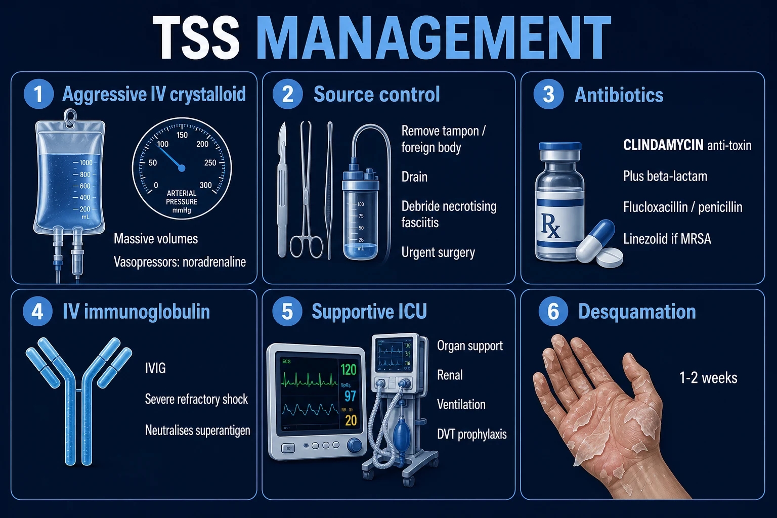
Treatment of TSS is the application of four ideas drawn directly from the pathophysiology: (1) stop the toxin being made (clindamycin + source control), (2) stop the toxin reaching more T-cells (IVIG), (3) support the organs the toxin has injured (resuscitation, vasopressors, organ support) and (4) kill the organism (a cell-wall-active antibiotic — beta-lactam or glycopeptide).[1][2] The Surviving Sepsis Campaign hour-1 bundle applies in full — TSS is a form of septic shock and should be resuscitated as such — but the additions of clindamycin, aggressive source control and IVIG are what make the difference.
Acute management of toxic shock syndrome — the first 6 hours
- RECOGNISE — any patient with HIGH fever + diffuse erythroderma + hypotension + multi-organ failure (or severe soft-tissue pain with shock) is TSS until proven otherwise. Do NOT attribute the picture to "viral illness", "drug fever" or "ordinary sepsis with a rash". Apply CDC criteria at the bedside. If there is a tampon, nasal pack, contraceptive diaphragm or retained foreign body in situ, find it now
- REMOVE THE SOURCE AT THE BEDSIDE (do not delay for theatre) —
- Vaginal examination and remove any tampon (menstrual TSS)
- Remove nasal packing (post-ENT surgery)
- Remove retained barrier contraceptive (diaphragm/sponge) or foreign body
- Remove infected central/venous catheter if line-source suspected
- Swab every removed surface and send for culture — this may be the only positive specimen in staphylococcal TSS
- RESUSCITATE using the SSC hour-1 bundle — high-flow oxygen; two large-bore cannulae; 20-30 mL/kg crystalloid bolus repeated to target MAP ≥65, lactate clearance, urine output ≥0.5 mL/kg/h. Anticipate massive capillary leak — 5-10 L of fluid in the first hours is common; albumin is a reasonable second-line after crystalloid given the profound hypoalbuminaemia. Start noradrenaline early for vasodilatory shock; add vasopressin (0.01-0.03 U/min) and/or adrenaline for catecholamine-refractory shock. An arterial line and (if unstable) central access are mandatory
- EMPIRICAL ANTIBIOTICS WITHIN 1 HOUR — must include a TOXIN-SUPPRESSOR:
- Clindamycin 600-900 mg IV q8h (the 50S inhibitor that stops toxin synthesis — NON-NEGOTIABLE in suspected TSS)
- PLUS a cell-wall agent against both staph and strep until the organism is known: flucloxacillin 2 g IV q6h (MSSA/GAS) OR vancomycin 15-20 mg/kg IV q12h (if MRSA suspected/healthcare-associated) OR linezolid 600 mg IV q12h (MRSA + second toxin-suppressor)
- PLUS broad gram-negative cover (e.g. piperacillin-tazobactam 4.5 g IV q6h or meropenem 1 g IV q8h) if the source is unclear or polymicrobial necrotising infection possible
- For confirmed streptococcal TSS: penicillin G 2.4 g IV q4h (GAS universally penicillin-susceptible) + clindamycin
- SURGICAL SOURCE CONTROL FOR STREPTOCOCCAL TSS — THE SCALPEL IS THE TREATMENT:
- Necrotising fasciitis/myonecitis → immediate surgical exploration and debridement of all non-viable tissue ("incise to fascia, debride to bleeding healthy muscle"). Time to debridement is the single biggest determinant of survival and limb salvage
- Re-look in theatre at 24-48 h: necrotising infections nearly always need a second debridement
- Do NOT delay surgery for imaging (CT confirms but does not treat); a finger-test or bedside incision revealing "dishwater" fluid and non-contractile muscle is enough
- IVIG 1-2 g/kg IV (single dose or divided over 2-3 days) for severe/refractory disease, especially streptococcal TSS with shock and multi-organ failure. Pooled human IgG contains neutralising antibodies to TSST-1, SEB/SEC and SpeA/SpeC. The Darenberg 2003 randomised trial was stopped early for recruitment but showed a 3.6-fold mortality signal in placebo and a significant SOFA-score improvement with IVIG; current guidelines recommend it for severe STSS
- INVESTIGATE AND MONITOR — blood cultures ×2 (sterile site), plus cultures from cervix/vagina/wound/nose/sputum/urine; FBC, coagulation (watch for DIC — D-dimer, fibrinogen), U&E, LFTs, CK (rhabdomyolysis), lactate, albumin, troponin (cytokine myocarditis); group and save. Imaging: CT of the relevant soft-tissue region for necrotising infection (subcutaneous gas, fascial thickening, fat stranding; LRINEC score to support). Continuous: arterial line, ECG, SpO2, urine output
- ORGAN SUPPORT — intubation and lung-protective ventilation for ARDS (common — the cytokine storm injures the lung); vasopressors as above; renal replacement therapy for AKI; transfusion / cryoprecipitate / FFP for coagulopathy and bleeding; analgesia for the severe pain of necrotising infection; thromboprophylaxis once not bleeding; early enteral nutrition
- DE-ESCALATE AT 48-72 h guided by cultures: narrow to organism-specific therapy (flucloxacillin/penicillin + clindamycin). Continue clindamycin for the toxin effect for at least 5-7 days or until clinical recovery. Total antibiotic duration is guided by source and response (typically 10-14 days). Consider hyperbaric oxygen only after adequate surgical debridement and never as a substitute for it (controversial; not standard). Arrange follow-up for desquamation, renal recovery, and — for staphylococcal TSS — counsel against high-absorbency tampon use to prevent recurrence
Why clindamycin, and why add it to a beta-lactam
This is the most commonly asked viva question on TSS. Clindamycin is a 50S ribosomal inhibitor (binds the 23S rRNA of the 50S subunit, blocking peptidyl-transferase and translocation). Because TSST-1, the staphylococcal enterotoxins and SpeA/SpeC are all proteins, shutting off bacterial protein synthesis stops further toxin production within hours — something no cell-wall-active beta-lactam can do.[9] Three further advantages make clindamycin the cornerstone:
- Activity against stationary-phase bacteria. Beta-lactams require active cell-wall synthesis (i.e. dividing organisms) to kill; in a necrotic wound or abscess the organisms are in stationary phase and beta-lactams fail. Clindamycin retains activity against non-dividing organisms and penetrates necrotic tissue and abscesses well.
- Inhibition of M-protein synthesis in GAS. M protein is the major anti-phagocytic virulence factor of S. pyogenes; suppressing its production restores neutrophil-mediated clearance.
- Anti-inflammatory effect. Clindamycin suppresses monocyte synthesis of TNF-alpha and IL-1, dampening the very cytokine cascade driving the shock.[9]
The classic experimental evidence is Zimbelman 1999 (mouse model of GAS myositis): clindamycin produced 100% survival versus 20-70% for penicillin and 40% for vancomycin + gentamicin, and worked even when given late, whereas penicillin efficacy collapsed with delayed administration (the "Eagle effect" — penicillin fails in high-inoculum stationary-phase infections).[9] The clinical translation: clindamycin is always given as an ADD-ON to, never a replacement for, a cell-wall agent, because each covers what the other misses — the beta-lactam kills dividing organisms and achieves bactericidal concentrations, clindamycin silences toxin and kills stationary-phase organisms.
Caveats. S. aureus and GAS clindamycin resistance is mediated by the erm gene (MLS_B phenotype, inducible or constitutive) — check the D-test on susceptibility panels; if resistant, substitute linezolid, which is also a 50S inhibitor (binds the 50S peptidyl-transferase centre), also suppresses toxin, and adds reliable MRSA cover, at the cost of thrombocytopenia with prolonged use and the need to avoid serotonergic drugs.[1][2]
IVIG — neutralising the superantigen
Pooled intravenous immunoglobulin is prepared from the plasma of thousands of donors and contains a polyclonal mix of IgG that, by chance, includes neutralising antibodies against the common staphylococcal and streptococcal superantigens (TSST-1, SEB, SEC, SpeA, SpeC). It is thought to work by three mechanisms: direct neutralisation of circulating toxin (so it can no longer bridge MHC II and TCR); blockade of Fc-gamma receptors on macrophages (anti-inflammatory); and anti-idiotypic modulation of pathogenic T-cell clones.[7]
The evidence base is exactly the kind examiners like to probe. The Darenberg 2003 European double-blind placebo-controlled RCT randomised 21 patients with STSS to IVIG vs placebo; it was stopped early for slow recruitment but showed a 3.6-fold higher 28-day mortality in placebo, a significant fall in SOFA score on days 2-3 with IVIG, and a measurable rise in plasma neutralising activity against the patient's own GAS superantigen.[7] A subsequent meta-analysis and Cochrane review concluded the evidence is biologically persuasive but statistically underpowered — and IVIG is therefore recommended for severe/refractory STSS (profound shock, multi-organ failure, necrotising fasciitis) at 1-2 g/kg, usually as a single infusion or divided over 2-3 days, with the practical caveats of cost, limited supply, and infusion-reaction/haemolysis risk.[2][7]
Staphylococcal vs streptococcal TSS — the side-by-side
| Feature | Staphylococcal TSS | Streptococcal TSS (STSS) |
|---|---|---|
| Organism | Staphylococcus aureus | Streptococcus pyogenes (Group A Strep) |
| Key toxin | TSST-1 (also SEB/SEC) | SpeA / SpeC (phage-encoded) |
| Classic source | Tampon, nasal pack, surgical wound, burn, barrier contraceptive | Necrotising fasciitis / myositis, cellulitis, postpartum, pneumonia, varicella lesion |
| Needs tissue invasion? | No (TSST-1 crosses intact mucosa) | Yes (deep soft-tissue infection) |
| Blood cultures | Usually negative (toxin from localised coloniser) | Positive in ~60% (GAS bacteraemia) |
| Pain | Mild / myalgia only | Severe, out of proportion to visible findings |
| Rash | Diffuse erythroderma, desquamates 1-2 weeks (palms/soles) | Variable / often absent |
| Defining CDC criteria | Fever + rash + desquamation + hypotension + ≥3 organs | Hypotension + ≥2 organs (+ GAS isolated) |
| Empiric cell-wall agent | Flucloxacillin (± vancomycin if MRSA risk) | Penicillin G (GAS universally susceptible) |
| Toxin-suppressor | Clindamycin (or linezolid) | Clindamycin (or linezolid) |
| IVIG role | Severe / refractory cases | Stronger recommendation — severe STSS |
| Source control | Remove tampon/pack/foreign body; drain wound | EMERGENCY surgical debridement of necrotic tissue |
| Mortality | 3-5% | 30-60% |
| Recurrence | Yes (~5-30% if antibody-naive / re-exposure) | Generally no (single clonal infection) |
Antibiotic regimens in TSS — agent, role, dose and rationale
| Agent | Class / target | Role in TSS | Adult dose | Key points |
|---|---|---|---|---|
| Clindamycin | Lincosamide; 50S ribosomal inhibitor | TOXIN SUPPRESSION (core agent); stationary-phase kill; anti-inflammatory; inhibits GAS M-protein | 600-900 mg IV q8h | Check D-test (erm/MLS_B resistance rising); never monotherapy |
| Flucloxacillin / dicloxacillin | Anti-staphylococcal penicillin (cell-wall) | Bactericidal against MSSA (most community staph TSS) | 2 g IV q6h | First-line cell-wall agent when MRSA not suspected |
| Penicillin G | Beta-lactam (cell-wall) | Bactericidal against GAS (universally susceptible) | 2.4 g (1.8 MU) IV q4h | First-line cell-wall agent for streptococcal TSS |
| Vancomycin | Glycopeptide (cell-wall) | Cover MRSA when healthcare-associated / colonised | 15-20 mg/kg IV q8-12h (trough 15-20) | No toxin suppression — always pair with clindamycin |
| Linezolid | Oxazolidinone; 50S ribosomal inhibitor | Alternative toxin-suppressor + MRSA cover | 600 mg IV q12h | Thrombocytopenia >14 d; avoid SSRIs/MAOIs (serotonin syndrome); excellent tissue penetration |
| Piperacillin-tazobactam / meropenem | Broad beta-lactam | Empiric gram-negative/anaerobic cover when source/polymicrobial unclear | 4.5 g q6h / 1 g q8h | De-escalate once source and organism known |
| IVIG | Pooled polyclonal IgG | Neutralises circulating superantigen; Fc blockade | 1-2 g/kg IV once (or over 2-3 d) | Severe/refractory STSS; cost/supply/haemolysis risk |
Key trials and evidence
Todd 1978 Lancet — the original description of toxic shock syndrome (PMID 82788)
Source
Lancet 1978;2:1116-1118 — the index case series that named the syndrome
Observation
Seven children with high fever, diffuse erythematous rash, desquamation, hypotension and multi-organ involvement, all colonised by phage-group-I Staphylococcus aureus
Significance
Defined toxic shock syndrome as a distinct toxin-mediated entity and pointed to a staphylococcal exotoxin as the cause
Clinical bottom line
The founding description — every TSS definition since descends from Todd's clinical criteria of fever + rash + desquamation + hypotension + multi-organ involvement
Shands 1980 NEJM — the CDC case-control study linking TSS to tampons (PMID 6965339)
Source
New England Journal of Medicine 1980;303:1436-1442 — the landmark Centers for Disease Control investigation
Design
Multistate case-control study of 52 women with menstrual toxic shock syndrome vs matched controls
Key result
Strong association with tampon use (especially continuous wear and high absorbency); withdrawal of the highest-absorbency products (Rely) was followed by a dramatic fall in menstrual TSS incidence
Clinical bottom line
Established tampon absorbency and duration of use as the modifiable risk factor for menstrual TSS — the public-health basis for removing the tampon at the bedside and counselling against continuous high-absorbency use
Stevens 1989 NEJM — severe GAS infection with a toxic-shock-like syndrome (PMID 2664504)
Source
New England Journal of Medicine 1989;321:1-7 — the paper that defined streptococcal toxic shock syndrome
Population
20 patients with severe invasive group A streptococcal infection and shock; most had soft-tissue infection (necrotising fasciitis/myositis)
Key result
83% of isolates produced scarlet fever toxin A (SpeA); hypotension and multi-organ failure mirrored staphylococcal TSS but with a much higher case-fatality; bacteraemia common
Clinical bottom line
Defined streptococcal TSS as a distinct GAS superantigen-mediated syndrome with necrotising soft-tissue infection as the source and a far higher mortality than staphylococcal TSS
Working Group on Severe Streptococcal Infections 1993 JAMA — consensus STSS case definition (PMID 8418350)
Source
JAMA 1993;269:390-391 — consensus definition adopted (with minor updates) by the CDC 2010 surveillance definition still used today
Key contribution
Standardised the diagnostic criteria for streptococcal TSS: hypotension + multi-organ involvement (renal, coagulation, hepatic, ARDS, rash, soft-tissue necrosis) + isolation of GAS
Clinical bottom line
The reference case definition — the basis of every STSS research study and the exam-standard criteria; lower rash threshold and higher soft-tissue emphasis than the staphylococcal definition
Darenberg 2003 Clin Infect Dis — IVIG for streptococcal TSS (PMID 12884156)
Source
Clinical Infectious Diseases 2003;37:333-340 — the only randomised double-blind placebo-controlled trial of IVIG in STSS
Design
European multicentre RCT: IVIG 1 g/kg on day 0 then 0.5 g/kg on days 1 and 2 vs placebo, in STSS
Population
21 patients (10 IVIG, 11 placebo) — trial terminated early for slow recruitment
Key result
3.6-fold higher 28-day mortality in placebo (not significant owing to small numbers); significant fall in SOFA score on days 2-3 with IVIG; significant rise in plasma neutralising activity against the patient's autologous superantigen
Clinical bottom line
Biologically persuasive but statistically underpowered — the evidence most often cited to justify IVIG in severe/refractory STSS despite the absence of a definitive trial
Zimbelman 1999 J Infect Dis — clindamycin vs penicillin in GAS myositis (PMID 9952378)
Source
Journal of Infectious Diseases 1999;179:566-571 — the experimental basis for clindamycin in invasive GAS disease
Design
Mouse model of group A streptococcal myositis comparing clindamycin, penicillin, and vancomycin + gentamicin, at varying delays to treatment
Key result
Clindamycin produced 100% survival vs 20-70% for penicillin and 40% for vancomycin + gentamicin; clindamycin retained efficacy even when given late, whereas penicillin's effect collapsed with delay (the Eagle effect in stationary-phase infection)
Clinical bottom line
The mechanistic rationale for ADDING clindamycin to a beta-lactam in invasive GAS / STSS: toxin suppression plus activity against stationary-phase organisms that beta-lactams miss
IDSA 2014 skin and soft-tissue infection guideline — necrotising fasciitis management (PMID 24947530)
Source
Stevens DL et al., Clinical Infectious Diseases 2014;59:e10-52 — IDSA practice guideline update
Key principle 1
Suspected necrotising fasciitis with systemic toxicity → prompt surgical exploration and debridement; imaging must not delay surgery
Key principle 2
Empiric antibiotic regimen: broad gram-positive (including MRSA) + gram-negative + anaerobic cover, PLUS clindamycin for toxin suppression — e.g. vancomycin/linezolid + piperacillin-tazobactam + clindamycin
Clinical bottom line
Codifies the dual requirement of immediate surgical source control and a toxin-suppressing antibiotic regimen for necrotising infection — the operative framework for managing the soft-tissue source of streptococcal TSS
Stevens 2021 IDSA guideline — streptococcal TSS management (PMID 34402722)
Source
Clinical Infectious Diseases 2021 — practice guideline for group A streptococcal infections
Key principle 1
Definite or probable STSS → penicillin + clindamycin as the core regimen
Key principle 2
IVIG considered for severe STSS with shock and multi-organ failure (acknowledging the limited but consistent evidence)
Key principle 3
Aggressive surgical debridement of any necrotising soft-tissue focus; supportive care per Surviving Sepsis
Clinical bottom line
The current standard-of-care reference for streptococcal TSS: penicillin + clindamycin + surgery + IVIG + sepsis-bundle resuscitation
Additional clinical pearls — pathophysiology, toxin biology and exam technique
Red flags — when TSS is at its most dangerous
[1]Viva / SAQ — worked example
SAQ — Streptococcal toxic shock syndrome complicating necrotising fasciitis
10 minutes · 10 marks
A previously well 42-year-old man presents 48 hours after a minor gardening abrasion to his left calf with severe pain (10/10) out of proportion to a mildly erythematous, swollen, tender leg. Observations: T 39.4 °C, HR 132, BP 84/46 (MAP 59), RR 28, SpO2 94% on room air. He is confused. Lactate 4.8 mmol/L, creatinine 210 µmol/L (baseline 80), platelets 84 × 10⁹/L, INR 1.8, CRP 340, CK 6,200. The leg skin is developing a dusky bulla. Blood cultures are drawn.
Summary — the exam one-liners
- TSS is a toxin-mediated, not infection-mediated illness: a superantigen bypasses antigen processing, binds MHC II + TCR V-beta directly, activates 20-30% of all T-cells and unleashes a cytokine storm.
- Staphylococcal TSS = TSST-1 (tampon, nasal pack, surgical wound); streptococcal TSS = SpeA/SpeC from GAS (necrotising fasciitis, myositis, postpartum).
- TSST-1 is the only staphylococcal superantigen that crosses intact mucosa — hence menstrual and nasal-packing disease with no tissue invasion.
- Diagnosis is CLINICAL (CDC criteria): fever ≥38.9 °C + diffuse erythroderma + desquamation (1-2 weeks later) + hypotension + ≥3 organ systems (staph); hypotension + ≥2 organ systems + GAS isolated (strep).
- Blood cultures negative in staphylococcal TSS, positive (~60%) in streptococcal TSS — the single best discriminator.
- Source control first: remove tampon/pack/foreign body at the bedside; emergency surgical debridement for necrotising fasciitis — the scalpel is the treatment.
- Always give a toxin-suppressor: clindamycin 600-900 mg IV q8h (50S inhibitor — stops toxin synthesis, kills stationary-phase organisms, anti-inflammatory, inhibits GAS M-protein) ON TOP OF a cell-wall agent.
- Staph: flucloxacillin + clindamycin (vancomycin/linezolid if MRSA). Strep: penicillin G + clindamycin.
- If clindamycin-resistant (D-test positive), switch to linezolid — another 50S / toxin-suppressor with MRSA cover.
- IVIG 1-2 g/kg for severe/refractory STSS — neutralises the superantigen; evidence is biologically persuasive but statistically underpowered (Darenberg 2003).
- Mortality: staphylococcal TSS 3-5%; streptococcal TSS 30-60%. Treat both as septic shock — SSC hour-1 bundle applies.
- Desquamation is retrospective (1-2 weeks); acute diagnosis rests on fever + erythroderma + hypotension + multi-organ + a toxin source — never wait for desquamation.
- Two scripts: 'tampon + fever + sunburn rash + shock' → staph TSS; 'severe pain + soft-tissue necrosis + shock' → strep TSS → debride, penicillin + clindamycin, IVIG. [1]
References
- [1]Lappin E, Ferguson AJ. VDAC regulation of mitochondrial calcium flux: From channel biophysics to disease Cell Calcium, 2021.PMID 33529977
- [2]Stevens DL, et al. Notum palmitoleoyl-protein carboxylesterase regulates Fas cell surface death receptor-mediated apoptosis via the Wnt signaling pathway in colon adenocarcinoma Bioengineered, 2021.PMID 34402722
- [3]Todd JK, Kapral FA, Fishman M, et al. Digoxin--more problems than solutions Lancet, 1978.PMID 82788
- [4]Shands KN, Schmid GP, Dan BB, et al. Prevalence of prior hysterectomy in the Seattle-Tacoma area Am J Public Health, 1980.PMID 6965339
- [5]Stevens DL, Tanner MH, Winship J, et al. Effect of hypertonic solutes upon the polysaccharide capsule in Cryptococcus neoformans Mycoses, 1989.PMID 2664504
- [6]The Working Group on Severe Streptococcal Infections. Ethical considerations in listing fetuses as candidates for neonatal heart transplantation JAMA, 1993.PMID 8418350
- [7]Darenberg J, Ihendyane N, Sjölin J, et al. Intravenous immunoglobulin G therapy in streptococcal toxic shock syndrome: a European randomized, double-blind, placebo-controlled trial Clin Infect Dis, 2003.PMID 12884156
- [8]Schlievert PM, Shands KN, Dan BB, Schmid GP, Nishimura RD. Early membrane injury in lethally irradiated salivary gland cells Int J Radiat Biol Relat Stud Phys Chem Med, 1981.PMID 6164661
- [9]Zimbelman J, Palmer A, Todd J. Cytomegalovirus infection and coronary heart disease: results of a german case-control study J Infect Dis, 1999.PMID 9952378
- [10]Stevens DL, Bisno AL, Chambers HF, et al. Practice guidelines for the diagnosis and management of skin and soft tissue infections: 2014 update by the infectious diseases society of America Clin Infect Dis, 2014.PMID 24947530
- [11]McCormick JK, Yarwood JM, Schlievert PM. Advances in the bacteriology of the coliform group: their suitability as markers of microbial water safety Annu Rev Microbiol, 2001.PMID 11544354