ICU · Neurocritical care / vascular
Acute Ischaemic Stroke — Thrombolysis, Thrombectomy & Hemicraniectomy
Also known as Acute ischaemic stroke · AIS · Thrombolysis · Alteplase · Tenecteplase · Mechanical thrombectomy · Large-vessel occlusion · LVO · DAWN trial · DEFUSE-3 trial · Decompressive hemicraniectomy · Malignant MCA infarct · NIHSS · ASPECTS · Haemorrhagic transformation
Acute ischaemic stroke is managed within strict time windows. The IV thrombolysis (alteplase 0.9 mg/kg, max 90 mg; within 4.5 hours of the onset) is for any measurable stroke without contraindications. The mechanical thrombectomy (within 6 hours for the large-vessel occlusion — the ICA, the M1 MCA, the basilar; extended to 24 hours for the selected patients with the CT perfusion mismatch per the DAWN and DEFUSE-3 trials) recanalises the large vessel directly. The post-stroke ICU care: the BP control (permissive hypertension below 220/120 for the non-thrombolysed; under 185/110 before and 180/105 for 24 hours after the thrombolysis), the aspirin after 24 hours, the statin, the swallow assessment, and the DVT prophylaxis. The decompressive hemicraniectomy (within 48 hours, for the patients under 60) for the malignant MCA infarct — the DESTINY, DECIMAL, and HAMLET trials showed an improved survival and a functional outcome.
On this page & tools
Your progress
Saved locally on this device.
8 MCQs with explanations
Target exams
Overview & definition
Acute ischaemic stroke (AIS) is the sudden neurological deficit from a cerebral arterial occlusion, managed within strict time windows: the IV thrombolysis (within 4.5 hours) and the mechanical thrombectomy (within 6 hours, extended to 24 hours with the imaging selection). The time is brain — every minute of the delay loses about 1.9 million neurons.[1]

The time windows
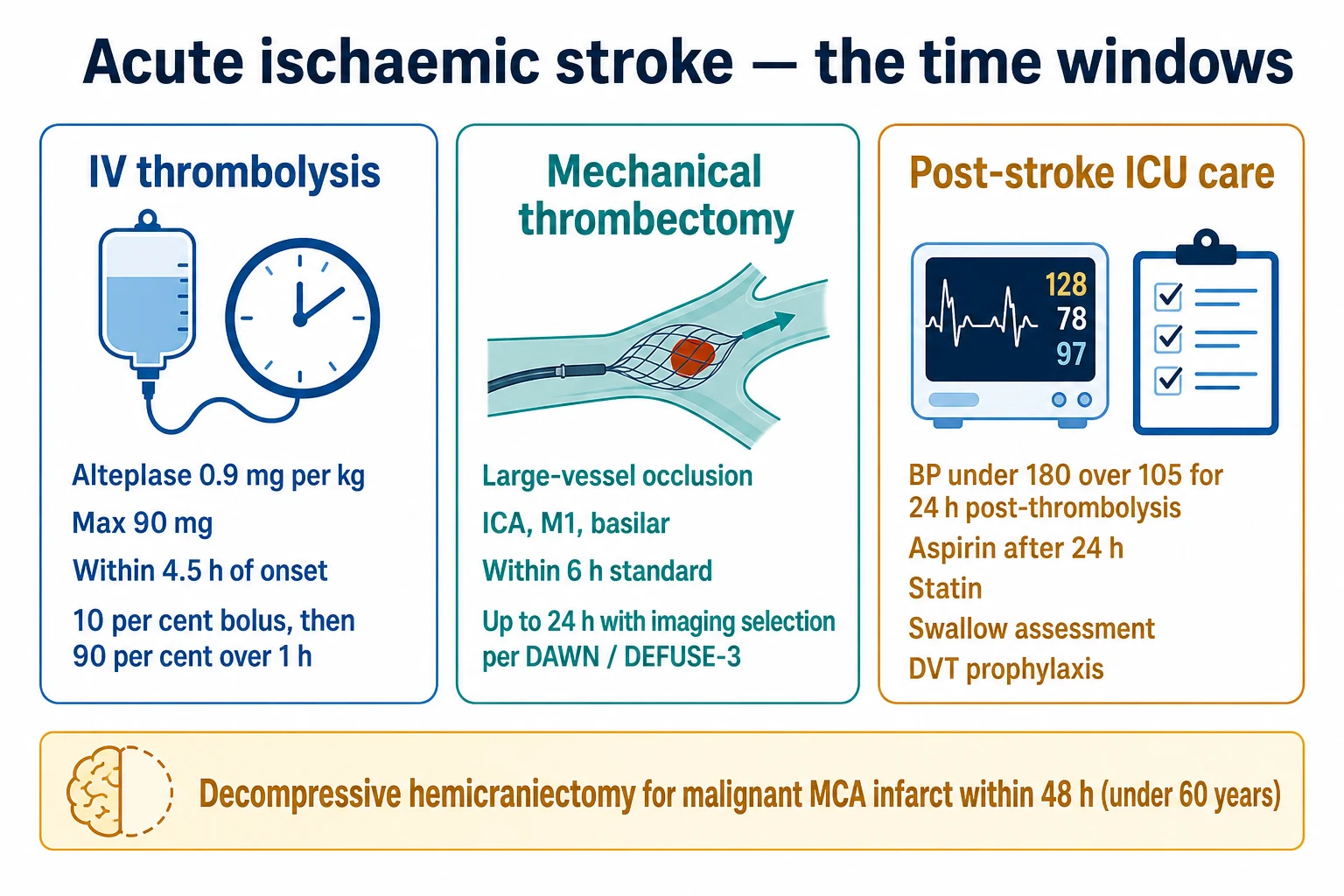
1. IV thrombolysis (alteplase) — within 4.5 hours
The agent: the alteplase (the recombinant tissue plasminogen activator — rtPA) at 0.9 mg/kg (maximum 90 mg) — 10 per cent as a bolus over 1 minute, then 90 per cent over 1 hour.[1]
The window: within 4.5 hours of the symptom onset (the ECASS III extended the 3-hour window to 4.5 hours; the benefit decreases with the time — the earlier the better — the "time is brain" principle).[2]
The inclusion: an acute ischaemic stroke with a measurable neurological deficit (the NIHSS above 0).[1]
The key exclusions:[17]
- A prior intracranial haemorrhage.
- A recent surgery or a major trauma (within 14 days).
- An active anticoagulation (the INR above 1.7, or the DOAC within 48 hours).
- The platelet count under 100.
- The BP over 185/110 uncontrolled.
- A rapid clinical improvement.
- A glucose under 2.7 or above 22 mmol/L.
2. Mechanical thrombectomy — within 6 hours (extended to 24)
For the large-vessel occlusion (LVO) — the internal carotid artery (ICA), the M1 middle cerebral artery, or the basilar artery.[7]
- Within 6 hours (the standard window — the MR CLEAN, the ESCAPE, the EXTEND-IA, the SWIFT-PRIME, and the REVASCAT trials all showed a dramatic benefit of the thrombectomy over the medical therapy alone for the LVO within 6 hours).[7]
- Extended to 24 hours (the DAWN and the DEFUSE-3 trials, NEJM 2018) — for the selected patients with a favorable imaging profile (the CT perfusion mismatch — a small core infarct and a large penumbra). The thrombectomy at 6-24 hours improved the functional outcome for these patients.[12]
The technique: the stent retriever (the stent-like device that ensnares the clot and pulls it out) or the aspiration (the large-bore catheter that suctions the clot). Both achieve the recanalisation in over 80 per cent of the cases.[8]
Post-stroke ICU care
- The BP control: after the thrombolysis, keep the SBP under 180 mmHg and the DBP under 105 mmHg for 24 hours (to prevent the haemorrhagic transformation). After the 24 hours, the standard stroke BP targets apply (lower the SBP by 10-15 per cent; a statin for the atherosclerotic stroke).[1]
- The antiplatelet: the aspirin after 24 hours (once a repeat CT excludes the haemorrhagic transformation).[1]
- The statin: a high-intensity statin (atorvastatin 80 mg) for the atherosclerotic stroke.[1]
- The normoglycaemia: avoid the hyperglycaemia (above 10 mmol/L) and the hypoglycaemia — both worsen the outcome. Use a sliding-scale insulin; avoid the intensive insulin (the NICE-SUGAR-type hypoglycaemia).[1]
- The normothermia: treat the fever (the paracetamol, the cooling). The fever worsens the outcome.[1]
- The swallow assessment: the bedside swallow screen before any oral intake; the formal speech-pathology assessment if the screen fails. The nil-by-mouth until the swallow is cleared.[1]
- The DVT prophylaxis: the intermittent pneumatic compression on admission; the LMWH after 24-48 hours (once the haemorrhage is excluded).[1]
The decompressive hemicraniectomy
For the malignant MCA infarct — the large MCA stroke that swells (the cerebral oedema, the mass effect, the midline shift, the herniation) within 24-48 hours, causing the coma and the death.[14]
- The surgery: the decompressive hemicraniectomy (the removal of a large bone flap on the affected side, allowing the swollen brain to expand outward rather than herniate inward) plus the durotomy (opening the dura).[14]
- The timing and the patient selection: within 48 hours, for the patients under 60 — the DESTINY, the DECIMAL, and the HAMLET trials showed a reduction of the mortality from 70 to 20 per cent, with the most survivors walking independently. For the patients over 60, the surgery reduces the mortality but many survivors are left with a severe disability (the mRS 4-5) — the decision is individualised with the family.[16]
Red flags
Deep dive — the IV thrombolysis pharmacology and the dosing
Alteplase (the recombinant tissue plasminogen activator, rt-PA) is a serine protease that converts the plasminogen to the plasmin, which then lyses the fibrin clot. It has a short half-life (about 5 minutes), so it is given as a bolus plus an infusion.[1]
The dose
| Parameter | Value | Note |
|---|---|---|
| The dose | 0.9 mg/kg | Total dose. |
| The maximum | 90 mg | Caps the total at 90 mg (the 100 kg patient gets 90 mg, not 100 mg). |
| The bolus | 10 per cent of the total dose | Over 1 minute (an IV push). |
| The infusion | 90 per cent of the total dose | Over 60 minutes (one hour). |
| Example (70 kg) | 63 mg total | 6.3 mg bolus, then 56.7 mg over 1 hour. |
The exclusion criteria — the absolute and the relative
The exclusion criteria were derived from the original NINDS trial and refined by the AHA/ASA 2019 guidelines.[17]
Absolute exclusions (do NOT thrombolyse):
- A prior intracranial haemorrhage (any).
- A known intracranial neoplasm, an AVM, or a ruptured aneurysm.
- An active internal bleeding.
- A suspected aortic dissection.
- An intracranial or an intraspinal surgery within 3 months.
- A serious head trauma or a stroke within 3 months.
- The platelet count under 100 × 10⁹/L.
- The anticoagulant use with an INR above 1.7 or a PT above 15 s.
- A low-molecular-weight heparin within 24 hours, or a direct oral anticoagulant within 48 hours (or any DOAC if the timing is unknown — unless the specific reversal or the normalised coagulation).
- The glucose under 2.7 mmol/L — treat first and re-assess; if the deficit resolves, it was the hypoglycaemia mimic.
- The BP above 185/110 — lower it with the IV labetalol or the nicardipine first; if it cannot be controlled, do not thrombolyse. [1]
Relative exclusions (weigh the risk and the benefit):
- A major surgery or a serious non-head trauma within 14 days.
- A recent GI or a GU bleed within 21 days.
- A seizure at the onset (the postictal deficit may mimic a stroke — confirm with the imaging).
- A pregnancy.
- An extensive hypodensity on the CT (the established infarct — beyond the window). [1]
The monitoring during and after the infusion
The alteplase infusion monitoring
Baseline
NIHSS, weight, CT excluded haemorrhage, BP, glucose, INR/platelets. Two large-bore IV cannulae. NO arterial punctures or central lines, NO urinary catheterisation, NO NG tube for 24 hours (bleeding risk). Strict nil by mouth until the swallow screen.
During the infusion (1 hour)
Monitor the BP and the neurological exam every 15 minutes. Watch for: angioedema (especially in patients on ACE inhibitors — stop infusion, give antihistamine/steroid/adrenaline), acute hypotension, anaphylaxis. If the patient deteriorates acutely (new headache, vomiting, decreasing GCS) — SUSPECT a symptomatic ICH: STOP the infusion, send for an urgent STAT CT.
For 24 hours after
BP every 15 min × 2 h, then every 30 min × 6 h, then hourly × 16 h. Keep SBP under 180 and DBP under 105. No antiplatelets or anticoagulants for 24 hours. Repeat CT at 24 hours — if no haemorrhage, start aspirin.
If symptomatic ICH occurs
STOP the alteplase immediately. Send CBC, coagulation, fibrinogen, group-and-save. Give cryoprecipitate (10 units) to replenish fibrinogen, plus tranexamic acid 1 g IV. Urgent CT. Neurosurgery consultation — though the surgical evacuation is rarely beneficial in thrombolysis-associated ICH.
Symptomatic intracranial haemorrhage (SICH) — the feared complication
The SICH is the bleeding into the infarcted territory causing a neurological deterioration. The incidence is about 2-6 per cent with the alteplase (vs about 0.6 per cent without). The risk factors: a high NIHSS, a high BP, a large infarct, the hyperglycaemia, the delayed treatment, and the prior antiplatelet use. The ECASS III defined the SICH as any blood seen on the CT with a clinical deterioration of 4 or more NIHSS points. The mortality of a SICH is around 50 per cent.[2]
Alteplase
rt-PA, the standard
- Dose 0.9 mg/kg (max 90 mg); 10% bolus over 1 min, 90% over 1 h
- Licensed and guideline-recommended for AIS within 4.5 h
- Short half-life (~5 min) — bolus + infusion required
- Cheaper; widely stocked; the NINDS, ECASS III, ENCHANTED evidence base
- Risk of SICH 2-6%; angioedema especially with ACE inhibitors
Tenecteplase
the genetically modified rt-PA
- Dose 0.25 mg/kg (max 25 mg) as a SINGLE 5-second bolus — no infusion
- Higher fibrin specificity, longer half-life (~22 min), less systemic fibrinolysis
- EXTEND-IA TNK (2018): better reperfusion and similar outcomes vs alteplase in LVO
- At least non-inferior to alteplase; favoured for the LVO bridging (faster, easier)
- AcT (2022): non-inferior for all thrombolysis-eligible strokes; increasingly adopted
Deep dive — the thrombolysis landmark trials
NINDS
NEJM 1995
624 pts with AIS within 3 h — IV alteplase vs placebo
Key finding
alteplase improved 3-month favourable outcome (50% vs 38%); benefit despite a small increase in SICH (6.4% vs 0.6%)
Practice change
Established IV alteplase as the standard for AIS within 3 h — the foundational trial
ECASS III
NEJM 2008
821 pts with AIS 3-4.5 h after onset — alteplase vs placebo
Key finding
alteplase improved functional independence (52.4% vs 45.2% mRS 0-2); more SICH (2.5% vs 0.3%); no mortality difference
Practice change
Extended the thrombolysis window from 3 to 4.5 hours
IST-3
Lancet 2012
3035 pts (incl. >80 y, the largest alteplase trial) within 6 h — alteplase vs control
Key finding
no overall benefit at 6 months but improved with earlier treatment; benefit even in the elderly within 3 h; more early deaths and SICH
Practice change
Removed the upper age limit for thrombolysis; reinforced "earlier is better"
ENCHANTED
NEJM 2016
3310 pts — low-dose alteplase (0.6 mg/kg) vs standard-dose (0.9 mg/kg)
Key finding
low dose non-inferior for the primary outcome (death/disability) and fewer SICH (1.0% vs 2.1%); failed strict non-inferiority for mRS 0-2
Practice change
Low-dose alteplase considered in Asian populations; standard dose remains the default in ANZ/US/Europe
EXTEND
NEJM 2019
225 pts 4.5-9 h after onset or wake-up with MRI/perfusion mismatch — alteplase vs placebo
Key finding
alteplase improved functional independence (35.4% vs 29.5%); more SICH (6% vs 1%)
Practice change
Thrombolysis may be extended to 9 h with CT/MRI perfusion selection
WAKE-UP
NEJM 2018
503 pts with wake-up stroke and MRI DWI-FLAIR mismatch — alteplase vs placebo
Key finding
alteplase better functional outcome (53.3% vs 41.8% mRS 0-1); more SICH (2% vs 0.4%)
Practice change
MRI-guided thrombolysis for wake-up strokes with a DWI-FLAIR mismatch
Deep dive — the mechanical thrombectomy
The patient selection
The thrombectomy is for the large-vessel occlusion (LVO) — the proximal anterior circulation (the intracranial ICA, the M1 MCA, sometimes the proximal M2) and the basilar artery. The distal occlusions and the lacunar strokes do NOT benefit (the clot is too small to retrieve).[7]
The imaging — the CTA first-line
- The non-contrast CT — excludes the haemorrhage; the ASPECTS score grades the early ischaemic change in the MCA territory (a score of 10 is normal; below 6 indicates an extensive established infarct and a poor prognosis for the intervention).
- The CT angiography (CTA) — the first-line to identify the LVO; assess from the aortic arch to the vertex.
- The CT perfusion (CTP) — used for the extended window (the 6-24 h) to demonstrate the core–penumbra mismatch: a small core infarct (the irreversibly dead tissue) and a large penumbra (the salvageable tissue) per the DAWN and the DEFUSE-3 criteria.[13]
The ASPECTS (Alberta Stroke Program Early CT Score)
The 10-point score grading the early ischaemic change on the non-contrast CT in the MCA territory. Ten regions (M1-M6, the caudate, the lentiform, the insula, the internal capsule) each score a point if normal; a point is subtracted for each region with the early ischaemia (the hypodensity or the swelling).[8]
| ASPECTS | Interpretation | The thrombectomy implication |
|---|---|---|
| 10 | No early ischaemia | Ideal candidate. |
| 7-9 | A small infarct | Good candidate — substantial penumbra. |
| 4-6 | A moderate infarct | Consider; the benefit is less certain. |
| 0-3 | An extensive established infarct | Poor candidate — the core is large, the futility is likely. |
The technique
Stent retriever
the current standard
- A self-expanding stent deployed across the clot — restores the flow immediately and ensnares the clot
- Withdrawn under the aspiration — the clot comes out with it
- Higher recanalisation rate (TICI 2b-3 in ~80-90%) — the SWIFT, the TREVO trials
- Examples: Solitaire, Trevo, Revive
Aspiration (ADAPT)
a direct suction
- A large-bore distal-access catheter advanced to the clot face and connected to the suction
- The clot is aspirated directly — fast in the favourable anatomy
- Faster, fewer passes, less endothelial trauma in some studies
- Often combined with the stent retriever for the large clots
Bridging therapy
IV then IA
- IV thrombolysis started on the CT table, THEN transfer to the angio suite
- Do NOT delay the thrombectomy to wait for the alteplase — give it concurrently
- Reduces the microvascular clot propagation; some patients recanalise before the thrombectomy
- For the direct-arrival LVO within 4.5 h, both are given ("bridging")
The anaesthesia for the thrombectomy — the general vs the sedation
The debate: the general anaesthesia (GA) gives a still patient but risks the hypotension (which worsens the outcome); the conscious sedation preserves the collateral perfusion but risks the patient movement. The current evidence (the SIESTA, the GOLIATH, the Sedation vs Intubation for Endovascular Stroke TreAtment meta-analyses) is mixed but leans toward the conscious sedation (or the monitored anaesthesia care) for the cooperative patients, reserving the GA for the agitated or the airway-compromised patients, with a strict avoidance of the hypotension (keep the SBP above 140).[1]
The thrombectomy workflow — the door-to-groin
Recognition & the CTA
Suspect the LVO on the high NIHSS (over 6). The non-contrast CT excludes the haemorrhage; the CTA confirms the LVO. Run the CTA from the arch — to assess the access (the tortuosity, the atherosclerosis).
Activate the angio team
Single-call activation of the interventional neuroradiologist, the anaesthetist, and the angio suite. The parallel processing — the IV thrombolysis on the CT table, the transfer to the angio suite, the groin prep.
The target — door-to-groin under 60 min
Aim for the arterial puncture within 60 min of the arrival. Every 10 min delay in the reperfusion loses about one month of disability-free life.
The procedure
Femoral (or radial) access, the guiding catheter to the carotid, the microcatheter across the clot, the stent retriever or the aspiration. The goal: a TICI 2b-3 reperfusion (over 50% of the territory). The first-pass success is around 50-70%.
Post-procedure ICU
Immediate NIHSS. BP: if the successful reperfusion, keep the SBP under 160 to avoid the hyperperfusion and the haemorrhage; if the unsuccessful, allow the permissive hypertension to the SBP 220. Monitor for the groin access complications.
Deep dive — the thrombectomy landmark trials
MR CLEAN
NEJM 2015
500 pts with proximal anterior circulation LVO within 6 h — intra-arterial treatment vs medical
Key finding
modified Rankin shifted favourably (OR 1.67); 13.5% absolute increase in functional independence
Practice change
The first positive thrombectomy RCT — established the thrombectomy for LVO within 6 h
ESCAPE
NEJM 2015
315 pts with LVO and good CT collateral imaging within 12 h — endovascular vs standard
Key finding
90-day functional independence 53% vs 29%; mortality 10% vs 19%; trial stopped early for benefit
Practice change
Thrombectomy with the imaging selection (ASPECTS, collaterals) for LVO
EXTEND-IA
NEJM 2015
70 pts with LVO within 4.5 h and CT perfusion mismatch — thrombectomy + alteplase vs alteplase alone
Key finding
early reperfusion 100% vs 37%; better 90-day functional outcome; trial stopped early
Practice change
Thrombectomy with the perfusion selection — added the CTP to the LVO workup
SWIFT-PRIME
NEJM 2015
196 pts with LVO within 6 h — stent retriever + alteplase vs alteplase alone
Key finding
functional independence 60% vs 35%; trial stopped early for benefit
Practice change
Confirmed the stent-retriever thrombectomy benefit within 6 h
REVASCAT
NEJM 2015
206 pts with proximal MCA/ICA occlusion within 8 h — thrombectomy vs medical
Key finding
functional independence 43.7% vs 28.2%; severity of the residual deficit reduced
Practice change
A fifth concurrent positive trial — solidified the thrombectomy as the standard
DAWN
NEJM 2018
206 pts 6-24 h after onset with a clinical-imaging mismatch (small core) — thrombectomy vs medical
Key finding
functional independence 49% vs 13%; mortality 19% vs 18%; trial stopped early
Practice change
Extended the thrombectomy window to 24 h with the imaging selection
DEFUSE-3
NEJM 2018
182 pts 6-16 h after onset with a CT/MRI perfusion mismatch — thrombectomy vs medical
Key finding
functional independence 45% vs 17%; mortality 14% vs 26%; trial stopped early
Practice change
Thrombectomy 6-16 h with the perfusion mismatch — core–penumbra selection
The blood pressure management — the permissive hypertension
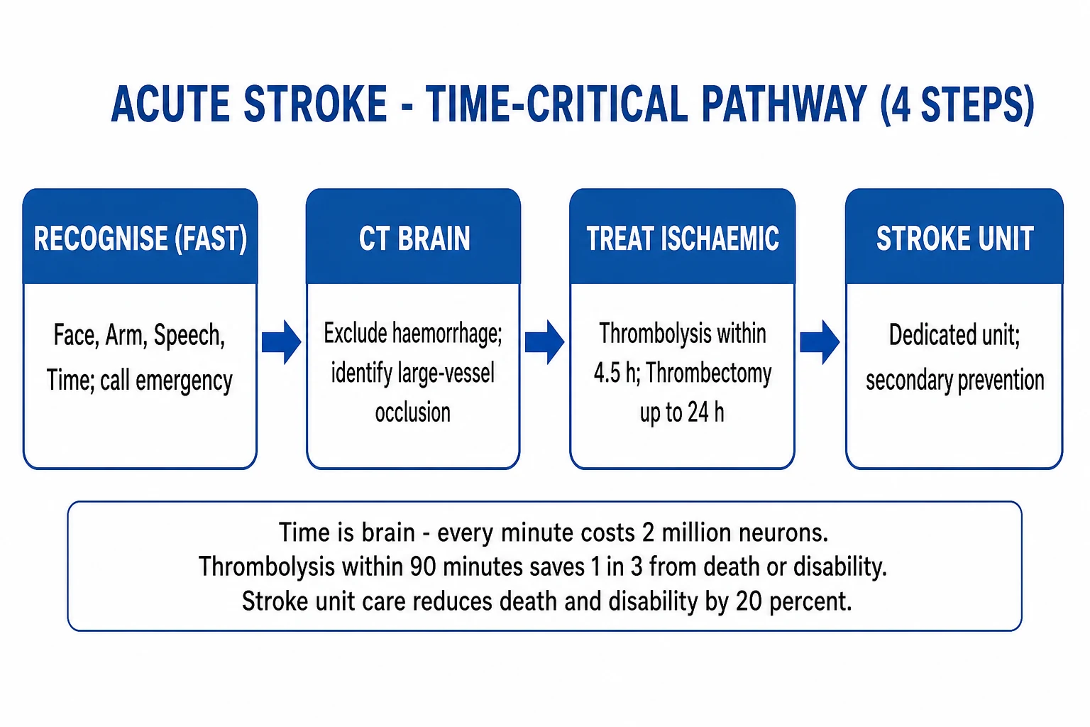
The BP management is one of the few modifiable factors in the ICU stroke care. The principle: maintain the collateral perfusion to the penumbra (the salvageable tissue around the dead core) by allowing the permissive hypertension — but NOT so high as to cause the haemorrhagic transformation.[17]
The three BP scenarios
| Scenario | The SBP/DBP target | The rationale |
|---|---|---|
| Before the thrombolysis | Under 185/110 | The alteplase cannot be given above this — lower it first with the IV labetalol or the nicardipine. |
| After the thrombolysis (24 h) | Under 180/105 | Prevents the haemorrhagic transformation. The strictest target — monitor every 15 min. |
| The non-thrombolysed stroke | Under 220/120 (permissive hypertension) | The collateral perfusion to the penumbra. Below 220/120 is safe; above, lower cautiously. |
| After the successful thrombectomy | Under 160 (some centres) | The hyperperfusion and the haemorrhagic transformation risk once the flow is restored. |
| After the failed thrombectomy | Permissive hypertension to 220 | Maintain the collateral perfusion; no flow restored. |
The agents
| Agent | The dose | The pros / the cons |
|---|---|---|
| Labetalol | 10-20 mg IV over 1-2 min, repeat q10min, max 300 mg; or 2-8 mg/min infusion | Combined α+β blocker; fast, titratable; avoid in the asthma, the heart block, the acute heart failure. |
| Nicardipine | 5 mg/h, titrate up by 2.5 mg/h q5min to max 15 mg/h | The most titratable; the smooth BP control; the favourite for the stroke; the phosphodiesterase vasodilator. |
| Clevidipine | 1-2 mg/h, double q90s to max 32 mg/h | Ultrashort-acting (1 min); ideal for the tight BP control; an egg/soy allergy contraindication. |
| Urapidil | 10-25 mg IV bolus, then 2-8 mg/h | α1 blocker + the 5-HT1A agonist; used widely in the ANZ/Europe. |
| Glyceryl trinitrate | The infusion (1-10 mg/h) | Useful for the concomitant ACS; can worsen the ICP (the vasodilatation) — use cautiously. |
The BP management after the thrombolysis
Baseline (0-15 min)
The BP on the arrival. If above 185/110 — lower it with the IV labetalol (10-20 mg) or the nicardipine BEFORE giving the alteplase. Do NOT give the alteplase until the BP is controlled.
During and after the infusion
Monitor every 15 min for the first 2 h, then every 30 min for 6 h, then hourly for 16 h. Target: the SBP under 180 and the DBP under 105.
If the BP rises above 180/105
IV labetalol 10 mg or the nicardipine infusion. Re-check in 15 min. If still high, increase the infusion. Do NOT allow a sustained elevation — the SICH risk.
After 24 hours
Transition to the standard stroke targets (lower the SBP by 10-15 per cent). Start the oral antihypertensives (the ACE inhibitor, the thiazide, the amlodipine). Continue the secondary prevention.
The complications of the acute stroke in the ICU
Cerebral oedema
days 2-5 (peak 48-72 h)
- The cytotoxic and the vasogenic oedema in the large MCA territory — peaks at 48-72 h
- The mass effect: the midline shift, the ventricular compression, the herniation (uncal, central)
- The predictors: the complete MCA infarct, the NIHSS above 20, the early CT hypodensity, the ASPECTS below 7
- The medical management: head up 30°, the normothermia, the normocapnia, the normoglycaemia, the osmotherapy (mannitol, the hypertonic saline)
- The definitive: the decompressive hemicraniectomy within 48 h (under 60)
Haemorrhagic transformation
0-24 h (post-thrombolysis) or days 3-7
- The bleeding into the infarcted tissue — from the petechiae (asymptomatic) to the frank haematoma (symptomatic SICH)
- The risk factors: the thrombolysis, the large infarct, the high BP, the hyperglycaemia, the cardioembolic stroke, the prior antiplatelet
- The SICH: an acute deterioration (headache, vomiting, decreased GCS, new deficit) — STOP the alteplase, give cryoprecipitate ± tranexamic acid, urgent CT
- The management: the reversal of the alteplase (cryoprecipitate 10 U, tranexamic acid 1 g), the BP control, the ICP management; the surgery rarely indicated
Seizures
early (first 7 d) or late
- The incidence: 5-10% in the acute stroke; higher in the cortical and the haemorrhagic strokes
- The early seizures (within 7 d) do not necessarily predict the epilepsy; treat if they occur
- Watch for the non-convulsive status epilepticus (NCSE) in the obtunded patient — the continuous EEG
- The treatment: levetiracetam (first-line), the valproate, the lacosamide. The prophylactic antiepileptics are NOT routinely recommended
Aspiration pneumonia
0-7 d
- The #1 infectious complication — the dysphagia in 50-80% of the strokes
- The strict nil-by-mouth until the swallow screen; the formal speech-pathology assessment
- The head of bed 30-45°; the good oral hygiene (chlorhexidine) reduces the ventilator-associated and the aspiration pneumonia
- The treatment: the broad-spectrum antibiotics (the ceftriaxone + the metronidazole, or the amoxicillin-clavulanate)
The cerebral oedema and the malignant MCA infarct — the detailed management
The malignant MCA infarct is the complete MCA territory stroke that swells to the point of the herniation in 24-72 hours. It occurs in 1-10 per cent of the supratentorial strokes but carries an 80 per cent mortality untreated.[14]
The medical management (the tiered approach):
- Tier 0 — the basics: head up 30° and the midline position (avoid the neck rotation and the tight endotracheal tube ties — they impede the venous drainage); the normothermia (the fever worsens the oedema); the normoglycaemia; the normocapnia (the PaCO₂ 35-40 mmHg — avoid the hyperventilation except as a temporary bridge); the normoxia; avoid the hypo- and the hypertension (the SBP 140-180).
- Tier 1 — the osmotherapy: the mannitol 0.25-1 g/kg IV over 15 min (or the 20% mannitol 1 g/kg) — pulls the water from the brain into the vasculature; check the serum osmolality (keep under 320 mOsm/kg) and the sodium; the hypertonic saline (3%, 5%, or the 23.4% bolus) — pulls the water and supports the BP; aim the Na 145-150 mmol/L.
- Tier 2 — the definitive: the decompressive hemicraniectomy (within 48 h, the under-60s) — the only intervention proven to reduce the mortality. The targeted temperature management and the barbiturates have NOT shown the benefit for the malignant MCA. [1]
The malignant MCA infarct — the management pathway
Identify the risk
The complete MCA infarct, the NIHSS above 20, the early CT hypodensity (ASPECTS below 7), the involvement of more than 50% of the MCA territory within 5 h, the midline shift on the CT. Admit to the ICU; monitor the GCS hourly.
The medical management
Head up 30°, midline. The normothermia, the normoglycaemia, the normocapnia. Avoid the hypotension. The serial CTs at 12, 24, 48 h to monitor the oedema and the midline shift.
Escalate if the GCS drops
If the GCS drops by 2 points or the pupils dilate — give the mannitol 0.5-1 g/kg or the 23.4% saline 30 mL bolus as a bridge. Repeat the CT. Insert an ICP monitor if the patient is being considered for the further escalation.
The decompressive hemicraniectomy
Within 48 hours for the patients under 60 (DESTINY, DECIMAL, HAMLET — mortality 70% to 20%). For the over 60s, the individualised decision (the mortality benefit but many survivors are the mRS 4-5). Discuss with the family. The surgery: a large fronto-temporo-parietal bone flap (at least 12 cm diameter) + the durotomy + the duraplasty.
Post-operative ICU
Monitor for the ongoing swelling (the bone flap bulge), the contralateral haematoma, the seizure. The cranioplasty (the bone flap replacement) at 6-12 weeks. The early rehabilitation.
DESTINY
Stroke 2007
32 pts under 60 with malignant MCA infarct — decompressive hemicraniectomy within 36 h vs medical
Key finding
mortality 25% vs 88%; more favourable outcomes (mRS 0-3) 47% vs 24%
Practice change
Early decompressive hemicraniectomy for the under-60 malignant MCA infarct
DECIMAL
Lancet Neurol 2007
Sequential-design RCT — early decompressive craniectomy vs medical in the malignant MCA
Key finding
trial stopped early — the surgery reduced the mortality and the severe disability
Practice change
Added to the body of evidence for the early hemicraniectomy
HAMLET
Lancet Neurol 2009
64 pts within 48 h of the malignant MCA — hemicraniectomy vs medical
Key finding
reduced the mRS 4-6 (OR 0.22); no clear benefit if the surgery was beyond 48 h (the earlier, the better)
Practice change
Confirmed the hemicraniectomy within 48 h; the pooled DESTINY+DECIMAL+HAMLET analysis is the definitive evidence
The neuroprotection and the supportive care — the "neuroprotective bundle"
No pharmacological neuroprotectant has shown the benefit in the human trials (despite hundreds of the animal studies). The best "neuroprotection" is the rapid reperfusion and the meticulous supportive care to avoid the secondary brain injury.[1]
| Parameter | The target | The rationale |
|---|---|---|
| The glucose | 6-10 mmol/L | Both the hypo- and the hyperglycaemia worsen the outcome. Avoid the intensive insulin (the NICE-SUGAR-type hypoglycaemia). |
| The temperature | 36-37°C (the normothermia) | The fever worsens the outcome (each 1°C rise increases the morbidity). Treat with the paracetamol and the cooling. |
| The oxygen | The SpO₂ above 94%, the PaO₂ above 60 mmHg | Avoid the hypoxia — each episode doubles the mortality. Supplemental O₂ only if needed (the routine high-flow O₂ does not help). |
| The CO₂ | 35-40 mmHg (the normocapnia) | Avoid the hypercapnia (the raised ICP) and the hypocapnia (the vasoconstriction, the reduced perfusion). |
| The sodium | 135-145 mmol/L (140-150 in the raised ICP) | Avoid the hyponatraemia (the cerebral oedema). The hypertonic saline may be used for the malignant oedema. |
| The BP | See the BP section | The permissive hypertension for the collateral perfusion; the strict control after the thrombolysis. |
| The head position | Head up 30°, midline | Promotes the venous drainage; reduces the ICP. Avoid the neck rotation and the tight ETT ties. |
The DVT prophylaxis and the early mobilisation
The stroke patients are at a high VTE risk (the immobility, the paresis). The intermittent pneumatic compression on the admission; the LMWH (the enoxaparin 40 mg SC daily) after 24-48 hours once the haemorrhage is excluded. The early mobilisation (within 24-48 h, if stable) improves the outcome — the AVERT trial showed that the very early intense mobilisation (within 24 h) may be harmful, but the gentle early mobilisation is beneficial.[1]
The specific scenarios
The wake-up stroke and the unknown onset
The wake-up stroke (about 25 per cent of the strokes) presents a challenge — the onset time is unknown, so the 4.5-hour window cannot be applied. The WAKE-UP trial (NEJM 2018) showed the alteplase benefit in the wake-up strokes with the MRI DWI-FLAIR mismatch (the DWI shows the infarct but the FLAIR has not yet brightened — indicating the infarct is less than 4.5 hours old). The EXTEND trial extended the perfusion-selected thrombolysis to 9 hours. The current practice: the MRI-guided (DWI-FLAIR) or the CT-perfusion-guided thrombolysis for the wake-up strokes.[6][5]
The posterior circulation stroke and the basilar occlusion
The basilar artery occlusion causes the brainstem stroke — the quadriparesis, the locked-in syndrome, the coma, the bilateral cranial nerve signs (the dysarthria, the dysphagia, the diplopia, the ataxia). The outcome without the recanalisation is fatal (over 80 per cent mortality). The thrombectomy (or the intra-arterial thrombolysis) is indicated regardless of the time window if the brainstem is salvageable (the CT/CTP or the MRI shows the salvageable tissue). The BASICS trial (2019, the Lancet) was neutral for the endovascular treatment within 6-24 h, but the BAOCHE trial (2022, the NEJM) showed the benefit for the basilar occlusion 6-24 h with the imaging selection. The basilar occlusion is the one situation where the recanalisation is attempted beyond 24 hours.[1]
The posterior circulation stroke — the subtle presentations
The brainstem strokes can present with the "crossed" signs (the ipsilateral cranial nerve palsy and the contralateral long tract sign), the vertigo, the nausea, the diplopia, the ataxia — easily mistaken for the vestibular or the GI pathology. The "HINTS" exam (the Head Impulse, the Nystagmus, the Test of Skew) helps distinguish the central (the stroke) from the peripheral (the vestibular) vertigo — a central cause has the NORMAL head impulse, the direction-changing nystagmus, and the skew deviation. The negative HINTS in a high-risk patient is a stroke until proven otherwise.[1]
The scorecards — the NIHSS and the mRS
| Score | The NIHSS (0-42) | The mRS (0-6) |
|---|---|---|
| 0 | No deficit | Asymptomatic |
| 1-4 | Minor | No significant disability (able to carry out all duties) |
| 5-15 | Moderate | Slight disability (able to look after self without assistance) |
| 16-20 | Moderate-severe | Moderate disability (requires some help, able to walk without assistance) |
| 21-42 | Severe | Moderately severe disability (unable to walk without assistance, unable to attend to own bodily needs) |
| 5 | — | Severe disability (bedridden, incontinent, requires constant nursing care) |
| 6 | — | Dead |
The NIHSS quantifies the stroke severity on the admission — it guides the thrombolysis and the thrombectomy decisions and predicts the outcome. The mRS (the modified Rankin Scale) is the 90-day outcome measure used in all the stroke trials — the mRS 0-2 is the "favourable" outcome (the functional independence), the mRS 4-6 the "unfavourable".[1]
[1] [1]Exam practice
SAQ — Acute ischaemic stroke — thrombolysis decision and post-lysis ICU care
10 minutes · 10 marks
A 68-year-old man is brought to the emergency department 90 minutes after his wife found him with a right-sided weakness and a slurred speech. His past history includes hypertension, type 2 diabetes, and paroxysmal atrial fibrillation for which he takes apixaban 5 mg twice daily (the last dose was 8 hours ago). On arrival his NIHSS is 12, blood pressure 192/104, capillary glucose 8.1 mmol/L, GCS 14. The non-contrast CT shows no haemorrhage and an ASPECTS of 9. You are the ICU registrar consulted for admission.
SAQ — Large-vessel occlusion, thrombectomy, and the malignant MCA infarct
10 minutes · 10 marks
A 54-year-old woman arrives 5 hours after the onset of a left-sided weakness and a gaze deviation. Her NIHSS is 18, BP 210/115, GCS 13. The non-contrast CT shows an ASPECTS of 8 with early loss of the right lentiform nucleus. The CT angiogram confirms a right M1 middle cerebral artery occlusion. The CT perfusion shows a small core (15 mL) with a large penumbra. She is not on any anticoagulant.
The references summary
The evidence base for the acute ischaemic stroke is one of the strongest in the neurocritical care. The key trials: NINDS (1995) established the IV alteplase within 3 h; ECASS III (2008) extended to 4.5 h; IST-3 (2012) showed the benefit even in the elderly within 3 h; ENCHANTED (2016) tested the low-dose alteplase; EXTEND (2019) and WAKE-UP (2018) extended the thrombolysis to 9 h and the wake-up strokes. The five 2015 thrombectomy trials (MR CLEAN, ESCAPE, EXTEND-IA, SWIFT-PRIME, REVASCAT) established the thrombectomy within 6 h; DAWN and DEFUSE-3 (2018) extended to 24 h. The DESTINY, DECIMAL, HAMLET (2007-2009) established the hemicraniectomy for the malignant MCA infarct. The AHA/ASA 2019 guidelines (Powers et al.) synthesise the evidence into the practice.[17]
References
- [1]The National Institute of Neurological Disorders and Stroke rt-PA Stroke Study Group Tissue plasminogen activator for acute ischemic stroke N Engl J Med, 1995.PMID 7477192
- [2]Hacke W, Kaste M, Bluhmki E, et al Thrombolysis with alteplase 3 to 4.5 hours after acute ischemic stroke N Engl J Med, 2008.PMID 18815396
- [3]The IST-3 collaborative group The benefits and harms of intravenous thrombolysis with recombinant tissue plasminogen activator within 6 h of acute ischaemic stroke (the third international stroke trial [IST-3]): a randomised controlled trial Lancet, 2012.PMID 22632908
- [4]Anderson CS, Robinson T, Lindley RI, et al (ENCHANTED) Low-Dose versus Standard-Dose Intravenous Alteplase in Acute Ischemic Stroke N Engl J Med, 2016.PMID 27161018
- [5]Ma H, Campbell BCV, Parsons MW, et al (EXTEND) Thrombolysis Guided by Perfusion Imaging up to 9 Hours after Onset of Stroke N Engl J Med, 2019.PMID 31067369
- [6]Thomalla G, Simonsen CZ, Boutitie F, et al (WAKE-UP) MRI-Guided Thrombolysis for Stroke with Unknown Time of Onset N Engl J Med, 2018.PMID 29766770
- [7]Berkhemer OA, Fransen PSS, Beumer D, et al (MR CLEAN) A randomized trial of intraarterial treatment for acute ischemic stroke N Engl J Med, 2015.PMID 25517348
- [8]Goyal M, Demchuk AM, Menon BK, et al (ESCAPE) Randomized assessment of rapid endovascular treatment of ischemic stroke N Engl J Med, 2015.PMID 25671798
- [9]Campbell BCV, Mitchell PJ, Kleinig TJ, et al (EXTEND-IA) Endovascular therapy for ischemic stroke with perfusion-imaging selection N Engl J Med, 2015.PMID 25671797
- [10]Saver JL, Goyal M, Bonafe A, et al (SWIFT-PRIME) Stent-retriever thrombectomy after intravenous t-PA vs. t-PA alone in stroke N Engl J Med, 2015.PMID 25882376
- [11]Jovin TG, Chamorro A, Cobo E, et al (REVASCAT) Thrombectomy within 8 hours after symptom onset in ischemic stroke N Engl J Med, 2015.PMID 25882510
- [12]Nogueira RG, Jadhav AP, Haussen DC, et al (DAWN) Thrombectomy 6 to 24 Hours after Stroke with a Mismatch between Deficit and Infarct N Engl J Med, 2018.PMID 29129157
- [13]Albers GW, Marks MP, Kemp S, et al (DEFUSE-3) Thrombectomy for Stroke at 6 to 16 Hours with Selection by Perfusion Imaging N Engl J Med, 2018.PMID 29364767
- [14]Jüttler E, Schwab S, Schmiedek P, et al (DESTINY) Decompressive Surgery for the Treatment of Malignant Infarction of the Middle Cerebral Artery (DESTINY): a randomized, controlled trial Stroke, 2007.PMID 17690310
- [15]Vahedi K, Vicaut E, Mateo J, et al (DECIMAL) Early decompressive surgery in malignant infarction of the middle cerebral artery: a pooled analysis of three randomised controlled trials Lancet Neurol, 2007.PMID 17303527
- [16]Hofmeijer J, Kappelle LJ, Algra A, et al (HAMLET) Surgical decompression for space-occupying cerebral infarction (the Hemicraniectomy After Middle Cerebral Artery infarction with Life-threatening Edema Trial [HAMLET]): a multicentre, open, randomised trial Lancet Neurol, 2009.PMID 19269254
- [17]Powers WJ, Rabinstein AA, Ackerson T, et al Guidelines for the Early Management of Patients With Acute Ischemic Stroke: 2019 Update to the 2018 Guidelines for the Early Management of Acute Ischemic Stroke: A Guideline for Healthcare Professionals From the American Heart Association/American Stroke Association Stroke, 2019.PMID 31662037