ICU · Neurocritical Care
Acute stroke: ischaemic and haemorrhagic
Also known as Acute ischaemic stroke (AIS) · Intracerebral haemorrhage (ICH) · Thrombolysis (alteplase/tenecteplase) · Mechanical thrombectomy · Large vessel occlusion (LVO) · Cerebral venous sinus thrombosis
Acute stroke is a neurological emergency. Time is brain — 1.9 million neurons lost per minute without reperfusion. Ischaemic stroke (85%): thrombolysis (alteplase 0.9 mg/kg up to 4.5h, or tenecteplase) + mechanical thrombectomy for large vessel occlusion (up to 24h with favourable imaging). Intracerebral haemorrhage (15%): BP control (SBP <140), reverse anticoagulation, surgical evacuation for cerebellar haemorrhage >3cm. ICU management: airway, BP control (permissive hypertension for ischaemic — SBP <185 for thrombolysis; reduce for haemorrhagic — SBP <140), glucose control (avoid hypo/hyperglycaemia), temperature control, DVT prophylaxis, swallowing assessment.
On this page & tools
Your progress
Saved locally on this device.
Target exams
Red flags

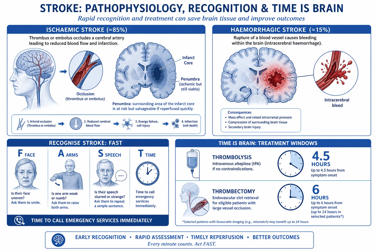
Recognition and prehospital — FAST / BEFAST and the time clock
Stroke is the second leading cause of death worldwide and the leading cause of acquired adult disability. The single most important determinant of outcome is time to reperfusion — every minute of large-vessel occlusion destroys ~1.9 million neurons, 13.8 billion synapses and 12 km of myelinated fibres. Recognition must therefore begin in the community, not in the CT scanner. [1]
Stroke recognition cascade (community → ED → scanner)
Recognise — FAST / BEFAST
FAST: Face drooping, Arm weakness, Speech difficulty, Time to call emergency services. BEFAST adds Balance (sudden vertigo/ataxia — posterior circulation) and Eyes (sudden vision loss / diplopia / hemianopia). Public-facing tools improve onset-time accuracy and prenotification, which shortens door-to-needle time. Negative FAST does NOT exclude posterior-circulation or purely sensory stroke.
Prenotify the receiving hospital
Prehospital stroke alert activates the stroke team (neurology, radiology, pharmacy) so the CT scanner and thrombolytic are prepared before arrival. Prenotification alone reduces door-to-CT and door-to-needle times by 10–20 min.
Establish "last known well" (LKW) time
The clock starts at LKW, NOT symptom discovery. For wake-up stroke, LKW = time last seen normal (e.g. before sleep). Determines the 4.5h thrombolysis and 6/24h thrombectomy windows. If LKW >4.5h, still assess for thrombectomy (imaging-based selection up to 24h).
Check fingerprick glucose IMMEDIATELY
Hypoglycaemia (and hyperglycaemia) is the most common stroke mimic — glucose <2.8 mmol/L can produce focal deficits identical to stroke. Treat first; re-examine. Glucose is the only bedside test that must precede thrombolysis decision-making.
Rapid ED assessment (target door-to-needle <45 min)
ABC, vital signs (BP — must be controlled before thrombolysis), brief focused history (onset, comorbidities, medications especially anticoagulants, recent surgery/trauma), NIHSS, then IMMEDIATE non-contrast CT brain. CT must be reported within 45 min of arrival.
NIHSS — the National Institutes of Health Stroke Scale
The NIHSS is the standardised, reproducible 11-item severity scale (score 0–42) that quantifies neurological deficit, guides thrombolysis/thrombectomy decisions, predicts outcome, and enables serial reassessment. It is one of the highest-yield examination skills for the CICM/FFICM neurology vivas. [1]
NIHSS scoring overview — what each item tests
1a–c Consciousness
0–9 points total
- 1a Level of consciousness (alert / drowsy / stupor / coma) — 0–3
- 1b LOC questions (month, age) — 0–2
- 1c LOC commands (open/close eyes, grip non-paretic hand) — 0–2
2–3 Gaze and visual fields
gaze palsy + hemianopia
- 2 Best gaze (horizontal forced deviation / partial palsy) — 0–2
- 3 Visual fields (quadrantanopia / hemianopia / bilateral blindness) — 0–3
- Tests anterior (MCA) vs posterior (PCA) territory involvement
4–6 Face, arm, leg
motor (left + right each)
- 4 Facial palsy (smile) — 0–3
- 5a/5b Arm drift (each side 0–4) — limb held 90° (arm) 10 s
- 6a/6b Leg drift (each side 0–4) — limb held 30° 5 s
- Highest single limb score predicts infarct volume and LVO
7–11 Language, sensation, neglect
cortical signs
- 7 Limb ataxia (finger-nose, heel-shin) — 0–2
- 8 Sensory (pinprick/face, arm, leg) — 0–2
- 9 Best language (name items, read sentence, describe picture) — 0–3
- 10 Dysarthria (read standard words) — 0–2
- 11 Extinction/inattention (double simultaneous stimulation) — 0–2
Severity bands and what they drive: NIHSS 1–4 = minor (consider dual antiplatelet CHANCE/POINT); 5–15 = moderate (thrombolysis ± thrombectomy); 16–20 = moderate–severe (thrombectomy candidate); >20 = severe — consider decompressive hemicraniectomy if MCA territory, age <60, within 48h. NIHSS >6 with a known LVO on CTA is the usual thrombectomy trigger. A dominant (left) hemisphere stroke scores higher than a non-dominant stroke of equal volume because language items (9, best language) inflate the score — a classic exam trap. [1]
Ischaemic vs haemorrhagic stroke — the fork in the road
The non-contrast CT (NCCT) brain is the decisive investigation: it dichotomises management completely. Blood on CT → no thrombolysis, tight BP control, reverse anticoagulation. No blood → reperfusion pathway. Everything downstream depends on this single image. [1]
Ischaemic stroke vs intracerebral haemorrhage
Ischaemic stroke (85%)
arterial occlusion
- Mechanism: thrombus (large-vessel atherosclerosis) or embolus (atrial fibrillation, valve disease)
- NCCT early: may be NORMAL first few hours; hyperdense MCA sign, loss of insular ribbon, loss of grey-white differentiation
- Later: wedge hypodensity in a vascular territory (MCA > PCA > ACA)
- BP strategy: PERMISSIVE hypertension (SBP ≤220, or <185/110 if thrombolysed) to perfuse penumbra
- Definitive: thrombolysis <4.5h + thrombectomy for LVO <6h (imaging-selected up to 24h)
- 30-day mortality 10–15%
Intracerebral haemorrhage (15%)
intraparenchymal bleed
- Mechanism: chronic hypertension (deep — basal ganglia, thalamus, pons, cerebellum) or cerebral amyloid angiopathy (lobar, elderly)
- NCCT: HYPERDENSE (bright) area within brain parenchyma; surrounding low-density oedema; ± intraventricular extension
- Risk of HAEMATOMA EXPANSION in first 24h (drives the aggressive BP target)
- BP strategy: AGGRESSIVE lowering — SBP <140 (INTERACT2) to prevent expansion
- Definitive: BP control, reverse anticoagulation, surgery for cerebellar >3cm / deteriorating lobar
- 30-day mortality 35–50% (highest of all stroke types)
The two share identical presentation (sudden focal deficit), identical first-line tool (NCCT), and identical ICU principles (airway, glucose, temperature, swallowing, DVT prophylaxis). They diverge entirely on BP, reperfusion, and surgery — hence CT first, always. [1]
Imaging pathway — NCCT, CT angiography, CT perfusion
Stroke imaging sequence
1. Non-contrast CT brain (NCCT) — within 25 min of arrival
PURPOSE: exclude HAEMORRHAGE (decides everything). Early ischaemic signs (within 6h): (a) HYPERDENSE MCA SIGN — thrombosed M1 appears bright (acute clot); (b) LOSS OF INSULAR RIBBON — grey-white differentiation lost in insular cortex; (c) LOSS OF GREY-WHITE DIFFERENTIATION; (d) effacement of cortical sulci. A >1/3 MCA territory hypodensity is a RELATIVE contraindication to thrombolysis (established infarct, higher bleed risk). Sensitivity for early ischaemia is modest — a normal early CT does NOT exclude ischaemic stroke.
2. CT angiography (CTA) — head and neck
PURPOSE: identify LARGE VESSEL OCCLUSION (LVO) — the thrombectomy target. Looks for cut-off of M1/M2 MCA, carotid-T (terminal ICA), basilar artery, or vertebral. CTA from aortic arch to vertex also evaluates carotid bifurcation stenosis (for secondary prevention / CEA) and underlying aneurysm/AVM in ICH. ASPECTS score on NCCT (10-point Alberta Stroke Programme Early CT Score) quantifies early ischaemia — ASPECTS ≤6 predicts poor outcome and LVO.
3. CT perfusion (CTP) — for late-window thrombectomy selection
PURPOSE: define CORE (irreversibly infarcted) vs PENUMBRA (salvageable, hypoperfused but viable) — the MISMATCH. Used when onset >6h (extended window) per DAWN/DEFUSE-3. Calculates rCBF (reduced in core), rCBV, MTT/Tmax (prolonged in penumbra). Favourable mismatch = small core + large penumbra → thrombectomy up to 24h. Unfavourable = matched defect (large core) → no benefit, higher bleed risk.
4. MRI (diffusion-weighted imaging — DWI) — selective
Role: (a) WAKE-UP stroke / unknown onset — DWI-FLAIR mismatch or DWI positivity identifies recent infarct <4.5h (WAKE-UP trial); (b) posterior-circulation stroke where CT is insensitive; (c) suspected stroke mimic (seizure post-ictal deficit). MR angiography (MRA) is an alternative to CTA if contrast contraindicated. MRI is NOT a first-line test — it delays reperfusion therapy.
Why not MRI first? MRI is more sensitive for early ischaemia (DWI changes within minutes), but adds 15–30 min, requires a stable patient, and most reperfusion decisions are made on NCCT + CTA. The only setting where MRI is time-critical is the wake-up/unknown-onset patient without a clear LKW, where DWI-FLAIR mismatch justifies thrombolysis (WAKE-UP).[17]
Ischaemic stroke management
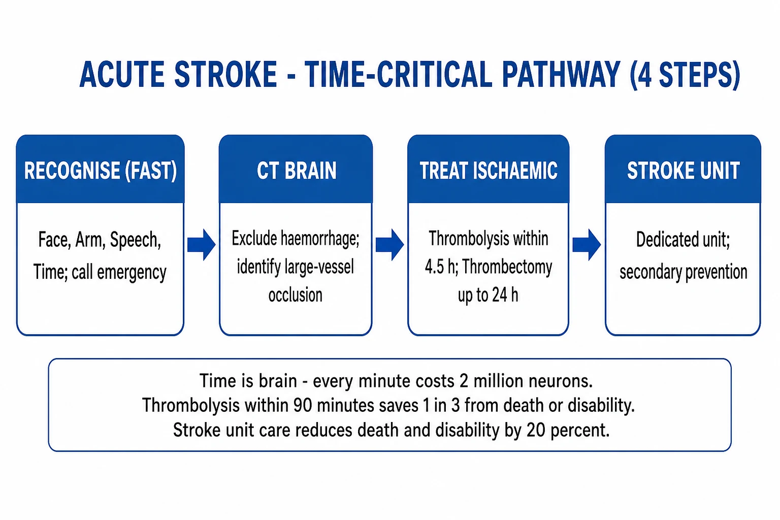
Acute ischaemic stroke protocol
Rapid assessment (door-to-needle <45 min)
ABC, glucose (hypoglycaemia mimics stroke), NIHSS score, last known well time. Non-contrast CT brain (exclude haemorrhage). CT angiography (identify LVO). CT perfusion (if >4.5h for thrombectomy selection).
Thrombolysis (if within 4.5h and no contraindication)
Alteplase 0.9 mg/kg IV (max 90 mg): 10% bolus over 1 min, rest over 60 min. OR Tenecteplase 0.25 mg/kg IV bolus (simpler, non-inferior — EXTEND-IA TNK). PREREQUISITE: BP <185/110 (lower with labatelol if needed). CONTRAINDICATIONS: haemorrhage on CT, recent surgery/trauma, active bleeding, INR >1.7, platelets <100k, recent intracranial haemorrhage.<Cite id="1" />}
Mechanical thrombectomy (if LVO and favourable imaging)
For large vessel occlusion (ICA, M1, M2, basilar). Up to 6h from onset (standard). Up to 24h if CT perfusion shows favourable penumbra (DAWN: up to 24h; DEFUSE 3: 6-16h). Gold standard: stent retriever or aspiration. Requires interventional neuroradiology.<Cite id="2" /><Cite id="3" />}
BP management
BEFORE thrombolysis: SBP <185/110 (labatalol, nicardipine). AFTER thrombolysis: SBP <180/105 for 24h (monitor every 15 min). WITHOUT thrombolysis: permissive hypertension (SBP <220) — maintain cerebral perfusion to penumbra. Do NOT lower BP rapidly unless extreme (>220).
Antiplatelet therapy
Aspirin 300 mg within 24h (after thrombolysis — give 24h AFTER alteplase, not before). If minor stroke (NIHSS <3) or TIA: dual antiplatelet (aspirin + clopidogrel) for 21 days (CHANCE/POINT trials). Then single antiplatelet long-term.
Secondary prevention
Statin (atorvastatin 80 mg). Carotid imaging (if anterior circulation stroke — CEA if >70% stenosis). AF detection (monitor rhythm 24h, or prolonged monitoring/implantable loop). Anticoagulation if AF (but NOT acutely — wait 2-14 days depending on severity).
Thrombolysis criteria — inclusion, exclusion, and dosing
Alteplase (recombinant tissue plasminogen activator, rt-PA) is the cornerstone of medical reperfusion. The evidence base spans three decades: NINDS (1995) established efficacy within 3h, ECASS III (2008) extended the window to 4.5h, and EXTEND (2019) showed benefit beyond 4.5h when selected by perfusion imaging.[5][6][12]
Alteplase (rt-PA) thrombolysis — inclusion and exclusion criteria
INCLUSION
who to lyse
- Age ≥18 years
- Diagnosis of ischaemic stroke causing measurable neurological deficit (NIHSS >0)
- Onset (last known well) within 4.5 HOURS
- Haemorrhage excluded on NCCT
ABSOLUTE EXCLUSION
do NOT lyse
- Intracerebral haemorrhage on CT (current or prior)
- Subarachnoid haemorrhage (SAH) — even suspected
- Active internal bleeding (e.g. GI bleed <21 days)
- Recent intracranial / intraspinal surgery (<3 months)
- Significant head trauma or prior stroke <3 months
- Platelets <100 × 10⁹/L; INR >1.7; aPTT >40 s (on anticoagulant)
- Glucose <2.7 mmol/L (treat first — may resolve the deficit)
RELATIVE EXCLUSION
individualise
- BP >185/110 — LOWER first with labetalol/nicardipine; lyse only if reduced
- Major surgery / non-procedure bleeding <14 days
- Recent arterial puncture at non-compressible site (<7 days)
- Seizure at onset (post-ictal deficit possible — but reasonable to lyse if residual deficit clearly stroke)
- Large infarct: early hypodensity >1/3 MCA territory on CT
- Pregnancy — weigh risk/benefit; not an absolute contraindication
- Age >80, severe stroke (NIHSS >25), on oral anticoagulant regardless of INR — these are caveats in the 4.5–9h window (ECASS III excluded >80s)
Dosing (alteplase): 0.9 mg/kg (maximum 90 mg) — give 10% as an IV bolus over 1 minute, then the remaining 90% as an IV infusion over 60 minutes. No heparin or antiplatelet for 24h afterwards; repeat CT at 24h to exclude asymptomatic haemorrhagic transformation before starting aspirin. Tenecteplase (0.25 mg/kg single IV bolus, max 25 mg) is increasingly preferred — it is a genetically modified variant with longer half-life, higher fibrin specificity, and a single-bolus administration; EXTEND-IA TNK confirmed non-inferiority to alteplase before thrombectomy.[11]
Key exclusion exam points: the four haemorrhage risk drivers are (1) recent surgery/trauma, (2) prior ICH, (3) uncontrolled BP >185/110, and (4) coagulopathy (INR >1.7, on anticoagulant). These are the ones most commonly tested and most frequently the reason thrombolysis is withheld. [1]
Mechanical thrombectomy — the extended window
Mechanical thrombectomy with a stent retriever or aspiration catheter is the greatest advance in acute stroke care in 25 years. The landmark 2015 thrombectomy trials (MR CLEAN, ESCAPE, SWIFT PRIME) established benefit within 6h of onset for LVO; DAWN and DEFUSE-3 extended the window to 24h with perfusion imaging.[7][8][9][2][3]
Thrombectomy windows — standard vs extended
Standard window (0–6h)
all 5 trials 2015
- LVO of anterior circulation (ICA terminus, M1, proximal M2)
- No upper age limit (within trial range); pre-stroke independent
- Imaging: NCCT (ASPECTS ≥6) + CTA showing LVO
- Benefit robust: NNT ~2.6 for functional independence (SWIFT PRIME)
Extended window (6–24h) — DAWN
imaging-selected
- 6–24h from last known well
- Clinical-imaging mismatch (deficit large, core small): core volume cut-offs by age (≥80 = <21 mL; <80 = <31 mL)
- Functional independence 49% vs 13% (NNT ~3)
- Cite: DAWN, Nogueira 2018 NEJM
Extended window (6–16h) — DEFUSE-3
perfusion mismatch
- 6–16h from last known well
- Target mismatch on CTP: core <70 mL AND mismatch ratio ≥1.8 AND mismatch volume ≥15 mL
- Stopped early for efficacy — mortality and disability both reduced
- Cite: DEFUSE-3, Albers 2018 NEJM
Practical rule: every suspected LVO stroke gets CTA at the same time as NCCT. If the LVO is identified and the patient is within 6h, proceed to thrombectomy regardless of perfusion. If beyond 6h, add CTP and apply DAWN/DEFUSE-3 criteria. Do not exclude a patient from thrombectomy purely on time up to 24h — imaging, not the clock, decides the extended window.[2][3]
Intracerebral haemorrhage (ICH) management
Intracerebral haemorrhage protocol
BP control — SBP target <140
INTERACT2 trial: intensive BP reduction (SBP <140 within 1h) improved functional outcome. Use IV nicardipine (5-15 mg/h) or labetalol. Avoid rapid reduction below 130 (may worsen perfusion). Target SBP 130-140.<Cite id="4" />}
Reverse anticoagulation
If on warfarin: stop, give vitamin K 10 mg IV + prothrombin complex concentrate (PCC 25-50 IU/kg — faster than FFP). If on DOAC: give specific antidote (idarucizumab for dabigatran, andexanet alfa for apixaban/rivaroxaban) OR PCC if antidote unavailable. Check coagulation.
ICP management
Head elevation 30 degrees. Maintain normocapnia, avoid hypoxia. Hyperosmolar therapy (3% NaCl or mannitol) if signs of raised ICP. Ventilate if GCS <8.
Surgical evacuation (selective)
CEREBELLAR haemorrhage >3 cm or with brainstem compression/hydrocephalus → urgent surgical evacuation (life-saving). Lobar supratentorial haemorrhage: consider craniotomy if deteriorating (STICH II). Deep (basal ganglia/thalamic) haemorrhage: surgery generally NOT beneficial.
Identify cause
Hypertension (#1 cause of deep ICH — basal ganglia, thalamus, pons, cerebellum). Cerebral amyloid angiopathy (#1 cause of lobar ICH in elderly). AVM, aneurysm, tumour, anticoagulation. CT angiography to exclude underlying vascular lesion (especially if atypical location or young patient).
Haemorrhagic stroke — deeper detail
The ICH score (0–6) is the validated prognostic score:[13]
| Component | 0 points | 1 point | 2 points |
|---|---|---|---|
| GCS | 13–15 | 5–12 | 3–4 |
| Volume (ABC/2) | <30 mL | ≥30 mL | — |
| Intraventricular extension | no | yes | — |
| Infratentorial origin | no | yes | — |
| Age | <80 | ≥80 | — |
30-day mortality by score: 0 → ~0%; 1 → ~13%; 2 → ~26%; 3 → ~72%; 4 → ~97%; 5+ → ~100%. Use it to counsel families and frame goals of care — but never to withdraw care in isolation. [1]
ATACH-2 (2016) compared SBP 110–139 vs 140–179 and found no additional benefit from ultra-intensive lowering, plus more renal adverse events — so the target is SBP 130–140, not lower.[10]
STICH II (2013, Mendelow): supratentorial lobar haemorrhage — no routine benefit from early surgery; a subset of superficial (<1 cm from cortex) lobar haematomas may benefit. Cerebellar haemorrhage is the exception — surgery is life-saving and must not be delayed.[14]
ICU management principles (both types)
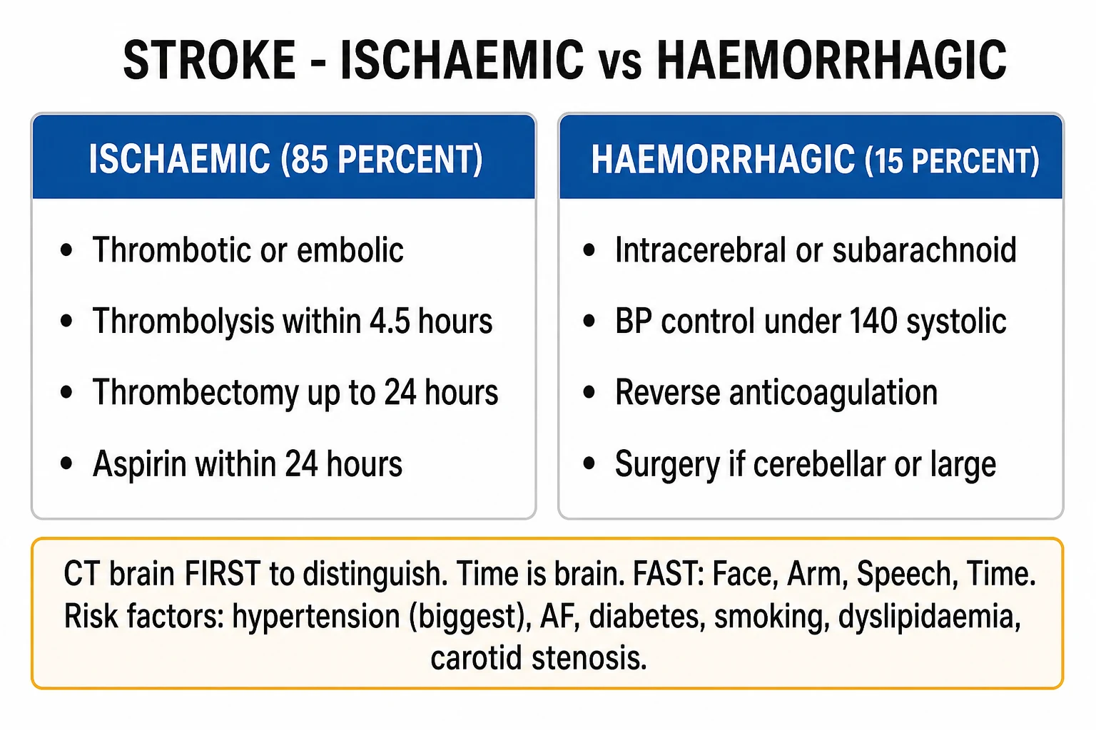
Ischaemic stroke
Maintain perfusion
- Permissive hypertension (SBP <220 without thrombolysis, <180/105 with)
- Do NOT lower BP unless extreme or for thrombolysis
- Glucose 6-10 mmol/L (avoid hypoglycaemia — worsens brain injury)
- Temperature <37.5C (fever worsens outcome — paracetamol, cooling)
- Swallow assessment before ANY oral intake (aspiration risk)
- DVT prophylaxis (enoxaparin after 24-48h)
Haemorrhagic stroke
Stop bleeding
- BP reduction SBP <140 (INTERACT2)
- Reverse anticoagulation immediately
- ICP management (head up, hyperosmolar therapy)
- Monitor for haematoma expansion (repeat CT if deteriorating)
- Surgery for cerebellar haemorrhage >3 cm
- DVT prophylaxis (after 48h, mechanical initially)
Blood pressure management — a unified view
BP is the one physiological variable managed oppositely in the two stroke types. Getting it wrong — by reflexively lowering an ischaemic stroke patient's BP, or by tolerating hypertension in ICH — is a common and avoidable cause of deterioration. [1]
Blood pressure targets by stroke type and phase
Ischaemic — NOT thrombolysed
permissive HTN
- Target: SBP ≤220 / DBP ≤120
- Rationale: collateral flow to the penumbra is pressure-dependent — lowering BP infarcts the penumbra
- Lower only if >220 (or organ damage — aortic dissection, ACS, severe pulmonary oedema)
- Use labetalol or nicardipine if treatment needed; avoid abrupt drops
Ischaemic — THROMBOLYSED
tight before & after
- BEFORE alteplase: SBP <185 AND DBP <110 (lower with labetalol 10–20 mg IV q10min, or nicardipine infusion)
- DURING + 24h AFTER: SBP <180 / DBP <105 — BP every 15 min for 2h, then 30 min for 6h, then hourly
- If BP exceeds limit during infusion → STOP alteplase, lower BP, repeat CT (rule out haemorrhagic transformation)
- Rationale: tight control prevents the main fatal complication of thrombolysis — symptomatic ICH
Intracerebral haemorrhage
stop expansion
- Target: SBP 130–140 within 1h (INTERACT2)
- Rationale: high BP drives haematoma expansion in first 24h — the main determinant of poor outcome
- Agents: IV nicardipine (5–15 mg/h), labetalol, clevidipine; AVOID nitroprusside (cerebral vasodilation → raised ICP)
- Do NOT go below 130 (ATACH-2 — no extra benefit, more renal injury)
Choice of antihypertensive: labetalol (combined α/β blocker, minimal cerebral vasodilation) and nicardipine/clevidipine (titratable calcium channel blockers) are first-line. Avoid hydralazine (reflex tachycardia), nitroglycerin (cerebral vasodilation, venodilation), and nitroprusside (raises ICP, cyanide toxicity). These are classic exam questions. [1]
Post-stroke complications
Stroke patients deteriorate more often from complications than from the index event. Anticipating and preventing them is the core of neurocritical care. [1]
Post-stroke complications — recognise and manage
Cerebral oedema (24–72h peak)
Cytotoxic oedema peaks at 24–72h. MALIGNANT MCA INFARCT: complete M1 occlusion with poor collaterals → >2/3 MCA territory infarcts and swells → midline shift → uncal herniation (mortality 80% untreated). MANAGEMENT: head up 30°, normocapnia, normothermia, hypertonic saline/ mannitol, and — for age <60, NIHSS >15, within 48h — DECOMPRESSIVE HEMICRANIECTOMY (DESTINY/DECIMAL/HAMLET: mortality 22% vs 71%).
Haemorrhagic transformation
Ischaemic infarct bleeds, either spontaneously (reperfusion of necrotic vessel) or after thrombolysis/thrombectomy. Asymptomatic petechial change is common and harmless; SYMPTOMATIC parenchymal haematoma (sudden GCS drop, NIHSS rise ≥4) is the feared thrombolysis complication (~2–6%). Management: stop alteplase, give cryoprecipitate/fibrinogen, lower BP, repeat CT, neurosurgical consult if evacuable.
Seizures (early <7d and late)
Early post-stroke seizures in ~5–10% (more with cortical, haemorrhagic, or large infarcts). May present as subtle twitching or coma (non-convulsive — consider cEEG if unexplained depressed consciousness). Treat with levetiracetam. Late seizures (>7d) recur and warrant long-term antiepileptic therapy. Prophylactic AEDs are NOT recommended.
Aspiration pneumonia
Dysphagia affects ~50% of stroke patients → aspiration is the leading cause of death in the subacute phase. PREVENT: NOTHING BY MOUTH until bedside swallow test; NG/NJ feeding if failed; head up 30°; good oral hygiene (chlorhexidine). Treat aspiration pneumonitis with supportive care; aspiration PNEUMONIA with broad-spectrum antibiotics.
Venous thromboembolism
Immobilised stroke patients have high DVT/PE risk. PROPHYLAXIS: intermittent pneumatic compression initially; LMWH (enoxaparin 40 mg SC) once haemorrhage risk acceptable — 24–48h post-thrombolysis, 48h+ post-ICH. Graduated stockings alone are inadequate.
Fever and hyperglycaemia (secondary brain injury)
Both fever and hyperglycaemia worsen ischaemic injury by increasing metabolic demand in compromised tissue. Keep temperature <37.5°C (paracetamol ± cooling) and glucose 6–10 mmol/L (avoid insulin-induced hypoglycaemia — NICE-SUGAR showed harm from tight control).
Pressure injury, contractures, depression
Repositioning q2h, heel protectors, early mobilisation (when stable), physiotherapy/OT, swallow/communication therapy. Post-stroke depression affects ~30% — screen and treat (SSRIs). Early stroke-unit multidisciplinary care reduces death and disability (Cochrane, NNT ~20).
Antiplatelet and secondary prevention
Secondary prevention is the long game that determines whether the patient has another stroke. Start it early — within 24h for ischaemic stroke (after the 24h post-thrombolysis delay), tailored to mechanism. [1]
Secondary prevention — by stroke mechanism
Non-cardioembolic (large-vessel, small-vessel, cryptogenic)
Single antiplatelet long-term: aspirin 75–300 mg/day OR clopidogrel 75 mg/day OR aspirin-dipyridamole. For minor stroke (NIHSS ≤3) or high-risk TIA: SHORT-COURSE DAPT (aspirin + clopidogrel) for 21 days then monotherapy — CHANCE and POINT showed reduced recurrence; POINT warned that >21 days increases major haemorrhage. Atorvastatin 80 mg; BP to <130/80; diabetes control; smoking cessation.
Cardioembolic (atrial fibrillation)
DOAC (apixaban, rivaroxaban, dabigatran — preferred over warfarin, lower ICH risk) OR warfarin (mechanical valve, severe MS, antiphospholipid syndrome, INR 2–3). TIMING: do NOT anticoagulate in first 24h — start at 2–14 days depending on infarct size and haemorrhagic transformation risk (small infarct ~3d; large ~2 weeks). Use the 1-3-6-12 rule as a guide.
Carotid disease (anterior circulation)
Carotid duplex/CTA. Symptomatic stenosis ≥70% (NASCET): carotid endarterectomy (CEA) within 2 weeks for maximum benefit (NASCET/ECST). 50–69%: individualise. <50%: medical therapy only. Stenting (CAS) is an alternative for high surgical risk / radiotherapy-induced stenosis.
Patent foramen ovale (PFO) — cryptogenic stroke <60y
If cryptogenic stroke in patient <60 with PFO and no other cause: PFO closure (DEVICE trials — RESPECT, REDUCE, CLOSE) reduces recurrence vs medical therapy. Medical therapy alone if >60 or alternative mechanism identified.
Lifestyle and risk-factor modification
Smoking cessation, alcohol moderation, Mediterranean/DASH diet, regular exercise, weight loss. Control hypertension (the single biggest modifiable risk factor), diabetes (HbA1c target), lipids (LDL <1.8 mmol/L). These reduce recurrence by ~30–50%.
Exam practice
SAQ — The stroke fork: ischaemic reperfusion vs intracerebral haemorrhage
10 minutes · 10 marks
A 72-year-old woman on warfarin 5 mg daily for atrial fibrillation (INR today 3.1) is brought to the emergency department 2 hours after the sudden onset of a right hemiparesis and aphasia. Her NIHSS is 16, blood pressure 168/92, capillary glucose 7.3 mmol/L, GCS 13. The non-contrast CT brain shows a 35 mL left basal ganglia haemorrhage with intraventricular extension but no midline shift. You are the intensive care registrar.
SAQ — Wake-up stroke and the extended-window thrombectomy decision
10 minutes · 10 marks
A 58-year-old man is brought to the emergency department at 07:00 having woken at 06:30 with a dense left hemiparesis and a left homonymous hemianopia. He was last known well at 22:30 the previous evening. His NIHSS is 14, blood pressure 176/96, GCS 14. The non-contrast CT shows no haemorrhage with an ASPECTS of 9. The CT angiogram confirms a right M1 middle cerebral artery occlusion. He takes no anticoagulant.
Clinical pearls
Red flags
[1]Prognosis and evidence
Landmark stroke trials and outcomes
NINDS (1995, NEJM): alteplase within 3h → improved outcomes (31% vs 20% no disability). Established thrombolysis.[5] ECASS III (2008, NEJM — Hacke): extended the thrombolysis window from 3h to 4.5h. The basis of the modern 4.5h limit.[6] EXTEND (2019, NEJM — Ma): thrombolysis beyond 4.5h (up to 9h) guided by perfusion imaging mismatch → improved functional outcome.[12] WAKE-UP (2018, NEJM — Thomalla): MRI-guided thrombolysis for unknown-onset stroke using DWI-FLAIR mismatch → improved outcome.[17] MR CLEAN (2015, NEJM — Berkhemer): first positive thrombectomy RCT — intra-arterial treatment within 6h for LVO.[7] ESCAPE (2015, NEJM — Goyal) and SWIFT PRIME (2015, NEJM — Saver): stent-retriever thrombectomy within 6h — large benefit, NNT ~2.6 for independence.[8][9] DAWN (2018, NEJM — Nogueira): thrombectomy 6–24h with deficit-infarct mismatch → functional independence 49% vs 13% (NNT ~3).[2] DEFUSE-3 (2018, NEJM — Albers): thrombectomy 6–16h with perfusion mismatch (core <70 mL, mismatch ratio ≥1.8) → improved outcome.[3] INTERACT2 (2013, NEJM — Anderson): ICH intensive BP (SBP <140) → favourable shift in functional outcome (OR 0.87).[4] ATACH-2 (2016, NEJM — Qureshi): SBP 110–139 vs 140–179 in ICH — no added benefit, more renal adverse events (target 130–140, not lower).[10] EXTEND-IA TNK (2018, NEJM — Campbell): tenecteplase 0.25 mg/kg non-inferior to alteplase before thrombectomy — simpler bolus, now preferred.[11] STICH II (2013, Lancet — Mendelow): supratentorial lobar ICH — no routine benefit from early surgery.[14] CHANCE (2013, NEJM — Wang) and POINT (2018, NEJM — Johnston): 21-day DAPT for minor stroke/high-risk TIA reduces recurrence; prolonged DAPT increases bleeding.[15][16] Hemphill ICH Score (2001, Stroke): validated 0–6 prognostic score — score 4+ mortality >90%.[13] Cochrane (Stroke Unit Trialists): organised stroke-unit care reduces death, death-or-disability, and death-or-institutionalisation (NNT ~20) — independent of age, sex, severity. Mortality (30-day): ischaemic 10–15%; ICH 35–50%; SAH 30–40%.
Pharmacology quick-reference
Acute stroke pharmacology — doses and cautions
Alteplase (rt-PA)
fibrinolytic
- Dose: 0.9 mg/kg IV (max 90 mg) — 10% bolus over 1 min + 90% infusion over 60 min
- Indication: ischaemic stroke within 4.5h (or imaging-selected beyond)
- Caution: symptomatic ICH 2–6%; oropharyngeal angio-oedema (esp. with ACE-inhibitor)
- Reversal (if bleed): stop infusion, cryoprecipitate to fibrinogen >1.5 g/L ± tranexamic acid
Tenecteplase
modified rt-PA
- Dose: 0.25 mg/kg IV bolus (max 25 mg) — SINGLE dose
- Advantage: longer half-life, higher fibrin specificity, bolus only — ideal for transfer
- EXTEND-IA TNK: non-inferior to alteplase before thrombectomy; now preferred first-line in many centres
Labetalol
α/β-blocker — BP
- Dose: 10–20 mg IV bolus q10min (max 300 mg), or 2–8 mg/min infusion
- First-line for BP control in stroke (minimal cerebral vasodilation, no rise in ICP)
- Cautions: bradycardia, heart block, LV failure, severe asthma
Nicardipine / Clevidipine
IV CCB — BP
- Nicardipine: 5–15 mg/h IV infusion (titratable, on in 5–10 min)
- Clevidipine: 1–6 mg/h (ultra-short acting, ester-hydrolysed — useful if rapid titration needed)
- First-line for ICH SBP <140 control; reflex tachycardia minor
Prothrombin complex concentrate (PCC)
reversal — warfarin
- Dose: 25–50 IU/kg IV (4-factor PCC) — reverses INR in minutes
- Plus vitamin K 10 mg IV slow (maintains reversal — PCC half-life short)
- Faster and more effective than FFP for warfarin-associated ICH; preferred
Idarucizumab / Andexanet alfa
reversal — DOAC
- Idarucizumab 5 g IV: specific reversal for dabigatran
- Andexanet alfa: reversal for apixaban/rivaroxaban (factor Xa inhibitors)
- If antidote unavailable: 4-factor PCC 50 IU/kg is a reasonable alternative
Prognosis summary
- Ischaemic stroke: 30-day mortality 10–15%; 30–50% of survivors have residual disability. Best predictors: NIHSS at admission (higher = worse; >20 poor), successful recanalisation (thrombolysis/thrombectomy salvages penumbra), age and comorbidity, time to treatment.
- ICH: 30-day mortality 35–50% (highest of all strokes). Best predictors: ICH score (score 4+ >90% mortality), haematoma volume (>30 mL worse), intraventricular extension (worse), GCS (lower worse), haematoma expansion (the modifiable one — drives aggressive BP control).[13]
- Functional recovery: most recovery in first 3 months; plateaus by 6 months. Stroke-unit care, early rehabilitation, and aggressive secondary prevention maximise independence.
References
- [1]Powers WJ, Rabinstein AA, Ackerson T, et al. Guidelines for the Early Management of Patients With Acute Ischemic Stroke: 2019 Update to the 2018 Guidelines for the Early Management of Acute Ischemic Stroke: A Guideline for Healthcare Professionals From the American Heart Association/American Stroke Association Stroke, 2019.PMID 31662037
- [2]Nogueira RG, Jadhav AP, Haussen DC, et al. Thrombectomy 6 to 24 Hours after Stroke with a Mismatch between Deficit and Infarct N Engl J Med, 2018.PMID 29129157
- [3]Albers GW, Marks MP, Kemp S, et al. Thrombectomy for Stroke at 6 to 16 Hours with Selection by Perfusion Imaging N Engl J Med, 2018.PMID 29364767
- [4]Anderson CS, Heeley E, Huang Y, et al. Rapid blood-pressure lowering in patients with acute intracerebral hemorrhage N Engl J Med, 2013.PMID 23713578
- [5]The National Institute of Neurological Disorders and Stroke rt-PA Stroke Study Group Tissue plasminogen activator for acute ischemic stroke N Engl J Med, 1995.PMID 7477192
- [6]Hacke W, Kaste M, Bluhmki E, et al. Thrombolysis with alteplase 3 to 4.5 hours after acute ischemic stroke N Engl J Med, 2008.PMID 18815396
- [7]Berkhemer OA, Fransen PSS, Beumer D, et al. A randomized trial of intraarterial treatment for acute ischemic stroke N Engl J Med, 2015.PMID 25517348
- [8]Saver JL, Goyal M, Bonafe A, et al. Stent-retriever thrombectomy after intravenous t-PA vs. t-PA alone in stroke N Engl J Med, 2015.PMID 25882376
- [9]Goyal M, Demchuk AM, Menon BK, et al. Randomized assessment of rapid endovascular treatment of ischemic stroke N Engl J Med, 2015.PMID 25671798
- [10]Qureshi AI, Palesch YY, Barsan WG, et al. Intensive Blood-Pressure Lowering in Patients with Acute Cerebral Hemorrhage N Engl J Med, 2016.PMID 27276234
- [11]Campbell BCV, Mitchell PJ, Churilov L, et al. Tenecteplase versus Alteplase before Thrombectomy for Ischemic Stroke N Engl J Med, 2018.PMID 29694815
- [12]Ma H, Campbell BCV, Parsons MW, et al. Thrombolysis Guided by Perfusion Imaging up to 9 Hours after Onset of Stroke N Engl J Med, 2019.PMID 31067369
- [13]Hemphill JC 3rd, Bonovich DC, Besmertis L, et al. The ICH score: a simple, reliable grading scale for intracerebral hemorrhage Stroke, 2001.PMID 11283388
- [14]Mendelow AD, Gregson BA, Rowan EN, et al. Early surgery versus initial conservative treatment in patients with spontaneous supratentorial lobar intracerebral haematomas (STICH II): a randomised trial Lancet, 2013.PMID 23726393
- [15]Wang Y, Wang Y, Zhao X, et al. Clopidogrel with aspirin in acute minor stroke or transient ischemic attack N Engl J Med, 2013.PMID 23803136
- [16]Johnston SC, Easton JD, Farrant M, et al. Clopidogrel and Aspirin in Acute Ischemic Stroke and High-Risk TIA N Engl J Med, 2018.PMID 29766750
- [17]Thomalla G, Simonsen CZ, Boutitie F, et al. MRI-Guided Thrombolysis for Stroke with Unknown Time of Onset N Engl J Med, 2018.PMID 29766770