ICU · Neurocritical care / monitoring
Brain Tissue Oxygen (PbtO2) & Multimodality Neuromonitoring
Also known as Brain tissue oxygen · PbtO2 · Licox · Multimodality monitoring · Cerebral microdialysis · Jugular venous oxygen saturation · SjvO2 · Pressure reactivity index · PRx · BOOST trial · Lactate-pyruvate ratio
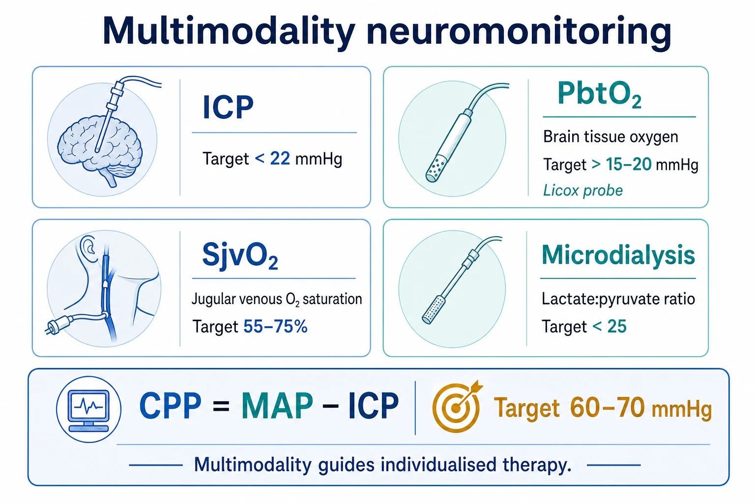
No single neuromonitor is sufficient. The ICP monitoring alone misses the brain tissue hypoxia (the PbtO2 can be low even when the ICP is normal). Multimodality neuromonitoring integrates the ICP, the PbtO2 (brain tissue oxygen, measured by the Licox probe; normal 20-40 mmHg, target above 15-20 mmHg), the SjvO2 (the jugular venous oxygen saturation, 55-75 per cent; below 50 is the cerebral hypoxia), the cerebral microdialysis (the lactate-pyruvate ratio above 25 indicates the ischaemia or the metabolic crisis), the continuous EEG (for the non-convulsive status), the transcranial Doppler (the Lindegaard ratio for the vasospasm), and the PRx (the pressure reactivity index for the optimal CPP). The PbtO2-guided therapy (the BOOST-2 trial) targets a PbtO2 above 20 mmHg in addition to the ICP and the CPP, reducing the brain tissue hypoxia. The PRx (the pressure reactivity index) identifies the optimal CPP — the MAP at which the cerebral autoregulation is best preserved.
On this page & tools
Your progress
Saved locally on this device.
Target exams
Red flags
Overview & definition
Multimodality neuromonitoring integrates several monitors to provide a comprehensive picture of the cerebral pathophysiology in the severely brain-injured patient. No single monitor is sufficient — the ICP alone misses the brain tissue hypoxia, the metabolic crisis, and the non-convulsive seizures. The multimodality approach (ICP + PbtO2 + SjvO2 + microdialysis + continuous EEG + TCD) guides the individualised therapy.[1][2]
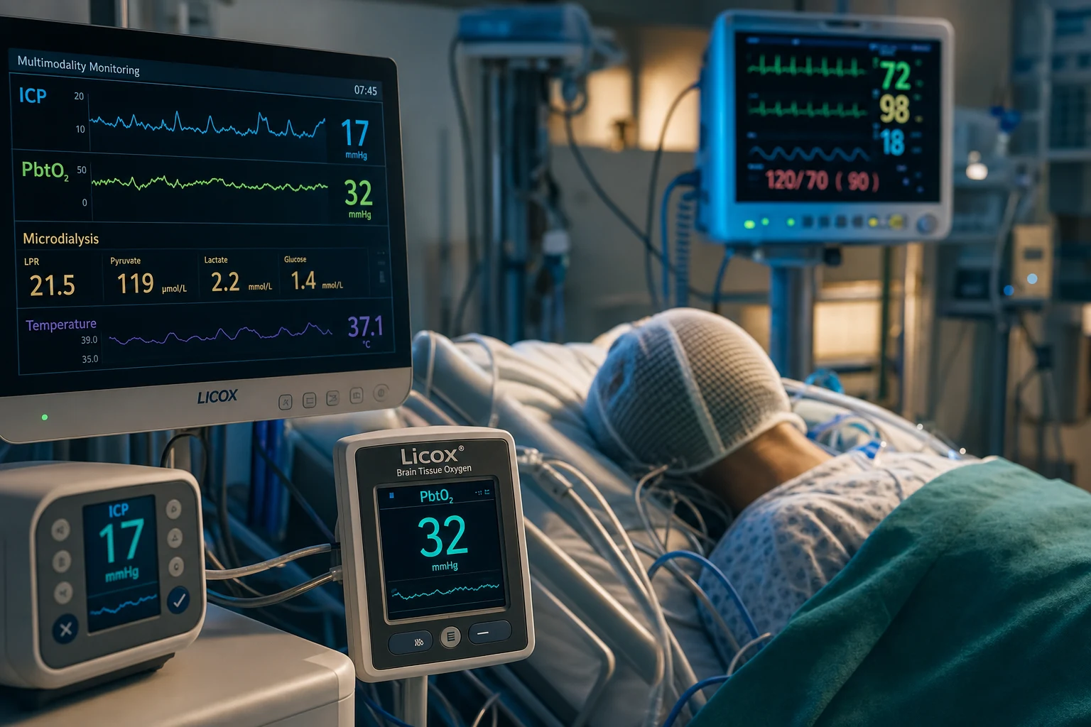
ICP monitoring — the foundation: EVD vs intraparenchymal (Codman / Spiegelberg)
The ICP monitoring is the standard of care for the severe traumatic brain injury (the GCS 3-8 with an abnormal CT, or a normal CT with two or more of: the age above 40, the motor posturing, the systolic BP below 90). The Brain Trauma Foundation target: the ICP below 22 mmHg, the CPP 60-70 mmHg (the CPP = the MAP minus the ICP).[1][1]
There are two families of the intracranial pressure device, and the choice is a favourite viva question. [1]
The external ventricular drain (EVD) — the gold standard
A catheter placed in the frontal horn of the lateral ventricle, connected to an external pressure transducer (referenced to the foramen of Monro — the external auditory meatus / the tragus). It measures the true ventricular CSF pressure, AND — uniquely among the ICP devices — it can drain the CSF (the single fastest bedside way to lower the ICP; opening the drain and letting 5-10 mL of the CSF out drops a critical ICP within seconds). It also provides the full pressure waveform (the P1/P2/P3 and the Lundberg waves). The trade-off is the infection — the ventriculitis in 5-15 per cent — which is why the aseptic insertion, the prophylactic antibiotics, the tunnelled and the antibiotic-impregnated catheters, and the minimising of the breaks into the system are mandatory.[1][8]
The intraparenchymal probe (Codman / Spiegelberg / Camino / Raumedic) — the workhorse
A miniature sensor advanced into the brain parenchyma (usually the right frontal lobe) via a bedside twist-drill bolt. The Codman Microsensor is a piezoelectric strain-gauge; the Spiegelberg uses an air-pouch pressure transducer (zeroed in situ, which mitigates the drift); the Camino / Raumedic use the fiberoptic technology. These are easier to place (especially in the slit ventricles of the diffuse brain swelling), have a lower infection rate (~1-2 per cent), and suffer only a minor zero-drift (1-3 mmHg per week). The trade-off: they cannot drain the CSF (the diagnostic-only device), and most provide a numeric mean and a trend rather than the true waveform.[8][1]
The ICP monitoring devices — the gold standard vs the workhorse
| Feature | External ventricular drain (EVD) | Intraparenchymal probe (Codman / Spiegelberg / Camino) |
|---|---|---|
| Site | The frontal horn of the lateral ventricle | The parenchymal sensor (usually the right frontal) |
| Accuracy | GOLD STANDARD — the true ventricular CSF pressure | Good; the minor zero-drift (1-3 mmHg/week); the Spiegelberg auto-zeroes in situ |
| Therapeutic? | YES — drains the CSF (the fastest bedside way to lower the ICP) | NO — the diagnostic only |
| Infection risk | HIGHEST — the ventriculitis 5-15 per cent | LOWER — ~1-2 per cent |
| Ease of placement | Harder — requires the ventricular puncture (the slit ventricles difficult) | Easier — the bedside twist-drill |
| Waveform | The full waveform (the P1/P2/P3, the Lundberg waves) | The numeric mean and the trend (no true waveform on most) |
| Best for | The hydrocephalus / the large ventricles; the need to drain; the need for the waveform | The slit ventricles; the emergency; the monitoring only |
| Transducer reference | The foramen of Monro (the external auditory meatus / the tragus) | Zeroed at the insertion (the in-built) |
The ICP waveform — P1, P2, P3
The normal ICP trace shows three peaks per the cardiac cycle:[8]
- P1 (the percussion wave) — the arterial pressure transmitted from the choroid plexus; tall and fairly constant.
- P2 (the tidal wave) — reflects the intracranial compliance; the key compliance-dependent wave.
- P3 (the respiratory wave) — the venous/respiratory fluctuation.
The P2 above the P1 = the exhausted intracranial compliance. As the compliance is lost, the P2 wave rises until it overtakes the P1 — a premonitory sign that often precedes the numeric mean ICP crossing 22 mmHg. A rising P2 is the early warning to escalate the osmotherapy, to drain the CSF, or to re-image BEFORE the mean number deteriorates.[8]
The Lundberg ICP waves — the pathological trends
| Wave | The pattern | The significance |
|---|---|---|
| A (the plateau) | The sustained ICP above 50 mmHg for 5-20 min | The vasodilatory cascade — the low CPP drives the cerebral vasodilation, the increased intracranial blood volume, the raised ICP, the lower CPP, the vicious cycle. Ominous — the severely impaired autoregulation, the exhausted compliance; the poor outcome |
| B | The rhythmic 0.5-2/min oscillations of 20-50 mmHg | The impaired pressure autoregulation; the warning that the system is becoming unstable |
| C | The low-amplitude, the higher-frequency oscillations | Less clinically significant |
PbtO2 (brain tissue oxygen tension)
PbtO2 is the partial pressure of the oxygen dissolved in the brain interstitium — a focal measure of the tissue oxygenation around the probe (typically placed in the at-risk penumbra).[1]
The device: the Licox probe (a Clark / polarographic electrode) placed into the brain parenchyma via a bolt. It measures the PbtO2 continuously. Target: above 15-20 mmHg (below 15 is the brain tissue hypoxia; below 10 is severe).[1]
The relationship to CPP: the PbtO2 is dependent on the CPP (the perfusion pressure) and the PaO2 (the arterial oxygenation). A low CPP or a low PaO2 reduces the PbtO2. The PbtO2 is a more direct measure of the cerebral tissue oxygenation than the CPP alone — it measures what the tissue actually receives.[1]
The PbtO2-guided therapy (the BOOST trials): the BOOST-2 trial (Okonkwo, JAMA Neurology 2017) compared the PbtO2-guided therapy (target PbtO2 above 20, in addition to the ICP and the CPP) versus the ICP/CPP-guided therapy in the severe TBI. The PbtO2-guided group had a reduced risk of the brain tissue hypoxia and a trend toward a better neurological outcome. The BTF guidelines recommend the PbtO2 monitoring as an option in centres with the expertise, alongside the ICP/CPP monitoring.[3]
The Licox probe in depth
The Licox is a polarographic (Clark) oxygen sensor — the oxygen diffuses across a membrane and is reduced at a gold cathode, generating a current proportional to the tissue oxygen tension. The alternative is the Neurotrend (a fluorescence-based fiberoptic sensor, now discontinued but still examined). The probe is sited in the at-risk penumbra: the right frontal lobe in the diffuse injury, or the ipsilateral penumbra around a focal lesion (the contralateral, "better," hemisphere under-reads the global oxygenation). It needs a 30-60 minute run-in to equilibrate after the insertion.[2]
The thresholds:[3]
- 20-40 mmHg — the normal range (some texts cite 23-35).
- 15-20 — the borderline; treat if trending down.
- below 15 — the brain tissue hypoxia (intervene).
- below 10 — the critical (strongly associated with the death and the poor outcome).
What the PbtO2 actually reflects: it is NOT simply the arterial PaO2 — it is the composite of the cerebral blood flow, the arterial oxygen content (the haemoglobin × the saturation), and the cerebral metabolic rate. A low PbtO2 can therefore arise from the low CPP, the anaemia, the hypoxaemia, the vasospasm, OR the increased demand (the seizures, the fever).[2]
The therapeutic levers to raise the PbtO2 — and the trap
| Lever | The mechanism | The caution |
|---|---|---|
| ↑ the FiO2 / the PaO2 | The direct increase in the arterial oxygen content | The simplest first step; the high FiO2 for the prolonged periods has the pulmonary toxicity |
| ↑ the CPP (the noradrenaline → the higher MAP) | The raised cerebral blood flow to the ischaemic regions | Cap the CPP at 70 (above 70 — the ARDS risk, no added benefit) |
| ↓ the ICP (the osmotherapy, the CSF drainage) | Lowers the resistance to the cerebral perfusion | — |
| Transfuse if the Hb below 90 g/L | Increases the oxygen carrying capacity | Balance against the transfusion-related complications |
| ↓ the metabolic demand (the sedation, the anticonvulsants, the normothermia) | The less oxygen consumed → the higher residual tissue O2 | Treat any seizures on the cEEG aggressively |
| AVOID the hyperventilation | — | Lowers the ICP but VASOCONSTRICTS → worsens the PbtO2 (the Pyrrhic victory) |
BOOST-2 — Okonkwo 2017, Crit Care Med (PMID 29028696)
Study design
The Phase II multicentre RCT — 119 severe TBI patients across 10 US ICUs
Population
The severe TBI; randomised to the ICP + PbtO2-guided protocol vs the ICP-only-guided protocol
Intervention
The tiered management informed by the ICP + the brain tissue oxygen (the PbtO2) vs the ICP alone
Primary outcome
The burden of the brain tissue hypoxia — REDUCED from the proportion of time 0.45 (the ICP only) to 0.16 (the ICP + the PbtO2), p<0.0001
Key finding
The PbtO2-directed therapy reduced the brain tissue hypoxia with a TREND toward the lower mortality and the more favourable outcomes; the ICP control was similar in both arms; no procedure-related complications
Clinical bottom line
The multimodal ICP + PbtO2 monitoring is feasible, safe, and reduces the brain hypoxia — supports the integration paradigm and justified the Phase III BOOST-3 trial (NCT03754114). Not powered for efficacy
SjvO2 (jugular venous oxygen saturation)
A global measure of the cerebral oxygen extraction. A fiberoptic catheter is placed retrograde in the internal jugular vein, with the tip in the jugular bulb (above the entry of the facial vein, so the blood is from the brain, not the face).[1]
Target: 55-75 per cent.[1]
- Below 50 per cent (desaturation) = the brain is extracting more oxygen than is being delivered (from a low flow, a low PaO2, a severe anaemia, or a high metabolism — fever, seizures). Treat: raise the CPP, the PaO2, or the Hb; reduce the fever, the seizures.
- Above 85 per cent = the luxury perfusion (a hyperaemia from the lost autoregulation) or the impaired extraction (a severe brain injury where the cells cannot use the oxygen).[1]
The SjvO2 interpretation — the global oxygen balance
| The SjvO2 | The interpretation | The action |
|---|---|---|
| 55-75 per cent | NORMAL — the delivery matched to the consumption | Maintain |
| below 50 per cent | The cerebral hypoxia — the extraction exceeds the delivery | ↑ the CPP (the noradrenaline), ↑ the FiO2/PaO2, transfuse if anaemic, ↓ the ICP, AVOID the hyperventilation, treat the seizures |
| above 90 per cent | The hyperaemia / the luxury perfusion or the AV shunting | The loss of the autoregulation; modestly ↓ the MAP within the CPP-safe range, the head-up, consider the brief moderate hyperventilation |
| The AJDO2 (the arterio-jugular difference) | Normally ~5 vol% | The widening = the increased extraction (the ischaemia); the narrowing = the hyperaemia |
The limitation: the SjvO2 is a global average — a focal ischaemic region may be diluted out by the well-perfused tissue. It is technically demanding (the catheter position must be confirmed radiologically; the contamination with the extracranial blood misleads). Largely superseded by the PbtO2 in many centres but still valuable for the global oxygenation trends and as a check on the regional PbtO2 reading.[2][4]
Cerebral microdialysis
A semipermeable membrane catheter in the brain parenchyma that samples the interstitial fluid, providing real-time biochemical information about the tissue metabolism.[1]
- Lactate — high = the anaerobic metabolism (ischaemia or the mitochondrial dysfunction).
- Pyruvate — low = the impaired oxidative metabolism.
- The lactate-pyruvate ratio (LPR) — above 25 indicates a metabolic crisis (ischaemic or non-ischaemic — the mitochondria are failing). The most sensitive marker of the secondary brain injury.
- Glycerol — high = the cell membrane breakdown (the tissue necrosis).
- Glutamate — high = the excitotoxicity (the massive release in the ischaemia).
The microdialysis markers — what each one means
| The marker | Normal | Abnormal | The meaning |
|---|---|---|---|
| The lactate/pyruvate ratio (LPR) | below 25 | above 25 = concern; above 40 = the significant distress | The master marker. The high LPR + the LOW pyruvate = the ischaemia; the high LPR + the NORMAL pyruvate = the mitochondrial dysfunction |
| Glucose | 1-2 mmol/L | below 0.7 = the metabolic crisis | The substrate depletion / the hypoxia — the brain is starving |
| Glutamate | Low | Rising = the excitotoxicity | The excitotoxic amino acid released with the ischaemic cell stress → the NMDA-mediated calcium influx → the cell death |
| Glycerol | Low | Rising = the cell membrane breakdown | The phospholipid breakdown product — the membrane destruction (the severe ischaemia, the cell death) |
| Lactate | Variable | Rising in the ischaemia | The end-product of the anaerobic glycolysis |
Hutchinson 2015 — the International Microdialysis Forum consensus (Intensive Care Med, PMID 26194024)
Document type
The consensus statement from the 2014 International Microdialysis Forum (Cambridge, UK)
Scope
The technical standards, the catheter placement, the perfusion flow rate, the marker interpretation (the LPR, the glucose, the glutamate, the glycerol), the clinical applications in the TBI, the SAH and the anoxic injury
Key thresholds
The lactate/pyruvate ratio above 25-40 = the metabolic distress; the brain glucose below 0.7-1.0 mmol/L = the metabolic crisis; the rising glycerol = the cell membrane breakdown; the LPR is the most robust marker (independent of the recovery rate)
Clinical bottom line
The authoritative reference for the cerebral microdialysis interpretation. Use the LPR above 25-40 and the low glucose to detect the ischaemia/the energy failure before the structural damage — and to individualise the CPP/the PbtO2 targets
Continuous EEG (cEEG)
The purpose: to detect the non-convulsive status epilepticus (the NCSE), which is clinically silent in the sedated or the comatose patient but drives up the cerebral metabolic demand, raises the ICP, raises the LPR on the microdialysis, and worsens the outcome.[5]
The indication: 24-48 h of the cEEG for any comatose neuro-ICU patient (the TBI, the SAH, the ICH, the post-anoxic) with the unexplained depressed consciousness, the fluctuating examination, or the refractory intracranial hypertension.[5]
The NCSE is doubly dangerous during the targeted temperature management (TTM) and the barbiturate coma — the neuromuscular blockade MASKS the motor seizures, so the only way to know the brain is seizing is the EEG. The EEG is mandatory during the barbiturate coma (the target burst suppression, 3-5 bursts/min) both to titrate the infusion AND to detect the breakthrough seizures.[1]
The treatment of the NCSE: the benzodiazepine trial (the lorazepam/midazolam with the EEG response), load with the levetiracetam 60 mg/kg, add the valproate or the fosphenytoin; if refractory, the propofol or the midazolam infusion to the burst suppression, then wean once seizure-free on the cEEG.[5]
Transcranial Doppler (TCD) — the Lindegaard ratio for the vasospasm
The TCD measures the blood flow velocity in the major intracranial arteries (the middle cerebral artery via the temporal bone window) using the non-invasive Doppler ultrasound, first described by Aaslid in 1982.[11]
The key use in the neuro-ICU: the detection of the vasospasm after the subarachnoid haemorrhage (SAH). The cerebral vasospasm (the days 3-14 after the bleed) narrows the arterial lumen, and the blood flow velocity rises (the continuity principle — the same flow through a narrower tube moves faster).[10]
The Lindegaard ratio (Lindegaard 1989) is the ratio of the MCA velocity to the ipsilateral extracranial internal carotid artery velocity. It distinguishes the true vasospasm from the hyperaemia (both raise the MCA velocity):[10]
The Lindegaard ratio — the vasospasm vs the hyperaemia
| The Lindegaard ratio (the MCA / the ICA) | The interpretation |
|---|---|
| below 3 | The normal — no spasm |
| 3-6 | The mild-to-moderate vasospasm |
| above 6 | The severe vasospasm |
| The high MCA velocity + the low Lindegaard ratio (below 3) | The hyperaemia (the increased flow, not the spasm) — the MCA and the ICA velocities both rise together |
The other uses of the TCD:[11]
- The embolus detection (the high-intensity transient signals — the HITS).
- The diagnosis of the brain death — the reverberating (the to-and-fro) flow pattern.
- The assessment of the cerebral autoregulation — the TCD-based reactivity (the breath-holding, the CO2 challenge).
- The estimation of the ICP trends (the pulsatility index).
The PRx (the pressure reactivity index)
A moving correlation coefficient between the MAP and the ICP over a time window (typically 5 minutes).[1]
- PRx below 0 = the intact autoregulation (the ICP does not follow the MAP — the cerebral vasculature constricts and dilates appropriately; the pressure changes are buffered).
- PRx above 0.3 = the impaired autoregulation (the ICP follows the MAP — the vasculature is non-reactive; the pressure changes passively transmit to the brain).
- The optimal CPP (the CPPopt) is the CPP at which the PRx is at its lowest — the MAP at which the autoregulation is best preserved. Targeting the CPPopt (rather than a fixed 60-70) provides the individualised haemodynamic management.[1]
The autoregulation indices in depth
The pressure reactivity index (the PRx) is the moving Pearson correlation between the slow waves of the MAP and the ICP. When the autoregulation is intact, a rise in the MAP causes the vasoconstriction (to maintain the constant cerebral blood flow), which REDUCES the intracranial blood volume and LOWERS the ICP — hence the negative correlation (the PRx below 0). When the autoregulation is lost, the vessels are passive, and a rise in the MAP directly raises the ICP — hence the positive correlation (the PRx above 0).[9]
- The PRx — the correlation of the MAP and the ICP.
- The TOx — the correlation of the TCD flow velocity and the MAP (the TCD-based).
- The COx — the correlation of the NIRS and the MAP (the non-invasive).
- The LAx — the long pressure reactivity index (the longer time window).
The autoregulation indices — the intact vs the impaired
| The index | The intact autoregulation | The impaired autoregulation |
|---|---|---|
| The PRx (the MAP-ICP correlation) | below 0 (the negative) | above 0.3 (the positive) |
| The CPPopt | The CPP at the most negative PRx | The lowest PRx is still positive — no safe window |
| The clinical implication | The patient tolerates the higher CPP | The higher CPP causes the hyperaemia and the raised ICP |
Czosnyka 1997 — the pressure reactivity index (Neurosurgery, PMID 9218290)
Study design
The observational cohort — the continuous MAP and ICP recording in the head-injured patients
Key contribution
Defined the PRx — the moving correlation coefficient between the slow waves of the MAP and the ICP — as a continuous, bedside index of the cerebral pressure autoregulation
Key finding
The PRx below 0 = the intact autoregulation (the favourable outcome); the PRx above 0.3 = the impaired autoregulation (the worse outcome). The CPPopt — the CPP at the lowest PRx — individualises the haemodynamic target
Clinical bottom line
The foundational reference for the autoregulation-guided CPP. Targeting the CPPopt rather than a fixed 60-70 provides the individualised haemodynamic management — a discriminating point for the higher-scoring candidates
The integration — treat the PATIENT, not the NUMBER
How to integrate the ICP + the CPP + the PbtO2 + the microdialysis + the cEEG at the bedside
Read the SYSTEM, not the single number
Every assessment: the ICP + the waveform, the MAP → the CPP (= the MAP minus the ICP), the PbtO2, and (if available) the microdialysis LPR and the cEEG. Ask four questions each time: (1) Is there the INTRACRANIAL HYPERTENSION (the ICP above 22)? (2) Is there the ISCHAEMIA (the low PbtO2 below 15, the high LPR above 25, the low SjvO2 below 50)? (3) Are there the SEIZURES (the cEEG)? (4) Is there the DISTRESSED METABOLISM (the low glucose, the high glycerol)? Treat each abnormality.
Resolve the common conflicts
The high ICP + the low PbtO2: treat the ICP (the osmotherapy, the CSF drainage) but AVOID the hyperventilation (lowers the ICP but worsens the PbtO2 by the vasoconstriction) — prefer the hypertonic saline, the CSF drainage, the sedation. The normal ICP + the low PbtO2: the ischaemia is NOT pressure-driven — raise the MAP/the CPP (the noradrenaline to 60-70), increase the FiO2, transfuse if anaemic, treat the seizures, check for the vasospasm (the TCD). The high ICP + the high PbtO2: the possible hyperaemia (the loss of the autoregulation) — modestly lower the MAP within the CPP-safe range, the head-up, consider the brief moderate hyperventilation. The rising LPR with the normal ICP/the PbtO2: the impending mitochondrial/cellular failure — escalate the ischaemic work-up and optimise the delivery.
Individualise the targets
Use the autoregulation-guided CPP (the PRx) — set the CPP at the patient's optimal autoregulatory range rather than a fixed number. Treat the PATIENT'S thresholds: some patients herniate at the ICP 20; others tolerate 25. The TREND, the WAVEFORM (the P2 above the P1), and the DOWNSTREAM MARKERS (the PbtO2, the LPR) are more informative than any single cut-off.
Treat the whole brain, not just the pressure
The integration paradigm (the Le Roux 2014 consensus) holds that the ICP/the CPP/the PbtO2/the metabolism are INTERDEPENDENT — a management decision that lowers the ICP but worsens the PbtO2 (e.g. the aggressive hyperventilation) is a Pyrrhic victory. Optimise the integrated bundle: the normoxia, the normocapnia (the PaCO2 35-40), the CPP 60-70, the PbtO2 above 20, no seizures, no fever, no hyperglycaemia (6-10), no anaemia (the Hb above 80-90).
Confirm the reading is REAL before treating the artefact
A sudden ICP rise with the preserved normal waveform and no clinical change suggests a SYSTEM problem (the kinked line, the air in the transducer, the patient coughing/straining, the malposition). Flush (aseptically), re-level, re-zero, observe — over-treating an artefact with the osmotherapy or the hyperventilation causes the real harm. The same discipline applies to a wildly swinging PbtO2 (check the probe position) or a "flat" EEG (check the electrode impedances).
The integration pitfalls — the conflict matrix
| The scenario | The apparent "fix" | Why it is wrong | The correct approach |
|---|---|---|---|
| The high ICP + the low PbtO2 | Hyperventilate to the PaCO2 below 30 | The vasoconstriction lowers the ICP but STRANGLES the PbtO2 → the worse ischaemia | The hypertonic saline, the CSF drainage, the sedation; keep the PaCO2 35-40 |
| The normal ICP + the low PbtO2 | "The ICP is fine, no action" | The ICP is blind to the regional/diffuse ischaemia | Raise the MAP/the CPP, ↑ the FiO2, transfuse, treat the seizures, check the TCD for the spasm |
| The refractory "ICP" + the sedated patient | More osmotherapy | Could be the non-convulsive status driving the ICP up | The cEEG — treat the NCSE and the ICP may fall |
| The high ICP + the high PbtO2 | Push the CPP higher with the fluids | The loss of the autoregulation → the hyperaemia; the fluids → the ARDS | Modestly lower the MAP within the CPP-safe range, the head-up |
| The single high ICP number | Treat immediately | May be the coughing/straining/the artefact | Confirm the waveform + the trend before treating |
The management targets
The multimodal neuromonitoring-guided therapy
The ICP-guided therapy (the standard)
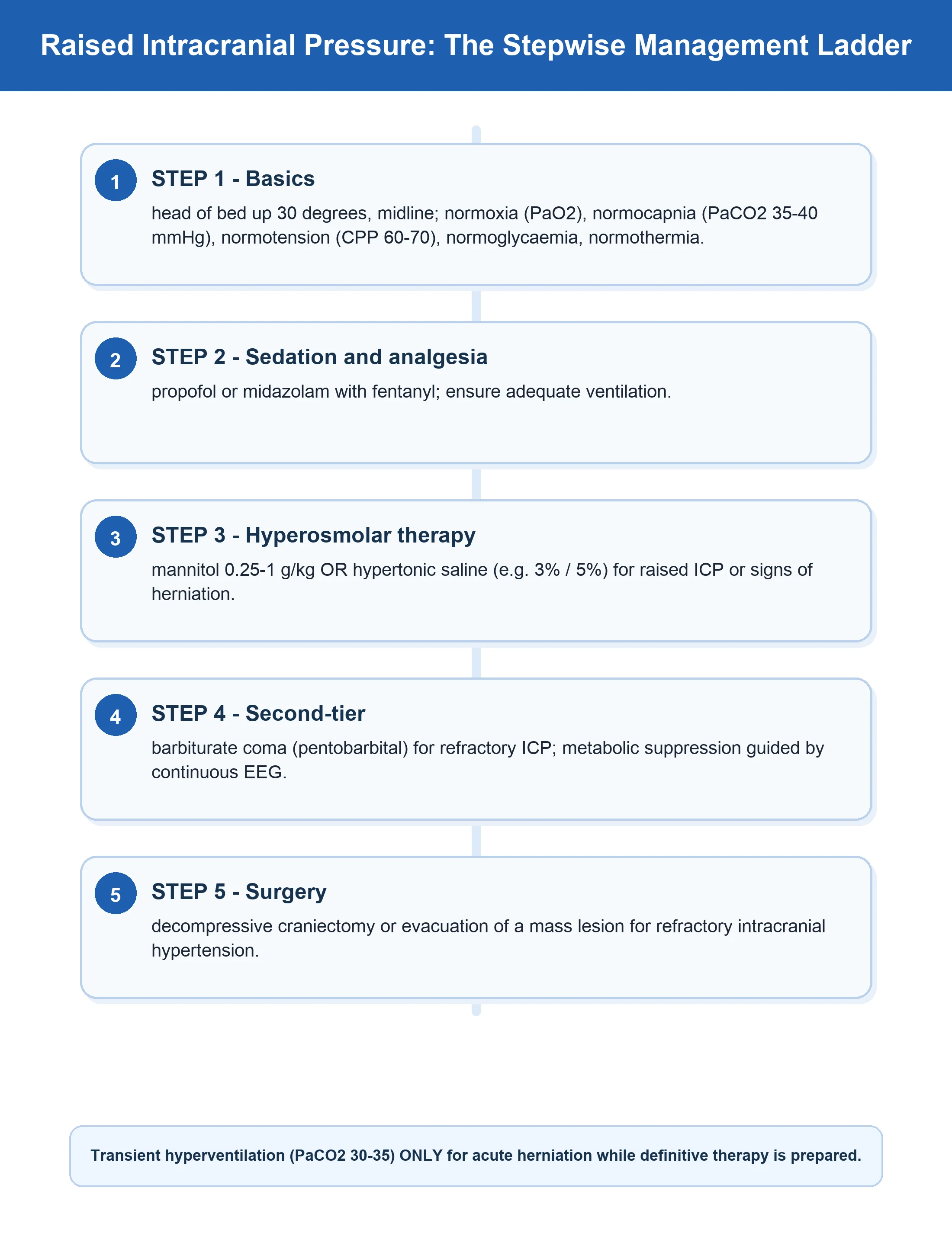
The ICP below 22 mmHg. The CPP 60-70 mmHg. The interventions: the head elevation 30°, the normocapnia (the PaCO2 35-40), the normoxia, the normoglycaemia, the normothermia, the sedation, the hyperosmolar therapy (the 3% NaCl, the mannitol), the barbiturate coma (the refractory), the decompressive craniectomy (the refractory).
The PbtO2-guided therapy (the additional, per the BOOST-2)
If the PbtO2 below 15 mmHg: (1) Increase the FiO2 (the target PaO2 above 80). (2) Increase the CPP (the noradrenaline to raise the MAP — improves the cerebral blood flow). (3) Reduce the ICP (the hyperosmolar therapy). (4) Transfuse if the Hb below 80 (improve the oxygen carrying capacity). (5) Reduce the metabolic demand (the sedation, the temperature control). The BOOST-2 trial: combining the ICP + the PbtO2 targets reduced the brain hypoxia and improved the functional outcomes.
The microdialysis-guided therapy
If the LPR above 25 (the ischaemia): check the ICP/the CPP/the PbtO2 (is the ischaemia global or regional?). Optimise the perfusion (the CPP, the vasopressors). If the low glucose: check the serum glucose (the target 6-10), ensure the adequate nutrition. If the high glutamate: consider the barbiturate coma (suppresses the metabolism). If the high glycerol: the poor prognosis (the cell death occurring).
The EEG-guided therapy
If the non-convulsive seizures/the NCSE detected: (1) Load with the levetiracetam 60 mg/kg IV. (2) If ongoing: the propofol infusion (the 3 mg/kg/h — suppresses the seizure activity). (3) If refractory: the midazolam infusion or the pentobarbital coma (the burst suppression on the EEG). (4) Continue the cEEG to confirm the seizure cessation. (5) Maintain the anticonvulsant for 7 days minimum, then wean if seizure-free.
The key trials and the evidence
Claassen 2004 — the cEEG detection of the non-convulsive seizures (Neurology, PMID 15159471)
Study design
The prospective observational cohort — 570 critically ill patients undergoing the continuous EEG monitoring (the Columbia University)
Population
The critically ill neuro-ICU and the general ICU patients with the altered mental status or the coma
Key finding
The electrographic seizures detected in 19 per cent (110/570); 92 per cent were exclusively NON-CONVULSIVE (no clinical signs). The coma was the strongest predictor (the OR 7.7). Most seizures (88 per cent) detected within the first 24 h, but the comatose patients often needed more than 24 h
Clinical bottom line
A comatose ICU patient can be in the continuous electrographic status with no motor signs. Monitor with the cEEG (24-48 h, longer if comatose), especially during the TTM, the barbiturate coma and the deep sedation when the paralysis masks the motor seizures
BEST:TRIP — Chesnut 2012, NEJM (PMID 23234472)
Study design
The multicentre, controlled RCT — 324 severe TBI patients (the GCS 3-8) in the Bolivia/Ecuador
Population
The severe TBI; randomised to the ICP-monitor-guided (the target ICP 20 or below) vs the imaging-and-clinical-examination-guided management
Key finding
NO significant difference in the composite outcome or the 6-month mortality (39% vs 41%) between the ICP-guided and a rigorous imaging-clinical-exam protocol
Clinical bottom line
Often misread as 'the ICP monitoring doesn\'t matter' — the correct interpretation is that the structured PROTOCOL drives the outcome as much as the monitor; the ICP monitoring remains the standard of care (needed for the sedated/unexaminable patient). The lesson reinforces the multimodal monitoring: the MONITOR is only as good as the PROTOCOL around it
Le Roux 2014 — the International Multidisciplinary Consensus on the Multimodality Monitoring (Intensive Care Med, PMID 25138226)
Document type
The international multidisciplinary consensus statement — the neurointensivists, the neurosurgeons, the neurologists
Scope
The integration of the ICP, the CPP, the PbtO2, the microdialysis, the SjvO2, the cEEG, the TCD and the autoregulation indices into a coherent monitoring strategy
Key recommendations
No single monitor is sufficient; the modalities are complementary; the thresholds are guides not absolutes; treat the integrated picture; the autoregulation-guided targets (the PRx) individualise the care
Clinical bottom line
The reference consensus for the integration paradigm — 'treat the patient, not the number.' Cite when justifying the multimodal monitoring beyond the ICP alone
Lindegaard 1989 — the TCD and the vasospasm (Acta Neurochir, PMID 2683600)
Study design
The observational study — the TCD blood velocity measurements and the angiography in the SAH patients
Key contribution
Defined the Lindegaard ratio — the MCA velocity divided by the ipsilateral extracranial ICA velocity — to distinguish the true vasospasm from the hyperaemia
Key thresholds
The ratio below 3 = the normal; 3-6 = the mild-to-moderate spasm; above 6 = the severe spasm
Clinical bottom line
The foundational reference for the non-invasive vasospasm surveillance after the SAH. Both the vasospasm and the hyperaemia raise the MCA velocity — the Lindegaard ratio is the discriminator
Exam practice
SAQ — Normal ICP, low PbtO2: the rationale for multimodality monitoring
10 minutes · 10 marks
A 24-year-old man is in the neurocritical care unit 48 hours after a severe traumatic brain injury (GCS 6 at the scene). He is intubated, sedated and on a norepinephrine infusion. He has an external ventricular drain, a Licox PbtO2 probe in the right frontal lobe, a jugular venous oximetry catheter, and a cerebral microdialysis catheter. His current readings are: ICP 14 mmHg, CPP 64 mmHg, PbtO2 11 mmHg, SjvO2 48 per cent, microdialysis lactate-pyruvate ratio 32, brain glucose 0.6 mmol/L.
SAQ — ICP monitoring devices and the waveform in severe TBI
10 minutes · 10 marks
A 45-year-old woman with a severe diffuse TBI (GCS 5) is admitted to the neurocritical care unit. The CT shows diffuse brain swelling with effaced basal cisterns but no surgical lesion. The neurosurgical team asks you which intracranial pressure monitoring device to insert and why. Her ventricles are slit-like on the CT.
Clinical pearls
Red flags
Prognosis
The multimodal prognostic markers in the severe TBI
| The marker | The threshold / the pattern | The prognostic implication |
|---|---|---|
| The ICP sustained above 22 mmHg | Refractory to the tiered therapy | The doubled mortality; each 10 mmHg above 20 worsens the outcome |
| The ICP above 40 mmHg (the severe) | Sustained | The very high mortality; the imminent herniation risk |
| The Lundberg A (the plateau) waves | The ICP above 50 mmHg for 5-20 min | The severely impaired autoregulation; the poor outcome |
| The CPP below 60 mmHg sustained | The ischaemic threshold | The secondary injury; the worse neurological outcome |
| The PbtO2 below 15 / below 10 mmHg | The brain tissue hypoxia burden | Strongly associated with the death and the unfavourable outcome (the BOOST-2) |
| The lactate-pyruvate ratio above 25-40 | The metabolic distress | The impending cellular energy failure; the worse outcome |
| The rising glycerol | The cell membrane breakdown | The cell death in progress; the grave prognosis |
| The brain glucose below 0.7 mmol/L | The metabolic crisis | The substrate depletion; the worse outcome |
| The SjvO2 below 50 per cent sustained | The global cerebral hypoxia | The increased extraction; the worse outcome |
| The cEEG: the non-convulsive status | The refractory NCSE | The independent predictor of the poor outcome (treatable if detected) |
| The loss of the pupil reactivity (the NPi below 3) | The automated pupillometry | The strong predictor of the unfavourable outcome; suggests the herniation |
| The PRx persistently above 0.3 | The impaired autoregulation | The loss of the pressure reactivity; the worse outcome |
The integrated bundle — the one-sentence exam answer
References
- [1]Carney N, Totten AM, O'Reilly C, et al. Guidelines for the Management of Severe Traumatic Brain Injury, Fourth Edition Neurosurgery, 2017.PMID 27654000
- [2]Oddo M, Villa F, Citerio G. Brain multimodality monitoring: an update Curr Opin Crit Care, 2012.PMID 22322259
- [3]Okonkwo DO, Shutter LA, Moore C, et al. Brain Oxygen Optimization in Severe Traumatic Brain Injury Phase-II: A Phase II Randomized Trial Crit Care Med, 2017.PMID 29028696
- [4]Le Roux P, Menon DK, Citerio G, et al. Consensus summary statement of the International Multidisciplinary Consensus Conference on Multimodality Monitoring in Neurocritical Care : a statement for healthcare professionals from the Neurocritical Care Society and the European Society of Intensive Care Medicine Intensive Care Med, 2014.PMID 25138226
- [5]Claassen J, Mayer SA, Kowalski RG, Emerson RG, Hirsch LJ. Detection of electrographic seizures with continuous EEG monitoring in critically ill patients Neurology, 2004.PMID 15159471
- [6]Hutchinson PJ, O'Connell MT, Al-Rawi PG, et al. Consensus statement from the 2014 International Microdialysis Forum Intensive Care Med, 2015.PMID 26194024
- [7]Chesnut RM, Temkin N, Carney N, et al. A trial of intracranial-pressure monitoring in traumatic brain injury N Engl J Med, 2012.PMID 23234472
- [8]Stocchetti N, Zoerle T, Carbonara M. Intracranial pressure management in patients with traumatic brain injury: an update Curr Opin Crit Care, 2017.PMID 28157822
- [9]Czosnyka M, Smielewski P, Kirkpatrick P, et al. Continuous assessment of the cerebral vasomotor reactivity in head injury Neurosurgery, 1997.PMID 9218290
- [10]Lindegaard KF, Nornes H, Bakke SJ, Sorteberg W, Nakstad P. Cerebral vasospasm diagnosis by means of angiography and blood velocity measurements Acta Neurochir (Wien), 1989.PMID 2683600
- [11]Aaslid R, Markwalder TM, Nornes H. Noninvasive transcranial Doppler ultrasound recording of flow velocity in basal cerebral arteries J Neurosurg, 1982.PMID 7143059