ICU · Neurocritical care / infectious
CNS Infections — Meningitis, Encephalitis & Brain Abscess
Also known as Bacterial meningitis · Viral encephalitis · HSV encephalitis · Brain abscess · CNS infection · Ceftriaxone · Acyclovir · Dexamethasone meningitis · Lumbar puncture · Cerebrospinal fluid · CSF analysis · Door-to-antibiotic
The CNS infections in the ICU are the bacterial meningitis, the viral encephalitis, and the brain abscess. The bacterial meningitis presents with the fever, the neck stiffness, and the altered consciousness (the classic triad — present together in only about 44 per cent, but at least one feature in 95 per cent). The CSF shows the raised opening pressure, the cloudy appearance, the neutrophil predominance, the raised protein, and the low glucose. The empirical therapy is the ceftriaxone 2 g IV BD plus the vancomycin plus the dexamethasone (given before or with the first antibiotic dose) plus the acyclovir (if the HSV is suspected). The door-to-antibiotic time is under 1 hour — the antibiotics are NOT delayed for the CT or the LP. The HSV encephalitis (the temporal lobe on the MRI) is treated with the acyclovir 10 mg/kg TDS for 14 to 21 days, started empirically. The brain abscess (the ring-enhancing collection) is treated with the ceftriaxone plus the metronidazole for 6 weeks, with the surgical drainage if over 2.5 cm or deteriorating.
On this page & tools
Your progress
Saved locally on this device.
8 MCQs with explanations
Target exams
Overview & definition
The central nervous system infections in the ICU are the bacterial meningitis, the viral encephalitis (most commonly the herpes simplex, HSV), and the brain abscess. All three present with a fever and a neurological dysfunction, and all three are time-critical — the empirical therapy is started within 1 hour of the presentation, before the microbiological confirmation. The principle: treat first, investigate after — never delay the antibiotics for the CT or the lumbar puncture.[1]
Bacterial meningitis
The presentation
The classic triad is the fever, the neck stiffness, and the altered consciousness — but all three are present together in only about 44 per cent of the cases. At least one feature is present in 95 per cent, and 99 per cent have at least one of the six features (fever, neck stiffness, altered consciousness, headache, rash, seizure). The petechial or purpuric rash suggests the meningococcal meningitis (the medical emergency — the septic shock and the adrenal haemorrhage).[1][1]
The CSF analysis
The lumbar puncture confirms the diagnosis (after the CT excludes the mass effect, if indicated). The CSF findings in the bacterial meningitis:[1]
| Parameter | Bacterial | Viral |
|---|---|---|
| Opening pressure | Raised | Normal or mildly raised |
| Appearance | Cloudy | Clear |
| White cells | Raised (neutrophils) | Raised (lymphocytes) |
| Protein | Raised (over 1 g/L) | Mildly raised |
| Glucose (CSF:serum ratio) | Low (under 0.4) | Normal |
| Gram stain | Often positive | Negative |
The empirical therapy — within 1 hour
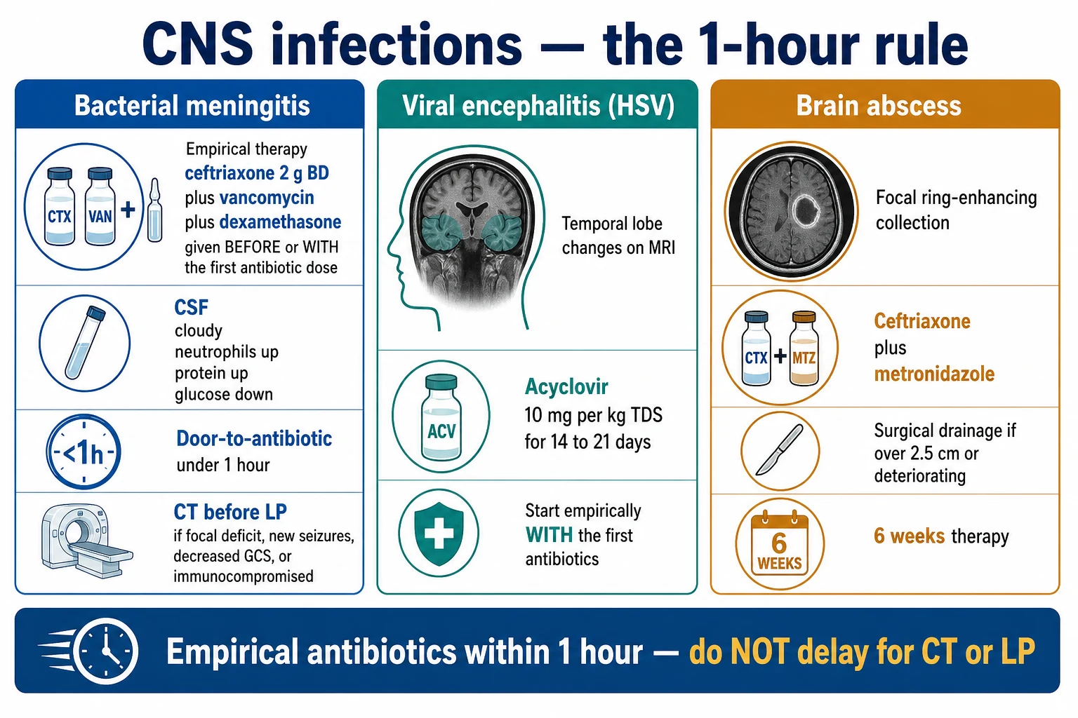
- The ceftriaxone 2 g IV twice daily (covers the meningococcus, the pneumococcus, and the Haemophilus influenzae) plus the vancomycin (for the penicillin-resistant pneumococcus).[1]
- The ampicillin added if the listeria is suspected (the age over 50, the immunocompromised, the pregnancy, the alcoholism).[1]
- The acyclovir 10 mg/kg IV three times daily added if the HSV encephalitis is suspected — until the CSF PCR is negative.[1]
- The dexamethasone 10 mg IV four times daily for 4 days — given before or with the first antibiotic dose (the de Gans trial, NEJM 2002 — reduced the mortality and the hearing loss in the pneumococcal meningitis). The dexamethasone given after the antibiotics is not effective (the dexamethasone reduces the inflammatory cascade from the lysed bacteria).[1]
The door-to-antibiotic — under 1 hour
The empirical antibiotics are given within 1 hour of the suspected meningitis. Do NOT delay the antibiotics for the CT or the lumbar puncture — the blood cultures are taken first, the antibiotics are given, then the CT and the LP follow. The delay worsens the outcome.[1]
The CT before the LP — the criteria
The CT is performed before the LP if any of:[1]
- An immunocompromised state.
- A new-onset seizure.
- A decreased consciousness (the GCS under 13).
- A focal neurological deficit.
- A papilloedema.
- A severe or prolonged headache.
In all these cases, the antibiotics and the dexamethasone are given before the CT and the LP.[1]
Viral encephalitis — the HSV
The herpes simplex encephalitis is the most common sporadic viral encephalitis. It affects the temporal lobes (the MRI shows the temporal lobe oedema and the haemorrhage; the CT is often normal early). The CSF shows the lymphocytes, the mildly raised protein, and the red cells (a haemorrhagic CSF). The CSF PCR is the diagnostic test. The treatment is the acyclovir 10 mg/kg IV three times daily for 14 to 21 days (renal-dosed). The acyclovir is started empirically with the first antibiotics — do not wait for the PCR (the untreated HSV encephalitis has a 70 per cent mortality).[1]
Brain abscess
The brain abscess is a focal intracerebral collection of pus. It presents with a headache, a fever, and a focal deficit (often subtle). The sources are the contiguous spread (the otitis, the mastoiditis, the sinusitis, the dental infection), the haematogenous spread (the endocarditis, the congenital heart disease), or the post-traumatic or the post-surgical. The CT or the MRI with the contrast shows a ring-enhancing lesion with the central necrosis and the surrounding oedema.[1]
The management:[1]
- The empirical antibiotics — the ceftriaxone plus the metronidazole (for the anaerobes) for 6 weeks (the streptococci and the anaerobes are the common organisms from the contiguous sources; the staphylococci from the post-surgical).[1]
- The surgical drainage (the aspiration or the excision) if the abscess is over 2.5 cm, if the patient is deteriorating, or if there is no response to the antibiotics.[1]
- The ICP management (the abscess and the surrounding oedema raise the ICP — the head elevation, the mannitol or the hypertonic saline if needed; the dexamethasone for the mass effect is controversial but used for the severe oedema).[1]
- The search for the source (the dental, the ENT, the cardiac echo for the endocarditis).[1]
Red flags
The microbiology by host — who gets what
The empirical antibiotic choice is governed by who the patient is as much as by the syndrome. The three community-acquired pathogens in the immunocompetent adult are Streptococcus pneumoniae (50–70 per cent of cases, the commonest by a margin), Neisseria meningitidis (15–25 per cent, peaks in adolescents and young adults, the meningo-coccal rash), and Haemophilus influenzae type b (now rare where conjugate vaccination is universal).[5] Listeria monocytogenes becomes a major pathogen in three groups — the age over 50, the immunocompromised (transplant, malignancy, HIV, anti-TNF therapy, corticosteroids), and pregnancy — and it is the only common cause of bacterial meningitis that is intrinsically resistant to the cephalosporins, mandating the addition of ampicillin (or high-dose benzylpenicillin) to the empirical regimen in these patients.[5][3]
| Host / scenario | Likely pathogens | Empirical cover to ADD or modify |
|---|
| Host / scenario | Likely pathogens | Empirical cover to ADD or modify |
|---|---|---|
| Immunocompetent adult, community-acquired | S. pneumoniae, N. meningitidis, H. influenzae | Ceftriaxone 2 g IV BD + vancomycin + dexamethasone (the backbone) |
| Age over 50, OR immunocompromised, OR pregnant, OR alcoholic | All of the above PLUS Listeria monocytogenes | ADD ampicillin 2 g IV 4-hourly (or benzylpenicillin). Cephalosporins do NOT cover listeria |
| Post-neurosurgery, post-trauma with CSF leak, OR CSF shunt | Coagulase-negative staphylococci, Staphylococcus aureus, Gram-negative bacilli (including Pseudomonas), propionibacterium acnes | Switch to vancomycin + ceftazidime or meropenem (anti-pseudomonal). Add intraventricular vancomycin for shunt infection |
| HIV / CD4 < 100 | Cryptococcus neoformans, Toxoplasma, CMV, VZV, JB virus | ADD liposomal amphotericin B + flucytosine if cryptococcus suspected; empiric TMP-SMX if toxoplasma |
| Tropical / returned traveller (ANZ, SE Asia) | Burkholderia pseudomallei (melioidosis), Mycobacterium tuberculosis, Salmonella (sickle cell), Streptococcus suis (pig exposure) | ADD ceftazidime + co-trimoxazole for suspected melioidosis; melioid meningitis is uniformly fatal if missed (the glycolipid capsule blocks cephalosporin entry to CSF)[17] |
| Penicillin allergy (anaphylaxis) | As above | Substitute vancomycin + meropenem (avoid ceftriaxone if type-1 hypersensitivity; meropenem cross-reactivity <1 per cent) |
| Neonate (not ICU here, but exam-worthy) | Group B strep, E. coli, Listeria | Ampicillin + cefotaxime + aminoglycoside |
The CSF analysis — a deeper dive
The single most diagnostic investigation is the lumbar puncture with CSF analysis — but its timing relative to the antibiotics, the imaging, and the clinical state is what examiners probe. The opening pressure (normal under 20 cmH₂O in the lateral decubitus position) is raised in bacterial meningitis in 80–90 per cent and is the single best discriminator between bacterial and viral meningitis when the cell count is borderline. The CSF glucose should always be interpreted as a ratio to the simultaneous serum glucose — a ratio under 0.4 is strongly bacterial, and a ratio under 0.3 is essentially diagnostic of a pyogenic, mycobacterial, or fungal process.[5][10]
| Parameter | Bacterial (pyogenic) | Viral (aseptic / HSV) | Tuberculous | Cryptococcal (fungal) |
|---|
| Parameter | Bacterial (pyogenic) | Viral (aseptic / HSV) | Tuberculous | Cryptococcal (fungal) |
|---|---|---|---|---|
| Opening pressure | Raised (often >30 cmH₂O) | Normal or mildly raised | Raised (often marked) | Markedly raised — the ICP is the proximate cause of death |
| Appearance | Cloudy / turbid | Clear (or faintly xanthochromic in HSV) | Often viscous, web/coagulum forming | Clear or slightly turbid |
| White cells | Markedly raised, NEUTROPHILS (often >1000/μL) | Raised, LYMPHOCYTES (10–500/μL); may have neutrophils early | Raised, LYMPHOCYTES (after first day); mixed early | Raised, LYMPHOCYTES (often modest, <100/μL) |
| Glucose (CSF:serum ratio) | Low (<0.4), often <0.3 | Normal (>0.4) | Very low (<0.3) — a hallmark | Low (<0.4) |
| Protein | Raised (>1.0 g/L, often >2.0) | Mildly raised (0.4–0.8 g/L) | Markedly raised (often >1.5 g/L) | Raised (0.5–1.5 g/L) |
| Gram stain | Often positive (60–90 per cent) | Negative | Negative in 80 per cent (acid-fast positive in <30 per cent) | Negative (India ink positive in ~60 per cent; crag positive) |
| PCR | Pneumococcal / meningococcal PCR | HSV-1/2 PCR (diagnostic, sensitivity ~96 per cent) | Xpert MTB/RIF (sensitivity ~80 per cent) | Cryptococcal antigen (CrAg) in CSF and serum |
A CSF that is "paucicellular but otherwise bacterial" (cloudy, low glucose, high protein, but few cells) is NOT reassuring — it is seen in the immunocompromised host, in overwhelming pneumococcal meningitis in the elderly (the cells have not yet mounted), and after partial antibiotic treatment. A nationwide Danish series confirmed that normocellular community-acquired bacterial meningitis is a real entity (3–4 per cent of cases) and carries a higher case-fatality than the cellular form, precisely because clinicians dismiss it.[15]
The empirical therapy — the adult community-acquired pathway
The 1-hour empiric bundle for suspected community-acquired bacterial meningitis
- Recognise and act on the syndrome within minutes. Suspect bacterial meningitis in any patient with at least two of: fever, neck stiffness, altered mental status, headache, or a petechial rash. The triad of fever, neck stiffness, and altered consciousness is present together in only 44 per cent — but at least one feature is in 95 per cent, and 99 per cent have at least one of six features (the triad plus headache, rash, seizure). Do NOT wait for confirmation.[5]
- Draw two sets of blood cultures (and a EDTA blood for PCR later) — this takes 5 minutes and must not delay therapy. Then give the empirical antibiotics AND the dexamethasone within 1 hour of suspicion. The dexamethasone (10 mg IV) is given immediately before or with the first antibiotic dose — this timing is the de Gans trial finding that changed practice.[2]
- The empiric regimen — memorise the four-drug bundle: (a) ceftriaxone 2 g IV 12-hourly (covers pneumococcus, meningococcus, H. influenzae); (b) vancomycin 15–25 mg/kg IV loading then AUC-guided (covers penicillin-resistant pneumococcus); (c) ampicillin 2 g IV 4-hourly ADDED if listeria plausible (age >50, immunocompromised, pregnant, alcoholic); (d) acyclovir 10 mg/kg IV 8-hourly ADDED if viral encephalitis plausible (altered consciousness, temporal-lobe signal, seizures).[3][10]
- Imaging and LP after the antibiotics. Perform a non-contrast CT brain first ONLY if any "CT-before-LP" criterion is met (immunocompromised, new seizure, GCS <13, focal deficit, papilloedema, prolonged headache). Otherwise, proceed directly to the LP. A normal CT does NOT exclude raised ICP in the context of infection — the LP is still deferred if the GCS is <9 or there is clinical brain-shift.[11][12]
- De-escalate at 48–72 hours when the organism and susceptibility return. Narrow to the narrowest effective agent (e.g., benzylpenicillin for penicillin-sensitive pneumococcus; ampicillin for listeria; ceftriaxone for meningococcus). Total duration is typically 7 days for meningococcus, 10–14 days for pneumococcus, 14–21 days for listeria, and >21 days for Gram-negative bacilli.[3]
The specific syndromes
Listeria monocytogenes meningitis and rhombencephalitis
Listeria is a Gram-positive bacillus acquired from contaminated food (soft cheese, deli meats, unpasteurised milk, smoked seafood). Beyond the classical meningitis picture, Listeria has an affinity for the brainstem — the rhombencephalitis syndrome (cranial-nerve palsies, cerebellar signs, nystagmus, respiratory depression) is essentially pathognomonic and is missed on a routine CT. The organism is intrinsically resistant to all cephalosporins (the third-generation cephalosporin is the backbone for everything else in meningitis — listeria is the exception). The treatment is ampicillin 2 g IV 4-hourly (or high-dose benzylpenicillin) PLUS gentamicin for synergy in the first week (the aminoglycoside is stopped after 7 days for toxicity, as the synergy benefit is early). High-dose co-trimoxazole (20 mg/kg/day of TMP in two divided doses) is the alternative in penicillin-allergic patients. Meropenem is the carbapenem that has listeria activity (imipenem does NOT reliably). Mortality is 20–30 per cent despite correct therapy.[5][3]
Tuberculous meningitis (TBM)
The most lethal form of tuberculosis (mortality 25–30 per cent even with treatment, and over 50 per cent in HIV). TBM is a subacute-to-chronic basilar meningitis that progresses through three stages: stage 1 (vague prodrome, headache, low-grade fever, no deficit); stage 2 (cranial-nerve palsies, lethargy, meningism, focal deficits); stage 3 (coma, seizures, dense hemiplegia). The CSF shows the classic lymphocytic pleocytosis with a very low glucose (<2.2 mmol/L or ratio <0.3) and a markedly raised protein (often >1.5 g/L). The CSF is often viscous and forms a "spider-web" coagulum on standing. Acid-fast bacilli are seen in fewer than 30 per cent; the Xpert MTB/RIF Ultra on CSF (sensitivity ~80 per cent) is the modern rapid test; culture takes 4–6 weeks.[8]
The treatment is the four-drug RIPE regimen (rifampicin 10 mg/kg, isoniazid 5 mg/kg, pyrazinamide 25 mg/kg, ethambutol 15 mg/kg daily) for 2 months then rifampicin + isoniazid for 9–12 months total. Adjunctive dexamethasone reduces mortality (Thwaites 2004 NEJM, confirmed in the long-term follow-up and in the Cochrane review) — the WHO-endorsed tapering regimen starts at 0.4 mg/kg/day and tapers over 6–8 weeks. Stage 3 disease benefits less. Intensified high-dose IV rifampicin (15 mg/kg/day) may improve survival (Brouwer 2013) but is not yet universal. Cerebral oedema, hydrocephalus (communicating), hyponatraemia (cerebral salt-wasting), and vasculitic stroke are the four killers — neurosurgical shunting for hydrocephalus and aggressive ICP management are part of intensive care.[7][8][9][13]
Cryptococcal meningitis (the HIV / immunocompromised host)
Cryptococcus neoformans and the more virulent C. gattii cause a subacute lymphocytic meningitis with a markedly raised opening pressure (the ICP is the proximate cause of death). The CSF India ink is positive in ~60 per cent; the cryptococcal antigen (CrAg) in CSF and serum is the diagnostic test (sensitivity >99 per cent). The IDSA regimen (Perfect 2010): induction with liposomal amphotericin B 3–4 mg/kg/day IV PLUS flucytosine 25 mg/kg 6-hourly for at least 2 weeks (the flucytosine halves the 2-week sterilisation-failure rate), then consolidation with fluconazole 400–800 mg/day for 8 weeks, then maintenance fluconazole 200 mg/day until immune reconstitution. Therapeutic lumbar punctures daily to keep the opening pressure under 20 cmH₂O (and a lumbar drain or ventriculostomy for refractory cases) is non-negotiable — the raised ICP, not the organism, kills the patient.[14]
Post-neurosurgical, post-traumatic, and shunt-related meningitis
The microbiology is entirely different from community-acquired meningitis: coagulase-negative staphylococci (especially Staphylococcus epidermidis), Staphylococcus aureus (including MRSA), Gram-negative bacilli (including Pseudomonas aeruginosa, E. coli, Klebsiella), and — uniquely for shunts — Propionibacterium acnes (a slow-growing anaerobe that requires anaerobic culture held for 10–14 days). The empirical regimen is vancomycin PLUS ceftazidime or meropenem (anti-pseudomonal) — ceftriaxone is inadequate. Intraventricular or intrathecal vancomycin (10–20 mg/day, via the shunt or EVD) is added for shunt infection because systemic vancomycin penetrates the CSF poorly (1–5 per cent). The shunt or EVD is removed or externalised — antibiotics alone do not sterilise an infected foreign body. Dexamethasone is NOT given in nosocomial meningitis (no evidence of benefit; the de Gans trial was community-acquired pneumococcal).[4]
HSV encephalitis — a deeper dive
The herpes simplex virus type 1 (HSV-1) is the most common cause of sporadic viral encephalitis (incidence ~1 in 250,000 per year) and the most devastating if missed (70 per cent mortality untreated, falling to under 20 per cent with prompt acyclovir — the Whitley 1986 NEJM trial that established acyclovir over vidarabine).[6]
The virus has a tropism for the medial temporal lobes and the orbitofrontal cortex — the classical MRI (the most sensitive imaging) shows T2/FLAIR hyperintensity in the temporal lobe, often bilateral and asymmetric, with restricted diffusion and sometimes haemorrhage. The CT is often normal in the first 48 hours. The CSF shows lymphocytes, a mildly raised protein, and red cells (a haemorrhagic CSF is a strong clue — it does not mean a traumatic tap). The CSF HSV PCR is the diagnostic test (sensitivity ~96 per cent, specificity ~99 per cent) but the PCR can be falsely negative in the first 24–72 hours of illness and again may wane after the first week of antivirals — a negative PCR in a high-probability case is repeated at 4–7 days rather than dismissing the diagnosis. The EEG shows periodic lateralised epileptiform discharges (PLEDs) over the temporal lobe in ~80 per cent — historically a key clue before MRI was universal. [1]
The HSV encephalitis pathway — recognising and treating the worst-of-the-worst
- Suspect on the constellation: fever, altered consciousness, behavioural change, seizures, and temporal-lobe signal on MRI. The behavioural change can be subtle (hallucinations, anomia, amnesia) or florid (agitation, psychosis) — families often report 'they are not themselves'. Do not be reassured by an early normal CT — get an MRI.[6]
- Start acyclovir 10 mg/kg IV 8-hourly (renal-dosed) empirically with the first antibiotics — within 1 hour. Do NOT wait for the PCR. The cost of a few days of empirical acyclovir (with renal monitoring) is trivial against the 70 per cent untreated mortality.
- Send the CSF HSV PCR (and store an extra aliquot for repeat at day 4–7 if initially negative in a high-suspicion case). Send a paired serum and CSF for HSV antibody (a CSF-to-serum antibody ratio >20 is suggestive of intrathecal synthesis in the subacute case).
- Treat for 14–21 days if the PCR is positive or the suspicion remains high. Shorter courses relapse. Continue a full 21 days for any relapse (relapses occur in ~5 per cent, often within a month, and may be immune-mediated rather than replicative).
- Monitor the creatinine every other day — acyclovir crystallises in the renal tubules (especially with rapid infusion, dehydration, or concurrent nephrotoxins). Pre-hydrate, infuse over 1 hour, and renal-dose-adjust. Monitor for neutropenia, thrombocytopenia, and hepatic enzyme rises.
- Look for and treat seizures (clinical or on continuous EEG) — post-HSV epilepsy is common and the temporal lobe is highly epileptogenic. Non-convulsive status is not uncommon in the comatose HSV patient; continuous EEG should be considered.
Brain abscess — a deeper dive
A brain abscess is a focal, encapsulated collection of pus within the brain parenchyma. It is NOT meningitis — the CSF may even be normal early, and an LP in a patient with a mass lesion and raised ICP risks cerebellar tonsillar herniation. The diagnosis is made on contrast CT or MRI, which shows a ring-enhancing lesion with central diffusion restriction (a high signal on DWI) — the restricted diffusion distinguishes an abscess from a necrotic tumour (which does not restrict). The lesion is surrounded by vasogenic oedema and produces mass effect.[1]
| Lesion | Clue | Confirmatory test |
|---|
| Lesion | Clue | Confirmatory test |
|---|---|---|
| Bacterial brain abscess | Primary source (sinus, ear, dental, endocarditis); restricted DIFFUSION on DWI | Aspiration culture |
| Toxoplasma (HIV, CD4 <100) | Multiple ring lesions, baseline ring-enhancement, positive Toxo IgG | Empirical anti-toxo therapy; biopsy if no response in 2 weeks |
| CNS lymphoma (EBV-associated in HIV) | Periventricular, homogeneous uptake on thallium SPECT/PET; reduces with steroids (so do NOT give steroids before the biopsy) | Stereotactic biopsy |
| Tuberculoma | Endemic area, TBM coexists; often solid caseating nodule | Biopsy / AFB; response to RIPE therapy |
| Neurocysticercosis | Multiple lesions of varying stages (vesicular, colloidal, granular, calcified); pig exposure / endemic travel | Cysticercus serology; classic 'scolex' on MRI |
| Metastasis | Primary tumour; usually multiple, at the grey-white junction; does NOT restrict on DWI | Biopsy / known primary |
| Demyelinating pseudotumour (tumefactive MS) | Young adult, open ring (incomplete), perivenular 'Dawson fingers' elsewhere | MRI brain, CSF oligoclonal bands |
The brain abscess pathway — antibiotics, drainage, and the search for the source
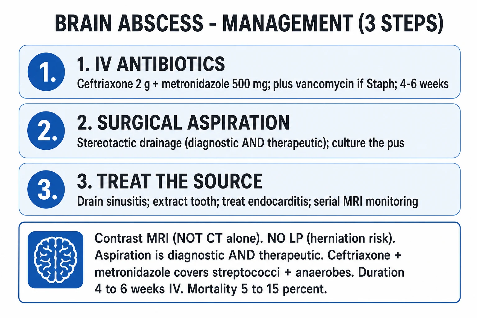
- Establish the diagnosis with a contrast MRI (preferred — better cortical detail, multiplanar, DWI sequences). The CT is the screening test. Do NOT perform an LP — it adds nothing and risks herniation.[1]
- Drainage is both diagnostic and therapeutic. Stereotactic aspiration (the standard) provides pus for culture AND debulks the mass. Drainage is indicated for any abscess >2.5 cm, any abscess with mass effect, any abscess that is deteriorating, and any abscess not responding to 2 weeks of antibiotics. Solitary, small (<2.5 cm), multiple-but-stable abscesses can be managed medically.
- Empirical antibiotics — guided by the source: (a) contiguous spread (otitis, mastoiditis, sinusitis, dental): ceftriaxone + metronidazole — covers streptococci (the S. milleri group), anaerobes (Bacteroides, Peptostreptococcus), and gram-negatives; (b) haematogenous (endocarditis, congenital cyanotic heart disease): ceftriaxone + metronidazole + consider anti-MRSA cover (vancomycin) for endocarditis; (c) post-surgical / post-trauma: vancomycin + ceftazidime (or meropenem) — anti-pseudomonal, anti-staph. Tailor to culture at 48–72 hours.[1]
- Treat for 6 weeks IV (4–6 weeks for a fully-drained small abscess; 6–8 weeks for undrained or large). Oral step-down is rarely used for brain abscess (poor CSF/abscess-cavity penetration of most oral agents).
- Search for and treat the source — the dental OPG, the ENT review (mastoid, sinus), the transthoracic AND transoesophageal echo for endocarditis (TTE is insufficient for the small vegetations of subacute endocarditis), and the immunocompromise screen. An unrepaired source predicts recurrence.
- Manage the ICP and the surrounding oedema — head-of-bed elevation 30 degrees, normocapnia, hypertonic saline or mannitol for impending herniation, and anticonvulsants (the abscess is highly epileptogenic, especially if cortical). The role of dexamethasone for the vasogenic oedema is controversial — it reduces the capsule integrity and penetrability of antibiotics, and is reserved for severe mass effect, used only transiently, and tapered as the mass effect resolves.
- Serial imaging at 2, 4, and 8 weeks (and at any clinical deterioration) — the abscess cavity can transiently enlarge on antibiotics before it shrinks, so a slight increase in size at 2 weeks is not by itself an indication to drain; a persistent increase, new loculation, or clinical deterioration is.
Adjunctive dexamethasone — the trials that changed practice
de Gans & van de Beek 2002 (NEJM) — dexamethasone in adults with bacterial meningitis
Design
Prospective, randomised, double-blind, multicentre European trial; 301 adults with suspected bacterial meningitis, started antibiotics + dexamethasone or placebo before or with the first antibiotic dose
Intervention
Dexamethasone 10 mg IV QDS for 4 days (started before or with the first antibiotic dose) vs placebo
Primary outcome
**Favourable outcome at 8 weeks: dexamethasone 40% vs placebo 26%** (RR 0.59). **Mortality: dexamethasone 7% vs placebo 15%** (RR 0.48). The benefit was confined to **pneumococcal meningitis** (mortality 14% vs 34%); no benefit in meningococcal (already low mortality)
What it changed
**Dexamethasone 10 mg QDS for 4 days became the standard** in suspected bacterial meningitis, given BEFORE or WITH the first antibiotic dose. The mechanism: dexamethasone blunts the inflammatory cascade triggered by antibiotic-induced bacterial lysis (the surge of TNF-α, IL-1, IL-6 in the CSF). Given AFTER the antibiotic, the cascade is already underway and the dexamethasone is ineffective.
Caveats
Benefit is for **community-acquired pneumococcal meningitis**. The follow-up Thwaites 2007 trial in Vietnamese adults (predominantly TBM, but a bacterial arm too) showed **NO benefit in confirmed bacterial meningitis in a low-income setting with high HIV/TB co-prevalence** — the dogma is now: give dexamethasone in confirmed pneumococcal meningitis in a high-income setting; re-consider in suspected TBM (where the dexamethasone is given for the TBM, not the bacterial).
Thwaites 2004 (NEJM) — dexamethasone in tuberculous meningitis
Design
Randomised, double-blind, placebo-controlled trial in Vietnam; 545 adolescents and adults with definite or probable TBM
Intervention
Dexamethasone IV/PO taper (starting 0.4 mg/kg/day, tapering over 6–8 weeks) vs placebo, both with standard four-drug anti-TB therapy
Primary outcome
**Death by 9 months: dexamethasone 31% vs placebo 41%** (RR 0.69, P=0.001). Benefit greatest in grade I/II disease; the **severe (grade III, comatose) subgroup did NOT benefit**
What it changed
Adjunctive **dexamethasone is now the standard of care for TBM** (WHO-endorsed), but it does **not improve survival in severe (grade III) disease** — it reduces death in the lesser-grade patient. Severe disability in survivors was unchanged.
Prasad 2008 Cochrane — corticosteroids for tuberculous meningitis (meta-analysis)
Design
Cochrane systematic review and meta-analysis; 7 trials, 1140 participants (including Thwaites 2004, the largest contributor)
Comparison
Corticosteroids (dexamethasone, methylprednisolone, or hydrocortisone) vs placebo/no adjunct in confirmed or probable TBM
Primary outcome
**Death: corticosteroids RR 0.77 (95% CI 0.63–0.94)**; **Death OR severe disability: RR 0.73 (0.57–0.94)**. No significant effect on severe disability in survivors alone
What it changed
Corticosteroids reduce death and the composite of death-or-disability in TBM — should be the routine adjunct. The dose, formulation, and exact regimen are less clear; the Thwaites 6–8 week taper is the most-quoted.
Special situations
The 'CT-before-LP' decision — modern evidence
- The default is LP-first. A normal CT is not needed in most patients with suspected meningitis and no focal signs. Doing imaging on everyone delays both the LP and (paradoxically) the antibiotic, and a CT before LP does NOT change the outcome in those without features of raised ICP.[11][12]
- The CT-before-LP criteria (memorise): (1) immunocompromised state (HIV, transplant, chemo, anti-TNF, chronic steroids); (2) a new-onset seizure within the past week; (3) decreased consciousness (GCS <13); (4) a focal neurological deficit (including dilated pupil, gaze palsy, hemiparesis, ataxia); (5) papilloedema on funduscopy; (6) a severe or prolonged headache with features of raised ICP.
- The antibiotics and dexamethasone come BEFORE the CT and the LP. Blood cultures → antibiotics + dexamethasone → CT (if indicated) → LP. This sequence is non-negotiable — the door-to-antibiotic target is under 1 hour.[2]
- A normal CT does NOT exclude raised ICP in CNS infection. If the GCS is <9, there is a rapidly declining level of consciousness, or clinical signs of brain-shift (anisosocoria, Cushing's triad, posturing), the LP is still deferred and the patient is treated as presumed meningitis with empirical antibiotics. A delayed LP after antibiotics still yields diagnostic value (the cell count, glucose, and protein remain abnormal for hours; the culture may be sterile but the antigen PCR is often still positive).[15]
Managing the raised ICP in CNS infection
- Elevate the head of the bed to 30 degrees, head midline — improves jugular venous drainage and is the cheapest, fastest intervention.[1]
- Maintain normocapnia (PaCO₂ 35–40 mmHg) — hyperventilation to a low PaCO₂ causes cerebral vasoconstriction and is NOT recommended as a routine measure (the recovery of vascular tone reverses any benefit).
- Osmotherapy for impending herniation — 3% hypertonic saline 250 mL bolus (or mannitol 0.5–1 g/kg) — both draw water out of the brain. Hypertonic saline is preferred in sepsis (it is also a volume expander). Monitor sodium (target 145–155 mmol/L) and osmolarity (<320 mOsm/L).
- Treat seizures aggressively — clinical and electrographic seizures markedly increase cerebral metabolic demand and ICP. Continuous EEG in any patient who is not waking as expected.
- Avoid hypoglycaemia and hyperglycaemia — both worsen the injured brain; target glucose 6–10 mmol/L.
- Consider an intracranial pressure monitor or external ventricular drain for the patient with suspected raised ICP and a GCS <8 — particularly in TBM with hydrocephalus (where an EVD is both diagnostic and therapeutic).
- In refractory raised ICP — consider decompressive craniectomy (rarely used in CNS infection, but reported for cerebellar abscess with posterior-fossa pressure or for massive HSV-related cerebral oedema).
Public health and chemoprophylaxis for the contacts of bacterial meningitis
- Notify the case to public health immediately (suspected meningococcal or Haemophilus — statutory notification).[10]
- Identify the contacts requiring chemoprophylaxis for meningococcal disease: household contacts, intimate kissing contacts, and 'mouth-to-mouth' resuscitation contacts — within 7 days (the highest attack-rate window is the first 48 hours after symptom onset in the index case).
- The chemoprophylaxis options for N. meningitidis: ciprofloxacin 500 mg PO single dose (the modern choice, no age limit) OR rifampicin 600 mg PO BD for 2 days (the classic, but stains secretions, reduces OCP efficacy, and is contraindicated in pregnancy) OR ceftriaxone 250 mg IM single dose (preferred in pregnancy). Ciprofloxacin is the practical choice in the alert, ambulatory contact.
- For H. influenzae type b — rifampicin 600 mg PO daily for 4 days for all household contacts if there is an unvaccinated child under 4 in the household.
- The healthcare worker — prophylaxis only after mouth-to-mouth exposure to the index case's secretions without PPE (the 'intubation without a closed circuit' scenario). Routine care of a meningococcal patient with PPE is NOT an exposure.
- Vaccination — conjugate vaccines (MenACWY, MenB, Hib, PCV13) are part of the public-health response and routine childhood schedules; the ICU role is to recognise and notify, not to deliver.
Exam practice
SAQ — Bacterial meningitis: the 1-hour bundle and the CT-before-LP decision
10 minutes · 10 marks
A 55-year-old man with a 2-day history of fever, headache, and photophobia presents with a GCS of 12, a stiff neck, and a petechial rash on his lower limbs. His blood pressure is 95/60, heart rate 120, temperature 39.1°C. He has no known immunocompromise. You are the ICU registrar called to the emergency department.
SAQ — HSV encephalitis: recognition and the empirical acyclovir
10 minutes · 10 marks
A 28-year-old previously well woman is brought to the emergency department by her family with a 3-day history of fever, headache, and increasingly bizarre behaviour. This morning she had a generalised tonic-clonic seizure. On arrival her GCS is 10, temperature 38.7°C, and she has intermittent myoclonic jerks. A non-contrast CT brain is normal. The lumbar puncture shows 80 white cells (lymphocyte predominant), protein 0.8 g/L, glucose 3.2 mmol/L (serum 6.5), and 200 red cells.
Clinical pearls — high-yield CICM/FFICM/EDIC points
Additional red flags
[1] [1]References
- [1]van de Beek D, de Gans J, Tunkel AR, Wijdicks EFM Community-acquired bacterial meningitis in adults N Engl J Med, 2006.PMID 16394301
- [2]de Gans J, van de Beek D Dexamethasone in adults with bacterial meningitis N Engl J Med, 2002.PMID 12432041
- [3]Tunkel AR, Hartman BJ, Kaplan SL, et al. Practice guidelines for the management of bacterial meningitis Clin Infect Dis, 2004.PMID 15494903
- [4]van de Beek D, Drake JM, Tunkel AR Nosocomial bacterial meningitis N Engl J Med, 2010.PMID 20071704
- [5]van de Beek D, Brouwer MC, Thwaites GE, Tunkel AR Community-acquired bacterial meningitis Nat Rev Dis Primers, 2016.PMID 27808261
- [6]Whitley RJ, Alford CA, Hirsch MS, et al. Vidarabine versus acyclovir therapy in herpes simplex encephalitis N Engl J Med, 1986.PMID 3001520
- [7]Thwaites GE, Nguyen DB, Nguyen HD, et al. Dexamethasone in Vietnamese adolescents and adults with bacterial meningitis N Engl J Med, 2007.PMID 18077808
- [8]Prasad K, Singh MB Corticosteroids for managing tuberculous meningitis Cochrane Database Syst Rev, 2008.PMID 18254003
- [9]Thwaites GE, Bhavnani SM, Chau TTH, et al. Dexamethasone and long-term outcome of tuberculous meningitis in Vietnamese adults and adolescents PLoS One, 2011.PMID 22174748
- [10]Roos KL, van de Beek D Update on community-acquired bacterial meningitis: guidance and challenges Clin Microbiol Infect, 2017.PMID 28478238
- [11]Bodilsen J, Dalager-Park C, Schonheyder HC, et al. Lumbar Puncture Performed Promptly or After Neuroimaging in Acute Bacterial Meningitis in Adults: A Prospective National Cohort Study Evaluating Different Guidelines Clin Infect Dis, 2018.PMID 29020334
- [12]Bodilsen J, Brouwer MC, van de Beek D Cranial Imaging and Lumbar Puncture in Patients With Suspected Central Nervous System Infection Clin Infect Dis, 2020.PMID 31437271
- [13]Brouwer MC, Heckenberg SGB, de Gans J, et al. Intensified intravenous rifampicin in tuberculous meningitis Lancet Infect Dis, 2013.PMID 23103176
- [14]Perfect JR, Dismukes WE, Dromer F, et al. Clinical practice guidelines for the management of cryptococcal disease: 2010 update by the infectious diseases society of america Clin Infect Dis, 2010.PMID 20047480
- [15]Bodilsen J, Storgaard M, Larsen L, et al. Normocellular Community-Acquired Bacterial Meningitis in Adults: A Nationwide Population-Based Case Series Ann Emerg Med, 2021.PMID 32747082
- [16]Bodilsen J, Dalager-Park C, Nielsen H Risk factors and prognosis of seizures in adults with community-acquired bacterial meningitis in Denmark: observational cohort studies BMJ Open, 2019.PMID 31266843
- [17]Scarborough M, Gordon SB, Whitty CJM, et al. Neurological melioidosis: seven cases from the Northern Territory of Australia Clin Infect Dis, 1992.PMID 1617057