ICU · Neurocritical care / monitoring
Continuous EEG & Non-Convulsive Status Epilepticus
Also known as Continuous EEG · cEEG · Non-convulsive status epilepticus · NCSE · Salzburg criteria · PLEDs · Periodic lateralised epileptiform discharges · Ictal-interictal continuum · Status epilepticus · LPDs / GPDs / LRDA / GRDA · SIRPIDs · Burst suppression · Quantitative EEG
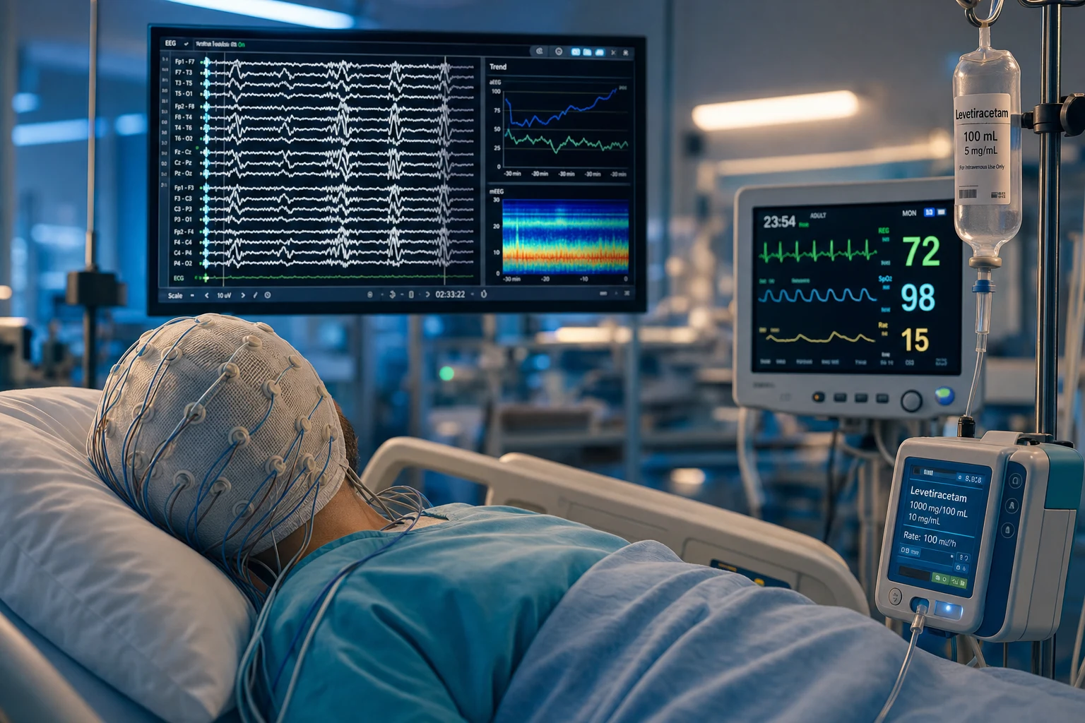
Continuous EEG (cEEG) monitoring in the ICU detects the non-convulsive status epilepticus (NCSE) — a continuous or recurring epileptiform activity on the EEG without the clinical motor convulsions, producing a persistent impaired consciousness. NCSE is common in the comatose ICU patient (10-30 per cent of the comatose patients, especially after a convulsive SE, a TBI, a SAH, or a cardiac arrest) and is detected only by the EEG — the clinical examination cannot distinguish it from the metabolic encephalopathy. The Salzburg criteria diagnose the NCSE (the epileptiform activity plus a response to the IV antiepileptic), and the ACNS terminology standardises the description of the ICU EEG patterns (LPDs, GPDs, LRDA, GRDA, SIRPIDs). cEEG is also the tool for the post-arrest prognostication (background reactivity, malignant patterns), for the seizure monitoring during the status epilepticus treatment, and for the burst-suppression verification during the anaesthetic coma. The treatment: 1st line IV benzodiazepine, 2nd line levetiracetam or valproate or fosphenytoin, 3rd line a continuous infusion (midazolam or propofol) for the refractory. Untreated, the NCSE worsens the neurological outcome.
On this page & tools
Your progress
Saved locally on this device.
Target exams
Red flags
Overview & definition
Non-convulsive status epilepticus (NCSE) is a continuous or recurring epileptiform activity on the EEG without the clinical motor convulsions, producing a persistent impaired consciousness. It is common in the comatose ICU patient (10-30 per cent of the comatose patients) and is detected only by the continuous EEG (cEEG) — the clinical examination cannot distinguish it from the metabolic encephalopathy or the post-ictal state. Untreated, the NCSE worsens the neurological outcome (the ongoing epileptiform activity is neurotoxic).[1]
Continuous EEG (cEEG) is the prolonged (≥24 hours) digital EEG recording from scalp electrodes in the critically ill patient. It is the gold-standard tool for: (1) the detection of the non-convulsive seizures and the NCSE; (2) the seizure monitoring during and after the treatment of the convulsive status epilepticus; (3) the post-cardiac-arrest prognostication (background, reactivity, malignant patterns); (4) the sedation-depth monitoring and the burst-suppression verification during the anaesthetic coma; and (5) the detection of the ischaemia (delayed cerebral ischaemia after SAH) through the gradual background attenuation. The ACNS consensus lists the indications and the technical specifications.[2]
Indications for cEEG
The cEEG is indicated for the comatose ICU patient with:[2]
- A persistent unexplained altered consciousness (the comatose patient with no clear cause, or a fluctuating conscious level).
- After a convulsive status epilepticus — to confirm the seizure has stopped (the NCSE may continue after the convulsions resolve).
- After a severe TBI (the NCSE worsens the secondary brain injury).
- After a subarachnoid haemorrhage (the seizures are common; the NCSE may indicate the vasospasm and the ischaemia).
- After a cardiac arrest (the cEEG assesses the background reactivity and the myoclonus; the NCSE or the status myoclonus is a poor prognostic sign).
- In the metabolic encephalopathy (the hepatic or the renal failure can produce the epileptiform activity).[1]
The five core roles of cEEG in the ICU
The ACNS consensus defines five distinct indications, each with a different question the cEEG is asked to answer:[2]
- NCSE detection — the unexplained or the fluctuating coma. This is the commonest indication. The question: is the coma due to an ongoing seizure? The yield: the non-convulsive seizures are found in roughly 8-10 per cent of the comatose ICU patients screened, rising to 20-30 per cent in those with an acute brain injury and an altered consciousness.[7]
- Post-arrest prognostication — the cEEG is one of the multimodality tools (with the SSEP, the imaging, the biomarkers) used to prognosticate after the cardiac arrest. The background (reactive vs unreactive, continuous vs suppressed), the malignant patterns (suppression, burst-suppression, status myoclonus, generalised periodic discharges), and the seizure burden all inform the outcome prediction.[5]
- Seizure monitoring in status epilepticus — after the convulsive SE is treated, the cEEG confirms the electrographic seizure has stopped (the motor signs stop before the electrical activity). It also guides the titration of the anaesthetic infusion in the refractory SE.
- Sedation-depth and burst-suppression verification — in the refractory SE treated with the anaesthetic coma, the clinical signs (the pupil reactivity, the motor response) are abolished by the anaesthetic and the neuromuscular blockade. The cEEG is the only way to verify the target (the burst suppression or the seizure cessation) is achieved and maintained.
- Ischaemia detection (SAH) — the gradual attenuation of the background activity on the cEEG can detect the delayed cerebral ischaemia (DCI) after the SAH before the clinical deterioration (a fall in the alpha/delta ratio, the loss of the reactivity).
cEEG indications mapped to the clinical scenario
| Clinical scenario | The question cEEG answers | Typical finding | Yield |
|---|---|---|---|
| Unexplained coma | Is there an ongoing seizure? | NCSE, LPDs, GPDs | Non-convulsive seizures in ~10% of comatose patients |
| After convulsive SE | Has the seizure truly stopped? | Persistent electrographic seizure (NCSE) | NCSE in ~14-50% who do not fully wake |
| Post-cardiac arrest | What is the prognosis? | Reactive vs malignant background | Standardised EEG predicts outcome (Westhall 2016) |
| Severe TBI | Is there occult seizure / secondary injury? | NCSE, periodic discharges | Seizures (mostly non-convulsive) in ~20% |
| SAH (high grade) | Is there DCI / occult seizure? | Background attenuation, NCSE | Non-convulsive seizures in ~10-20% |
| Refractory SE (anaesthetic coma) | Is burst suppression achieved/maintained? | Burst-suppression pattern, seizure cessation | Mandatory — cannot titrate without EEG |
| Paralysed + sedated patient | Are there electrographic seizures? | NCSE (clinical signs abolished) | cEEG is the only detection method |
The clinical signs of NCSE — subtle and fluctuating
The clinical hallmark of the NCSE is the subtlety: there are no tonic-clonic convulsions. The patient is simply comatose or encephalopathic, and the signs that do exist fluctuate over minutes to hours. The clinical examination alone cannot reliably distinguish the NCSE from the metabolic encephalopathy or the post-ictal state — hence the dependence on the cEEG. Recognise the following subtle signs and use them as a trigger to request the cEEG, not as a substitute for it:[1]
- A fluctuating consciousness — the waxing and waning of the responsiveness, not a stable coma. The patient may briefly localise to pain, then become unresponsive, then open the eyes to voice. This fluctuation is the most consistent clue.
- Eye deviation — a sustained conjugate deviation of the eyes (a subtle ictal sign), or the periodic eye-opening and eye-cycling.
- Automatisms — the repetitive, semi-purposeful movements: the lip-smacking, the chewing, the picking at the sheets, the fumbling with the tubes, the swallowing. These are the same frontal/temporal automatisms of the focal impaired-awareness seizures, but muted by the critical illness.
- Nystagmus / eyelid twitching — the periodic nystagmoid eye movements or the rhythmic eyelid fluttering (the eyelid myoclonia).
- Myoclonus — the subtle, focal, or multifocal myoclonic jerks (a finger, a corner of the mouth, a shoulder). After the cardiac arrest, the status myoclonus is a malignant sign.
- Autonomic instability — the unexplained tachycardia, the blood-pressure surges, the pupillary dilatation, the diaphoresis that coincide with the EEG ictal activity.
- Atonic periods — the brief loss of the postural tone (a head drop, a limb slump) in a patient who otherwise holds a posture. [1]
The examination pearl: if the comatose patient has any of these signs, or simply fails to wake as expected after a convulsive SE or a sedation wean, request the cEEG. The NCSE is a clinical-Electroencephalographic diagnosis — the EEG is not an adjunct, it is the test. [1]
[1]The Salzburg criteria for NCSE
The IFNS-Euroclonus (2013) Salzburg criteria for the NCSE diagnosis:[3]
- The epileptiform activity on the EEG (the spikes, the sharp waves, the rhythmic activity of an epileptiform morphology).
- Plus one of:
- A response to the IV antiepileptic (the benzodiazepine or the valproate/phenytoin — the EEG improves; but caution: the benzodiazepine may also sedate and confound the clinical assessment).
- A subtle clinical improvement (a small motor, a cognitive, or an autonomic change after the antiepileptic).
- A typical EEG pattern (the ictal-interictal continuum — the rhythmic activity that is not clearly ictal but is more than the interictal).[3]
The Salzburg criteria add the formal definitions for the four sub-patterns of the suspected NCSE: (a) the focal NCSE (the focal rhythmic activity with the epileptiform morphology); (b) the generalised NCSE (the generalised spike-wave or the rhythmic activity); (c) the NCSE in the coma (the special case where the patient is already comatose — apply the criteria strictly, and beware the false positives from the sedation, the metabolic encephalopathy, and the post-anoxic patterns); and (d) the subtle / electrographic SE (the deeply comatose patient with the minimal or the no clinical signs).[3]
The ictal-interictal continuum
Not every periodic or rhythmic pattern is a seizure, and not every non-seizure is benign. The ictal-interictal continuum (IIC) is the grey zone — the rhythmic or the periodic discharges that are more than the background but not clearly ictal. To decide whether a pattern on the IIC is treatable, the Salzburg criteria suggest a trial of a fast-acting antiepileptic (a small benzodiazepine or a loading dose of an intravenous antiepileptic): an improvement in both the EEG and the clinical state confirms the NCSE; an EEG improvement with clinical deterioration (from the sedation) is inconclusive; no change is consistent with the interictal. The neurophysiologist's structured interpretation is essential.[3]
The ACNS standardised terminology for the ICU EEG
The American Clinical Neurophysiology Society (ACNS) standardised terminology provides a common language to describe the EEG patterns in the critically ill, replacing the older non-standard terms (PLEDs, PLEDs+, BiPLEDs, GPEDs, SIRPIDs) with a structured nomenclature. The 2012/2018 terminology describes each pattern by its main term (the morphology) and the modifiers (the frequency, the amplitude, the polarity, the evolution, the stimulus-dependence).[4]
The two main terms are: [1]
- Periodic discharges — the discharges that repeat at a nearly regular interval (a consistent inter-discharge interval), e.g. every 1-2 seconds. The morphology is a spike, a sharp wave, or a slow wave.
- Rhythmic activity — the activity that repeats with a regular waveform and a uniform morphology, but without the distinct inter-discharge interval (a continuous train rather than discrete discharges). [1]
The location prefix narrows the pattern: lateralised (one hemisphere) or generalised (bilaterally synchronous) or bilateral independent (two independent foci). Combining the main term and the location gives the four common ICU patterns:[4]
- LPDs (lateralised periodic discharges) — formerly PLEDs. Periodic spikes/sharp waves over one hemisphere at ~1 Hz. The classic structural-focal pattern (a stroke, a haemorrhage, an abscess, a tumour, HSV encephalitis).
- GPDs (generalised periodic discharges) — formerly GPEDs. Bilaterally synchronous periodic discharges. The causes include the toxic-metabolic encephalopathy (the triphasic waves of the hepatic failure), the post-anoxic injury, the CJD, and the severe sepsis.
- LRDA (lateralised rhythmic delta activity) — the slow rhythmic activity over one hemisphere, often a marker of the focal dysfunction or the non-convulsive seizure.
- GRDA (generalised rhythmic delta activity) — the bilaterally synchronous slow rhythmic activity, often a non-specific encephalopathy. [1]
The modifiers add the critical detail: the frequency (Hz — >2.5 Hz is more likely ictal); the plus (+) features (superimposed fast activity or rhythmic activity that makes the pattern more ictal-looking — LPDs+, GPDs+); the stimulus-induced modifier (SIRPIDs — the stimulus-induced rhythmic, periodic, or ictal discharges, provoked by suctioning, turning, or noise); the sharpness, the amplitude, and the evolution (a pattern that evolves in frequency, morphology, or location is more likely a true seizure).[4]
The ACNS ICU EEG patterns — the old name, the new term, and the meaning
| ACNS term | Old name | Morphology | Typical cause | Treat? |
|---|---|---|---|---|
| LPDs | PLEDs | Periodic discharges, one hemisphere, ~1 Hz | Focal structural (stroke, ICH, abscess, tumour, HSV) | Trial an AED (ictal-interictal continuum) |
| GPDs | GPEDs / triphasic waves | Bilaterally synchronous periodic discharges | Toxic-metabolic, post-anoxic, CJD, sepsis | Treat the cause; AED trial if uncertain |
| LRDA | — | Lateralised rhythmic delta, 1-2.5 Hz | Focal dysfunction, non-convulsive seizure | Often treat (esp. with + or evolution) |
| GRDA | Frontal intermittent RDA (FIRDA) | Bilaterally synchronous rhythmic delta | Diffuse encephalopathy (non-specific) | Usually not an AED target |
| SIRPIDs | — | Any periodic/rhythmic pattern provoked by a stimulus | Critical illness, sedation, stimulation | Reduce stimulation; AED if recurrent |
| (+ modifier) | PLEDs+, GPEDs+ | Pattern + superimposed fast/rhythmic activity | More ictal-looking, higher seizure risk | More likely to treat |
| Seizure | Electrographic seizure | Evolving rhythm ≥10 s (freq/morphology/location) | Ongoing seizure | Treat |
Frequency and the ictal likelihood
The discharge frequency is the single most useful modifier. A frequency of >2.5 Hz strongly suggests an ictal pattern (treat). A frequency of 1-2.5 Hz is the grey zone (the ictal-interictal continuum — apply the Salzburg criteria and consider an AED trial). A frequency of <1 Hz is more often interictal (the periodic discharges of a chronic or a recovering lesion). The duration matters too: a pattern that evolves (changes in frequency, morphology, or field over ≥10 seconds) and then resolves is an electrographic seizure, regardless of the morphology.[4]
The EEG patterns in the ICU
- Periodic lateralised epileptiform discharges (PLEDs / LPDs) — periodic spikes or sharp waves, lateralised to one hemisphere, at 1-2 Hz. They indicate a focal structural lesion (a stroke, a haemorrhage, an infection, a tumour). They are on the ictal-interictal continuum (they may represent the NCSE in some patients — test the response to the antiepileptic).[1]
- Generalised periodic discharges (GPDs) — the bilateral, synchronous periodic discharges (the triphasic waves of the hepatic encephalopathy, or the toxic-metabolic).[1]
- Lateralised rhythmic delta activity (LRDA) — a slow, rhythmic activity that may represent the non-convulsive seizure or the structural lesion.[1]
- The ictal-interictal continuum — the rhythmic activity that is not clearly a seizure but is more than the background; the response to the antiepileptic determines whether it is the NCSE or the interictal.[1]
- Burst suppression — the bursts of the mixed-frequency activity alternating with the flat (suppressed) periods. It is the target pattern for the anaesthetic coma in the refractory SE, and a malignant pattern if it occurs spontaneously after the cardiac arrest. The depth of the suppression (the inter-burst interval) reflects the depth of the anaesthesia.
- The malignant post-anoxic patterns — the generalised suppression (<10 μV), the burst-suppression (without the anaesthesia), the status myoclonus, and the alpha/theta coma. These are highly predictive of the poor outcome after the cardiac arrest.[5]
The impact of the seizures and the NCSE on the ICU outcomes
The non-convulsive seizures and the NCSE are not benign EEG findings — they worsen the neurological outcome. The mechanism is the ongoing excitotoxicity: the sustained epileptiform activity releases the glutamate, drives the calcium influx, and produces the neuronal injury and the secondary brain damage. The longer the NCSE continues, the worse the outcome — which is why the early detection (the cEEG) and the early treatment matter. [1]
After the convulsive status epilepticus — up to 14-50 per cent of the patients who do not promptly regain the consciousness have the NCSE (the electrographic seizure continues after the motor convulsions stop). The untreated NCSE prolongs the coma, increases the ICU length of stay, and worsens the functional outcome. The lesson: every patient who does not wake after the convulsive SE needs the cEEG to confirm the seizure has stopped.[7]
After the traumatic brain injury — the non-convulsive seizures occur in roughly 20 per cent of the severe TBI patients on the cEEG (the majority are purely electrographic — no motor signs). The seizures raise the intracranial pressure, increase the metabolic demand, and worsen the secondary brain injury. The early detection and the treatment are associated with the better outcomes.[2]
After the cardiac arrest — the seizures and the status myoclonus within the first 24-72 hours are a malignant prognostic sign. The standardised EEG interpretation (the Westhall criteria — the highly malignant patterns of the generalised suppression, the burst-suppression without the reactivity, and the status myoclonus) predicts the poor outcome with a very high specificity when combined with the absent N20 SSEP and the neuron-specific enolase. The reactive continuous background is reassuring.[5]
After the subarachnoid haemorrhage — the non-convulsive seizures occur in 10-20 per cent and are associated with the worse outcome, partly because they signal the cortical irritation and the ischaemia. The cEEG can also detect the delayed cerebral ischaemia (the DCI) early through the gradual background attenuation and the fall in the alpha/delta ratio, before the clinical deterioration.[2]
[1]The impact of the seizures and the NCSE on the ICU outcomes — the evidence
Claassen 2004 (Neurology, n=570): continuous EEG in the critically ill detected the non-convulsive seizures in 8 per cent; the NCSE in the coma was common and often occurred without any motor signs. The non-convulsive seizures independently prolonged the coma and the hospital stay.[7] Westhall 2016 (Neurology): the standardised EEG interpretation accurately predicted the prognosis after the cardiac arrest. The highly malignant patterns (the generalised suppression, the burst-suppression, the status myoclonus, the unreactive background) predicted the poor outcome with a high specificity; the reactive continuous background was reassuring. The EEG is one of the most reliable prognostic tools when interpreted with the strict criteria.[5] ACNS consensus 2015 (Herman et al., J Clin Neurophysiol): the non-convulsive seizures are found in roughly 8-10 per cent of the comatose ICU patients screened, and up to 20-30 per cent in those with an acute brain injury and an altered consciousness — supporting the early and the liberal use of the cEEG.[2] Clinical bottom line: the seizures worsen the outcome after the TBI, the SAH, the convulsive SE, and the cardiac arrest. The early cEEG detection and the treatment are the intervention that changes the outcome.
Treatment of NCSE
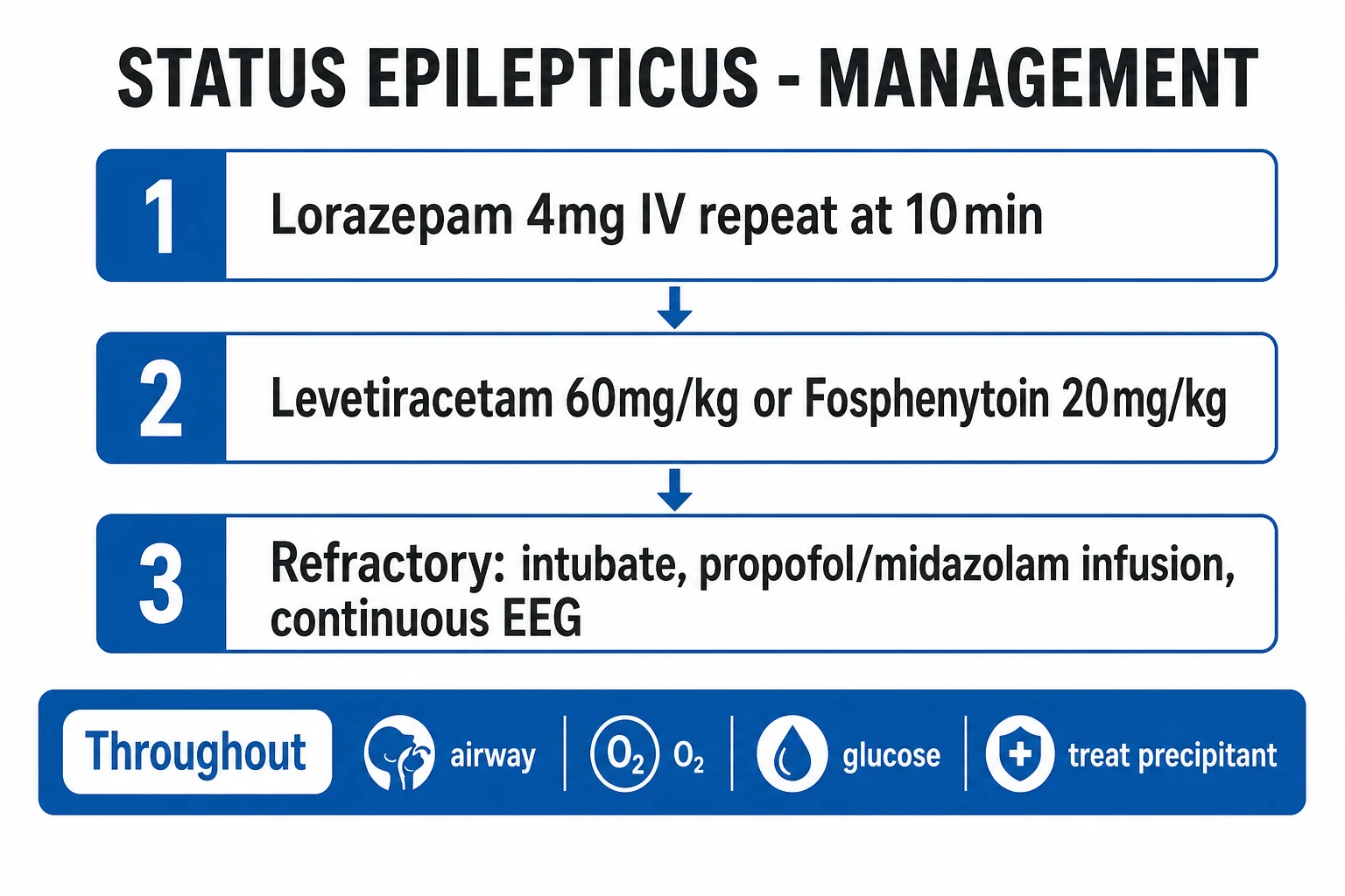
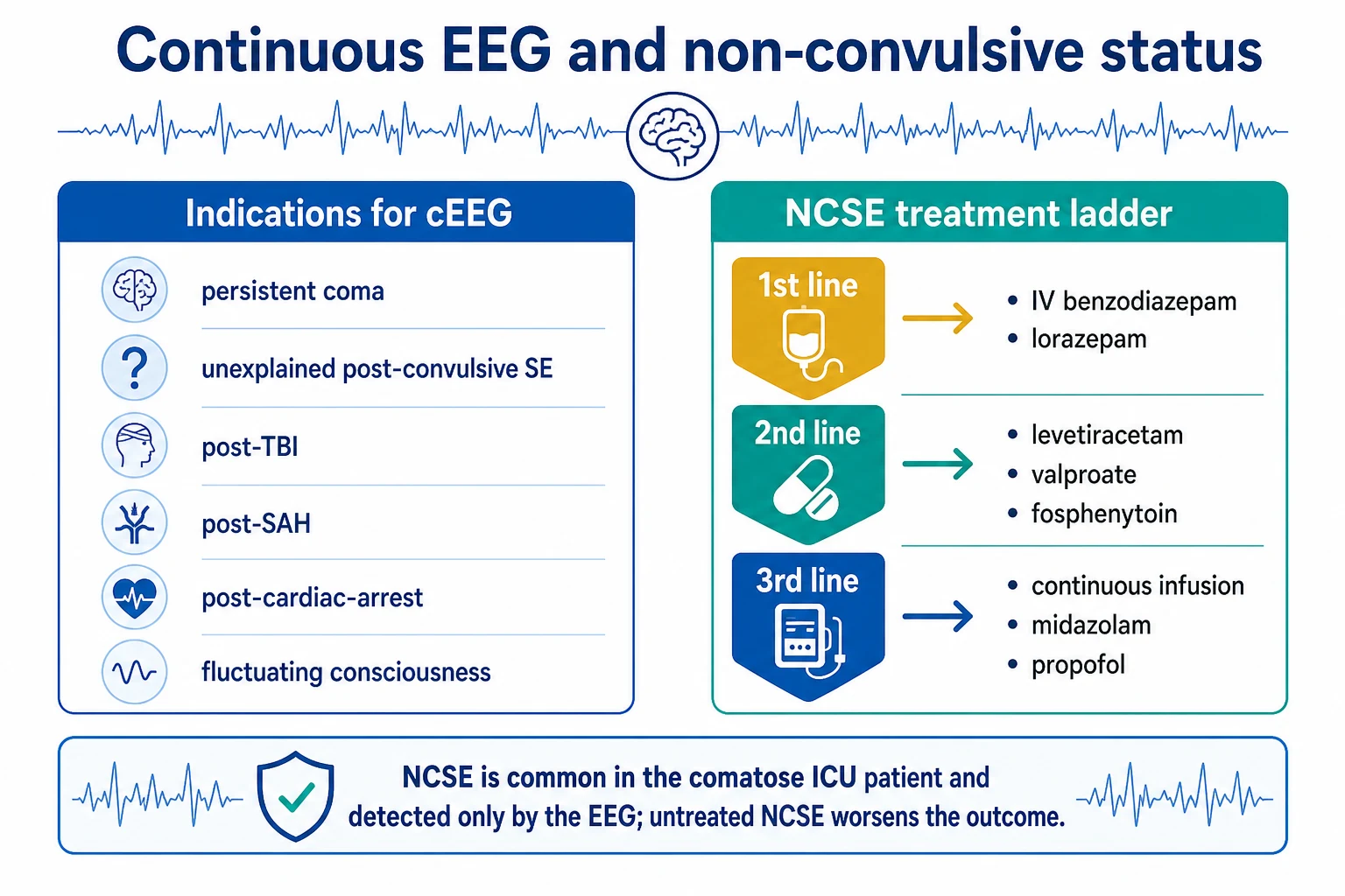
1st line — IV benzodiazepine:[1]
- Lorazepam 2-4 mg IV (or the diazepam 5-10 mg IV). If the EEG improves, the NCSE is confirmed and the benzodiazepine provides the initial treatment.
- Caution: the benzodiazepine causes a sedation and a respiratory depression (the patient may need the intubation); and it may sedate the EEG background (making it harder to interpret). Use the smallest effective dose.
2nd line — the IV antiepileptic:[6]
- Levetiracetam (60 mg/kg IV — the loading dose, then 500 mg twice daily).
- Valproate (20-40 mg/kg IV — the loading dose, then 400 mg three times daily). Caution: the hepatotoxicity, the thrombocytopenia, and the pregnancy.
- Fosphenytoin (20 mg PE/kg IV — the loading dose; faster and safer than the phenytoin — the risk of the infusion-related hypotension and the arrhythmia is lower).
- The choice depends on the patient (the levetiracetam for the hepatic impairment; the valproate for the migraine/bipolar; the fosphenytoin for the focal structural lesion).
- The ESETT trial (2019, NEJM) showed that the levetiracetam, the fosphenytoin, and the valproate were equivalent for the convulsive SE that failed the benzodiazepine (~45-50 per cent each stopped the seizure). The choice is therefore driven by the patient factors, not by a superiority of one agent.[6]
3rd line — the continuous infusion for the refractory:[1]
- Midazolam infusion (0.05-2 mg/kg/h).
- Propofol infusion (the sedation and the antiepileptic effect; caution: the propofol infusion syndrome at the high doses and the prolonged duration).
- Thiopental (the most potent; the deepest suppression; the most side effects — the hypotension, the infection, the paralytic ileus).
- The goal of the continuous infusion: the burst suppression (the EEG pattern of the bursts of activity alternating with the flat suppression) — maintained for 24-48 hours before the weaning and the re-assessment.
The benzodiazepine trial — the diagnostic and the therapeutic test
The benzodiazepine trial is the bedside test that serves both to diagnose the NCSE and to begin the treatment. A small, serial dose of a short-acting benzodiazepine is given while the cEEG records continuously: a clear resolution of the epileptiform pattern (with or without a clinical improvement) confirms the NCSE. The protocol: (1) record a baseline EEG epoch; (2) give the lorazepam 1 mg IV (or the midazolam 1-2 mg IV) in aliquots every 2-5 minutes, up to a defined maximum (e.g. the lorazepam 4 mg total); (3) record the EEG after each aliquot; (4) stop at the resolution of the pattern, or at the clinical deterioration (the hypotension, the respiratory depression), or at the maximum dose.[3]
Interpreting the benzodiazepine trial
| EEG response | Clinical response | Interpretation | Action |
|---|---|---|---|
| Epileptiform pattern resolves | Clinical improvement | NCSE confirmed | Continue the maintenance AED; load the second-line agent |
| Epileptiform pattern resolves | No clinical change (or sedation) | NCSE probable (sedation confounds) | Treat as NCSE; maintain the AED; reassess |
| Pattern attenuates diffusely (background flattens) | Sedation / hypotension | Inconclusive (the drug sedated the background) | Re-assess when the drug clears; trial a non-sedating AED |
| No change in the pattern | No clinical change | NCSE not benzodiazepine-responsive (or not NCSE) | Trial the second-line IV AED (levetiracetam / valproate / fosphenytoin) |
The pitfalls: (a) the benzodiazepine sedates the background, so an apparent "improvement" may be the diffuse slowing of the sedation rather than the resolution of the ictal pattern — have the neurophysiologist interpret the change; (b) a negative trial does not exclude the NCSE (the NCSE may be refractory to the benzodiazepine — the trial of the second-line agent); (c) the benzodiazepine causes the respiratory depression and the hypotension — ensure the airway, the breathing, and the circulation are secure before the trial, and have the vasopressors ready.[1]
The prognostic value of the cEEG
The cEEG provides the prognostic information:[1]
- The reactivity — a reactive EEG (the background changes with the stimulus — a noise, a pain) suggests an intact brainstem-thalamus-cortex circuit and a better prognosis.
- The continuity — a continuous background is better than a discontinuous or a suppressed one.
- The absence of the epileptiform activity — a good sign.
- After the cardiac arrest, the cEEG is part of the multimodality prognostication (the cEEG, the somatosensory evoked potentials, the imaging, the biomarkers).[5]
The post-cardiac-arrest EEG categories (the Westhall criteria)
The standardised interpretation of the post-arrest EEG (the Westhall criteria) groups the patterns into the prognostic categories, used alongside the SSEP, the biomarkers, and the imaging in the multimodality prognostication:[5]
- Highly malignant (predict the poor outcome with a high specificity): the generalised suppression (<10 μV), the burst-suppression without the anaesthesia, the status myoclonus (the continuous or the near-continuous myoclonus with the corresponding EEG spikes), the generalised periodic discharges.
- Malignant (associated with the poor outcome but less specific): the unreactive background, the discontinuous background, the low-voltage background, the epileptiform activity without the status.
- Benign (associated with the good outcome): the reactive continuous background, the normal voltage, the normal sleep transients. [1]
The key principle: a single malignant EEG pattern is not in itself sufficient to prognosticate a poor outcome — the EEG is interpreted as part of the multimodality bundle (the absent N20 SSEP, the highly elevated neuron-specific enolase, the diffuse cortical injury on the imaging), and the prognostication is delayed until at least 24-72 hours after the arrest to avoid the self-fulfilling prophecy.[5]
The SSEP and the EEG together
The bilateral absence of the N20 cortical response of the somatosensory evoked potentials is one of the most specific predictors of the poor outcome after the cardiac arrest. The N20 and the EEG are complementary: the N20 tests the integrity of the somatosensory pathway (the median nerve → the dorsal columns → the thalamus → the cortex), while the EEG reflects the global cortical function. A combined approach (the malignant EEG plus the absent N20) provides the highest specificity for the poor outcome prediction.[9]
The practical setup of the cEEG
A technically adequate cEEG is the prerequisite for the reliable interpretation. The setup, the montage, the duration, and the review process all matter; a poorly applied cEEG produces the artefact-laden tracings that the neurophysiologist cannot read, and that miss the seizures. [1]
The practical setup of the cEEG in the ICU
- THE INDICATION AND THE REQUEST — Confirm the indication (the unexplained coma, the post-convulsive SE, the post-arrest prognostication, the burst-suppression verification, the SAH/DCI monitoring) and the clinical question. A focused request ("exclude the NCSE", "verify the burst suppression", "prognosticate at 48 hours") guides the duration and the interpretation. Obtain the consent (or the surrogate consent) where required; the cEEG itself is non-invasive.[2]
- THE ELECTRODE APPLICATION — Use the full international 10-20 system (21 electrodes minimum — Fp1/Fp2, F3/F4, C3/C4, P3/P4, O1/O2, F7/F8, T3/T4, T5/T6, Fz, Cz, Pz). Apply with the collodion and the conductive gel (the gold standard — secure for days) or the cup electrodes with the paste (faster but less secure). The cup/disc electrodes are preferred over the needle electrodes for the prolonged monitoring. Check the impedances (the target <5 kΩ, balanced between the paired electrodes) — the high or the asymmetric impedances produce the artefacts.[2]
- THE MONTAGE AND THE RECORDING — Record with the referential and the bipolar montages. The bipolar longitudinal (the "double banana") is the standard review montage. Set the filters (the high-frequency 35-70 Hz, the low-frequency 0.5-1 Hz; the 60-Hz/50-Hz notch filter for the mains interference). Sample at ≥256 Hz (≥512 Hz preferred for the spike morphology).[2]
- THE DURATION — The ACNS consensus recommends at least 24 hours to exclude the NCSE with a reasonable confidence (the non-convulsive seizures are intermittent; a shorter recording misses them). The 30-minute rule: roughly half of the non-convulsive seizures are detected in the first 30 minutes, but the remainder need the longer recording; the post-anoxic coma and the high-risk patients need ≥48 hours. Continue until the clinical question is answered (the NCSE excluded, the burst suppression achieved, the prognostic epoch recorded).[2]
- THE QUANTITATIVE EEG (qEEG) TRENDS — Enable the quantitative EEG trends (the compressed spectral array, the density spectral array, the alpha/delta ratio, the asymmetry index, the amplitude-integrated EEG) to allow the bedside staff to screen for the changes over hours. The qEEG does not replace the raw-EEG review (the trends can miss the brief seizures and produce the false positives from the artefacts) but it is the practical tool for the trending and the alarm setting.[2]
- THE PERSONNEL AND THE REVIEW — The cEEG needs the trained personnel: the neurophysiology technologist (the application, the daily electrode checks, the troubleshooting), the neurophysiologist (the daily interpretation, the ictal-pattern calls), and the bedside nurse (the input the events, the recognising the artefacts, the maintaining the electrodes). Review the raw EEG at least daily by the neurophysiologist; review the qEEG trends more frequently (the bedside screening). Document the sedation, the neuromuscular blockade, the temperature, and the medications at the time of the epochs (they all affect the EEG).[2]
- THE ARTEFACT RECOGNITION — The ICU is the electrically hostile environment. The common artefacts: the sweat (the slow baseline drift), the movement (the patient, the staff, the turning), the muscle (the chewing, the shivering, the myoclonus), the ECG and the pacemaker spikes, the IV drip chamber, the ventilator, the bed vibration, and the electrode pops. The artefacts can mimic the seizures (especially the rhythmic movement artefact) — the neurophysiologist distinguishes them with the phase-reversal analysis and the multiple montages. Do not treat an artefact as a seizure.[4]
The NCSE diagnostic workup — from the suspicion to the treatment
- SUSPECT THE NCSE — Any comatose ICU patient with an unexplained or a fluctuating consciousness, or a failure to wake after the convulsive SE or the sedation wean. Recognise the subtle signs (the eye deviation, the automatisms, the nystagmus, the myoclonus, the autonomic surges). Do not attribute the coma to the "sedation" or the "metabolic" cause without the cEEG.[7]
- CORRECT THE REVERSIBLE CAUSES — Check the glucose, the sodium, the calcium, the magnesium, the renal and the hepatic function, the ammonia, the drug levels, the sepsis screen, and the neuroimaging (the CT to exclude the mass lesion, the haemorrhage, the infarct). Treat the hypoglycaemia, the hyponatraemia, the hypoxia, and the sepsis. A reversible metabolic cause does not exclude the NCSE (both can co-exist).[1]
- REQUEST THE cEEG — Request the urgent cEEG (the 10-20 system, the full montage, ≥24 hours). If the cEEG is not immediately available, a routine 30-minute EEG is the interim test (it detects roughly half of the non-convulsive seizures — better than nothing, but not definitive). Document the indication and the clinical question on the request.[2]
- INTERPRET THE PATTERN — Apply the ACNS terminology (LPDs, GPDs, LRDA, GRDA, SIRPIDs) and the Salzburg criteria. Determine whether the pattern is a clear seizure (the evolving rhythm), the ictal-interictal continuum, or the interictal. Consider the modifiers (the frequency >2.5 Hz, the + features, the evolution, the SIRPIDs).[3]
- THE AED TRIAL (if on the ictal-interictal continuum) — Perform the structured benzodiazepine trial (the lorazepam 1 mg aliquots to 4 mg, or the non-sedating AED trial with the levetiracetam or the valproate). Interpret the EEG and the clinical response (the resolution confirms the NCSE; the inconclusive response — treat as the NCSE and reassess; the negative — trial the second-line agent).[3]
- TREAT THE NCSE — Follow the ladder: the 1st-line benzodiazepine (the lorazepam), the 2nd-line IV AED (the levetiracetam / the valproate / the fosphenytoin — ESETT, all equivalent), the 3rd-line continuous infusion (the midazolam / the propofol) for the refractory, titrated to the burst suppression. Continue the maintenance AED (2-3 agents) to allow the anaesthetic wean.[6]
- FIND AND TREAT THE CAUSE — The NCSE is rarely idiopathic in the ICU. Look for the structural cause (the stroke, the haemorrhage, the tumour, the abscess, the HSV encephalitis — the MRI), the autoimmune cause (the anti-NMDA, the LGI1 — the antibodies, the immunotherapy), the infectious cause (the encephalitis, the meningitis — the CSF), the metabolic cause, and the drug withdrawal (the alcohol, the benzodiazepine, the AED non-adherence).[1]
- MONITOR AND WEAN — Continue the cEEG throughout the treatment and the wean. Confirm the seizure cessation for 24-48 hours before the gradual anaesthetic taper. Re-assess the EEG at each step of the wean (a recurrence restarts the infusion and extends the duration).[1]
cEEG vs the routine EEG — when each is appropriate
| Feature | Routine EEG (20-30 min) | Continuous EEG (≥24 h) |
|---|---|---|
| Detection of the non-convulsive seizures | Detects ~50% (misses the intermittent) | Detects ~90%+ over 24-48 h |
| Best for | The first-line screen; the post-ictal assessment | The suspected NCSE; the post-arrest prognostication; the burst-suppression verification |
| The intermittent seizure risk | High miss rate | Low miss rate |
| The qEEG trending | Not applicable | Enables the bedside trending and the alarms |
| The personnel demand | Low (a single recording) | High (the technologist, the daily neurophysiology review) |
| The cost / the access | Lower, widely available | Higher, requires the specialist infrastructure |
| The recommendation | The interim test if the cEEG is unavailable | The standard for the comatose ICU patient with the suspected NCSE |
Exam practice
SAQ — Persistent coma after convulsive SE: detecting NCSE
10 minutes · 10 marks
A 62-year-old man was admitted after a 25-minute generalised convulsive status epilepticus that terminated with 8 mg IV lorazepam and levetiracetam 60 mg/kg. Twelve hours later he remains comatose (GCS 7). He has no motor seizure activity but you notice occasional subtle eyelid twitching and a fluctuating conscious level — he briefly opens his eyes to voice, then becomes unresponsive. The cEEG shows continuous rhythmic 3 Hz activity with lateralised periodic discharges over the left temporal region.
SAQ — Post-cardiac-arrest EEG prognostication and the malignant patterns
10 minutes · 10 marks
A 58-year-old man is in the ICU 48 hours after an out-of-hospital cardiac arrest. He was cooled to 36°C for 24 hours and is now normothermic, off sedation for 12 hours. His GCS is 4 (opens eyes to pain, extensor posturing, no verbal response). The cEEG shows a suppressed background (under 10 microvolts) with no reactivity to the painful stimulus, and there are brief bursts of activity alternating with the flat periods. There is intermittent myoclonus. The bilateral somatosensory evoked potentials show absent N20 cortical responses.
Clinical pearls
Red flags
[1]Prognosis
The evidence and the outcomes for the cEEG and the NCSE
The NCSE prevalence: the non-convulsive seizures are detected by the cEEG in roughly 8-10 per cent of the comatose ICU patients screened, rising to 20-30 per cent in those with the acute brain injury and the altered consciousness (Claassen 2004; ACNS consensus 2015).[7] The NCSE after the convulsive SE: up to 14-50 per cent of the patients who do not promptly wake after the convulsive SE have the NCSE.[7] The Westhall post-arrest categories (2016, Neurology): the highly malignant patterns (the suppression, the burst-suppression, the status myoclonus, the GPDs) predict the poor outcome with a high specificity; the reactive continuous background is reassuring. The EEG is the reliable prognostic tool when interpreted with the strict criteria and the multimodality bundle.[5] The ESETT (2019, NEJM): the levetiracetam = the fosphenytoin = the valproate for the convulsive SE that failed the benzodiazepine (~45-50 per cent each). The second-line choice is driven by the patient factors, not the efficacy.[6] The Treiman VA cooperative (1998, NEJM): the early benzodiazepine (the lorazepam) is the effective first-line for the convulsive SE — the foundation of the staged treatment that the NCSE inherits.[8] The clinical bottom line: the seizures and the NCSE worsen the outcome after the TBI, the SAH, the convulsive SE, and the cardiac arrest. The early cEEG detection and the staged treatment (the benzodiazepine → the second-line AED → the anaesthetic infusion) are the interventions that change the outcome.
Key takeaways
- The NCSE is common (10-30 per cent of the comatose ICU patients) and detectable only by the cEEG. The clinical signs are subtle and fluctuating; the EEG is the test, not the adjunct.
- Request the cEEG for the unexplained or the fluctuating coma, after the convulsive SE (confirm the seizure stopped), after the cardiac arrest (the prognostication), and during the anaesthetic coma (the burst-suppression verification).
- The ACNS terminology (LPDs, GPDs, LRDA, GRDA, SIRPIDs) standardises the description; the Salzburg criteria diagnose the NCSE (the epileptiform activity plus the AED response).
- The frequency determines the ictal likelihood — the >2.5 Hz favours the treatment; the ictal-interictal continuum needs the AED trial.
- The treatment ladder: the 1st-line benzodiazepine, the 2nd-line levetiracetam / valproate / fosphenytoin (the ESETT — all equivalent), the 3rd-line anaesthetic infusion titrated to the burst suppression.
- The seizures worsen the outcome after the TBI, the SAH, the convulsive SE, and the cardiac arrest — the early detection and the treatment are the intervention that changes the outcome.
- The cEEG is mandatory in the paralysed patient — the clinical signs are abolished; without the EEG the NCSE is undetectable.
- Do not prognosticate on a single EEG pattern — use the multimodality bundle (the EEG, the SSEP, the NSE, the imaging) and the delay to 24-72 hours. [1]
References
- [1]Maganti R, Gerber P, Drees C, Chung S Nonconvulsive status epilepticus. Epilepsy & behavior : E&B, 2008.PMID 18248774
- [2]Herman ST, Abend NS, et al. Consensus statement on continuous EEG in critically ill adults and children, part I: indications. J Clin Neurophysiol, 2015.PMID 25626778
- [3]Beniczky S, Hirsch LJ, et al. Unified EEG terminology and criteria for nonconvulsive status epilepticus (Salzburg). Epilepsia, 2013.PMID 24001066
- [4]Maciel CB, Hirsch LJ, et al. Definition and classification of periodic and rhythmic patterns (ACNS). J Clin Neurophysiol, 2018.PMID 29718827
- [5]Westhall E, Rossetti AO, et al. Standardized EEG interpretation accurately predicts prognosis after cardiac arrest. Neurology, 2016.PMID 26865516
- [6]Kapur J, Elm J, et al. (ESETT) Randomized trial of three anticonvulsant medications for status epilepticus. N Engl J Med, 2019.PMID 31774955
- [7]Claassen J, Mayer SA, et al. Detection of electrographic seizures with continuous EEG monitoring in critically ill patients. Neurology, 2004.PMID 15159471
- [8]Treiman DM, Meyers PD, et al. A comparison of four treatments for generalized convulsive status epilepticus (VA cooperative). N Engl J Med, 1998.PMID 9738086
- [9]Ruijter BJ, et al. Association between somatosensory evoked potentials and EEG in comatose patients after cardiac arrest. Clin Neurophysiol, 2019.PMID 31541979