ICU · Neurocritical care / neuromuscular
Critical-Illness Polyneuropathy & Myopathy (CIPNM) — ICU-Acquired Weakness
Also known as Critical illness polyneuropathy · CIP · Critical illness myopathy · CIM · CIPNM · ICU-acquired weakness · ICUAW · Failure to wean · Thick-filament myosin loss · MRC sum score · Van den Berghe · NICE-SUGAR · Early mobilisation
The critical-illness polyneuropathy and myopathy (CIPNM, the ICU-acquired weakness) is an acquired weakness of the critically ill patient, developing after the sepsis, the multi-organ failure, or the prolonged ventilation. It comprises two overlapping entities — the critical illness polyneuropathy (the CIP, an axonal sensorimotor neuropathy) and the critical illness myopathy (the CIM, a myosin-loss myopathy, classically from the corticosteroids plus the neuromuscular blockers in the asthma and the ARDS). The clinical hallmark is the failure to wean from the ventilation, with the flaccid weakness, the areflexia, and the muscle wasting — the cranial nerves are SPARED (a useful discriminator from the Guillain-Barre). The only proven prevention is the glucose control (the Van den Berghe trial) and the early mobilisation; minimise the sedation, the corticosteroids, and the neuromuscular blockers. The recovery is over weeks to months and is often incomplete (a long-term disability).
On this page & tools
Your progress
Saved locally on this device.
8 MCQs with explanations
Target exams
Overview & definition
The critical-illness polyneuropathy and myopathy (CIPNM) — also called the ICU-acquired weakness (ICUAW) — is an acquired weakness that develops in the critically ill patient during the ICU stay, after the sepsis, the systemic inflammation, the multi-organ failure, or the prolonged ventilation. It comprises two overlapping entities:[1]
- The critical illness polyneuropathy (CIP) — an axonal sensorimotor polyneuropathy (the distal predominance).[1]
- The critical illness myopathy (CIM) — a myopathy with the thick-filament (myosin) loss, classically from the combination of the corticosteroids and the neuromuscular blockers in the severe asthma and the ARDS (the proximal predominance).[1]
The clinical hallmark is the failure to wean from the ventilation in the awakening patient, with the flaccid weakness, the areflexia, and the muscle wasting — the cranial nerves are spared (a useful discriminator from the Guillain-Barre).[1]
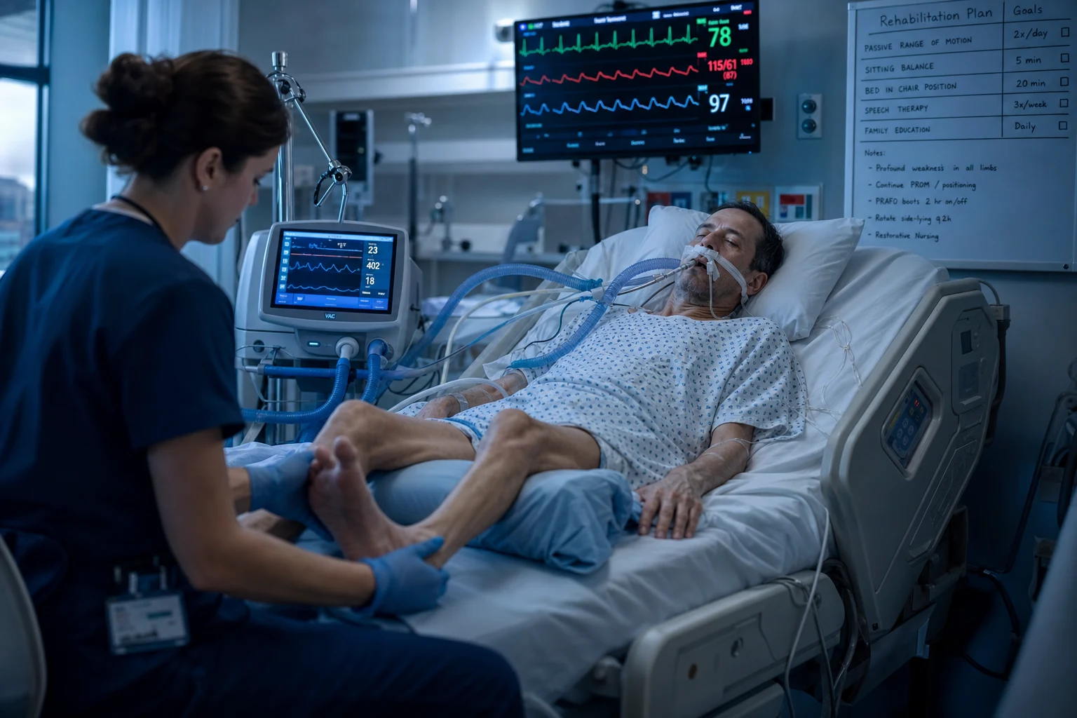
The risk factors
- The sepsis and the systemic inflammation (SIRS) — the central driver (the inflammatory mediators damage the nerve and the muscle).[1]
- The multi-organ failure.[1]
- The prolonged ventilation and the immobility.[1]
- The corticosteroids plus the neuromuscular blockers — the classic combination for the critical illness myopathy (especially in the severe asthma and the ARDS).[1]
- The hyperglycaemia (a modifiable factor — see the prevention).[1]
- The vasopressors, the renal failure, the female sex, the older age.[1]
The clinical recognition
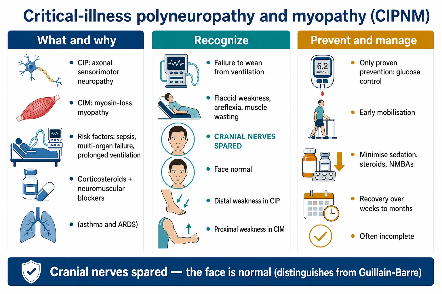
- The failure to wean from the ventilation — the commonest ICU sign (the weak respiratory muscles cannot sustain the spontaneous breathing).[1]
- The flaccid weakness, the areflexia, and the muscle wasting — in the awakening patient. The weakness is the distal in the CIP and the proximal in the CIM.[1]
- The cranial nerves are SPARED — the face is normal. This is a key discriminator from the Guillain-Barre (the facial diplegia) and an alternative diagnosis. A facial weakness in the suspected CIPNM suggests another cause.[1]
- The sensory loss — present but hard to assess in the sedated or the encephalopathic ICU patient.[1]
- The deep tendon reflexes are reduced or absent.[1]
The diagnosis
The diagnosis is clinical (in the awakening ICU patient who fails to wean, with the flaccid weakness and the areflexia), supported by the investigations:[1]
- The nerve conduction studies and the EMG — the CIP shows the axonal sensorimotor neuropathy (the reduced CMAP and SNAP, the normal conduction velocity); the CIM shows the small CMAP with the normal sensory potentials (the direct muscle stimulation distinguishes the nerve from the muscle origin). The myosin-loss myopathy is characteristic.[1]
- The creatine kinase — mildly raised in the CIM (a marked rise suggests a necrotising myopathy or the rhabdomyolysis).[1]
- The MRC sum score (the Medical Research Council) — a 0 to 5 grade in the 6 muscle groups (3 upper, 3 lower limbs), the sum out of 60; under 48 defines the weakness.[1]
- Exclude the others: the residual neuromuscular-blocker effect (the train-of-four), the Guillain-Barre (the pre-ICU onset, the albuminocytologic dissociation, the facial diplegia), the electrolyte disorders (the potassium, the phosphate, the magnesium), the spinal cord lesion, the central weakness from the stroke.[1]
The prevention
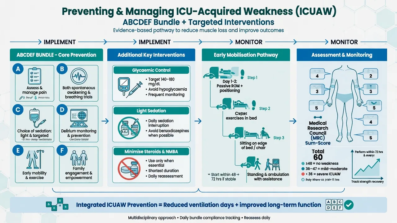
The prevention is the best strategy — once established, the CIPNM is only supportive:[1]
- The glucose control — the only intervention with the proven prevention (the Van den Berghe Leuven trial, NEJM 2001, showed the intensive insulin reduced the CIP/CIM). However, the subsequent NICE-SUGAR trial (NEJM 2009) showed that the tight control (the 4.4 to 6.1) caused the hypoglycaemia with no outcome benefit, so the moderate control (the 8 to 10) is now the standard — which does not prevent the CIPNM as strongly. Avoid the hyperglycaemia.[1]
- The early mobilisation and the rehabilitation — the regular passive and the active movement, the sitting, the standing, and the early walking; reduces the weakness and the duration.[1]
- Minimise the sedation (the daily sedation interruptions, the protocolised weaning).[1]
- Minimise the corticosteroids and the neuromuscular blockers (especially the combined use).[1]
- The spontaneous breathing trials, the early nutrition.[1]
The management and the prognosis
- The supportive care — the respiratory support (the weaning, the tracheostomy if the prolonged), the prevention of the complications (the DVT, the pressure areas, the contractures), the nutrition, the pain control.[1]
- The rehabilitation — the intensive, multidisciplinary physiotherapy.[1]
- The recovery — over the weeks to the months; the CIP recovers better than the CIM; the recovery is often incomplete, with a long-term disability (the ICU-acquired weakness persists in many survivors for the months to the years, with the reduced quality of life).[1]
Red flags
CIP versus CIM — the two entities distinguished
The CIP and the CIM are the two overlapping entities within the CIPNM/ICUAW syndrome. They share the same clinical milieu (the sepsis, the multi-organ failure, the prolonged ventilation) but differ in the primary target (the nerve versus the muscle), the histology, the electrophysiology, and the recovery. In practice they coexist in the majority of the severe cases — the term critical illness neuromyopathy (CINM) is used when both are present.[8][1]
CIP versus CIM — the two entities of the CIPNM syndrome
| Feature | Critical illness polyneuropathy (CIP) | Critical illness myopathy (CIM) |
|---|---|---|
| Primary target | The peripheral nerve (the axon) | The skeletal muscle fibre |
| Onset | After 1-3 weeks of the critical illness (often the sepsis or the MOF) | Days to weeks — classically after the steroids plus the NMBAs (the asthma, the ARDS) |
| Histology | The axonal degeneration (the distal-predominant) of the motor AND the sensory fibres — no demyelination | The thick-filament (myosin) loss — selective loss of the myosin filaments with relative sparing of the actin; may be a necrotising or a myosin-loss type |
| Weakness pattern | The distal > proximal (the foot drop, the hand weakness) — sensorimotor | The proximal > distal (the hip and the shoulder girdle, the neck flexors) — pure motor |
| Sensation | Reduced (the distal sensory loss in the glove-and-stocking) — but hard to assess in the ICU | Normal (the pure myopathy) |
| Deep tendon reflexes | Reduced or absent (especially distally) | Reduced or absent |
| Cranial nerves | SPARED (the face is normal) | SPARED (the face is normal) |
| CK | Normal or mildly raised | Mildly to moderately raised (a marked rise suggests the necrotising myopathy or the rhabdomyolysis) |
| Nerve conduction (CMAP) | Reduced (the axonal loss) | Reduced (the muscle fibre loss) |
| Nerve conduction (SNAP) | Reduced (the sensory axonal loss — KEY: distinguishes from CIM) | Normal (the sensory nerve is unaffected) |
| Conduction velocity | Normal (the axonal, NOT the demyelinating — distinguishes from the GBS) | Normal |
| Needle EMG | The fibrillation potentials and the positive sharp waves (the denervation); reduced recruitment | The fibrillation potentials and the small, short, polyphasic motor-unit potentials (the myopathic units) |
| Direct muscle stimulation | The CMAP is preserved (the muscle responds normally to the direct stimulation) | The CMAP is reduced even with the direct stimulation (the muscle itself is diseased) — the gold standard to separate the CIM from the CIP |
| Respiratory muscle involvement | Yes — the diaphragm and the intercostals (the failure to wean) | Yes — even more pronounced (the diaphragm myopathy) |
| Recovery | Better (the axonal regrowth at 1 mm/day — weeks to months) | Slower and often incomplete (the myosin regeneration is slow) |
| Classic setting | The sepsis, the multi-organ failure, the prolonged ventilation | The severe asthma or the ARDS treated with the corticosteroids and the neuromuscular blockers |
Incidence and epidemiology
The CIPNM is the commonest neuromuscular disorder of the ICU. The incidence depends on the population studied and the diagnostic method (the clinical MRC sum score versus the electrophysiology):[5][8]
- Around 25-50% of the general ICU patients who are ventilated for more than 7 days develop a clinically detectable ICU-acquired weakness (the MRC sum score under 48).[5]
- Up to 100% of the severe ARDS patients develop an electrophysiological CIPNM if the ICU stay is prolonged (the sepsis, the steroids, the NMBAs, and the prolonged ventilation all converge).[1]
- Around 60-80% of the patients with the septic shock and the multi-organ failure have the electrophysiological evidence of the CIP/CIM by the second week.[8]
- The CIM is more common than the CIP when the steroids and the NMBAs are used (the asthma and the ARDS); the CIP is more common in the pure sepsis and the multi-organ failure.
- The CIPNM is under-recognised — the sedation, the encephalopathy, and the bed-bound state mask the weakness until the patient fails to wean.
Pathophysiology
The CIPNM is not a single disease but a final common pathway of the nerve and the muscle damage from the critical illness. The key mechanisms:[1][7][8]
- The microvascular and the metabolic derangement — the sepsis and the systemic inflammation cause the microcirculatory failure (the capillary leak, the oedema, the hypoxia) in the vasa nervorum and the muscle capillaries. The nerve and the muscle become energy-starved and the axonal transport fails.
- The hyperglycaemia and the mitochondrial dysfunction — the hyperglycaemia damages the mitochondria of the nerve and the muscle (the oxidative stress); the intensive insulin (the Van den Berghe trial) reduced the CIPNM by restoring the mitochondrial function.[2]
- The channelopathy and the membrane depolarisation — the inflammatory mediators (the TNF, the IL-1, the IL-6) and the steroid-induced catabolism cause the inexcitable nerve and muscle membranes (the sodium-potassium pump failure, the resting membrane potential depolarisation). This produces the weakness BEFORE the structural damage is visible.
- The muscle wasting (the acute sarcopenia) — the Puthucheary study (JAMA 2013) showed that the rectus femoris cross-sectional area on the ultrasound drops by around 12-15% in the first week of the critical illness, and by over 30% by day 10 in the severe cases. The wasting is driven by the proteolysis (the ubiquitin-proteasome pathway) and the impaired protein synthesis.[7]
- The myosin-loss myopathy (the CIM) — the high-dose corticosteroids, especially combined with the NMBAs, cause a selective loss of the thick (myosin) filaments. The mechanism is the steroid-induced calpain activation and the disuse (the NMBAs abolish the muscle contraction → the muscle-disuse atrophy is amplified).
- The disuse and the immobilisation — the bed rest alone causes a 1-1.5% loss of the muscle mass per day; the critical illness amplifies this many-fold.
Risk factors — detailed
The risk factors for the CIPNM — modifiable versus non-modifiable
| Factor | Modifiable? | Mechanism | Relative risk / note |
|---|---|---|---|
| The sepsis and the systemic inflammation (SIRS) | Partially (the source control, the antibiotics) | The inflammatory mediators damage the nerve and the muscle; the central driver | The strongest single risk factor — present in the majority of the cases |
| The multi-organ failure | Partially | The cumulative catabolic and the hypoxic burden | The risk rises with the number of the failing organs |
| The hyperglycaemia | YES — the glycaemic control | The mitochondrial dysfunction, the oxidative stress | The Van den Berghe trial: the intensive insulin reduced the CIPNM by ~44%[2] |
| The prolonged immobilisation / the bed rest | YES — the early mobilisation | The disuse atrophy (1-1.5% mass/day), the contractures | The early mobilisation (the TEAM trial) is the prevention[13] |
| The prolonged deep sedation | YES — the sedation minimisation | The immobility, the suppressed muscle activity | The daily sedation interruptions (Kress 2000) reduce the ventilation duration[10] |
| The corticosteroids | YES — minimise | The myosin-loss myopathy (the calpain activation, the catabolism) | Especially combined with the NMBAs (the asthma, the ARDS) |
| The neuromuscular blocking agents (NMBAs) | YES — minimise, short courses only | The disuse, the upregulation of the acetylcholine receptors | The ACURASY trial showed a 48h cisatracurium infusion improved the ARDS survival but the ROSE trial did NOT confirm this — the routine prolonged NMBA is NOT recommended[11][12] |
| The prolonged mechanical ventilation | Partially (the early weaning) | The diaphragm dysfunction (the VIDD — the ventilator-induced diaphragm dysfunction) | The diaphragm loses 6-7% of its force per day of the controlled ventilation |
| The female sex, the older age | No | The lower baseline muscle mass | Increases the risk and the severity |
| The vasopressors, the renal failure, the low albumin | Partially | The cumulative severity marker | The markers of the severe illness |
| The vitamin D deficiency | Possibly (the supplementation) | The muscle weakness, the immune dysfunction | The association is observational; the supplementation trials are inconclusive |
Diagnosis — the systematic approach
The diagnosis is clinical (in the awakening ICU patient who fails to wean, with the flaccid weakness and the areflexia), supported by the electrophysiology and the exclusion of the mimics. The 2014 American Thoracic Society guideline (Fan et al) defines the diagnostic criteria.[8]
Diagnostic workup for the suspected CIPNM
- SUSPECT THE CIPNM when an awakening ICU patient (after the sepsis, the multi-organ failure, or the prolonged ventilation) develops the failure to wean with the flaccid limb weakness, the areflexia, and the muscle wasting. The cranial nerves are SPARED (the face normal). The weakness is the distal (the CIP) or the proximal (the CIM).
- CONFIRM COOPERATIVITY — the MRC sum score requires a cooperative patient who can follow the commands. The delirium and the residual sedation confound the assessment. Use the CAM-ICU to exclude the delirium; allow a sedation hold before the testing.
- THE MRC SUM SCORE (the bedside gold standard):
- Grade 6 muscle groups bilaterally: the shoulder abduction, the elbow flexion, the wrist extension (upper); the hip flexion, the knee extension, the ankle dorsiflexion (lower).
- Each 0-5 (0 = no contraction, 5 = normal power against resistance).
- Sum out of 60. Under 48 = the ICU-acquired weakness.
- A mean MRC grade under 4 (out of 5) across the 12 groups is an alternative threshold.
- Document the trend (the daily or the alternate-day scoring) — the trajectory matters.
- THE BLOODS:
- The creatine kinase (CK) — mildly raised in the CIM (a marked rise suggests the necrotising myopathy, the rhabdomyolysis, or the statin myopathy). Normal or near-normal in the pure CIP.
- The electrolytes — the potassium, the phosphate, the magnesium, the calcium (the hypokalaemia, the hypophosphataemia, the hypomagnesaemia, and the hypercalcaemia/hypocalcaemia all cause the weakness).
- The magnesium, the urea, the creatinine (the renal failure and the uraemia contribute).
- The TSH and the free T4 (the hypothyroid myopathy).
- The B12 and the folate (the subacute combined degeneration).
- The albumin (the low albumin is a marker of the severity).
- THE NERVE CONDUCTION STUDIES (NCS) AND THE NEEDLE EMG — the gold-standard electrophysiology:
- The CIP: the axonal sensorimotor neuropathy — the reduced CMAP and SNAP, the normal conduction velocity (the axonal, NOT the demyelinating). The needle EMG shows the fibrillation potentials (the denervation).
- The CIM: the small CMAP with the NORMAL sensory potentials (SNAP). The needle EMG shows the small, short, polyphasic motor-unit potentials (the myopathic units) and the early recruitment.
- The direct muscle stimulation (DMS) — the nerve is blocked and the muscle is stimulated directly. If the CMAP is preserved with the DMS but reduced with the neural stimulation → the CIP. If the CMAP is reduced EVEN with the DMS → the CIM (the muscle itself is diseased). This is the definitive way to separate the CIM from the CIP.
- The NCS may be normal in the first week (the membrane inexcitability precedes the structural damage) — repeat if initially normal.
- THE MUSCLE AND THE NERVE BIOPSY — rarely needed (the research or the diagnostic uncertainty). The CIM shows the thick-filament (myosin) loss; the CIP shows the axonal degeneration. Reserve for the atypical cases (the suspected necrotising myopathy, the vasculitic neuropathy).
- THE IMAGING:
- The muscle ultrasound — the increased echogenicity and the reduced rectus femoris cross-sectional area (the wasting). Useful for the serial monitoring.
- The MRI of the spine — only if a cord lesion is suspected.
- The chest X-ray and the CT brain — to exclude the alternative diagnoses.
- EXCLUDE THE MIMICS (the critical step before labelling the CIPNM):
- The residual neuromuscular-blocker effect — the train-of-four (aim for 4 of 4 twitches, or the post-tetanic count), the head lift for 5 seconds, the qualitative peripheral nerve stimulator. Allow the full recovery of the NMBA before the assessment.
- The Guillain-Barre syndrome (GBS) — the pre-ICU onset (the ascending weakness over the days before the admission), the facial diplegia (the cranial nerve INVOLVEMENT — the opposite of the CIPNM), the CSF albuminocytologic dissociation, the demyelinating (not the axonal) NCS in the AIDP variant.
- The electrolyte disorders — the hypokalaemia, the hypophosphataemia (especially the refeeding), the hypomagnesaemia, the hypercalcaemia. Correctable.
- The spinal cord lesion — the transverse myelitis, the cord compression, the epidural abscess. A sensory level, the sphincter dysfunction, and the upper motor neuron signs (the hyperreflexia, the Babinski) below the level distinguish it from the CIPNM (which is the lower motor neuron).
- The central weakness — the stroke, the intracerebral haemorrhage, the subdural haematoma. The imaging excludes this.
- The myasthenia gravis — the fluctuating weakness, the ptosis, the fatigability, the positive anti-AChR antibodies.
- The necrotising myopathy or the rhabdomyolysis — the marked CK rise, the myoglobinuria, the renal failure.
- The drug-induced — the statins, the colchicine, the antimalarials, the aminoglycosides, the vincristine.
- CONFIRM THE DIAGNOSIS — the CIPNM is a diagnosis of the clinical syndrome (the awakening patient, the failure to wean, the flaccid weakness, the areflexia, the cranial nerves spared) plus the supportive electrophysiology (the axonal neuropathy and/or the myopathy), having excluded the mimics. There is no single confirmatory test.
The electrophysiology — the CIP versus the CIM versus the GBS
| Parameter | CIP | CIM | GBS (AIDP) |
|---|---|---|---|
| CMAP (compound muscle action potential) | Reduced | Reduced | Reduced (or normal early) |
| SNAP (sensory nerve action potential) | Reduced (the key: the sensory axon is lost) | Normal (the sensory nerve is spared) | Reduced (or normal early) |
| Conduction velocity | Normal (the axonal) | Normal | Slow (the demyelinating) |
| Distal latency | Normal | Normal | Prolonged |
| F-waves | Normal or mildly abnormal | Normal | Prolonged or absent (the proximal demyelination — the earliest sign) |
| Conduction block | Absent | Absent | Present (the hallmark of the demyelination) |
| Needle EMG | Fibrillation + the reduced recruitment (the denervation) | Small, short, polyphasic units + the early recruitment (the myopathy) | Fibrillation + the reduced recruitment |
| Direct muscle stimulation | The CMAP preserved (the muscle is normal) | The CMAP reduced (the muscle is diseased) | The CMAP preserved |
| CSF protein | Normal | Normal | Elevated (the albuminocytologic dissociation) |
| Cranial nerves | SPARED | SPARED | Involved (the facial diplegia 50%) |
Prevention — the evidence bundle
The prevention is the only effective strategy — once established, the CIPNM has no specific treatment and is only supportive. The prevention targets the modifiable risk factors. The evidence base:[8]
The prevention strategies — the evidence for each
| Strategy | Evidence | Mechanism | Recommendation |
|---|---|---|---|
| The glycaemic control | The Van den Berghe Leuven surgical ICU trial (NEJM 2001): the intensive insulin (4.4-6.1 mmol/L) reduced the CIP/CIM by 44% and the mortality. BUT the NICE-SUGAR trial (NEJM 2009) showed the tight control caused the hypoglycaemia with no mortality benefit → the moderate control (8-10 mmol/L) is now the standard. The moderate control does NOT prevent the CIPNM as strongly as the intensive — but the intensive is too dangerous. | The mitochondrial protection, the reduced oxidative stress | Avoid the hyperglycaemia. The target 8-10 mmol/L (NICE-SUGAR).[2][4] |
| The early mobilisation | The Schweickert trial (Lancet 2009): the early PT/OT in the mechanically ventilated patients returned more patients to independent function at hospital discharge (59% vs 35%). The TEAM trial (NEJM 2022, Hodgson): the early, goal-directed active mobilisation was SAFE but did NOT improve the outcome at 180 days (the primary outcome was neutral). | The prevention of the disuse atrophy, the maintenance of the muscle mass | The early mobilisation is SAFE and recommended, but the aggressive protocolised mobilisation (TEAM) did not improve the outcomes — the benefit is modest.[6][13][14] |
| The sedation minimisation | The Kress trial (NEJM 2000): the daily sedation interruptions reduced the ventilation duration and the ICU stay. The PADIS guidelines (2018) recommend the light sedation, the daily interruptions, and the protocolised weaning. | The reduced immobility, the earlier awakening, the earlier mobilisation | The light sedation, the daily interruptions, the SAT + SBT (the awakening and the breathing trials).[10] |
| The minimisation of the corticosteroids and the NMBAs | The combination of the high-dose steroids and the NMBAs causes the CIM (the myosin loss). The ACURASY trial (NEJM 2010, Papazian) showed a 48h cisatracurium infusion improved the severe ARDS survival, BUT the ROSE trial (NEJM 2019) did NOT confirm this → the routine prolonged NMBA is NOT recommended. | The prevention of the myosin-loss myopathy | Use the NMBAs for the shortest possible course; avoid the combination with the high-dose steroids if possible.[11][12] |
| The spontaneous breathing trials (SBTs) | The daily SBTs reduce the ventilation duration. | The earlier weaning → the shorter ICU stay → the less disuse | The daily SBTs as soon as the patient is ready. |
| The early nutrition | The early enteral nutrition (within 48h) supports the muscle mass. | The reduced catabolism | The early enteral nutrition; the high-protein (1.5-2.0 g/kg/day) for the catabolic state. |
| The vitamin D | The observational association between the low vitamin D and the ICUAW; the supplementation trials are inconclusive. | The muscle function, the immune modulation | The supplementation is reasonable if the deficiency is documented; not a proven prevention. |
| The intensive insulin (the intensive control) | The Van den Berghe Leuven trials — but the hypoglycaemia risk and the NICE-SUGAR refutation make this UNACCEPTABLE in the modern practice. | — | NOT recommended — the moderate 8-10 control is the standard. |
Impact on outcomes
The CIPNM is not a benign complication — it has a profound impact on the short-term and the long-term outcomes. The weakness is a major driver of the prolonged ventilation, the longer ICU and the hospital stay, the higher mortality, and the long-term disability.[5][9]
The impact of the CIPNM on the outcomes
| Outcome | Effect of the CIPNM | Mechanism / note |
|---|---|---|
| The prolonged mechanical ventilation | Doubled or tripled | The weak respiratory muscles (the diaphragm and the intercostals) cannot sustain the spontaneous breathing → the failure to wean → the prolonged ventilation. The CIPNM is the commonest neurological cause of the failure to wean. |
| The prolonged ICU stay | Increased by 5-15 days | The weakness delays the mobilisation and the weaning. |
| The prolonged hospital stay | Increased by 2-4 weeks | The rehabilitation and the slow recovery. |
| The mortality | Increased by around 1.5-2x in the severe cases | The weakness is a marker of the severe illness, AND the weakness itself contributes (the failed weaning, the VAP, the immobility complications — the DVT, the pressure areas, the infections). The De Jonghe study and the cohort studies show the independent association. |
| The 1-year mortality | Increased | The CIPNM at the ICU discharge predicts the 1-year mortality. |
| The long-term disability | Persistent in many survivors | The Herridge 5-year ARDS study (NEJM 2011) showed that the ICU survivors had the persistent weakness, the reduced 6-minute-walk distance (only 76% of the predicted at 5 years), the chronic pain, the fatigue, and the reduced quality of life. The muscle weakness was the dominant problem. |
| The quality of life | Reduced for the months to the years | The weakness, the fatigue, the anxiety, the depression, the PTSD. |
| The ICU readmission | Increased | The residual weakness and the deconditioning. |
| The tracheostomy rate | Increased | The failed weaning → the tracheostomy for the prolonged ventilation. |
| The VAP (the ventilator-associated pneumonia) | Increased | The prolonged ventilation → the VAP risk. |
| The DVT and the PE | Increased | The immobility and the hypercoagulability. |
| The pressure areas and the contractures | Increased | The immobility and the reduced movement. |
Management — the ICU protocol
There is no specific treatment for the established CIPNM — the management is the supportive care, the rehabilitation, and the prevention of the further weakness. [1]
The CIPNM management protocol in the ICU
- CONFIRM THE DIAGNOSIS AND EXCLUDE THE MIMICS (see the diagnostic workup). The most reversible mimics are the residual NMBA effect (the train-of-four), the electrolyte disorders (the potassium, the phosphate, the magnesium), and the drug-induced myopathy (the statins — stop them if the CK is raised).
- OPTIMISE THE GLYCAEMIC CONTROL — the target 8-10 mmol/L (the NICE-SUGAR). Avoid the hyperglycaemia (which worsens the weakness) AND the hypoglycaemia (which is dangerous). The intensive control (4.4-6.1) is NOT recommended (the hypoglycaemia risk).
- MINIMISE THE SEDATION — the light sedation (the Richmond Agitation-Sedation Scale 0 to -1), the daily sedation interruptions (the SAT), the protocolised weaning. Avoid the benzodiazepines (the delirium risk) — prefer the propofol or the dexmedetomidine.
- STOP OR MINIMISE THE CORTICOSTEROIDS AND THE NMBAs — if the steroids are necessary (the ARDS, the refractory shock), use the lowest effective dose and the shortest course. The NMBAs should be the short courses only (not the routine prolonged infusion).
- THE EARLY MOBILISATION AND THE REHABILITATION — the bed mobility, the sitting on the edge of the bed, the standing, the marching on the spot, the early walking as the patient tolerates. The passive range-of-motion if the patient cannot move actively. The in-bed cycling ergometry. The electrical muscle stimulation (the experimental). The TEAM trial (NEJM 2022) showed the aggressive protocolised mobilisation is safe but the outcome benefit is modest — the regular early mobilisation remains the recommendation.
- THE RESPIRATORY SUPPORT AND THE WEANING — the spontaneous breathing trials (the daily SBTs), the pressure support weaning, the early tracheostomy if the prolonged ventilation is expected (the failed weaning). The respiratory muscle training (the inspiratory muscle trainer).
- THE NUTRITION — the early enteral nutrition (within 48h), the high-protein (1.5-2.0 g/kg/day) for the catabolic state, the avoidance of the overfeeding (which causes the refeeding syndrome and the hyperglycaemia). Consider the dietitian review.
- THE PREVENTION OF THE COMPLICATIONS OF THE IMMOBILITY — the DVT prophylaxis (the LMWH — enoxaparin 40 mg SC daily, plus the sequential compression devices), the pressure-area care (the regular turning, the pressure-relieving mattress), the prevention of the contractures (the splints, the passive stretching), the eye care (the lubrication if the eyes cannot close), the bowel and the bladder care.
- THE PAIN MANAGEMENT — the neuropathic pain (the gabapentin, the pregabalin, the amitriptyline) and the nociceptive pain (the paracetamol; avoid the NSAIDs if the renal failure; the opioids sparingly — they cause the sedation and the constipation).
- THE PSYCHOLOGICAL SUPPORT — the anxiety, the depression, and the PTSD are common in the ICU survivors. The communication aids (the writing boards, the speech therapy), the early involvement of the family, the psychiatric review if severe.
- THE DELIRIUM MANAGEMENT — the delirium is a barrier to the mobilisation and the cooperation. Treat the underlying causes (the infection, the hypoxia, the metabolic), avoid the deliriogenic drugs (the benzodiazepines), ensure the sleep-wake cycle, the orientation cues, the early mobilisation.
- THE PLANNING FOR THE DISCHARGE — the early involvement of the rehabilitation medicine, the physiotherapy, the occupational therapy, the speech therapy. The functional goals. The home assessment. The family education. The follow-up clinic (the post-ICU clinic) for the long-term recovery.
- THE LONG-TERM FOLLOW-UP — the recovery is over the weeks to the months (and may be incomplete for the years). The CIP recovers better than the CIM (the axonal regrowth at 1 mm/day versus the slow myosin regeneration). The serial MRC sum scores to monitor the recovery. The 6-minute walk test, the grip strength, the quality-of-life measures.
Exam practice
SAQ — Failure to wean: CIPNM and the CIP-versus-CIM distinction
10 minutes · 10 marks
A 58-year-old woman is in the ICU on day 18 of a severe ARDS admission. She was treated with cisatracurium for 48 hours and received high-dose methylprednisolone for the ARDS. She is now awake and cooperative but has failed three spontaneous breathing trials. On examination she has a flaccid quadriparesis (MRC sum score 36/60), areflexia, and marked muscle wasting. Her face and extraocular movements are normal. Her creatine kinase is 450 U/L, potassium 4.1 mmol/L, phosphate 0.9 mmol/L, magnesium 0.85 mmol/L.
SAQ — ICU-acquired weakness: the MRC sum score and the mimics
10 minutes · 10 marks
A 65-year-old man is in the ICU on day 14 after a complicated abdominal sepsis with a prolonged ICU stay. He is now awake but has diffuse weakness. You suspect ICU-acquired weakness and need to systematically assess him and exclude the mimics. His train-of-four shows 4 of 4 twitches, potassium 3.9 mmol/L, phosphate 0.7 mmol/L, magnesium 0.6 mmol/L.
Clinical pearls
Additional red flags
Key trials and evidence
Van den Berghe Leuven Surgical ICU trial — intensive insulin reduced the CIP/CIM (NEJM 2001, PMID 11794168)
Study design
Single-centre RCT — 1548 surgical ICU patients (Leuven, Belgium)
Population
Adults admitted to the surgical ICU (predominantly post-cardiac surgery)
Arms
(1) Intensive insulin (blood glucose 4.4-6.1 mmol/L). (2) Conventional (glucose 10-11.1 mmol/L, insulin only if >11.9)
Primary outcome
ICU mortality: 4.6% (intensive) vs 8.0% (conventional) — significant in the >5-day-stay subgroup
Key finding (CIPNM)
Intensive insulin reduced the NEW CIP/CIM (on electrophysiology) from 51.9% to 28.7% (a 44% relative reduction)
Safety
Hypoglycaemia: 5.1% (intensive) vs 0.8% (conventional)
Clinical bottom line
The intensive insulin REDUCED the CIP/CIM and the mortality — the first (and only) intervention to clearly prevent the CIPNM. BUT the subsequent NICE-SUGAR trial refuted the mortality benefit and confirmed the hypoglycaemia risk → the moderate 8-10 control is now the standard.
NICE-SUGAR trial — the moderate 8-10 control is the modern standard (NEJM 2009, PMID 19318384)
Study design
Multicentre international RCT — 6104 patients (42 ICUs, Australia/NZ/Canada/USA)
Population
Adults expected to remain in the ICU >3 days (mixed medical-surgical)
Arms
(1) Intensive glucose control (4.4-6.1 mmol/L). (2) Conventional (glucose target <10 mmol/L, insulin if >10)
Primary outcome
90-day mortality: 27.5% (intensive) vs 24.9% (conventional) — the intensive was WORSE (not statistically significant but no benefit)
Safety
Severe hypoglycaemia (glucose <2.2): 6.8% (intensive) vs 0.5% (conventional) — a 14-fold increase
Key finding
The intensive control did NOT reduce mortality and caused the dangerous hypoglycaemia
Clinical bottom line
The tight 4.4-6.1 control is NOT recommended. The modern standard is the moderate control (8-10 mmol/L). The moderate control prevents the CIPNM less strongly than the intensive but is safer.
Schweickert trial — early PT/OT in the mechanically ventilated (Lancet 2009, PMID 19446324)
Study design
Single-centre RCT — 104 mechanically ventilated patients
Population
Adults on mechanical ventilation, expected to need >72h
Arms
(1) Early physical and occupational therapy (within the first 48-72h of the ICU stay, combined with the daily sedation interruptions). (2) Standard care (the therapy at the discretion of the team)
Primary outcome
Return to independent functional status at hospital discharge (the ADLs and the 6-minute walk)
Key finding
59% of the early-therapy group versus 35% of the standard group returned to independent function — a 24% absolute improvement
Secondary
Shorter duration of the delirium, more ventilator-free days
Clinical bottom line
The early combined PT/OT (with the daily sedation interruptions) improved the functional outcomes — the landmark trial for the early ICU mobilisation.
TEAM trial — the aggressive protocolised early mobilisation (NEJM 2022, PMID 36286256)
Study design
International multicentre RCT — 750 mechanically ventilated adults (ANZICS CTG)
Population
Adults on mechanical ventilation, expected to need >48h
Arms
(1) Early, goal-directed active mobilisation (the structured protocol, the increasing intensity, the daily targets). (2) Standard care (the usual mobilisation at the discretion of the team)
Primary outcome
The functional status at 180 days (the PROMIS-29 utility score)
Key finding
The aggressive protocolised mobilisation was NEUTRAL — no significant difference in the 180-day functional outcome. No increase in the adverse events (the safety was confirmed).
Secondary
No difference in the mortality, the ventilation duration, the ICU or the hospital stay
Clinical bottom line
The aggressive protocolised early mobilisation is SAFE but does NOT improve the outcomes. The regular early mobilisation remains the recommendation — the benefit is real but smaller than the Schweickert trial suggested, and the structured protocol adds little over the usual care.
De Jonghe paresis study — the MRC sum score and the incidence (JAMA 2002, PMID 12472328)
Study design
Prospective multicentre cohort — 135 patients ventilated for >7 days (5 French ICUs)
Objective
To determine the incidence, the risk factors, and the outcome of the ICU-acquired paresis
Diagnostic method
The MRC sum score (0-60, the cooperative patient) — the under-48 defined the ICU-acquired weakness
Key finding (incidence)
25.3% of the patients ventilated for 7 or more days developed the clinically detectable paresis
Risk factors
The female sex, the number of days of the mechanical ventilation, the corticosteroids, the dialysis, the Diagnostic and Statistical Manual of Mental Disorders-IV criteria for the catatonia
Outcome impact
The ICU-acquired paresis independently predicted the prolonged ventilation and the longer ICU stay
Clinical bottom line
The De Jonghe study established the MRC sum score as the bedside diagnostic standard, quantified the incidence (25%), and showed the outcome impact — the foundational study for the ICUAW.
ACURASY and ROSE — the NMBAs in the severe ARDS (NEJM 2010 and 2019, PMIDs 20843245 and 31112383)
ACURASY (Papazian 2010)
Multicentre RCT — 340 patients with the severe early ARDS (PaO2/FiO2 <150). The 48h cisatracurium infusion versus the placebo. The mortality at 90 days: 23.7% (cisatracurium) vs 33.3% (placebo) — significant. The NMBA improved the ARDS survival.
ROSE (PETAL Network 2019)
Multicentre RCT — 1006 patients with the moderate-to-severe ARDS (PaO2/FiO2 <150, PEEP >=8). The early 48h cisatracurium versus the usual care (the lighter sedation). The mortality at 90 days: 42.5% (cisatracurium) vs 42.8% (usual care) — NO difference. The routine NMBA did NOT improve the outcomes.
Key discrepancy
The ACURASY was positive; the ROSE (the larger and the more modern) was negative. The differences: the ACURASY used the deep sedation in BOTH arms; the ROSE used the lighter sedation in the usual-care arm (which may be the beneficial component, not the NMBA itself).
Clinical bottom line
The routine prolonged NMBA in the ARDS is NOT recommended. The NMBAs cause the disuse and (combined with the steroids) the CIM. Use the short courses for the severe oxygenation failure (the refractory hypoxaemia, the dangerous dyssynchrony) — NOT the routine infusion.
Puthucheary muscle-wasting study — the acute sarcopenia is rapid (JAMA 2013, PMID 24108501)
Study design
Prospective observational cohort — 63 ICU patients (with the multiple-organ failure, >=7 days ICU stay)
Method
The serial ultrasound of the rectus femoris (the cross-sectional area) at day 1, day 7, and day 10
Key finding
The rectus femoris cross-sectional area dropped by a mean of 12.5% in the first 7 days; over 30% in the severe cases by day 10. The wasting was most rapid in the first week and in the lowest-BMI patients.
Mechanism
The proteolysis (the ubiquitin-proteasome pathway) and the impaired protein synthesis — the acute sarcopenia of the critical illness
Outcome link
The greater the muscle wasting, the worse the functional outcome at the ICU discharge and the longer the recovery
Clinical bottom line
The muscle wasting is RAPID (visible within a week on the ultrasound) and is the substrate of the ICUAW. The early mobilisation, the high-protein nutrition, and the minimisation of the catabolic triggers aim to slow this.
Kress daily sedation interruption — the foundational sedation trial (NEJM 2000, PMID 10816184)
Study design
Single-centre RCT — 128 mechanically ventilated medical ICU patients
Arms
(1) Daily interruption of the sedative infusions (until the patient was awake or uncomfortable). (2) Standard sedation (the discretion of the team)
Primary outcome
Duration of the mechanical ventilation: 4.9 days (interruption) vs 7.3 days (standard) — significant reduction
Secondary
Shorter ICU stay, fewer diagnostic tests for the altered mental status, no increase in the complications (the self-extubation, the tracheostomy)
Clinical bottom line
The daily sedation interruptions reduce the ventilation duration and the ICU stay — the foundational trial for the sedation minimisation and the prevention of the CIPNM (via the earlier awakening and the earlier mobilisation).
The ATS guideline — the diagnosis of the ICU-acquired weakness (AJRCCM 2014, PMID 25496103)
Source
Fan E, et al — the American Thoracic Society Clinical Practice Guideline
Scope
The diagnosis of the intensive care unit-acquired weakness (the CIP, the CIM, the CINM) in the adults
Key recommendations
(1) The manual muscle strength testing (the MRC sum score) in the cooperative patients at the awakening and the ICU discharge. (2) The electrophysiology (the NCS and the needle EMG) for the diagnostic confirmation and the subtype (the CIP vs the CIM) — but NOT required for the clinical diagnosis. (3) The exclusion of the alternative diagnoses (the GBS, the electrolyte disorders, the residual NMBA, the cord lesion). (4) The muscle biopsy and the direct muscle stimulation for the research or the atypical cases.
Definition
The ICUAW is the MRC sum score under 48 (or the mean MRC under 4) in a cooperative patient, AFTER the exclusion of the alternative causes.
Clinical bottom line
The ATS guideline standardised the diagnosis — the MRC sum score is the bedside tool, the electrophysiology is the confirmation, and the exclusion of the mimics is the critical step.
References
- [1]Bolton CF, Gilbert JJ, Hahn AF, Sibbald WJ Polyneuropathy in critically ill patients J Neurol Neurosurg Psychiatry, 1984.PMID 6094735
- [2]Van den Berghe G, Wouters P, Weekers F, et al Intensive insulin therapy in critically ill patients N Engl J Med, 2001.PMID 11794168
- [3]Van den Berghe G, Wilmer A, Hermans G, et al Intensive insulin therapy in the medical ICU N Engl J Med, 2006.PMID 16452557
- [4]Finfer S, Chittock DR, Su SY, et al (NICE-SUGAR Study Investigators) Intensive versus conventional glucose control in critically ill patients N Engl J Med, 2009.PMID 19318384
- [5]De Jonghe B, Sharshar T, Lefaucheur JP, et al Paresis acquired in the intensive care unit: a prospective multicenter study JAMA, 2002.PMID 12472328
- [6]Schweickert WD, Pohlman MC, Pohlman AS, et al Early physical and occupational therapy in mechanically ventilated, critically ill patients: a randomised controlled trial Lancet, 2009.PMID 19446324
- [7]Puthucheary ZA, Rawal J, McPhail M, et al Acute skeletal muscle wasting in critical illness JAMA, 2013.PMID 24108501
- [8]Fan E, Cheek F, Chlan L, et al An official American Thoracic Society Clinical Practice guideline: the diagnosis of intensive care unit-acquired weakness in adults Am J Respir Crit Care Med, 2014.PMID 25496103
- [9]Herridge MS, Tansey CM, Matte A, et al Functional disability 5 years after acute respiratory distress syndrome N Engl J Med, 2011.PMID 21470008
- [10]Kress JP, Pohlman AS, O'Connor MF, Hall JB Daily interruption of sedative infusions in critically ill patients undergoing mechanical ventilation N Engl J Med, 2000.PMID 10816184
- [11]Papazian L, Forel JM, Gacouin A, et al (ACURASY Study) Neuromuscular blockers in early acute respiratory distress syndrome N Engl J Med, 2010.PMID 20843245
- [12]Moss M, Huang DT, Brower RG, et al (ROSE PETAL Network) Early Neuromuscular Blockade in the Acute Respiratory Distress Syndrome N Engl J Med, 2019.PMID 31112383
- [13]TEAM Study Investigators; Hodgson CL, Bailey M, Bellomo R, et al Early Active Mobilization during Mechanical Ventilation in the ICU N Engl J Med, 2022.PMID 36286256
- [14]Schaller SJ, Scheffenbichler FT, Bein T, et al Guideline on positioning and early mobilisation in the critically ill by an expert panel Intensive Care Med, 2024.PMID 39073582