MedVellum
MCQsExamsAtlas
DashboardPricing
MBBS / Core medicine✳Dermatology✳ICU Fellowship (CICM)✳Anaesthesia✳Emergency Medicine✳Psychiatry Fellowship✳Paediatrics Fellowship✳Physician Medicine✳MCQs✳SAQs✳Vivas✳OSCE✳Evidence-first✳MBBS / Core medicine✳Dermatology✳ICU Fellowship (CICM)✳Anaesthesia✳Emergency Medicine✳Psychiatry Fellowship✳Paediatrics Fellowship✳Physician Medicine✳MCQs✳SAQs✳Vivas✳OSCE✳Evidence-first✳

MedVellum.

The folio

Exam-exhaustive medical education across every specialty — evidence-graded topics, engraved plates, and practice in every written and oral format. Educational content only — not medical advice.

llms.txt · psychiatry LLM catalog · sitemap

Atlas

  • Specialty atlas
  • MBBS / Core medicine
  • Dermatology
  • ICU Fellowship (CICM)
  • Anaesthesia
  • Emergency Medicine
  • Psychiatry Fellowship
  • Paediatrics Fellowship
  • Physician Medicine

Study & account

  • MCQ practice
  • Practice alias
  • Exam tools
  • Dashboard
  • Pricing
  • Sign in

© 2026 MedVellum. For education only — not a substitute for clinical judgement.

Folio edition · Set in Instrument Serif & Archivo

ICU TopicsNeurocritical Care

ICU · Neurocritical Care

Guillain-Barré syndrome and neuromuscular emergencies

Also known as Guillain-Barré syndrome (GBS) · Acute inflammatory demyelinating polyneuropathy (AIDP) · Myasthenia gravis crisis · Miller Fisher syndrome · IVIG vs plasmapheresis

Guillain-Barré syndrome (GBS) is an acute, ascending, symmetrical flaccid paralysis with areflexia caused by autoimmune demyelination of peripheral nerves — classically preceded by Campylobacter jejuni or viral infection (1-3 weeks prior). ICU admission is for respiratory monitoring (20-30% require mechanical ventilation), autonomic dysfunction (arrhythmia, BP fluctuation), and disease-modifying therapy (IVIG or plasmapheresis — equally effective, no benefit of combined therapy). Intubation criteria: FVC <20 mL/kg, MIP < -30 cmH2O, MEP <40 cmH2O, or bulbar weakness with aspiration risk. Myasthenia gravis crisis: respiratory failure from neuromuscular weakness — treat with IVIG/plasmapheresis + cholinesterase inhibitors.

high8 referencesUpdated 3 July 2026
On this page & tools

Your progress

Saved locally on this device.

Practise this topic

8 MCQs with explanations

Target exams

CICMFFICMEDIC

Red flags

FVC &lt;15 mL/kg or rapidly declining trend = impending respiratory failure — intubate BEFORE arrestAutonomic dysfunction (arrhythmia, BP swings) is the leading cause of death in GBS — monitor continuouslyDysautonomia can cause fatal brady/asystolic arrest — have atropine and pacing readyDo NOT combine IVIG + plasmapheresis — no additional benefit over monotherapyDo NOT use corticosteroids as monotherapy — proven ineffective in GBSA normal CSF in week 1 does NOT exclude GBS — albuminocytological dissociation evolves over 1-2 weeks

Your progress

Saved locally on this device.

Practise this topic

8 MCQs with explanations

Target exams

CICMFFICMEDIC

Red flags

FVC &lt;15 mL/kg or rapidly declining trend = impending respiratory failure — intubate BEFORE arrestAutonomic dysfunction (arrhythmia, BP swings) is the leading cause of death in GBS — monitor continuouslyDysautonomia can cause fatal brady/asystolic arrest — have atropine and pacing readyDo NOT combine IVIG + plasmapheresis — no additional benefit over monotherapyDo NOT use corticosteroids as monotherapy — proven ineffective in GBSA normal CSF in week 1 does NOT exclude GBS — albuminocytological dissociation evolves over 1-2 weeks
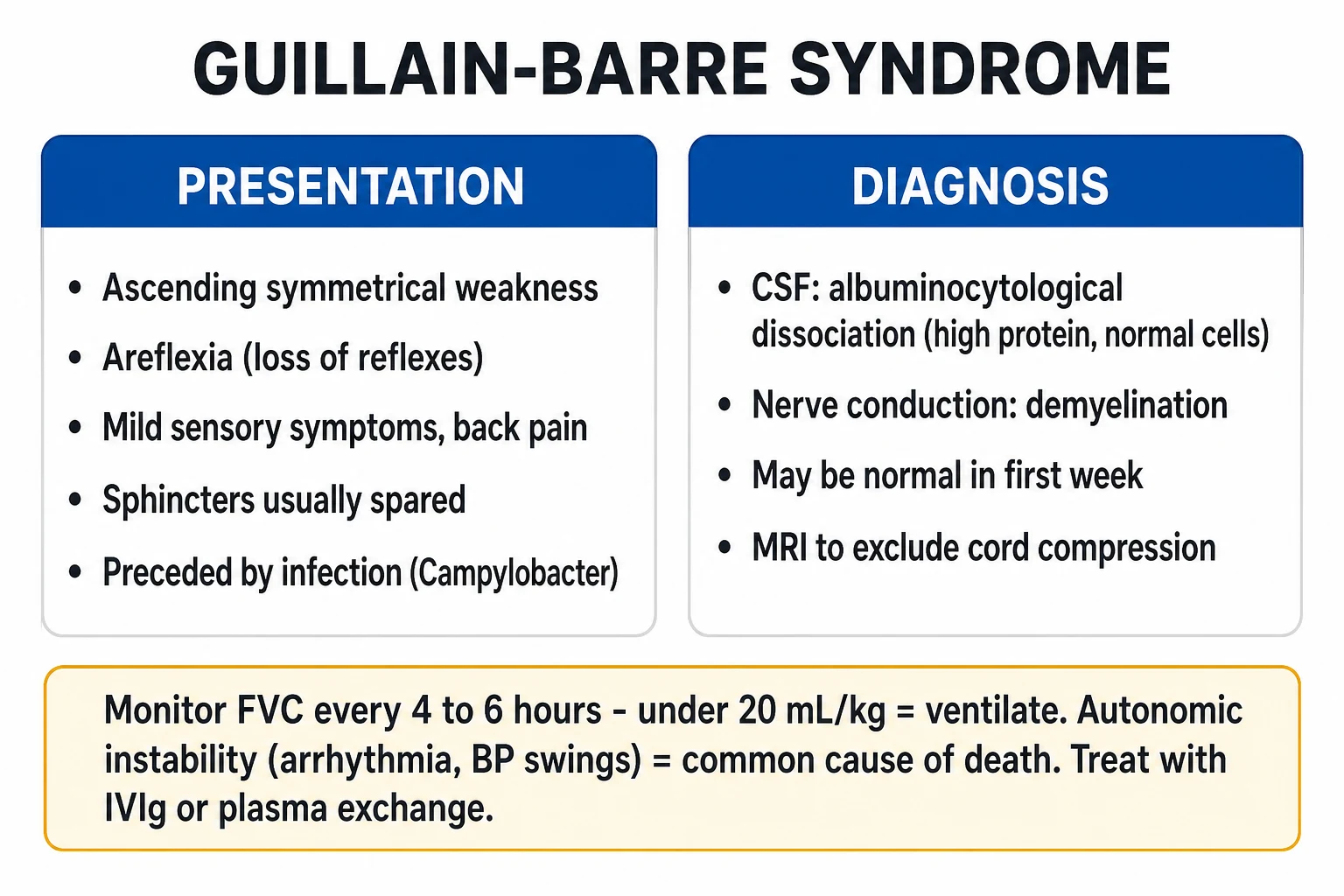
Cinematic ICU scene of a Guillain-Barré patient with ascending flaccid paralysis, a bedside spirometer showing a falling FVC, a cardiac monitor flagging dysautonomia, an IVIG infusion running, clinical-blue lighting, medical educational, no faces, no text
FigureGuillain-Barré — the acute, ascending, symmetrical flaccid paralysis with areflexia, autoimmune demyelination, classically post-Campylobacter or viral. Twenty to thirty per cent need ventilation. Intubate when the FVC <15–20 mL/kg, the MIP worse than −30, the MEP <40, or bulbar failure threatens the airway. IVIG or plasmapheresis (equally effective, never combined). Autonomic instability is the leading cause of death — continuous cardiac monitoring.
[1]

In one line

GBS = acute ascending flaccid paralysis + areflexia, autoimmune demyelination. Preceded by infection (Campylobacter, viral) 1-3 weeks prior. 20-30% need mechanical ventilation. ICU monitoring: serial FVC (intubate if <15-20 mL/kg), MIP (<-30 cmH2O), MEP (<40), bulbar function. Treatment: IVIG (0.4 g/kg/day x 5 days) OR plasmapheresis (5 sessions over 1-2 weeks) — equally effective, do NOT combine. Autonomic dysfunction = leading cause of death — continuous cardiac monitoring, have atropine/pacing ready. Myasthenia crisis: treat with IVIG/plasmapheresis + pyridostigmine.

[1]

Pathophysiology and antecedent triggers
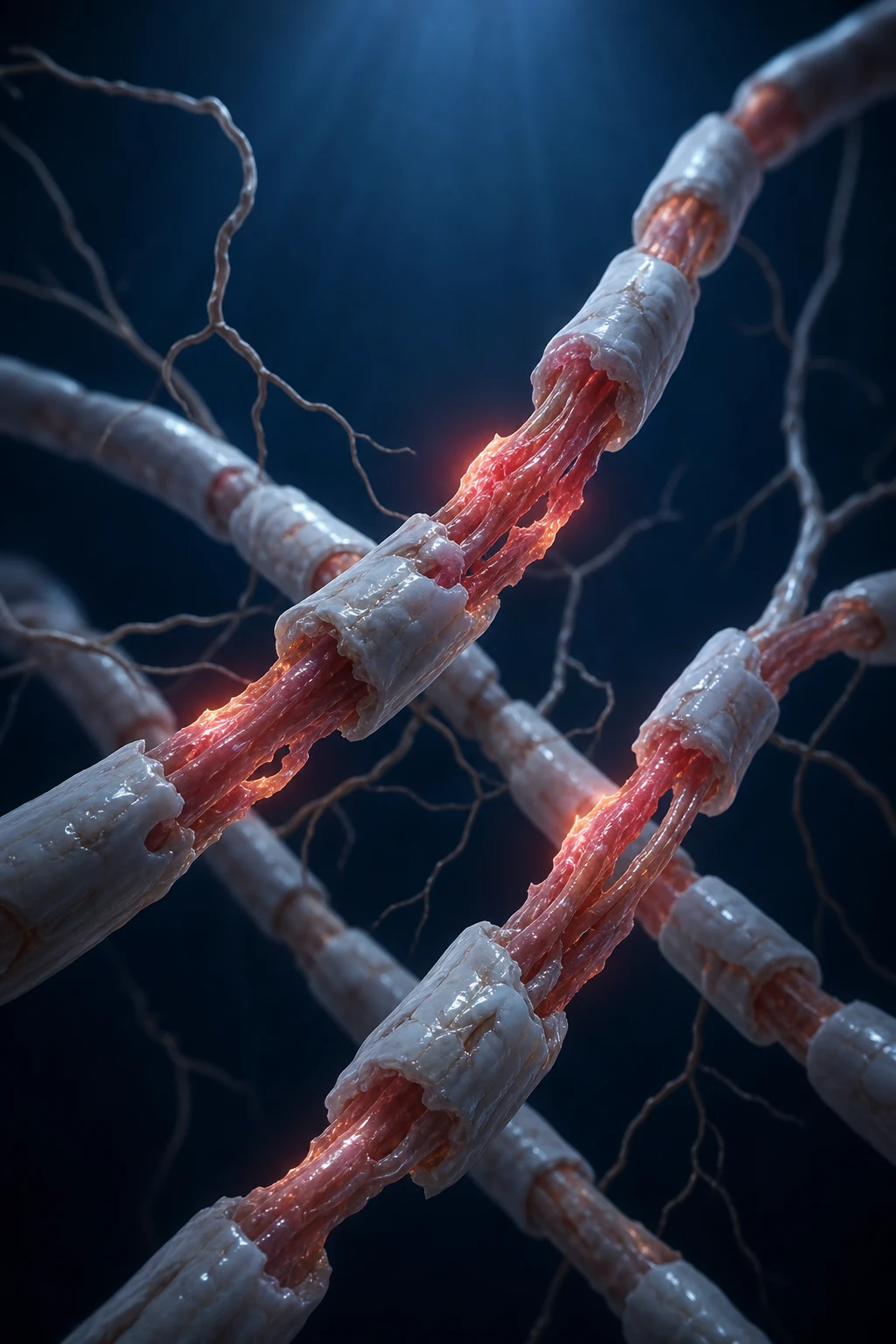
Educational schematic of Guillain-Barré molecular mimicry: Campylobacter or viral epitopes cross-react with peripheral-nerve gangliosides, complement attack on myelin or axon, conduction block and albuminocytological dissociation
FigureMolecular mimicry — pathogen epitopes resemble nerve gangliosides (GM1, GD1a, GQ1b). Cross-reactive antibody activates complement, injures myelin (AIDP) or axon (AMAN), and protein leaks into CSF while cells stay low.

Molecular mimicry — the unifying mechanism of GBS

GBS is a post-infectious, immune-mediated polyradiculoneuropathy. The dominant mechanism is molecular mimicry: oligosaccharide epitopes on an antecedent pathogen structurally resemble gangliosides on the surface of human peripheral nerves (especially GM1, GD1a, GT1a, GQ1b). The resulting cross-reactive humoral response (IgG anti-ganglioside antibodies) activates complement, forms membrane attack complex (C5b-9), and injures the myelin sheath (AIDP) or the axon itself (AMAN/AMSAN).[1][2]

The injury localises to the ventral spinal roots, the nodal/paranodal regions of motor fibres, and the blood-nerve barrier, producing conduction block (clinical weakness out of proportion to axonal loss early in the course). The classic albuminocytological dissociation in CSF reflects root sleeve permeability — protein leaks into the CSF while cellular infiltration remains low.[1]

Campylobacter jejuni

#1 antecedent (~30%)

  • Antecedent diarrhoeal illness 1-3 weeks prior; underdiagnosed (request stool culture/PCR)
  • Strongly associated with anti-GM1 and anti-GD1a antibodies
  • Predominantly motor GBS — may be axonal (AMAN) variant, more common in Asia
  • Higher incidence of severe weakness, mechanical ventilation, and slower recovery

CMV

~10-15%

  • Preceding upper-respiratory or flu-like illness
  • Associated with anti-GM2 antibodies
  • Often severe sensory involvement and prominent cranial nerve + autonomic dysfunction

EBV / Mycoplasma

Less common

  • EBV — non-specific prodrome, can mimic any GBS subtype
  • Mycoplasma pneumoniae — classically linked to Bickerstaff brainstem encephalitis and anti-GQ1b/GT1a antibodies

Influenza & other viruses

Recognised triggers

  • Influenza A/B, SARS-CoV-2 (COVID-19), Zika virus, dengue, hepatitis E, HIV seroconversion
  • Zika-associated GBS is predominantly AIDP with a monophasic course
  • Influenza vaccine association is marginal (≈1 excess case per million doses) — far below the risk of GBS after influenza infection itself

Surgery / trauma / drugs

Non-infectious triggers

  • Recent surgery, bone marrow transplant, and immunotherapy can precede GBS
  • Implicated drugs: checkpoint inhibitors (nivolumab, pembrolizumab) — immune-related adverse event
[1] [2]

Clinical features

Classic GBS (AIDP)

90% of cases

  • Ascending symmetrical flaccid paralysis (legs → trunk → arms → cranial nerves)
  • Areflexia (absent deep tendon reflexes)
  • Progressive over days to 4 weeks (nadir by 4 weeks)
  • Preceded by infection 1-3 weeks prior (Campylobacter jejuni #1, CMV, EBV, influenza)
  • Sensory: paraesthesia, sensory loss (less prominent than motor)
  • Pain: neuropathic pain common (back, thighs)

Variants

Less common forms

  • Miller Fisher syndrome: ophthalmoplegia + ataxia + areflexia (anti-GQ1b antibody)
  • AMAN (acute motor axonal neuropathy): pure motor, axonal — common in Asia
  • AMSAN (acute motor and sensory axonal neuropathy): axonal motor + sensory
  • Pharyngeal-cervical-brachial variant: bulbar weakness
  • Bickerstaff brainstem encephalitis: altered consciousness + external ophthalmoplegia + ataxia
[1] [2] [1]

Diagnosis — investigations

The diagnostic triad — clinical pattern + CSF + NCS

GBS is a clinical diagnosis supported by two investigations — neither is required to start treatment if the phenotype is typical.[1]

1. CSF (lumbar puncture): Albuminocytological dissociation — elevated CSF protein (often >0.55 g/L, can exceed 2-3 g/L) with a normal cell count (<5 white cells/µL). This pattern reflects blood-nerve barrier disruption at the spinal roots.

  • Timing: CSF protein is normal in ~50% of patients in week 1 and rises over the second and third weeks. A normal early LP does NOT exclude GBS — repeat after 1 week if suspicion remains.
  • Caveat — cytoalbuminologic dissociation with cells: >50 cells/µL should prompt search for an alternative diagnosis (HIV seroconversion, Lyme, polio, West Nile, meningeal carcinomatosis, lymphoma). [1]

2. Nerve conduction studies (NCS): demonstrate the demyelinating phenotype — prolonged distal latencies, conduction block, temporal dispersion, slowed velocities, and prolonged/absent F-waves. As with CSF, NCS may be normal in the first 1-2 weeks and should be repeated if initially non-diagnostic. [1]

3. MRI: gadolinium enhancement of the cauda equina and spinal nerve roots on post-contrast MRI is supportive when the diagnosis is uncertain. MRI brain excludes brainstem stroke in Miller Fisher / Bickerstaff overlap. [1]

4. Autoantibodies: anti-GQ1b (Miller Fisher, Bickerstaff — >90% specific), anti-GM1/GD1a (AMAN, C. jejuni-associated). These are confirmatory, not screening.

[1]

Brighton diagnostic criteria for GBS (level 1-3 certainty)

  • Level 1 (highest): Bilateral flaccid limb weakness + reduced/absent deep tendon reflexes in weakened limbs + NCS consistent with GBS + CSF albuminocytological dissociation
  • Level 2: Bilateral flaccid weakness + areflexia + either NCS or CSF consistent with GBS
  • Level 3: Bilateral flaccid weakness + areflexia (clinical pattern alone, monophasic course, nadir 12h-28d)
  • Level 4 (limited): As Level 3 but unable to exclude alternative diagnosis
[1]

Mimics you must NOT miss

Areflexic weakness is not always GBS. Actively exclude:

  • Acute intermittent porphyria / variegate porphyria — abdominal pain, seizures, psychosis; check urine PBG (porphobilinogen) — a true GBS mimic that is worsened by the drugs used for GBS sedation.
  • Transverse myelitis / spinal cord compression — check for a sensory level and sphincter involvement; GBS has no sensory level.
  • Tick paralysis — ascending weakness that resolves with tick removal; examine scalp and groin.
  • Botulism — descending paralysis, dilated pupils, dry mouth; check for tainted food/wound.
  • Hypokalaemic periodic paralysis — rapid onset, low K+, resolves with K+ replacement, areflexic.
  • Acute-onset CIDP (A-CIDP) — meets GBS criteria at onset but continues to progress or relapses ≥8 weeks after onset; treated as CIDP (sustained steroids ARE effective in CIDP, unlike GBS).[8]

Respiratory monitoring — when to intubate

20/30/40 rule — intubation criteria for GBS

Monitor EVERY SHIFT:

  • FVC <20 mL/kg (or <1.5 L in average adult) — or declining trend
  • Maximum Inspiratory Pressure (MIP) < -30 cmH2O
  • Maximum Expiratory Pressure (MEP) <40 cmH2O (indicates weak cough → secretion clearance failure) [1]

Intubate EARLY — do NOT wait for respiratory arrest. Elective intubation is safer than emergency. Also intubate if:

  • Bulbar weakness (aspiration risk — ineffective swallow, weak cough)
  • Autonomic instability causing haemodynamic compromise
  • Rapidly progressive weakness (FVC declining >30% in 24h)
[1]

EGRIS — Erasmus GBS Respiratory Insufficiency Score (predicts need for intubation within 1 week)

Validated score (Walgaard 2010) summing three predictors measured at admission to ICU/hospital:[6]

PredictorScore 0Score 1Score 2Score 3
Days between onset and admission>7 days4-7 days—≤3 days
Presence of facial and/or bulbar palsyAbsentPresent——
MRC sum score (0-60, sum of 3 muscle groups × both sides)60-5150-4140-31≤30

Total 0-7. Risk of mechanical ventilation within 1 week:

  • 0-2 → low risk (~1-6%)
  • 3-4 → intermediate (~10-25%)
  • 5-7 → high risk (>40%, up to ~90% at score 7) [1]

A short interval from onset to admission, bulbar weakness, and severe limb weakness (low MRC sum) are the three independent predictors. EGRIS does not replace serial FVC/MIP — use it to triage monitoring intensity and admission location.

[1]

NIV → intubation → weaning pathway in GBS

1

1. Baseline + serial respiratory surveillance

On ICU admission measure FVC, MIP (NIF), MEP and single breath count EVERY shift (q4-6h, more often if declining). Trend is more important than any single value. Calculate EGRIS at admission to gauge trajectory. Document bulbar function (cough, swallow, speech) every shift.

2

2. Non-invasive ventilation (NIV) is NOT a substitute for intubation

In GBS the weakness is rapidly progressive and NIV does NOT protect the airway from bulbar/aspiration risk. NIV (BiPAP) may be used briefly to buy time or to bridge while preparing for intubation, or as an adjunct to secretion clearance, but should NEVER delay definitive airway control when 20/30/40 thresholds are crossed. Mask ventilation is poorly tolerated in weak, areflexic patients and risks insufflation of the stomach.

3

3. Elective intubation BEFORE arrest

Intubate when FVC <15-20 mL/kg (or <1.5 L), MIP worse than -30 cmH2O, MEP <40 cmH2O, bulbar weakness with aspiration, or rapid decline (>30% FVC fall in 24h). Use modified RSI. Anticipate a hyperkalaemic response to suxamethonium — AVOID suxamethonium in GBS (risk of hyperkalaemic arrest from up-regulated acetylcholine receptors); use rocuronium with sugammadex reversal available.

4

4. Ventilator settings — protect the lungs

Lung-protective ventilation (Vt 6 mL/kg PBW, plateau <30 cmH2O, PEEP 5-8). Patients are typically fully conscious — ensure adequate analgesia and sedation, and use early tracheostomy (7-14 days) if prolonged ventilation is anticipated (median ventilation in severe GBS is 3-4 weeks). Avoid heavy sedation that confounds neurological exam.

5

5. Weaning criteria

Wean when: FVC >10-15 mL/kg off the ventilator on a T-piece, MIP more negative than -40 cmH2O, MEP >50 cmH2O (effective cough), bulbar function recovered (swallow intact, secretions manageable), and the underlying disease is recovering (MRC sum score rising). Use progressive T-piece / pressure support trials; percutaneous dilatational tracheostomy facilitates slow weaning.

[1] [6]

Autonomic dysfunction

Autonomic dysfunction — the leading cause of death in GBS

Up to 65% of GBS patients have autonomic dysfunction:[1][2]

  • Cardiac arrhythmias: bradycardia, asystole, sinus tachycardia, AF
  • Blood pressure instability: hypertension AND hypotension (rapid swings)
  • Ileus, urinary retention
  • SIADH (hyponatraemia)

Management: continuous cardiac monitoring. Have atropine and temporary pacing available. Treat hypertension cautiously (short-acting agents — avoid sustained antihypertensives as BP may swing to hypotension). Treat hypotension with cautious IV fluids.

[1] [1]

ICU management protocol

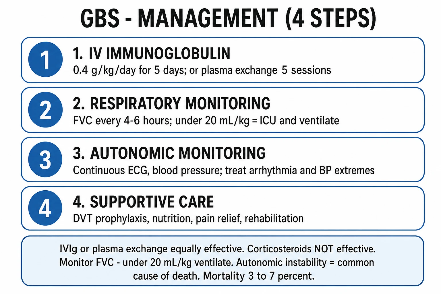
Educational ICU management infographic for Guillain-Barré: serial spirometry intubation thresholds, IVIG or plasma exchange not both, continuous monitoring for dysautonomia
FigureGBS ICU protocol — serial FVC/MIP/MEP and bulbar checks drive intubation; disease-modifying therapy is IVIG or plasma exchange (equally effective, never combined first-line); continuous cardiac monitoring because dysautonomia kills.

GBS ICU management protocol

1

1. RESPIRATORY — the #1 priority

Serial FVC, MIP, MEP and bulbar assessment every 4-6h. Apply the 20/30/40 rule and EGRIS. Intubate electively before arrest. Avoid suxamethonium. Plan for median 3-4 weeks ventilation in severe cases — early tracheostomy discussion at day 7-10.

2

2. AUTONOMIC — continuous cardiac monitoring

Continuous ECG + arterial line. Bedside atropine, isoprenaline, and transcutaneous pacer. Short-acting agents only for BP swings. Correct SIADH-induced hyponatraemia (fluid restriction; avoid hypertonic saline unless seizures).

3

3. DVT prophylaxis — mandatory

All immobile GBS patients are high-risk for VTE. Enoxaparin 40 mg SC daily (dose-adjust for renal function) + intermittent pneumatic compression. Continue until independently mobile.

4

4. DISEASE-MODIFYING THERAPY — start within 2-4 weeks of onset

IVIG 0.4 g/kg/day × 5 days OR plasma exchange (5 sessions over 1-2 weeks, 1.0-1.5 plasma volumes each). Do NOT combine. Treatment shortens time to recover walking by ~40%. Efficacy declines after 4 weeks from onset.

5

5. PAIN — actively treat

Neuropathic pain (back, thighs, paraesthesia) is the most under-treated symptom. First-line gabapentin/pregabalin or amitriptyline; add simple analgesia. AVOID opioids as sole agent (constipation + sedation compound ileus and delirium). Carbamazepine is second-line.

6

6. SUPPORTIVE bundle

Early enteral nutrition (nasogastric). Eye protection (lubricant + tape/tarsorrhaphy if facial diplegia → corneal abrasion). Pressure-area care. Passive ROM physiotherapy to prevent contractures; early active rehab once stable. Bowel regimen (paralytic ileus common). Psychological support — patient is awake and terrified while intubated. Communication aids (letter boards).

7

7. AVOID harm

No suxamethonium, no long-acting antihypertensives, no corticosteroids as monotherapy, no combined IVIG + PLEX. Minimise sedation to preserve neurological exam. Avoid aminoglycosides and magnesium if MG overlap is considered.

[1]

Treatment

GBS treatment protocol

1

IVIG 0.4 g/kg/day for 5 days

Standard first-line in most centres. IVIG neutralises autoantibodies and modulates immune function. Equally effective as plasmapheresis. Advantages: easier to administer, fewer vascular access complications. Side effects: headache, thrombosis (check for hyperviscosity), renal failure (rare).

2

OR Plasmapheresis (5 sessions over 1-2 weeks)

Removes circulating autoantibodies. Equally effective as IVIG. Disadvantages: requires central venous access, vascular instability during exchange, more nursing resources. Preferred if IVIG contraindicated (IgA deficiency, severe renal failure).

3

Do NOT combine IVIG + plasmapheresis

No additional benefit over monotherapy (large RCT showed no difference). Adding IVIG to plasmapheresis does not improve outcomes. Choose ONE treatment modality.

4

Supportive care

DVT prophylaxis (GBS patients are immobilised). Pressure area care. Eye protection (if facial weakness — corneal abrasion risk). Physiotherapy (passive ROM, prevent contractures). Pain management (neuropathic pain — gabapentin, pregabalin). Nutrition (early enteral feeding). Psychological support.

5

Corticosteroids are NOT effective

Oral or IV steroids do NOT improve outcomes in GBS and should NOT be used as monotherapy. May be used for neuropathic pain or in specific overlap syndromes (CIDP).

[1]

Dutch GBS Trial — IVIG vs plasma exchange (the landmark RCT)

Study

van der Meche FG, Schmitz PI; Dutch Guillain-Barre Study Group. NEJM 1992

Design

Multicentre randomised trial — 150 adults with GBS unable to walk independently, within 2 weeks of onset

Arms

IVIG 0.4 g/kg/day × 5 days vs plasma exchange (5 sessions over 8-13 days)

Primary outcome

Improvement of one GBS disability grade at 4 weeks

Key finding

IVIG was AT LEAST as effective as plasma exchange (more patients improved with IVIG), and was simpler and safer — establishing IVIG as first-line

Clinical bottom line

IVIG and plasma exchange are EQUIVALENT — pick one, do not combine

[4]

French Cooperative Group — plasma exchange vs supportive care

Study

French Cooperative Group on Plasma Exchange in GBS. Ann Neurol 1987

Design

Multicentre RCT establishing PE as the first disease-modifying therapy for GBS

Key finding

Plasma exchange significantly improved outcome vs supportive care alone — shortened time to wean from ventilation and time to walk, and reduced residual disability

Replacement fluid caveat

Albumin was superior to fresh-frozen plasma as replacement fluid (fewer side effects)

Historical significance

First proof that an immune-directed therapy changes the natural history of GBS — the benchmark against which IVIG was later shown non-inferior

[5]

EGRIS — predicting respiratory failure in GBS

Study

Walgaard C, Lingsma HF, Ruts L, et al. Ann Neurol 2010

Design

Prospective derivation & validation cohort (n=395) of a bedside score to predict need for mechanical ventilation within 7 days

Predictors

(1) Days onset→admission, (2) Facial/bulbar palsy, (3) MRC sum score

Key finding

A score of 0-7 stratifies ventilation risk from <1% (score 0) to ~90% (score 7); discriminates well (AUC ~0.83)

Clinical bottom line

EGRIS complements (does not replace) serial FVC/MIP — use it to triage monitoring intensity and admission location

[6] [1]

Myasthenia gravis crisis

Myasthenia crisis

Respiratory failure from MG

  • Weakness of respiratory muscles (diaphragm, intercostals)
  • Bulbar weakness (dysphagia, aspiration)
  • Triggered by: infection, surgery, pregnancy, drugs (aminoglycosides, beta-blockers, magnesium)
  • Treatment: IVIG or plasmapheresis + pyridostigmine + treat trigger
  • May need temporary mechanical ventilation (NIV or intubation)
  • Thymectomy if thyma present
[3]

Prognosis and recovery

Recovery, relapse and mortality in GBS

GBS is monophasic — once the nadir is reached (by 4 weeks), the disease stabilises and recovery begins. Treatment-related fluctuations (a second worsening after initial response to IVIG/PLEX) occur in ~5-10%; recurrent or持续 progression beyond 8 weeks should trigger reassessment for acute-onset CIDP (A-CIDP), which is treated differently — sustained corticosteroids ARE effective in CIDP.[8]

Mortality is 3-7% even in modern ICUs (commonly quoted ~5%); deaths are from autonomic dysfunction, pulmonary embolism, sepsis, and respiratory arrest.[1][7]

Functional recovery:

  • ~80% recover fully or with minor residual deficits, but recovery is slow — months to 1-2 years.
  • ~20% remain unable to walk at 6 months; ~5-10% have persistent severe disability.
  • Predictors of poor outcome (Walgaard 2011): age >40, preceding diarrhoeal illness (C. jejuni), axonal subtype on NCS, low MRC sum score at admission, need for mechanical ventilation, and severe disability at nadir.[7]
  • Early disease-modifying therapy (within 2-4 weeks of onset) shortens time to independent walking.

Exam practice

SAQ — Guillain-Barré syndrome

10 minutes · 10 marks

A 45-year-old man presents with progressive leg weakness over 4 days. He had a diarrhoeal illness 2 weeks ago. Examination: flaccid paralysis in all 4 limbs (power 2/5), absent reflexes, facial diplegia. FVC 1.2 L (predicted 4.5 L). MIP -25 cmH2O. HR 48, BP fluctuating 90/50 to 160/90.

[1]

SAQ — Diagnosis and disease-modifying therapy choice

8 minutes · 8 marks

A 32-year-old woman presents with ascending weakness over 5 days following a flu-like illness. FVC 25 mL/kg, MIP -40 cmH2O, MEP 60 cmH2O. Power 4/5 in legs, 5/5 arms, reflexes absent at ankles and knees. CSF: protein 1.2 g/L, 2 lymphocytes/µL. NCS pending.

[1]

SAQ — Ventilation strategy and weaning

8 minutes · 8 marks

A 58-year-old man with biopsy-confirmed GBS has been intubated and ventilated for 18 days via tracheostomy. He received IVIG on day 2. Over the last week his MRC sum score has risen from 20 to 42. Current: FVC 12 mL/kg on T-piece, MIP -45 cmH2O, MEP 55 cmH2O, bulbar function recovered, weak but effective cough.

[1]

Clinical pearls

High-yield GBS points for the CICM/FFICM exam

  1. GBS = ascending flaccid paralysis + areflexia, preceded by infection (Campylobacter #1).[2]
  2. 20-30% need mechanical ventilation. Monitor serial FVC/MIP/MEP.
  3. Intubation criteria (20/30/40 rule): FVC <20 mL/kg, MIP < -30 cmH2O, MEP <40 cmH2O.[1]
  4. IVIG OR plasmapheresis — equally effective. Do NOT combine.
  5. Corticosteroids are NOT effective in GBS.
  6. Autonomic dysfunction = leading cause of death. Continuous cardiac monitoring. Atropine/pacing ready.
  7. CSF: albuminocytological dissociation (high protein, normal WBC). May be normal in first week.
  8. Miller Fisher: ophthalmoplegia + ataxia + areflexia. Anti-GQ1b antibody.
  9. Intubate EARLY — elective intubation safer than emergency.
  10. Campylobacter jejuni is #1 antecedent infection (diarrhoeal illness 1-3 weeks prior).
  11. DVT prophylaxis essential (immobilised patient at high risk).
  12. Recovery: starts 2-4 weeks after nadir. Complete recovery in ~80% but may take months to years.
  13. Myasthenia crisis: respiratory failure from neuromuscular weakness. Treat with IVIG/plasmapheresis + pyridostigmine.[3]
  14. Avoid drugs that worsen MG: aminoglycosides, beta-blockers, magnesium, neuromuscular blockers.

Advanced neurocritical-care pearls — beyond the basics

  1. AIDP is the dominant subtype in the West (~85-90%); AMAN/AMSAN (axonal) dominate in Asia and are linked to C. jejuni and anti-GM1/GD1a antibodies.[2]
  2. Molecular mimicry is the unifying mechanism: pathogen oligosaccharides mimic nerve gangliosides (GM1, GD1a, GQ1b); complement-mediated injury at nodes of Ranvier → conduction block.
  3. EGRIS (Erasmus score) predicts ventilation within 7 days from three bedside variables — short onset-to-admission interval, facial/bulbar palsy, low MRC sum score.[6]
  4. CSF protein is normal in ~50% of cases in week 1 — a normal early LP does NOT exclude GBS; repeat after 1 week. Conversely, >50 CSF WBC/µL should trigger search for HIV seroconversion, Lyme, or polio/enterovirus.
  5. AVOID suxamethonium in GBS — up-regulated acetylcholine receptors cause dangerous hyperkalaemia and cardiac arrest on RSI. Use rocuronium (with sugammadex reversal available).
  6. NIV is NOT a substitute for intubation in progressive GBS — it does not protect the airway in bulbar weakness and delays definitive control. Use only to bridge while preparing for intubation.
  7. Median ventilation in severe GBS is 3-4 weeks — plan early tracheostomy (day 7-14) and slow weaning; the patient is typically awake, so communication aids and psychological support are essential.
  8. Anti-GQ1b IgG is >90% specific for Miller Fisher / Bickerstaff — supports the diagnosis in atypical brainstem presentations; consider MRI brain to exclude brainstem stroke.
  9. Plasma exchange replacement fluid: albumin is superior to fresh-frozen plasma (fewer side effects) — a finding from the original French Cooperative Group trial.[5]
  10. The Dutch 1992 trial (NEJM) established IVIG as first-line by proving it was at least as effective as plasma exchange, and simpler/safer — the foundation of modern GBS therapy.[4]
  11. A-CIDP masquerades as GBS — if weakness continues to progress or relapses beyond 8 weeks, rediagnose as acute-onset CIDP; unlike GBS, sustained corticosteroids ARE effective in CIDP.[8]
  12. Poor-prognosis predictors (Walgaard 2011): age >40, preceding diarrhoea (C. jejuni), axonal NCS subtype, low admission MRC sum score, need for ventilation, severe nadir disability.[7]
  13. SIADH complicates up to a third of severe GBS — hyponatraemia from autonomic/hypothalamic involvement; treat with fluid restriction, avoid rapid correction.
  14. Treatment-related fluctuation (~5-10%) after IVIG/PLEX is distinct from A-CIDP; a single re-treatment course is reasonable, but sustained relapse mandates CIDP workup.
  15. Mortality is 3-7% even in modern ICUs — autonomic dysfunction, pulmonary embolism, sepsis and respiratory arrest dominate; rigorous supportive care is as important as disease-modifying therapy.
  16. Checkpoint-inhibitor–associated GBS (nivolumab, pembrolizumab) is an increasingly recognised immune-related adverse event — manage jointly with oncology and consider steroids (immune-related AE context differs from classical GBS).

Red flags

Critical GBS points

  • Intubate EARLY — FVC <15-20 mL/kg or declining trend. Do NOT wait for respiratory arrest.[1]
  • Autonomic dysfunction is the leading cause of death — continuous cardiac monitoring. Have atropine/pacing ready.
  • Do NOT combine IVIG + plasmapheresis — no additional benefit over monotherapy.[2]
  • Corticosteroids are NOT effective in GBS — do NOT use as monotherapy.
  • Campylobacter jejuni antecedent infection — check stool culture.
  • Dysautonomia can cause fatal brady/asystolic arrest — have atropine and pacing immediately available.
  • AVOID suxamethonium at RSI — risk of hyperkalaemic arrest from up-regulated acetylcholine receptors.
  • A normal CSF in week 1 does NOT exclude GBS — albuminocytological dissociation evolves over 1-2 weeks; repeat the LP.
  • CSF >50 WBC/µL is atypical — exclude HIV seroconversion, Lyme, polio/enterovirus, meningeal malignancy.
  • NIV does NOT protect the airway — never use it to delay intubation in bulbar or rapidly progressive GBS.
  • Weakness progressing beyond 8 weeks = rethink — consider acute-onset CIDP (steroids ARE effective) rather than refractory GBS.[8]

References

  1. [1]Leonhard SE, Mandarakas MR, Gondim FAA, et al. Diagnosis and management of Guillain-Barré syndrome in ten steps Nat Rev Neurol, 2019.PMID 31541214
  2. [2]Willison HJ, Jacobs BC, van Doorn PA. Guillain-Barré syndrome Lancet, 2016.PMID 26948435
  3. [3]Lacomis D. Myasthenic crisis Neurocrit Care, 2005.PMID 16377829
  4. [4]van der Meche FG, Schmitz PI; Dutch Guillain-Barre Study Group. A randomized trial comparing intravenous immune globulin and plasma exchange in Guillain-Barré syndrome. Dutch Guillain-Barré Study Group N Engl J Med, 1992.PMID 1552913
  5. [5]French Cooperative Group on Plasma Exchange in Guillain-Barre syndrome. Efficiency of plasma exchange in Guillain-Barré syndrome: role of replacement fluids. French Cooperative Group on Plasma Exchange in Guillain-Barré syndrome Ann Neurol, 1987.PMID 2893583
  6. [6]Walgaard C, Lingsma HF, Ruts L, et al. Prediction of respiratory insufficiency in Guillain-Barré syndrome Ann Neurol, 2010.PMID 20517939
  7. [7]Walgaard C, Lingsma HF, Ruts L, et al. Early recognition of poor prognosis in Guillain-Barre syndrome Neurology, 2011.PMID 21403108
  8. [8]Ruts L, Drenthen J, Jacobs BC, et al. Distinguishing acute-onset CIDP from fluctuating Guillain-Barre syndrome: a prospective study Neurology, 2010.PMID 20427754