ICU · neurocritical-care
Guillain-Barre Syndrome — Comprehensive ICU Management
Also known as Guillain-Barre syndrome · GBS · Acute inflammatory demyelinating polyneuropathy · AIDP · Acute motor axonal neuropathy · AMAN · Miller-Fisher syndrome · MFS · Acute flaccid paralysis · Ascending paralysis · Neuromuscular respiratory failure
Guillain-Barre syndrome (GBS) — acute immune-mediated demyelinating polyradiculoneuropathy causing rapidly progressive (hours to 4 weeks) ascending flaccid paralysis + areflexia ± autonomic dysfunction ± respiratory failure ± cranial nerve involvement. ICU admission for: respiratory failure (20-30% require intubation), severe autonomic dysfunction (arrhythmia, BP swings), bulbar weakness (aspiration risk), or severe weakness (unable to walk). Variants: AIDP (90% — demyelinating), AMAN/AMSAN (axonal — Campylobacter jejuni), Miller-Fisher (ophthalmoplegia + ataxia + areflexia + GQ1b antibody), Bickerstaff brainstem encephalitis. Diagnostic: Brighton criteria (progressive weakness, areflexia, CSF albuminocytologic dissociation, NCS abnormalities). Treatment: IVIG (0.4 g/kg/day x 5 days) OR plasma exchange (5 sessions over 1-2 weeks) — BOTH EQUALLY EFFECTIVE — NO BENEFIT of combined therapy — corticosteroids are INEFFECTIVE (contraindicated). ICU management: monitor FVC + NIF every 4-6h (intubate when FVC <15 mL/kg or NIF < -30 cmH2O), autonomic monitoring (ECG, BP — watch for sudden asystole from vagal hypertonia), DVT prophylaxis, pain management (neuropathic pain common — gabapentin), early physiotherapy. Prognosis: 80% full recovery, 5-10% mortality (autonomic dysfunction, PE, sepsis), 20% residual disability. Erasmus GBS Respiratory Insufficiency Scale (EGRIS) predicts need for ventilation.
On this page & tools
Your progress
Saved locally on this device.
Target exams
Red flags
Overview
GBS is the most common cause of acute generalized paralysis in the post-polio era (incidence 1-2 per 100,000 per year). The intensivist encounters GBS in three scenarios: (1) the patient with rapidly progressive weakness (the diagnostic challenge — must distinguish from other causes of acute paralysis), (2) the patient with established GBS requiring ICU monitoring (respiratory function monitoring + autonomic monitoring + when to intubate), (3) the intubated GBS patient (prolonged ventilation, tracheostomy timing, autonomic crisis management, weaning). The key to good outcomes is EARLY recognition + EARLY treatment (IVIG or PLEX within 2-4 weeks of symptom onset) + proactive ICU monitoring (respiratory + autonomic).[1][5]
Variants of GBS
GBS variants — clinical features and prognosis
| Variant | Frequency | Mechanism | Clinical features | Antibody | Prognosis |
|---|---|---|---|---|---|
| AIDP (acute inflammatory demyelinating polyneuropathy) | 85-90% (Western) | Demyelination of motor + sensory nerves (Schwann cell attack) | Ascending weakness, areflexia, sensory symptoms (paraesthesia), cranial nerve (facial diplegia 50%) | Various (GM1, GD1a — less specific) | GOOD (80% full recovery in 6-12 months) |
| AMAN (acute motor axonal neuropathy) | 5-10% (Western); 30-50% (Asia — China, Japan) | AXONAL degeneration of motor nerves (macrophage invasion of node of Ranvier) | Pure MOTOR weakness (no sensory symptoms), rapid progression, respiratory failure common | Anti-GD1a, anti-GM1 | VARIABLE (slower recovery — axonal regrowth takes months-years — but many recover fully) |
| AMSAN (acute motor sensory axonal neuropathy) | 1-5% | AXONAL degeneration of motor AND sensory nerves | Severe motor + sensory loss, very slow recovery | Anti-GM1 | POOR (worst prognosis — prolonged disability) |
| Miller-Fisher syndrome (MFS) | 5% (more common in Asia) | Attack on oculomotor and proprioceptive nerves | TRIAD: ophthalmoplegia + ataxia + areflexia (DESCENDING weakness — starts in eyes/face then descends — OPPOSITE of AIDP) | Anti-GQ1b (95% positive — pathognomonic) | GOOD (usually mild — rarely requires ventilation) |
| Bickerstaff brainstem encephalitis | Rare | Extension of MFS into brainstem | MFS triad + altered consciousness (encephalopathy) + hyperreflexia (distinguishes from MFS which is areflexic) | Anti-GQ1b | Good (responds to IVIG) |
| Pharyngeal-cervical-brachial (PCB) variant | Rare | Weakness limited to upper body | Weakness of oropharynx, neck, upper limbs (SPARING legs) — mimics botulism or MG | Anti-GT1a | Moderate (can cause respiratory failure from bulbar weakness) |
Diagnosis — Brighton criteria and workup
Diagnostic workup for suspected GBS
- CLINICAL FEATURES — (a) Progressive bilateral weakness (usually ascending — starts in legs, progresses to arms, then trunk, then cranial nerves). (b) Areflexia (absent reflexes — particularly in affected limbs — REQUIRED for diagnosis). (c) Symmetrical (usually). (d) Progression over hours to <4 weeks (the 4-week rule distinguishes GBS from CIDP — chronic inflammatory demyelinating polyneuropathy — which progresses >8 weeks). (e) Preceding infection 1-3 weeks before (C. jejuni diarrhoea, URI, CMV, EBV, Zika, surgery)
- LUMBAR PUNCTURE — CSF analysis:
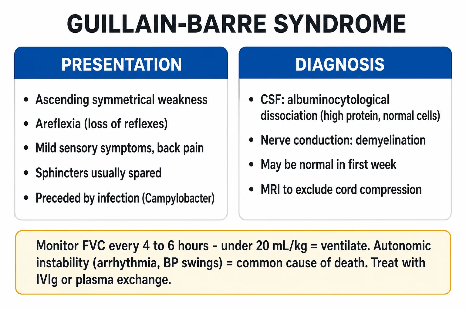
- Albuminocytologic dissociation: elevated protein (typically 0.55-2.0 g/L — normal <0.45) with NORMAL cell count (<5 × 10^6/L, occasionally up to 10). This is the HALLMARK of GBS — the elevated protein comes from inflammation of spinal nerve roots (dorsal + ventral) → breakdown of blood-nerve barrier → protein leak into CSF, but no cellular infiltration
- CSF protein may be NORMAL in first week (rises in week 2-3 — if suspicion is high and CSF is normal early, repeat LP in 1 week)
- If CSF pleocytosis >50 cells: RECONSIDER DIAGNOSIS — differential includes: HIV-associated GBS (HIV seroconversion), leptomeningeal carcinomatosis/lymphoma, Lyme disease, poliomyelitis, West Nile virus
- Send CSF for: cells, protein, glucose, culture, viral PCRs (if indicated)
- NERVE CONDUCTION STUDIES (NCS) + ELECTROMYOGRAPHY (EMG):
- AIDP: DEMYELINATING features — prolonged distal latencies, conduction block, temporal dispersion, slow conduction velocity (<70% of lower limit of normal), prolonged/absent F-waves (PROXIMAL demyelination — the earliest NCS sign)
- AMAN: AXONAL features — reduced CMAP amplitudes (low or absent compound muscle action potentials), normal conduction velocity, no conduction block
- NCS may be NORMAL in first week (demyelination may take 1-2 weeks to show on NCS) — repeat if initially normal and suspicion persists
- BLOODS — (a) Anti-ganglioside antibodies: anti-GQ1b (Miller-Fisher — 95% positive), anti-GM1 (AMAN), anti-GD1a (AMAN). These are DIAGNOSTIC for specific variants but NOT required for treatment. (b) HIV serology (HIV seroconversion can trigger GBS). (c) Stool culture + serology for C. jejuni. (d) Electrolytes (exclude hyponatraemia from SIADH — common in GBS). (e) CK (exclude rhabdomyolysis or myositis mimicking GBS). (f) Anti-AChR antibodies (if MG crisis is in the differential — MG has FLUCTUATING weakness, not progressive). (g) TSH, B12, folate (exclude other causes of neuropathy)
- MRI SPINE (if diagnostic uncertainty) — exclude spinal cord compression (transverse myelitis, epidural abscess, cord compression). GBS may show enhancement of nerve roots on post-contrast MRI (cauda equina, spinal nerve roots)
- BRIGHTON CRITERIA for diagnosis:
- Level 1 (highest diagnostic certainty): bilateral flaccid weakness + areflexia + CSF albuminocytologic dissociation + NCS abnormalities
- Level 2: bilateral flaccid weakness + areflexia + CSF OR NCS abnormalities
- Level 3: bilateral flaccid weakness + areflexia (clinical diagnosis only)
- Treatment should not be delayed for all criteria — if clinical picture is suggestive, start IVIG or PLEX
Respiratory monitoring — when to intubate
Respiratory failure from diaphragm and intercostal muscle weakness is the #1 reason for ICU admission in GBS (20-30% of patients require intubation). The key is PROACTIVE MONITORING — do not wait for the patient to be in trouble.[3]
Respiratory function monitoring in GBS — thresholds for intervention
| Parameter | Normal | Concerning | ACTION THRESHOLD | Intervene |
|---|---|---|---|---|
| FVC (forced vital capacity) | 60-70 mL/kg | 25-35 mL/kg | <20 mL/kg = start preparing for intubation | <15 mL/kg = intubate |
| NIF (negative inspiratory force) | < -50 cmH2O | -30 to -40 cmH2O | -25 to -30 cmH2O = concerning | < -30 cmH2O (i.e., more positive — e.g., -20) = intubate |
| PEF (peak expiratory flow) | >300 L/min | <200 L/min | <100 L/min = poor cough | Consider intubation if cough ineffective |
| Bulbar weakness | Normal swallow/cough | Dysphagia, weak cough, pooled secretions | Any bulbar weakness + declining FVC = VERY HIGH RISK | Intubate early (aspiration risk) |
| Single breath count | >30 | 15-20 | <10 | Prepare for intubation |
| Monitoring frequency | — | — | Every 4-6 hours (more frequently if declining) | Continuous trend monitoring |
Treatment — IVIG vs plasma exchange
Treatment options for GBS — head-to-head comparison
| Feature | IVIG | Plasma exchange (PLEX) | Corticosteroids |
|---|---|---|---|
| Efficacy | EQUIVALENT to PLEX (multiple RCTs — PSUNSHINE, Dutch GBS trial) | Effective (first proven therapy — 1985) | INEFFECTIVE — no benefit (multiple RCTs) — do NOT give |
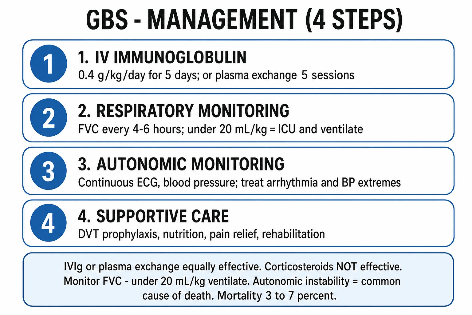
| Dose | 0.4 g/kg/day IV x 5 days (total 2 g/kg) | 5 sessions over 1-2 weeks (200-250 mL/kg total exchange) | — |
| Mechanism | Neutralises autoantibodies + modulates complement + inhibits macrophage Fc receptors | Physically removes autoantibodies and complement from circulation | Anti-inflammatory (but ineffective — may even be harmful) |
| Advantages | Simple (IV infusion — no central line), widely available, fewer complications, no plasma needed | Faster antibody removal, no transfusion-related risk | — |
| Disadvantages | Cost, thrombosis risk (hyperviscosity), AKI (especially with sucrose-containing formulations), headache, aseptic meningitis | Central venous catheter required (infection, thrombosis), bleeding (from plasma removal), hypotension (volume shifts), more nursing effort | Increased infection risk, myopathy — without benefit |
| Combination (IVIG + PLEX)? | NO added benefit — ICE trial showed combination is NOT superior to IVIG alone | — | — |
| Which to choose? | IVIG = FIRST CHOICE in most centres (simpler, safer, equally effective) | PLEX if IVIG contraindicated (IgA deficiency — anaphylaxis), or if IVIG unavailable | NEVER steroids |
| Timing | Start within 2-4 weeks of symptom onset (earlier is better — ideally within 2 weeks) | Same | — |
| Second course of IVIG? | If patient DETERIORATES after first course → consider second course of IVIG (0.4 g/kg/day x 5 days) — evidence emerging for benefit in poor-prognosis patients (I-SID-GBS trial) | If deterioration after PLEX → consider IVIG | — |
ICE trial — IVIG vs PLEX vs combination for GBS
Study design
International, multicentre RCT — 449 patients
Population
Adults with GBS unable to walk independently, within 4 weeks of onset
Arms
(1) IVIG alone (0.4 g/kg/day x 5d). (2) PLEX alone (5 sessions). (3) PLEX followed by IVIG
Primary outcome
Disability grade at 4 weeks: NO significant difference between the three arms
Key finding
IVIG alone = PLEX alone = PLEX + IVIG (combination is NOT superior)
Safety
IVIG had fewer complications than PLEX
Clinical bottom line
IVIG is FIRST-LINE for GBS — equally effective as PLEX, simpler, safer. Combination therapy has NO added benefit
ICU management
GBS ICU management protocol
-
RESPIRATORY MONITORING (the #1 priority):
- FVC + NIF every 4-6 hours (more frequently if declining)
- Intubate when FVC <15 mL/kg or NIF < -30 cmH2O or bulbar weakness with aspiration risk
- Use RSI with rocuronium (1.2 mg/kg) — NOT succinylcholine (hyperkalaemia risk from muscle denervation — upregulated acetylcholine receptors)
- Ventilation: assist-control or pressure support. PEEP 5. Target SpO2 94-98%, PaCO2 35-45. Wean as strength recovers
- Tracheostomy: consider at day 10-14 if no improvement (prolonged ventilation expected in 10-20%). Early tracheostomy does NOT improve outcomes (TracMan trial principles apply) but improves comfort and facilitates weaning [1]
-
AUTONOMIC DYSFUNCTION MONITORING (the #2 killer):
- Continuous ECG + BP monitoring (intra-arterial line) — autonomic storms can cause life-threatening arrhythmia or BP swings
- BRADYARRHYTHMIA: severe bradycardia or asystole from vagal hypertonia → atropine 0.5-1 mg IV, external/transvenous pacing. Have atropine and pacing equipment at bedside at ALL times
- TACHYARRHYTHMIA: AF, SVT from sympathetic overactivity → treat cautiously with short-acting beta-blockers (esmolol — titratable) — AVOID long-acting agents (may precip sudden bradycardia)
- HYPERTENSION: systolic can reach 200-240 → short-acting agents only (labetalol 10-20 mg IV, nicardipine infusion) — AVOID long-acting antihypertensives (BP may crash)
- HYPOTENSION: systolic can drop to 60-80 → fluids, vasopressors (metaraminol or noradrenaline — short-acting, titratable). AVOID aggressive volume loading (autonomic dysfunction + third space = pulmonary oedema)
- ILEUS: common (autonomic gut dysfunction) → nasogastric tube for decompression, minimal enteral feeding initially, prokinetics (metoclopramide — cautious — may worsen weakness)
- URINARY RETENTION: common (autonomic bladder dysfunction) → urinary catheter [1]
-
DISEASE-SPECIFIC TREATMENT:
- IVIG 0.4 g/kg/day x 5 days (first-line)
- OR plasma exchange (5 sessions) if IVIG contraindicated
- Start within 2-4 weeks of symptom onset (earlier = better) [1]
-
PAIN MANAGEMENT (often under-recognised):
- GBS causes severe NEUROPATHIC PAIN (burning, shooting, aching in back and legs — from nerve root inflammation)
- Gabapentin 300-1200 mg TDS (first-line for neuropathic pain)
- Pregabalin 75-300 mg BD
- Amitriptyline 10-50 mg nocte
- Tramadol (cautious — serotonergic — may interact with other drugs)
- AVOID NSAIDs (limited efficacy for neuropathic pain + nephrotoxicity — these patients are at risk for AKI) [1]
-
THROMBOPROPHYLAXIS:
- LMWH (enoxaparin 40 mg SC daily) — GBS patients are at HIGH risk for VTE (immobility + inflammation)
- Mechanical prophylaxis (sequential compression devices) — ALWAYS
- Check for contraindications (recent surgery, thrombocytopenia) [1]
-
NUTRITION:
- Early enteral nutrition (within 48h) — GBS is a catabolic state
- Nasogastric or nasojejunal tube (paralytic ileus is common — start trophic feeding, advance as tolerated)
- Monitor for aspiration (bulbar weakness + gastroparesis) [1]
-
EYE CARE:
- Facial weakness → inability to close eyes → exposure keratopathy
- Lubricating eye drops (hourly) + eye taping at night
- Ophthalmology review if corneal abrasion suspected [1]
-
PHYSIOTHERAPY + REHABILITATION:
- Passive range-of-motion exercises (prevent contractures)
- Early as strength allows
- Splints (prevent foot drop, wrist drop)
- Speech therapy (swallowing assessment, communication)
- Occupational therapy
- Psychological support (anxiety and depression common — the patient is "trapped in a paralysed body") [1]
-
MONITORING FOR CLINICAL DETERIORATION (treatment-related fluctuation):
- Some patients DETERIORATE after initial improvement post-IVIG/PLEX → "treatment-related fluctuation" (TRF)
- If TRF occurs: consider second course of IVIG
- Monitor: MRC sum score (0-60 — sum of 6 muscle groups bilaterally, 0-5 scale), GBS disability score (0-6)
- Overall trend should be improvement over weeks — if plateau or deterioration, reconsider diagnosis (CIDP overlap, alternative diagnosis)
Clinical pearls
Red flags
Prognosis
GBS prognosis — outcomes and timelines
| Outcome | Proportion | Timeline | Notes |
|---|---|---|---|
| Full recovery | 60-80% | 6-12 months | AIDP variant, mild-moderate weakness, younger patients |
| Residual disability (mild-moderate weakness, fatigue, pain) | 10-20% | Permanent | Axonal variants (AMAN/AMSAN), older age, severe weakness at nadir |
| Severe disability (wheelchair/bedbound) | 5-10% | Permanent | AMAN/AMSAN, prolonged ventilation, older age |
| Mortality | 5-10% | During acute illness | Causes: respiratory failure, autonomic dysfunction (arrhythmia), PE, sepsis |
| Time to nadir | — | 1-4 weeks | Time from onset to maximum weakness |
| Time to improvement | — | 2-4 weeks after nadir | Plateau phase (2-4 weeks at maximum weakness) then recovery |
| Time to walk independently | — | 3-6 months | AIDP: faster; AMAN: slower |
| Relapse/recurrence | 2-5% | Months-years | GBS is monophasic — recurrence suggests CIDP |
Key trials and evidence
Dutch GBS Trial — IVIG vs PLEX (the landmark trial)
Study design
Randomised, multicentre — 150 patients
Population
Adults with GBS, unable to walk independently, within 2 weeks of onset
Arms
IVIG (0.4 g/kg/day x 5d) vs PLEX (5 sessions over 8-13 days)
Primary outcome
Improvement in disability grade at 4 weeks: IVIG was SUPERIOR to PLEX (or at least equivalent)
Key finding
IVIG is AT LEAST AS EFFECTIVE as PLEX — and simpler/safer
Clinical bottom line
IVIG is first-line therapy for GBS — proven equivalent (or superior) to PLEX
EGRIS — Predicting respiratory failure in GBS (PMID 26246876)
Source
Erasmus MC, Rotterdam — prospective cohort study
Objective
Develop a clinical prediction model for mechanical ventilation in GBS
Variables
(1) Days from onset to admission (shorter = more aggressive disease). (2) MRC sum score at admission (lower = more weakness). (3) Cranial nerve involvement (facial/bulbar weakness)
EGRIS score
0-7 — higher score = higher probability of requiring ventilation
EGRIS >4
High probability of mechanical ventilation — proactive ICU monitoring + early intubation discussion
Clinical bottom line
The EGRIS score predicts the need for mechanical ventilation in GBS — use it to identify high-risk patients who need ICU admission and proactive respiratory monitoring
Examiner densify anchors
SAQ — Progressive ascending weakness and falling FVC
12 minutes · 10 marks
A 34-year-old man develops ascending leg then arm weakness over 4 days after a diarrhoeal illness. Reflexes are absent. FVC falls from 28 to 16 mL/kg over 12 hours; NIF is −28 cmH2O. CSF protein is 1.1 g/L with 4 cells. He needs intubation for progressive weakness and weak cough.
Practical ICU checklist (densify)
Bedside densify checklist
- Confirm diagnosis thresholds with numbers the examiner expects.
- Name the first therapy and the absolute contraindication.
- State monitoring frequency and escalation triggers.
- Cite one landmark paper/guideline and one limitation of the evidence.
- Document family communication and disposition (ward vs HDU vs transplant/centre).
References
- [1]van den Berg B, et al. Multi-channel purge and trap system coupled with ion chromatography for the determination of alkylamines in cosmetics Anal Chim Acta, 2012.PMID 22244166
- [2]Hughes RA, et al. Patterns of care for lung cancer in radiation oncology departments of Turkey Int J Radiat Oncol Biol Phys, 2008.PMID 18707825
- [3]Walgaard C, et al. Effect of Surgical Removal of Endometriomas on Cyclic and Non-cyclic Pelvic Pain Int J Fertil Steril, 2015.PMID 26246876
- [4]Chevret S, et al. Dermoscopic features of ocular and extraocular sebaceous carcinomas J Dermatol, 2017.PMID 28631273
- [5]Willison HJ, et al. Lesbian, Gay, Bisexual, and Transgender Health Disparities. In response Ann Intern Med, 2015.PMID 26666793
- [6]Fokke C, et al. Milk products in the dietary management of childhood undernutrition - a historical review Nutr Res Rev, 2018.PMID 29113618