ICU · Hepatobiliary / neurocritical care
Hepatic Encephalopathy — Ammonia, Lactulose, Rifaximin & the Precipitant
Also known as Hepatic encephalopathy · HE · Portosystemic encephalopathy · Ammonia · Asterixis · West Haven grade · Lactulose · Rifaximin · L-ornithine L-aspartate · LOLA · Astrocyte swelling · Cerebral oedema · Acute liver failure
The hepatic encephalopathy (HE) is a reversible syndrome of the impaired brain function in the patient with the advanced liver failure and the portosystemic shunting. The pathophysiology centres on the ammonia (the gut-derived ammonia bypasses the liver, crosses the blood-brain barrier, and is converted to the glutamine in the astrocytes, causing the astrocyte swelling and the cerebral oedema). In the chronic liver disease, the HE is precipitated (the GI bleed, the infection, the constipation, the hypokalaemia and the alkalosis, the dehydration, the sedatives); finding and treating the precipitant is the most important step. The treatment: the lactulose (the first-line, titrated to 2 to 3 soft stools a day), the rifaximin (550 mg BD, the add-on for the recurrent), the L-ornithine L-aspartate; the adequate protein (1.2 to 1.5 g/kg, NOT the restriction — an old myth); and the avoidance of the sedatives and the benzodiazepines. In the acute liver failure, the HE is from the acute necrosis, the cerebral oedema is the major threat, and the emergency transplant is the consideration. The West Haven classification grades the HE from I (the mild confusion) to IV (the coma).
On this page & tools
Your progress
Saved locally on this device.
Target exams
Overview & definition
Hepatic encephalopathy (HE) is a reversible syndrome of the impaired brain function in the patient with the advanced liver failure and the portosystemic shunting. It ranges from the minimal (covert) encephalopathy (only on the psychometric testing) to the overt confusion, the somnolence, and the coma. The two contexts:[1]
- The chronic liver disease (the cirrhosis) — the HE is precipitated (the GI bleed, the infection, the constipation, the hypokalaemia). Finding and treating the precipitant is the most important step.[1]
- The acute liver failure — the HE is from the acute hepatocyte necrosis (no precipitant needed), and the cerebral oedema is the major threat (the ICP management, the emergency transplant).[1]
The pathophysiology centres on the ammonia (the gut-derived ammonia bypasses the liver and crosses the blood-brain barrier).[1]

The pathophysiology
- The ammonia (the central mediator) — produced in the gut (the bacterial breakdown of the urea and the protein), normally cleared by the liver (the urea cycle). In the liver failure or the portosystemic shunting, the ammonia bypasses the liver, enters the systemic circulation, and crosses the blood-brain barrier.[1]
- In the brain, the ammonia is converted to the glutamine (by the astrocyte glutamine synthetase). The glutamine accumulation causes the astrocyte swelling (the osmotic stress) and the cerebral oedema, plus the neurotransmitter dysfunction (the GABAergic tone, the glutamate).[1]
- The inflammation and the manganese (in the chronic shunting) also contribute.[1]
- The muscle clears the ammonia too (the muscle glutamine synthetase) — the sarcopenia worsens the HE (and the adequate protein and the muscle mass help).[1]
The four overlapping hypotheses
Ammonia / glutamine
Central, best-supported
- Gut bacteria generate NH3 from dietary protein and urea; the failing liver or portosystemic shunt lets it reach the brain
- Astrocytic glutamine synthetase converts NH3 to glutamine; glutamine is osmotically active → astrocyte swelling (the Alzheimer type II change)
- In ALF the swelling is rapid and cytotoxic → raised ICP; in cirrhosis it is chronic and low-grade → neuronal dysfunction without overt oedema
- Ammonia also deranges the astrocyte-neuron lactate shuttle and glutamate uptake (excitotoxicity)
- Lowering ammonia (lactulose, rifaximin, LOLA) correlates with clinical improvement — the rationale for current therapy
GABA / neurosteroid
Increased inhibitory tone
- Increased GABAergic tone explains the lethargy, somnolence and the partial response to flumazenil
- Ammonia triggers astrocyte synthesis of neurosteroids (allopregnanolone) — potent positive allosteric modulators of the GABA-A receptor
- Autopsied HE brain tissue shows raised pregnenolone and allopregnanolone — the molecular basis of the "increased GABAergic tone"
- Endogenous benzodiazepine-like ligands also contribute; flumazenil helps a minority but with no overall survival/recovery benefit
Inflammation / oxidative
Synergist, not solo driver
- Systemic inflammation (infection, sepsis) dramatically amplifies the neurotoxicity of a given ammonia level
- Ammonia primes neutrophils → oxidative stress; the "inflammation + ammonia" synergy explains why infection is the #1 precipitant
- Inflammatory markers (CRP, cytokines) correlate with HE severity independent of ammonia
Manganese / neuropathy
Chronic shunting
- Portosystemic shunting lets manganese deposit in the basal ganglia (hyperintense T1 signal on MRI)
- Explains the extrapyramidal signs (rigidity, tremor, parkinsonism) in chronic HE
- Not a target for acute therapy, but largely reversible after transplant
The West Haven classification

| Grade | Features |
|---|---|
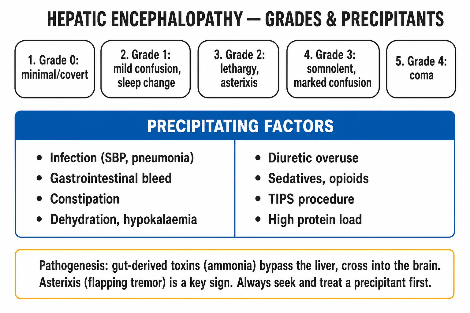
| I | A mild confusion, a euphoria or an anxiety, a shortened attention, a sleep disturbance, the plus-minus asterixis |
| II | A lethargy, an apathy, a disorientation, an obvious asterixis, a slurred speech |
| III | A somnolence but arousable, a gross disorientation, a bizarre behaviour, a marked asterixis |
| IV | A coma |
West Haven HE grading — click each
Somnolent — overt
Somnolent but arousable, gross disorientation (person / place), bizarre behaviour, marked asterixis, muscular rigidity, hyperreflexia, clonus. Airway at risk — intubate.
Covert HE (CHE)
Grades 0–I
- Minimal (grade 0, psychometric only) plus grade I
- Affects up to 60-80% of cirrhotics — usually undiagnosed
- Impairs driving, work, and quality of life; predicts overt HE
- Detected by psychometric testing (PHES), ICT, or Stroop-type apps
Overt HE (OHE)
Grades II–IV
- Clinically apparent encephalopathy — the typical ICU presentation
- Asterixis from grade II; airway threat from grade III
- Each overt episode worsens long-term cognitive function
Episodic
Precipitated
- Acute onset with an identifiable precipitant (the ICU default)
- Resolves when the precipitant is treated
Persistent / recurrent
Relapsing
- Recurrent (≥2 episodes in 6 months) → ADD rifaximin to lactulose
- Persistent (continuous cognitive impairment) → consider transplant evaluation
The precipitants (the chronic liver disease)
In the chronic liver disease, the HE is precipitated by:[1]
- The GI bleed (the blood in the gut is a large protein load, digested to the ammonia).[1]
- The infection or the sepsis.[1]
- The constipation (the increased time for the ammonia absorption).[1]
- The hypokalaemia and the alkalosis (the hypokalaemia increases the renal ammonia production; the alkalosis shifts the ammonia into the cells).[1]
- The dehydration or the diuretic overuse (the reduced renal perfusion, the azotaemia).[1]
- The sedatives, the benzodiazepines, the opiates (the direct CNS depression, the reduced clearance).[1]
- The TIPS or the surgical shunt (the increased portosystemic shunting).[1]
- The excess dietary protein.[1]
HEPATICHEPATIC — the precipitants of chronic-liver HE
Mechanism-ordered precipitants
Why each causes HE
- INFECTION (SBP, pneumonia, UTI): the #1 precipitant — systemic inflammation amplifies ammonia neurotoxicity and sepsis itself impairs BBB / astrocyte function
- GI BLEED: digested blood is a large protein load → bacterial generation of ammonia; also worsens portal perfusion and haemodynamics
- CONSTIPATION: prolonged colonic transit → more time for ammonia to be absorbed
- HYPOKALAEMIA / ALKALOSIS: hypokalaemia drives renal ammoniagenesis; alkalosis favours NH3 (lipophilic, crosses the BBB) over NH4+ (trapped)
- HYPOVOLAEMIA / DIURETIC OVERUSE: reduced renal perfusion → azotaemia (urea → ammonia) and electrolyte loss
- TIPS / SHUNT: iatrogenic increase in portosystemic shunting — HE develops in ~20-40% within the first 3-6 months
- SEDATIVES / BENZODIAZEPINES / OPIOIDS: direct CNS depression + increased GABAergic tone + constipation
- ALCOHOL intake or withdrawal: direct neurotoxicity; withdrawal mimics and worsens HE
- EXCESS DIETARY PROTEIN: especially after a protein binge in a sarcopenic, decompensated patient
The investigation bundle
Order on every HE presentation
- Blood cultures, urine culture, chest X-ray, and a DIAGNOSTIC ASCITIC TAP (SBP >250 neutrophils/mm³) — never skip the tap if ascites is present
- FBC, U&E (K, Na, creatinine), LFTs, glucose, coagulation (INR), venous or arterial ammonia
- CT brain — exclude a structural lesion, haemorrhage, or subdural (a coagulopathic cirrhotic who has fallen)
- Drug and alcohol screen; review the medication chart (look for the iatrogenic benzodiazepine or opioid)
- Complete the septic and metabolic workup BEFORE attributing the confusion to "just" HE
The diagnosis
The diagnosis is clinical (the impaired mentation plus the liver disease), supported by:[1]
- The asterixis (the flapping tremor) — a sign, not specific to the HE.[1]
- The ammonia — supportive, NOT necessary for the diagnosis. The level does NOT correlate well with the grade. The arterial ammonia is better than the venous. A normal ammonia does NOT exclude the HE.[1]
- The EEG — the triphasic waves (a characteristic but not specific pattern).[1]
- The psychometric tests (the number-connection test) — for the minimal (covert) HE.[1]
- Exclude the others — the glucose, the sodium, the sepsis screen, the CT brain, the drug screen. The differential: the metabolic encephalopathy (the uraemia, the hyponatraemia, the hypoxia), the sepsis, the intracranial lesion, the Wernicke, the drug toxicity.[1]
The diagnostic workup — confirm HE, find the precipitant, exclude the mimics
Step 1 — Confirm the clinical syndrome
Impaired cognition or alertness in a patient with known or suspected liver disease plus portosystemic shunting. Grade by West Haven. Look for asterixis (grade ≥II), constructional apraxia (draw a star / Rey figure), and day-night sleep reversal.
Step 2 — Exclude the mimics (this is the discriminator)
Hypoglycaemia (glucose), hyponatraemia (Na), uraemia, hypoxia or hypercapnia, sepsis, an intracranial lesion (CT brain), drug or alcohol toxicity, Wernicke encephalopathy (give thiamine), and seizures / post-ictal states. A cirrhotic can still have a subdural haematoma or meningitis — do NOT assume every confused cirrhotic has HE.
Step 3 — Find and treat the precipitant
Diagnostic ascitic tap (SBP >250 neutrophils/mm³), blood and urine cultures, chest X-ray, electrolytes, and a drug screen; review the medication chart. Infection and GI bleed are the top two — they must be actively sought in every case.
Step 4 — Ammonia (supportive, not diagnostic)
Arterial preferred over venous. Helps in diagnostic uncertainty (in ALF, an ammonia >100-150 µmol/L predicts cerebral oedema) and in tracking trends over time. NEVER use a single level to diagnose, grade, or exclude HE.
Step 5 — Adjuncts when needed
EEG (triphasic waves — supportive, not specific); psychometric tests (PHES, number-connection test) for covert HE; MRI brain if a structural or basal-ganglia (manganese) question arises. These are rarely needed in the acute ICU setting.
Asterixis (flapping tremor)
A sign, not a diagnosis
- Ask the patient to hold the arms outstretched with wrists dorsiflexed for ~30 s — a brief, arrhythmic "flap" is positive
- Indicates impaired motor coordination of metabolic origin; present from West Haven grade II
- NOT specific to HE — also seen in CO2 retention (respiratory failure), uraemia, hypomagnesaemia, and other metabolic encephalopathies
- Absent in grade I (covert) and usually in grade IV (comatose) — a negative test does not exclude HE
EEG triphasic waves
Supportive, not specific
- Bilateral, synchronous, symmetrical waves with a surface-positive first phase, maximal frontally
- Seen in ~25% of grade I-II and most grade III-IV HE
- NOT specific — also in uraemia, anoxia, hyponatraemia, lithium toxicity, and post-ictal states
- Resolution parallels clinical improvement — useful for the intubated / sedated patient where the exam is limited
The management
1. Find and treat the precipitant — the most important step (the chronic liver HE)
Search for and treat the precipitant: the GI bleed (the endoscopy), the infection (the cultures and the antibiotics), the constipation (the laxatives), the electrolyte correction (the potassium), the dehydration (the cautious fluids), and the cessation of the sedatives.[1]
2. The ammonia-lowering therapy
- The lactulose (a non-absorbable disaccharide) — the first-line. Titrated to 2 to 3 soft bowel movements a day. It works by acidifying the colon (the lactate converts the NH3 to the NH4+, which is trapped in the gut and excreted), the cathartic effect (the reduced transit time), and the reduced ammonia-producing bacteria. Given orally or via the nasogastric tube; the enema in the comatose. The lactitol is an alternative.[1]
- The rifaximin (a poorly-absorbed antibiotic) — 550 mg twice daily, as the add-on for the recurrent or the refractory HE (with the lactulose). Reduces the ammonia-producing gut bacteria.[1]
- The L-ornithine L-aspartate (LOLA) — enhances the urea cycle (the substrate for the ammonia detoxification); an alternative or an adjuvant.[1]
- The branched-chain amino acids — an adjuvant (theoretical benefit).[1]
3. The nutrition — AVOID the protein restriction (an old myth)
Do NOT restrict the protein. The adequate protein (1.2 to 1.5 g/kg/day) supports the hepatic regeneration and the muscle mass (the muscle clears the ammonia). The plant and the dairy protein are preferred (the lower ammonia load than the red meat). The protein restriction worsens the sarcopenia and the HE.[1]
4. Avoid the sedatives and the benzodiazepines
Avoid the sedatives, the benzodiazepines, and the opiates (they precipitate and worsen the HE). If the sedation is essential (the intubation), use the short-acting agents and the lowest effective dose. Avoid the benzodiazepine reversal (the flumazenil) except in the specific benzodiazepine overdose (it may transiently improve the HE but is not used routinely, due to the seizure risk).[1]
5. The airway and the ventilation
Intubate the grade III to IV HE (the comatose, the airway unprotected). The cautious sedation with the short-acting agents.[1]
6. The cerebral oedema management (the acute liver failure)
In the acute liver failure (not the chronic), the cerebral oedema is the major threat: the head elevation (30 degrees), the normoglycaemia, the normocapnia, the hypertonic saline (the Na 145 to 155) or the mannitol, the ICP monitoring in the select cases, the normothermia, and the urgent transplant referral. The lactulose and the rifaximin have a limited role here (the problem is the acute necrosis, not the precipitant).[1]
7. Key trials — the evidence behind the therapy
Bass et al. — Rifaximin for the prevention of HE recurrence (NEJM 2010, PMID 20335583)
Design
Multicentre, randomised, double-blind, placebo-controlled trial — 299 patients in remission from ≥2 episodes of overt HE
Intervention
Rifaximin 550 mg orally twice daily vs placebo for 6 months; most patients in BOTH arms (~90%) were also taking lactulose
Primary outcome
Time to first breakthrough HE episode: rifaximin significantly reduced the risk (HR 0.42; 95% CI 0.28–0.64; p<0.001) — a 58% relative risk reduction
Secondary outcome
Also reduced HE-related hospitalisation (HR 0.50; 95% CI 0.29–0.87; p=0.01)
Safety
Favourable; no excess of serious adverse events. The landmark study establishing rifaximin as add-on therapy for recurrent HE.
Bottom line
Rifaximin 550 mg BD is ADDED to (not substituted for) lactulose in patients with ≥2 episodes of overt HE. It is a PREVENTION dose — continue long-term.
Córdoba et al. — Normal-protein vs low-protein diet in episodic HE (J Hepatol 2004, PMID 15246205)
Design
Randomised trial — 30 cirrhotic patients admitted with an episode of acute HE
Intervention
Normal-protein diet (1.2 g/kg/day) vs low-protein diet (0 g/day for 3 days, then progressively increased) during recovery
Outcome
No difference in the time-course of HE recovery; the low-protein group had a worse nitrogen balance and did NOT recover faster
Bottom line
The pivotal trial debunking protein restriction — the normal-protein group recovered equally well without the catabolic harm. Underpins the modern mandate to NOT restrict protein (1.2-1.5 g/kg/day, per ESPEN).
Als-Nielsen et al. — Flumazenil for hepatic encephalopathy (Cochrane 2004, PMID 15106178)
Design
Systematic review and meta-analysis of 12 randomised trials (805 patients) of benzodiazepine-receptor antagonists (flumazenil) vs placebo in HE
Population
Cirrhotic patients with acute or chronic HE, grades I-IV
Findings
Flumazenil produced a short-lived improvement in a small subgroup (benefit detectable within minutes), but NO significant effect on overall recovery, survival, or long-term outcome. Adverse events included seizures, especially in those with alcohol-related or epileptic predisposition
Bottom line
Flumazenil is NOT a routine therapy for HE. A trial dose (0.2 mg IV, titrated) is reasonable ONLY when occult benzodiazepine use is suspected and the patient is refractory to standard therapy, in a monitored setting with seizure precautions
CANONIC — defining acute-on-chronic liver failure (Moreau, Gastroenterology 2013, PMID 23474284)
Design
Prospective observational study — 1,343 patients hospitalised with acute decompensation of cirrhosis across 29 European centres
Contribution
Operationally defined ACLF using the CLIF-C Organ Failure score (liver, kidney, brain, coagulation, circulation, lung). Graded ACLF 1/2/3 by the number and severity of organ failures
Key findings
ACLF is common (~30% at admission, ~20% develop it within 3 months), carries high 28-day mortality (ACLF grade 3 ~75%), and HE is one of the six defining organ failures. Precipitants: infection, alcohol, GI bleed
Bottom line
The framework for recognising the cirrhotic who is failing more than the brain — HE in ACLF is a multi-organ disease, not a single-organ problem, and changes the trajectory (escalate organ support, refer for transplant)
Boike et al. — North American Practice-Based Recommendations for TIPS (Clin Gastroenterol Hepatol 2022, PMID 34274511)
Source
Multidisciplinary consensus (Society of Interventional Radiology / hepatology)
Relevance to HE
New HE, or worsening of pre-existing HE, occurs after ~20-40% of TIPS procedures, typically within the first 3-6 months. Pre-existing overt HE uncontrolled by medical therapy is a relative contraindication
Management of post-TIPS HE
Standard therapy (lactulose ± rifaximin) is first-line; if refractory, options include shunt reduction or revision (narrowing the stent to restore some portal flow), and ultimately liver transplant evaluation
Bottom line
A TIPS is both a therapy and a precipitant — assess HE before TIPS, treat with standard agents after, and consider reduction / occlusion of the shunt if HE becomes intractable
Hepatorenal syndrome overlap
Hepatorenal syndrome (HRS) and HE frequently coexist — both are features of decompensated cirrhosis and acute-on-chronic liver failure, and both are driven by the same splanchnic vasodilation / effective arterial underfilling physiology. [1]
Why they coexist
Shared pathophysiology
- Splanchnic vasodilation (NO, splanchnic angiogenesis) → reduced effective arterial blood volume → renal vasoconstriction (HRS) and a hyperammonaemic, inflamed brain (HE)
- A precipitant (infection, GI bleed, or diarrhoea from over-titrated lactulose) often triggers BOTH simultaneously
- Azotaemia itself raises urea → gut ammonia → worsens HE; hypokalaemia from diuretics or diarrhoea drives renal ammoniagenesis
- HRS-AKI (formerly type 1 HRS) carries a grim prognosis (median survival ~2 weeks untreated) and is an independent predictor of death in HE
Practical management points
When HE and HRS coexist
- Diagnose HRS-AKI per ICA criteria: AKI (creatinine rise ≥26.5 µmol/L in 48 h, or ≥1.5x baseline) + no response to 2 days of albumin 1 g/kg + exclusion of shock, nephrotoxins, and structural disease
- Treat HRS with TERLIPRESSIN + albumin (splanchnic vasoconstriction; watch for respiratory failure) or a norepinephrine infusion
- STOP diuretics and nephrotoxins; cautious volume with 20-25% albumin
- Be careful with lactulose — over-titration causes diarrhoea → hypovolaemia → worsens HRS. Titrate to 2-3 soft stools, NOT watery diarrhoea
- Both HRS-AKI and grade III-IV HE are indications for liver transplant evaluation
ACLF, HE and HRS outcomes
Clinical pearls
Exam practice
SAQ — Hepatic encephalopathy in the cirrhotic
10 minutes · 10 marks
A 58-year-old man with alcohol-related cirrhosis presents with 3 days of confusion and drowsiness. GCS 12 (E3V3M6). Tense ascites and asterixis. Temp 38.3°C, HR 110, BP 96/58. Na 128, K 3.0, creatinine 140, INR 2.1. Ascitic tap: 360 neutrophils/mm³. Blood cultures pending.
Red flags
References
- [1]Vilstrup H, Amodio P, Bajaj J, et al. Hepatic encephalopathy in chronic liver disease: 2014 Practice Guideline by the American Association for the Study of Liver Diseases and the European Association for the Study of the Liver Hepatology, 2014.PMID 25042402
- [2]Bass NM, Mullen KD, Sanyal A, et al. Rifaximin treatment in hepatic encephalopathy N Engl J Med, 2010.PMID 20335583
- [3]Als-Nielsen B, Gluud LL, Gluud C. Benzodiazepine receptor antagonists for hepatic encephalopathy Cochrane Database Syst Rev, 2004.PMID 15106178
- [4]Häussinger D, Sies H, Wettstein M, et al. Pathomechanisms in hepatic encephalopathy Biol Chem, 2021.PMID 34049427
- [5]Plauth M, Bernal W, Dasarathy S, et al. ESPEN guideline on clinical nutrition in liver disease Clin Nutr, 2019.PMID 30712783
- [6]Córdoba J, López-Hellín J, Planas M, et al. Normal protein diet for episodic hepatic encephalopathy: results of a randomized study J Hepatol, 2004.PMID 15246205
- [7]Cabral CM, Burns DL. Low-protein diets for hepatic encephalopathy debunked: let them eat steak Nutr Clin Pract, 2011.PMID 21447768
- [8]Ahboucha S, Butterworth RF. The neurosteroid system: an emerging therapeutic target for hepatic encephalopathy Metab Brain Dis, 2007.PMID 17823858
- [9]Moreau R, Jalan R, Gines P, et al. Acute-on-chronic liver failure is a distinct syndrome that develops in patients with acute decompensation of cirrhosis Gastroenterology, 2013.PMID 23474284
- [10]Gustot T, Fernandez J, Garcia E, et al. Clinical Course of acute-on-chronic liver failure syndrome and effects on prognosis Hepatology, 2015.PMID 25877702
- [11]Boike JR, Varghese J, Bhatia S, et al. North American Practice-Based Recommendations for Transjugular Intrahepatic Portosystemic Shunts in Portal Hypertension Clin Gastroenterol Hepatol, 2022.PMID 34274511
- [12]Ntuli Y, Rennie K, Williams S, et al. Infection, inflammation and hepatic encephalopathy from a clinical perspective Metab Brain Dis, 2024.PMID 39212845
- [13]Thanapirom K, Suksawatamnuay S, Tanpowpong N, et al. Ammonia is associated with liver-related complications and predicts mortality in acute-on-chronic liver failure patients Sci Rep, 2024.PMID 38461166
- [14]Ahboucha S, Desjardins P, Chatauret N, et al. Increased levels of pregnenolone and its neuroactive metabolite allopregnanolone in autopsied brain tissue from cirrhotic patients who died in hepatic coma Neurochem Int, 2006.PMID 16563564
- [15]Higuera-de-la-Tijera F, Servín-Caam AI, Abigail Lorence LM, et al. Current vision on diagnosis and comprehensive care in hepatic encephalopathy Rev Gastroenterol Mex (Engl Ed), 2023.PMID 37127462