ICU · neurocritical-care
ICP Monitoring and Multimodal Neuromonitoring — Comprehensive ICU Management
Also known as Intracranial pressure monitoring · ICP monitor · External ventricular drain · EVD · Intraparenchymal bolt · Codman probe · Camino probe · Cerebral perfusion pressure · CPP · Brain tissue oxygen · PbtO2 · LICOX · Cerebral microdialysis · Jugular venous bulb oximetry · SjvO2 · Multimodality neuromonitoring · Monroe-Kellie doctrine · ICP waveform · Percussion wave · Tidal wave
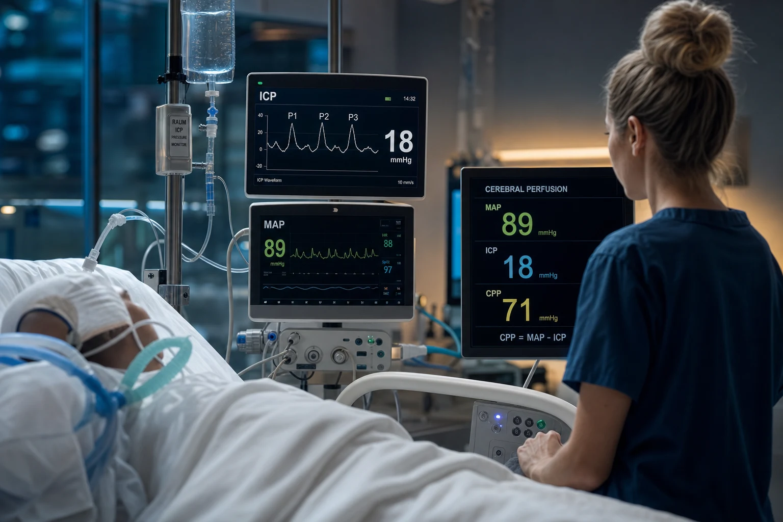
Intracranial pressure (ICP) monitoring is the cornerstone of neurocritical care for severe traumatic brain injury (TBI) and other intracranial hypertension states. The MONROE-KELLIE DOCTRINE governs the rigid cranial vault: volume = brain (80%) + blood (10%) + CSF (10%) — an increase in one component MUST be offset by a decrease in another, or ICP rises. Normal ICP is 5-15 mmHg (recumbent). INTRACRANIAL HYPERTENSION (ICH) = ICP 22 mmHg (BTF 4th edition treatment threshold). DEVICES: (1) EXTERNAL VENTRICULAR DRAIN (EVD) — the GOLD STANDARD — catheter in frontal horn of lateral ventricle, transduced against a reference (external auditory meatus) — MEASURES ICP AND drains CSF (therapeutic + diagnostic) — BUT highest infection rate (ventriculitis 5-15%); (2) INTRAPARENCHYMAL BOLT (Codman, Camino, Raumedic) — fibreoptic/strain-gauge probe bolted into brain parenchyma — LOWER infection (<2%), continuous reading, zero-drift over time, NO CSF drainage — most common in modern practice; (3) SUBDURAL/EPIDURAL sensors — LESS ACCURATE (overestimate/underestimate), rarely used now. ICP WAVEFORM: three peaks per cardiac cycle — P1 PERCUSSION (arterial pulsation — relatively constant), P2 TIDAL (brain COMPLIANCE — rises when compliance falls), P3 RESPIRATORY/DCOUP (venous/respiratory). P2 RISING ABOVE P1 = the brain has EXHAUSTED its compliance reserves = impending intracranial hypertension — a premonitory sign even if the MEAN ICP looks acceptable. CEREBRAL PERFUSION PRESSURE (CPP) = MAP − ICP; target 60-70 mmHg (BTF 4th ed). CPP <60 = cerebral ischaemia (secondary injury); CPP 70 = no added benefit AND increased ARDS risk (from the Robertson/Contant data — aggressive fluid/vasopressor-driven high CPP floods the injured lung). MULTIMODAL NEUROMONITORING recognises that ICP ALONE misses regional ischaemia — PbtO2 (brain tissue oxygen, LICOX/Neurotrend probe — target 20 mmHg — complements ICP), cerebral MICRODIALYSIS (lactate/pyruvate ratio 40 = metabolic distress; glycerol and glutamate = cell damage), JUGULAR VENOUS BULB OXIMETRY (SjvO2 <50% = global cerebral hypoxia, 90% = hyperaemia), and CONTINUOUS EEG (non-convulsive status detection). The integration paradigm: treat ICP + CPP + PbtO2 + metabolism TOGETHER — a normal ICP does NOT exclude ongoing brain ischaemia. KEY TRIALS: BTF 4th edition (Carney 2017), BEST:TRIP (Chesnut 2012 NEJM — ICP-guided care NOT superior to imaging-clinical exam in a Bolivian/Ecuadorian cohort, but monitoring is still standard of care in well-resourced settings), BOOST-2 (Okonkwo 2017 — PbtO2 + ICP monitoring reduced burden of brain hypoxia vs ICP alone, trend to better outcome — Phase III BOOST-3 ongoing).
On this page & tools
Your progress
Saved locally on this device.
Target exams
Red flags
Overview
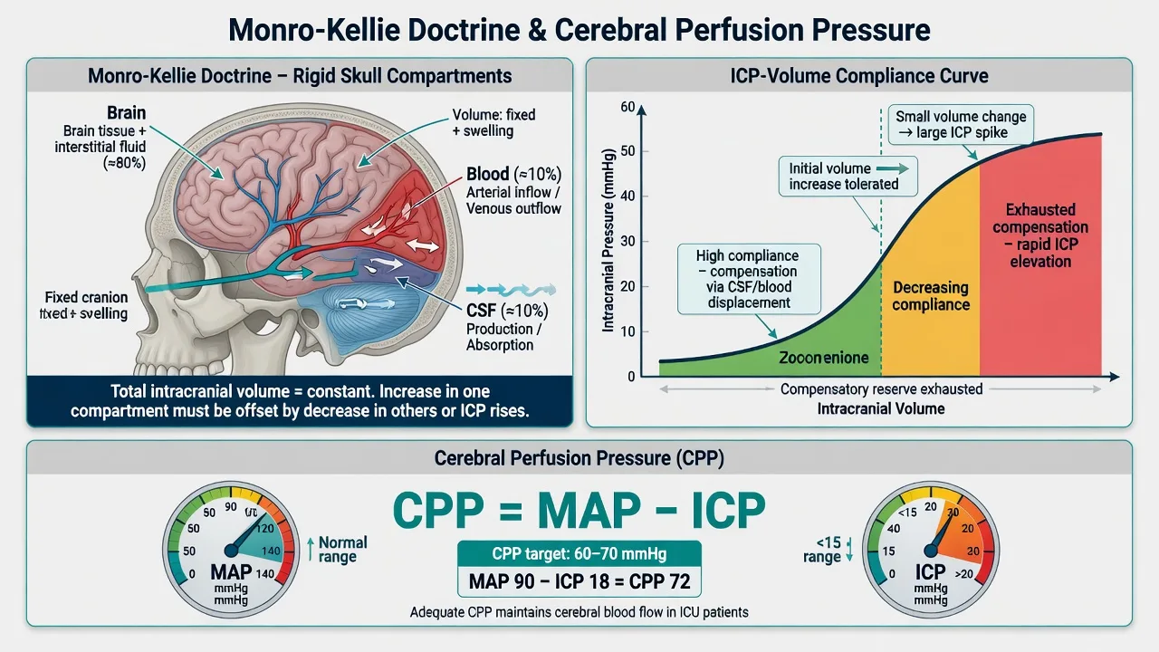
The Monroe-Kellie doctrine and intracranial compliance
The cranial vault — why ICP rises
| Concept | Mechanism | Clinical implication |
|---|---|---|
| Monroe-Kellie doctrine | The skull is a rigid box containing brain (80%) + blood (10%) + CSF (10%). Total volume is fixed — an increase in any one component MUST be offset by displacement of another | Any expanding lesion (haematoma, oedema) first displaces CSF and venous blood (compensated); once exhausted, ICP rises steeply |
| Intracranial compliance | Compliance = ΔVolume / ΔPressure. Early, compliance is high (CSF/venous blood shift out) — small pressure rise. Late, compliance is exhausted — same volume rise causes an explosive ICP rise | The patient who was 'fine' can herniate within minutes once compensation is exhausted — the compliance curve is exponential, not linear |
| Spatial compensation | CSF is displaced into the spinal theca; venous blood is displaced out of the dural venous sinuses | These are the FIRST buffers — they buy time but are finite (≈100-150 mL reserve in adults) |
| Pressure-volume curve | Flat initially (high compliance) → elbow → steep (low compliance) | On the steep part, removing a few mL of CSF (EVD drainage) or blood (evacuation) drops ICP dramatically — this is why EVDs are so effective |
ICP monitoring devices — the three options
ICP monitoring devices — comparison
| Device | How it works | Measures ICP | Drains CSF | Accuracy | Infection risk | Can re-zero | Drift |
|---|---|---|---|---|---|---|---|
| External ventricular drain (EVD) — GOLD STANDARD | Catheter tip in frontal horn of lateral ventricle, fluid-coupled to an external pressure transducer | YES | YES (therapeutic) — can drain CSF to lower ICP | Highest — true global ventricular pressure | HIGHEST (ventriculitis 5-15%) — foreign body in the ventricle + CSF is poor host defence | YES (transducer re-levelled to external auditory meatus) | No drift (external transducer) |
| Intraparenchymal bolt (Codman, Camino, Raumedic) | Fibreoptic or strain-gauge sensor bolted 1-2 cm into brain parenchyma | YES (continuous) | NO | High (local parenchymal pressure — close to ventricular if no gradient) | LOW (<2%) — parenchyma is more resistant to infection than CSF | NO (zeroed once at insertion) | YES — zero drift over days (1-3 mmHg/week) |
| Subdural / epidural sensor | Pressure sensor placed on (subdural) or outside (epidural) the dura | Approximate | NO | LEAST accurate — over/under-reads, dampened | Low | NO | Yes |
| Lumbar CSF pressure | Lumbar puncture / drain transducer | Indirect (only if basilar cisterns open) | YES | Poor correlation if gradient present | Low-moderate | YES | No drift |
External ventricular drain — practical points
- Insertion: frontal (Kocher's point — typically non-dominant hemisphere, 2-3 cm lateral to midline, 1 cm anterior to coronal suture) catheter passed into the frontal horn of the lateral ventricle.
- Transducer levelling: the external pressure transducer is levelled to the FORAMEN OF MONRO / external auditory meatus (the ventricle). The drainage bag height is set ABOVE this reference — e.g. set at 15 cmH2O to drain when ICP exceeds that level. NEVER lower the bag below the reference unless deliberately draining.
- Zeroing: the external transducer is zeroed to atmosphere and re-levelled if the patient's head moves.
- Closing the system to measure ICP: to obtain a true ICP reading, the drainage line is CLAMPED above the transducer so the column couples to the ventricle (continuous drainage under-reads the true peak ICP).
- Risks: ventriculitis (5-15%), haemorrhage (~1-2%), malposition, obstruction (debris, choroid plexus), over-drainage (causing upward herniation or re-bleed of a subdural).
Intraparenchymal bolt — practical points
- Insertion: bolt screwed into the skull via a twist-drill hole; parenchymal sensor (Codman MicroSensor, Camino, Raumedic Neurovent) advanced 1-2 cm into tissue.
- Zeroing: zeroed to atmosphere ONCE at insertion — CANNOT be re-zeroed in vivo (a key limitation).
- Drift: sensor zero-drift accumulates over days (typically 1-3 mmHg/week) — a limitation for prolonged monitoring. The Brain Trauma Foundation accepts a drift of up to ±2 mmHg as acceptable.
- Advantage: low infection, continuous reading, easy insertion even with small/slit ventricles (common in swollen brains where EVD placement is difficult).
- Limitation: measures LOCAL parenchymal pressure — may miss a global rise if the probe sits in a relatively spared region, and provides NO CSF drainage.
ICP waveform analysis — reading the pressure trace
The three components of the ICP waveform
| Wave | Origin | Amplitude behaviour | Clinical meaning |
|---|---|---|---|
| P1 — PERCUSSION wave | Arterial pulsation transmitted to the choroid plexus / intracranial arteries | Relatively CONSTANT regardless of compliance — the 'fixed' arterial input | Present as long as there is arterial inflow |
| P2 — TIDAL wave | Brain TISSUE compliance / recoil — the intracranial contents bouncing back | RISES as compliance FALLS — the dynamic, compliance-dependent component | P2 > P1 = EXHAUSTED COMPLIANCE = intracranial hypertension imminent |
| P3 — RESPIRATORY / dicrotic wave | Venous / respiratory fluctuation (the dicrotic component after aortic valve closure) | Small, varies with respiration and venous return | Less clinically used; affected by CVP, ventilation |
Lung-brain coupling: respiratory effects on ICP
- Mechanical ventilation raises intrathoracic pressure → impedes cerebral venous return → increases cerebral blood volume → raises ICP. Positive end-expiratory pressure (PEEP) and high mean airway pressures (inverse-ratio ventilation, high driving pressures) transmit to the intracranial compartment.
- Coughing, straining, bucking the ventilator, suctioning cause transient ICP spikes (often >40 mmHg) — adequate sedation/analgesia and pre-oxygenation before suctioning blunt these.
- The P3 (respiratory) wave visibly oscillates with each breath — large respiratory swing suggests high intrathoracic pressure transmission to the brain.
Lundberg waves — pathological ICP trends
- Lundberg A (plateau) waves: sustained ICP >50 mmHg for 5-20 minutes — vasodilatory cascade from transiently low CPP → cerebral vasodilation → rise in intracranial blood volume → higher ICP → lower CPP → vicious cycle. Ominous — indicate severely impaired autoregulation and exhausted compliance; associated with poor outcome.
- Lundberg B waves: rhythmic 0.5-2/min oscillations of 20-50 mmHg — reflect impaired pressure autoregulation; a warning that the system is becoming unstable.
- Lundberg C waves: low-amplitude, higher-frequency oscillations — less clinically significant.
ICP and CPP thresholds — the treatment targets
ICP and CPP thresholds — what the numbers mean
| Parameter | Range | Interpretation | Action |
|---|---|---|---|
| ICP | 5-15 mmHg | Normal (recumbent) | — |
| ICP | 15-20 mmHg | Borderline / mildly raised | Monitor closely, address exacerbating factors (position, seizures, fever, agitation) |
| ICP | 20-25 mmHg | Mild intracranial hypertension | Begin tiered therapy; BTF 4th ed treats >22 mmHg for >1 min |
| ICP | 25-40 mmHg | Moderate intracranial hypertension | Aggressive tiered therapy; consider second-line agents; re-image for a surgical lesion |
| ICP | >40 mmHg | SEVERE — imminent herniation | Neurosurgical emergency: maximal osmotherapy, anaesthesia/barbiturates, decompressive craniectomy, check for surgical lesion |
| CPP | <60 mmHg | Cerebral ischaemia — secondary injury | Raise MAP (noradrenaline) and/or lower ICP; transfuse if anaemic |
| CPP | 60-70 mmHg | TARGET (BTF 4th ed) | Maintain |
| CPP | >70 mmHg | No added benefit AND increased ARDS risk | Do not push CPP above 70 with fluids/vasopressors |
Why CPP > 70 is harmful — the ARDS link
Aggressive CPP-targeted therapy (maintaining CPP >70 mmHg with high-volume fluids and high-dose vasopressors, popularised in the 1990s) was associated with a 5× increase in ARDS (Robertson 1999; Contant 2001), with NO improvement in neurological outcome. The mechanism: the injured brain has regions of disrupted autoregulation where cerebral blood flow is pressure-passive; pushing CPP very high floods these regions, but the fluid and vasoconstrictor load needed to do so overwhelms the injured lung alveoli → ARDS. The BTF 4th edition therefore caps the CPP target at 60-70 mmHg — the ischaemia floor (60) matters more than any ceiling benefit above 70.
Indications for ICP monitoring (BTF 4th edition, Carney 2017)
Who gets an ICP monitor — BTF 4th edition indications
- SEVERE TBI (GCS 3-8 after resuscitation) AND an ABNORMAL head CT:
- Abnormal = haematomas, contusions, swelling, herniation, or compressed basal cisterns
- This is the clearest indication — these patients have a >50% chance of intracranial hypertension
- SEVERE TBI with a NORMAL CT if TWO OR MORE of:
- Age >40 years
- Unilateral OR bilateral motor posturing
- Systolic BP <90 mmHg
- (These features identify the ~30% of normal-CT severe TBI patients who will still develop raised ICP)
- OTHER indications (consensus / guideline-supported beyond TBI):
- Large spontaneous intracerebral haemorrhage with reduced GCS / midline shift
- Extensive subarachnoid haemorrhage with hydrocephalus or depressed GCS
- Subdural/epidural haematoma post-evacuation with brain swelling
- Anoxic brain injury with cerebral oedema (e.g. post-cardiac arrest with CT swelling)
- Fulminant hepatic failure with cerebral oedema
- Meningoencephalitis with signs of raised ICP
- Any patient in whom you cannot reliably examine neurologically (deep sedation) AND who is at risk of intracranial hypertension
- Contraindications (relative):
- Coagulopathy that cannot be corrected (INR >1.4-1.6, platelets <75-100, on therapeutic anticoagulation) — correct first, then insert
- Scalp infection at the insertion site — choose an alternative site
- Absolute: no contraindication outweighs the risk of unmonitored herniation in a patient who needs the monitor
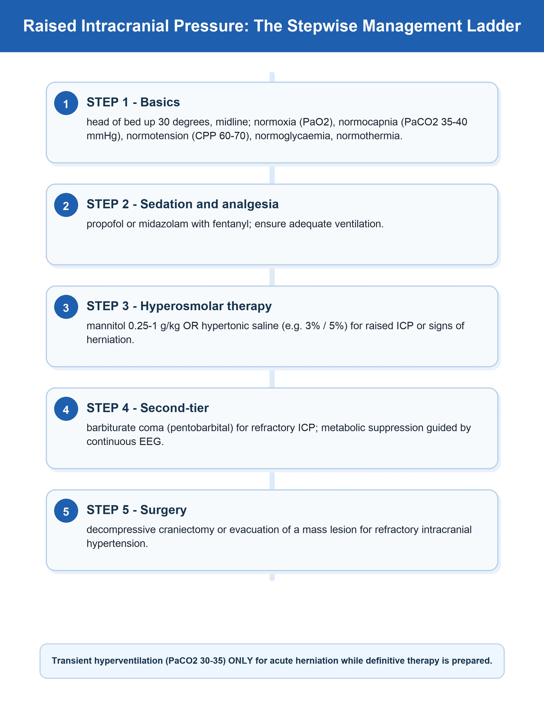
ICP management — the tiered (staircase) approach
Tiered ICP/CPP management protocol — severe TBI
- TIER 0 — FOUNDATIONS (prevent secondary injury, do no harm):
- Head of bed 30° (midline, neck neutral — optimises venous drainage)
- Normoxia (PaO2 >60, SpO2 >94%), normocapnia (PaCO2 35-40 mmHg) — AVOID prophylactic hyperventilation (causes cerebral vasoconstriction → ischaemia)
- Normotension (SBP >110 / MAP >80 for 50-69 yr; SBP >100 / MAP >80 for ≥14 yr — BTF 4th ed)
- Normothermia (avoid fever — raises cerebral metabolic demand), normoglycaemia (6-10 mmol/L), avoid anaemia (Hb >70-90)
- Adequate analgesia and sedation (propofol ± fentanyl) — pain and agitation raise ICP
- Seizure prophylaxis (levetiracetam) for high-risk patients
- TIER 1 — FIRST-LINE ICP >22 mmHg:
- Sedation and analgesia optimised (ensure the patient is not fighting the ventilator)
- CSF drainage if an EVD is in situ (drain 5-10 mL) — the most rapid, direct method
- Hyperosmolar therapy: MANNITOL 0.25-1 g/kg bolus (oncotic withdrawal of oedema fluid, onset 15-30 min; monitor osmolar gap <20, serum osmolality <320) OR HYPERTONIC SALINE (e.g. 3% / 5% bolus or infusion — preferred if hypotensive, longer duration, less diuresis, also restores intravascular volume)
- Re-image if ICP not controlled or if a surgical lesion is suspected
- TIER 2 — SECOND-LINE (ICP refractory to tier 1):
- Neuromuscular paralysis (cisatracurium / rocuronium infusion) — abolishes coughing/straining-induced ICP spikes; confirm with train-of-four
- Moderate hyperventilation (PaCO2 30-35 mmHg) — temporary measure ONLY (hours), while awaiting definitive therapy; causes cerebral vasoconstriction → lower cerebral blood volume → lower ICP, but risks ischaemia — use with PbtO2 monitoring if available
- Repeat hyperosmolar therapy / consider continuous hypertonic saline infusion
- Re-image — is there a surgical lesion to evacuate?
- TIER 3 — REFRACTORY INTRACRANIAL HYPERTENSION:
- Decompressive craniectomy (RESCUEicp — reduced mortality but higher rates of vegetative/severe disability vs medical therapy; DECRA — earlier bifrontal craniectomy worsened 6-month outcome) — individualise; discuss goals of care
- High-dose barbiturates (pentobarbital/thiopentol coma) — suppress cerebral metabolism → lower cerebral blood flow → lower ICP; monitor with continuous EEG for burst suppression; significant side effects (hypotension, infection, ileus)
- Optimise CPP — use noradrenaline to keep CPP 60-70 mmHg
- Targeted temperature management (33-36°C) — modest ICP reduction; evidence mixed
- Re-image / re-operate — evacuate new surgical lesions
Multimodal neuromonitoring — beyond ICP
The modalities — what each one measures
| Modality | What it measures | Target / threshold | What it detects that ICP misses | Key limitation |
|---|---|---|---|---|
| Brain tissue oxygen (PbtO2) — LICOX, Neurotrend | Local oxygen partial pressure at a parenchymal probe (mmHg) — reflects the balance of oxygen delivery and consumption at the tissue level | >20 mmHG normal; <15 = hypoxia; <10 = critical | Regional/diffuse ischaemia with a NORMAL ICP (microvascular dysfunction, vasospasm, low delivery) | Measures a SMALL region (~15-20 mm radius) — may miss a distant ischaemic territory; placement site matters |
| Cerebral microdialysis | Extracellular brain metabolites via a semi-permeable dialysis catheter | Lactate/pyruvate ratio (LPR) <25 normal; >40 = metabolic distress; lactate, pyruvate, glucose, glutamate, glycerol | Cellular energy failure / ischaemia and excitotoxicity before structural damage — LPR rises indicate mitochondrial hypoxia; glycerol rises = cell membrane breakdown; glutamate rises = excitotoxic injury | Regional (probe vicinity); delayed readout (hourly samples); technically demanding |
| Jugular venous bulb oximetry (SjvO2) | Oxygen saturation of venous blood sampled from the jugular bulb (global outflow) | 55-75% normal; <50% = cerebral hypoxia; >90% = hyperaemia or AV shunting | GLOBAL cerebral oxygenation imbalance — low SjvO2 = extraction exceeds delivery (ischaemia); high = luxury perfusion | GLOBAL average — masks focal ischaemia; technically fiddly (catheter position, contamination) |
| Continuous EEG (cEEG) | Cortinal electrical activity over hours-days | Normal background; detect seizures / NCSE | Non-convulsive status epilepticus (NCSE) — occult in >10-20% of comatose TBI / SAH / ICH patients, driving up metabolic demand and ICP | Requires expert interpretation; artefact in the ICU; regional sensitivity |
| Transcranial Doppler (TCD) | Blood flow velocity in major intracranial arteries | Velocities, pulsatility index, Lindegaard ratio (to distinguish vasospasm from hyperaemia) | Vasospasm (post-SAH), impaired autoregulation (PRx), raised ICP trends | Operator-dependent; ~10% inadequate acoustic windows |
| Near-infrared spectroscopy (NIRS) / automated pupillometry | Regional cerebral oxygen saturation (rSO2); pupil size/reactivity | rSO2 trends; Neurological Pupil index (NPi) | Non-invasive cerebral oxygenation trends; pupil reactivity as a prognostic and herniation marker | NIRS confounded by extracranial signal; trend more useful than absolute value |
Brain tissue oxygen (PbtO2) in detail
- Probe: a polarographic (Clark) or fluorescence oxygen sensor placed into parenchyma alongside the ICP bolt (LICOX, Neurotrend). Measures the O2 that diffuses from capillaries into tissue — the net of delivery minus consumption.
- Thresholds: >20 mmHg normal; 15-20 borderline; <15 hypoxia (treat); <10 critical (strongly associated with death and poor outcome).
- Therapeutic levers to raise PbtO2: increase FiO2 / PaO2; increase CPP (raise MAP with noradrenaline — but cap at 70); transfuse if anaemic (Hb <90); reduce metabolic demand (sedation, anticonvulsants, normothermia); avoid hyperventilation.
- BOOST-2 (Okonkwo 2017): a PbtO2 + ICP guided protocol REDUCED the burden of brain tissue hypoxia (proportion of time hypoxic 0.45 with ICP-alone vs 0.16 with ICP + PbtO2, p<0.0001) with a trend toward lower mortality and better outcome — not powered for efficacy, but justified the Phase III BOOST-3 trial.[3]
Cerebral microdialysis in detail
- Mechanism: a double-lumen dialysis catheter perfused with isotonic fluid; extracellular solutes diffuse across the semi-permeable membrane and are collected hourly for bedside analysis.
- Markers:
- Lactate / pyruvate ratio (LPR) — the master marker of metabolic distress. Pyruvate is the substrate; when mitochondria are hypoxic, pyruvate is shunted to lactate → LPR rises. LPR >25-30 concerning; >40 = significant metabolic distress (ischaemia or mitochondrial failure). A high LPR with LOW pyruvate = ischaemia; high LPR with normal pyruvate = mitochondrial dysfunction.
- Glucose — low brain glucose (<0.6-1.0 mmol/L) = substrate depletion / hypoxia.
- Glutamate — excitotoxic amino acid; rises with ischaemic cell stress.
- Glycerol — cell membrane phospholipid breakdown product; rises with membrane destruction (severe ischaemia, cell death).
- Use: detects energy failure and ischaemia earlier and more locally than global markers; useful to individualise CPP and PbtO2 targets. [1]
Jugular venous bulb oximetry (SjvO2) in detail
- Technique: a retrograde catheter advanced up the internal jugular vein into the jugular bulb (at the skull base), sampling cerebral venous outflow before mixing with extracranial venous blood.
- Interpretation: SjvO2 reflects global balance of cerebral oxygen delivery (DO2) and consumption (CMRO2). <50% = cerebral hypoxia (delivery insufficient — low CPP, hypoxaemia, anaemia, hyperventilation-induced vasoconstriction). >90% = hyperaemia / luxury perfusion or arteriovenous shunting (loss of autoregulation).
- Limitation: it is a GLOBAL average — a focal ischaemic region may be diluted out by well-perfused tissue. Largely superseded by PbtO2 in many centres but still used for global oxygenation trends.
Continuous EEG (cEEG)
- Purpose: detect non-convulsive status epilepticus (NCSE), which is clinically silent in a sedated/comatose patient but drives up cerebral metabolic demand, raises ICP, and worsens outcome. NCSE is found in >10-20% of comatose neuro-ICU patients (TBI, SAH, ICH, post-anoxic).
- Recommendation: 24-48h cEEG for any comatose neuro-ICU patient with unexplained depressed consciousness or fluctuating examination; many centres use routine cEEG for all severe TBI/SAH with depressed GCS.
Integration — the multimodal management paradigm
How to integrate ICP + CPP + PbtO2 + microdialysis at the bedside
- Read the SYSTEM, not the single number:
- Every assessment: ICP + waveform, MAP → CPP, PbtO2, and (if available) microdialysis LPR and cEEG
- Ask: is there INTRACRANIAL HYPERTENSION (ICP), ISCHAEMIA (low PbtO2 / high LPR / low SjvO2), SEIZURES (cEEG), or DISTRESSED METABOLISM — and treat each abnormality
- The common conflicts and how to resolve them:
- High ICP + low PbtO2: treat the ICP (osmotherapy, drainage), but AVOID hyperventilation (it lowers ICP but worsens PbtO2 by vasoconstriction) — prefer hypertonic saline, CSF drainage, sedation
- Normal ICP + low PbtO2: the ischaemia is not pressure-driven — raise MAP/CPP (noradrenaline to CPP 60-70), increase FiO2, transfuse if anaemic, treat seizures; check for vasospasm (TCD)
- High ICP + high PbtO2: possible hyperaemia (loss of autoregulation) — modestly lower MAP within the CPP-safe range, head-up position; consider moderate hyperventilation briefly
- Rising LPR with normal ICP/PbtO2: impending mitochondrial/cellular failure — escalate investigation (ischaemic work-up), optimise delivery, treat seizures
- Individualise the targets:
- Autoregulation-guided CPP (using PRx from TCD/ICP waveform analysis) — set CPP at the patient's optimal autoregulatory range rather than a fixed number
- Treat the PATIENT'S thresholds: some patients herniate at ICP 20; others tolerate 25. The trend, the waveform (P2 > P1), and the downstream markers (PbtO2, LPR) are more informative than any single cut-off
- Treat the whole brain, not just the pressure:
- The integration paradigm (Le Roux 2014 consensus) holds that ICP/CPP/PbtO2/metabolism are INTERDEPENDENT — a management decision that lowers ICP but worsens PbtO2 (e.g. aggressive hyperventilation) is a Pyrrhic victory. Optimise the integrated bundle: normoxia, normocapnia, CPP 60-70, PbtO2 >20, no seizures, no fever, no hyperglycaemia, no anaemia[5]
Complications of monitoring — and how to avoid them
Device-related complications
| Complication | Device | Rate | Prevention / management |
|---|---|---|---|
| Ventriculitis / catheter-related CNS infection | EVD (highest) | 5-15% | Aseptic insertion, prophylactic antibiotic at insertion, tunnelled catheter, minimise breaks into the system, avoid CSF leakage at the skin, replace if infected; antibiotic-impregnated catheters reduce infection |
| Intraparenchymal haemorrhage | EVD, bolt | 1-2% (clinically significant) | Correct coagulopathy before insertion (INR <1.4-1.6, platelets >75-100); avoid multiple passes |
| Zero drift (sensor inaccuracy) | Intraparenchymal bolt | 1-3 mmHg/week | Accept up to ±2 mmHg; if ICP reading seems inconsistent with the clinical picture, re-image or compare with an EVD if critical |
| Over-drainage / up-herniation | EVD | Low but catastrophic | Never set the drain below the reference level unintentionally; secure the system; avoid rapid CSF drainage |
| Catheter malposition / obstruction | EVD | 5-10% | Confirm position on CT; flush only with strict asepsis; re-site if obstructed |
| Skin infection / osteomyelitis | Bolt / EVD | Low | Aseptic insertion, site care |
SAQ — ICP and multimodal neuromonitoring
ICP crisis and CPP/PbtO2 integration
10 minutes · 10 marks
A ventilated severe TBI patient has ICP 28 mmHg, MAP 76 mmHg, PbtO2 15 mmHg. Outline interpretation and stepwise management.
Red flags
Prognosis
Prognostic markers in ICP-monitored severe TBI
| Marker | Threshold / pattern | Prognostic implication |
|---|---|---|
| ICP sustained >20-22 mmHg | Refractory to tiered therapy | Doubled mortality; each 10 mmHg increment above 20 worsens outcome |
| ICP >40 mmHg (severe) | Sustained | Very high mortality; imminent herniation risk |
| Lundberg A (plateau) waves | ICP >50 mmHg for 5-20 min | Severely impaired autoregulation; poor outcome |
| CPP <60 mmHg sustained | Ischaemic threshold | Secondary injury; worse neurological outcome |
| PbtO2 <15 mmHg / <10 mmHg | Brain tissue hypoxia burden | Strongly associated with death and unfavourable 6-month outcome (BOOST-2) |
| Lactate/pyruvate ratio >40 | Metabolic distress | Impending cellular energy failure; worse outcome |
| Loss of pupil reactivity (NPi <3) | Automated pupillometry | Strong predictor of unfavourable outcome; suggests herniation |
| cEEG: non-convulsive status | Refractory NCSE | Independent predictor of poor outcome; treatable if detected |
Outcome by duration/intensity of intracranial hypertension
| ICP pattern | Approximate mortality / unfavourable outcome | Notes |
|---|---|---|
| ICP controlled <22 mmHg throughout | Lowest | Best outcomes with intact autoregulation |
| Brief intracranial hypertension, readily controlled | Low-moderate | Good prognosis if CPP and PbtO2 maintained |
| Refractory intracranial hypertension (tier 3) | High | Mortality 50-80% without surgery; decompressive craniectomy reduces mortality but increases rates of vegetative/severe disability (RESCUEicp) |
| Bilateral fixed dilated pupils + ICP >40 | Very high | Imminent/established herniation; grave prognosis, but not universally fatal — some recover with aggressive therapy if the cause is reversible |
Key trials and evidence
BEST:TRIP — Chesnut 2012, NEJM (PMID 23234472)
Study design
Multicentre, controlled RCT — 324 severe TBI patients (GCS 3-8) in Bolivia/Ecuador
Population
Severe TBI in ICUs; randomised to ICP-monitor-guided (target ICP ≤20 mmHg) vs imaging-and-clinical-examination-guided management
Intervention
Intraparenchymal ICP-monitor-guided protocol vs structured ICE protocol (treatment triggered by CT and clinical signs)
Primary outcome
Composite of survival, consciousness, functional and neuropsychological status at 3 and 6 months — NO significant difference (score 56 vs 53, p=0.49)
Key finding
6-month mortality 39% (ICP) vs 41% (ICE); ICU stay similar; the ICE group received MORE brain-specific treatments (hyperosmolar therapy, hyperventilation)
Clinical bottom line
In this setting, ICP-guided care was NOT superior to a rigorous imaging-clinical exam protocol. It does NOT invalidate ICP monitoring in well-resourced ICUs (standard of care; needed for the sedated/unexaminable patient) — the lesson is that the structured PROTOCOL, not the monitor alone, drives outcome
BOOST-2 — Okonkwo 2017, Crit Care Med (PMID 29028696)
Study design
Phase II multicentre RCT — 119 severe TBI patients across 10 US ICUs
Population
Severe TBI; randomised to ICP + PbtO2-guided protocol vs ICP-only-guided protocol
Intervention
Tiered management informed by ICP + brain tissue oxygen (PbtO2) vs ICP alone
Primary outcome
Burden of brain tissue hypoxia — REDUCED from proportion of time 0.45 (ICP only) to 0.16 (ICP + PbtO2), p<0.0001
Key finding
PbtO2-directed therapy reduced brain tissue hypoxia with a TREND toward lower mortality and more favourable outcomes; ICP control was similar in both arms; no procedure-related complications
Clinical bottom line
Multimodal ICP + PbtO2 monitoring is feasible, safe, and reduces brain hypoxia — supports the integration paradigm and justified the Phase III BOOST-3 trial. Not powered for efficacy
BTF Guidelines 4th Edition — Carney 2017, Neurosurgery (PMID 27654000)
Document type
Evidence-based clinical practice guidelines (Brain Trauma Foundation / AANS / CNS)
Scope
Management of severe TBI — monitoring, thresholds, resuscitation, hyperosmolar therapy, anaesthetics, analgesia, nutrition, seizure prophylaxis
Key recommendations
ICP monitoring for severe TBI + abnormal CT (or normal CT with ≥2 risk factors); treat ICP >22 mmHg; target CPP 60-70 mmHg; avoid prophylactic hyperventilation (especially first 24h); hyperosmolar therapy with mannitol or hypertonic saline; early enteral nutrition
Clinical bottom line
The definitive reference for severe TBI management in the ICU — cite for thresholds (ICP >22, CPP 60-70), indications, and the tiered approach. Decompression recommendations updated in 2020 to integrate RESCUEicp and DECRA
References
- [1]Carney N, Totten AM, O'Reilly C, et al. Guidelines for the Management of Severe Traumatic Brain Injury, Fourth Edition Neurosurgery, 2017.PMID 27654000
- [2]Chesnut RM, Temkin N, Carney N, et al. A trial of intracranial-pressure monitoring in traumatic brain injury N Engl J Med, 2012.PMID 23234472
- [3]Okonkwo DO, Shutter LA, Moore C, et al. Brain Oxygen Optimization in Severe Traumatic Brain Injury Phase-II: A Phase II Randomized Trial Crit Care Med, 2017.PMID 29028696
- [4]Stocchetti N, Zoerle T, Carbonara M. Intracranial pressure management in patients with traumatic brain injury: an update Curr Opin Crit Care, 2017.PMID 28157822
- [5]Le Roux P, Menon DK, Citerio G, et al. Consensus summary statement of the International Multidisciplinary Consensus Conference on Multimodality Monitoring in Neurocritical Care : a statement for healthcare professionals from the Neurocritical Care Society and the European Society of Intensive Care Medicine Intensive Care Med, 2014.PMID 25138226
- [6]Chesnut RM, Temkin N, Dikmen S, et al. A Method of Managing Severe Traumatic Brain Injury in the Absence of Intracranial Pressure Monitoring: The Imaging and Clinical Examination Protocol J Neurotrauma, 2018.PMID 28726590