ICU · Neurocritical care / monitoring
ICP Monitoring & Waveforms
Also known as Intracranial pressure monitoring · ICP monitoring · ICP waveforms · Lundberg waves · Plateau waves · A-waves · P1 P2 P3 · External ventricular drain · EVD · Intraparenchymal pressure monitor · Codman · Cerebral perfusion pressure · CPP
Intracranial pressure (ICP) monitoring measures the pressure within the skull, guiding the management of raised ICP (target under 22 mmHg in TBI). The cerebral perfusion pressure (CPP = MAP minus ICP) is the key derived variable, with a target of 60-70 mmHg. The intraventricular catheter (external ventricular drain) is the gold standard (it measures, drains, and samples the CSF); the intraparenchymal microsensor (Codman) is accurate but cannot drain. The normal ICP waveform has three peaks: P1 (the arterial percussion), P2 (the tidal wave reflecting compliance — a P2 rising above P1 indicates a loss of compliance), and P3 (the venous dicrotic). The Lundberg A-wave (the plateau wave — a sustained rise to 50-100 mmHg for 5-20 minutes) indicates a critical loss of the intracranial compliance and requires an urgent treatment.
On this page & tools
Your progress
Saved locally on this device.
Target exams
Overview & definition
Intracranial pressure (ICP) monitoring measures the pressure within the rigid skull, guiding the management of the raised ICP (the target is under 22 mmHg in TBI per the 4th Brain Trauma Foundation guideline). The key derived variable is the cerebral perfusion pressure (CPP = MAP minus ICP), with a target of 60-70 mmHg. The ICP waveform and the Lundberg waves provide real-time information about the intracranial compliance.[1]

Indications for ICP monitoring
The Brain Trauma Foundation guidelines:[1]
- Severe TBI (GCS 3-8) with an abnormal CT scan (a haematoma, a contusion, a swelling, a compressed cisterns) — monitor the ICP.
- Severe TBI with a normal CT if 2 or more of: age over 40, unilateral or bilateral motor posturing, SBP under 90 mmHg.
- Also: the large haemorrhagic stroke, the subarachnoid haemorrhage with hydrocephalus, the fulminant hepatic failure, the post-craniotomy swelling — at the clinician's discretion.[1]
The monitoring devices
| Device | Advantages | Disadvantages |
|---|---|---|
| Intraventricular catheter (external ventricular drain, EVD) | The gold standard — measures, drains the CSF (treats the raised ICP), and allows the CSF sampling | The highest infection risk (ventriculitis); the most difficult to place if the ventricles are collapsed or shifted |
| Intraparenchymal microsensor (Codman) | Accurate; easy to place; low infection risk | Cannot drain the CSF; cannot be re-zeroed (the drift over time); the cost |
| Subdural or epidural | Less invasive | Less accurate (especially the epidural) — not recommended for the critical management |
| Lumbar drain | Less invasive | Only if the intracranial pressure is communicated with the lumbar CSF (risk of herniation if there is a pressure gradient) |
The intraventricular catheter is the gold standard because it measures, drains, and samples — the intraparenchymal cannot drain.[1]
The ICP waveform — P1, P2, P3
The normal ICP waveform has three peaks (like an arterial waveform but at a lower pressure):[1]
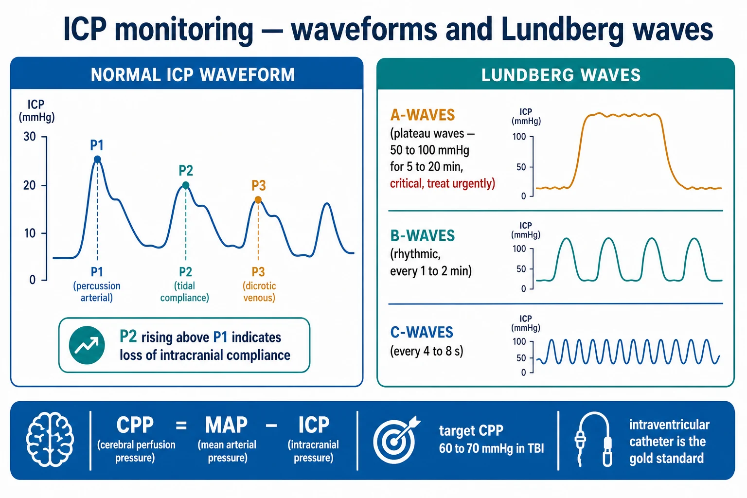
- P1 (the percussion wave) — the tallest, from the arterial pressure transmitted through the choroid plexus to the CSF. It reflects the arterial pulse.
- P2 (the tidal wave) — reflects the intracranial compliance (the ability of the intracranial contents to accommodate a volume change without a pressure rise). Normally P2 is lower than P1.
- P3 (the dicrotic wave) — from the venous pulse and the a-wave valve closure. [1]
The key sign: a P2 rising above P1 indicates a loss of the intracranial compliance — the intracranial volume is on the steep part of the pressure-volume curve. Each small volume increment (a cough, a head turn, a vasodilation) now causes a large ICP rise. This is the precursor to the decompensation — the intracranial reserve is exhausted.[1]
The Lundberg waves
- A-waves (the plateau waves) — a sustained rise to 50-100 mmHg for 5-20 minutes or more. They indicate a critical loss of the intracranial compliance (the intracranial volume is at the flat top of the compliance curve; a small vasodilatory stimulus triggers a large ICP rise that does not resolve until the stimulus is removed). They are pathological and require an urgent treatment (reduce the ICP — the head elevation, the CSF drainage, the mannitol/hypertonic saline, the sedation). Untreated, they cause cerebral ischaemia and death.[1]
- B-waves — rhythmic oscillations of 10-20 mmHg, every 0.5-2 minutes. Associated with the periodic breathing (the Cheyne-Stokes) and the CO2 fluctuation. Less concerning but may indicate a reduced compliance.[1]
- C-waves — small oscillations every 4-8 seconds, related to the cardiac cycle. Normal.[1]
Complications
- Infection (ventriculitis/meningitis) — the EVD has the highest risk (5-20 per cent). Prevent with the tunnelled insertion, the aseptic technique, and the prophylactic antibiotics (evidence mixed). Monitor the CSF (the cell count, the glucose, the culture) for the early detection.
- Haemorrhage — 1-2 per cent, from the catheter trajectory through the brain parenchyma. Higher in the coagulopathic patient.
- Malposition — the catheter not in the ventricle; may need a re-position.
- Obstruction — the blood, the debris, or the tissue blocking the catheter lumen.[1]
Red flags
The intracranial compliance (pressure-volume) curve — why ICP is non-linear
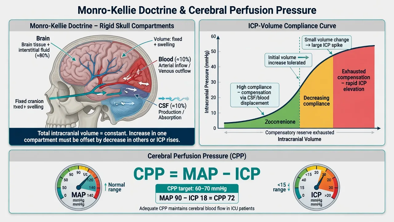
The Monro-Kellie doctrine is the foundation: the skull is a rigid box of fixed volume holding brain (~80 per cent) + blood (~10 per cent) + CSF (~10 per cent). An increase in the volume of one component requires an equal decrease in the others, or the ICP rises. In health the body compensates by displacing CSF (into the spinal thecal sac) and venous blood (into the jugulars). This compensation keeps the ICP flat and low (5-15 mmHg) over a wide range of intracranial volume — the flat, shallow portion of the pressure-volume curve (high compliance, low elastance).[1][5]
Once the compensatory reserve is exhausted (the displaceable CSF and venous blood are spent), the curve turns steep: each additional millilitre of volume now produces a large ICP rise. Compliance has fallen and elastance (the inverse — dP/dV, the change in pressure per unit change in volume) has risen. This is the critical zone — the patient looks the same but the intracranial reserve is gone, and any small volume increment (a cough, a head turn, a bout of agitation, a rise in PaCO₂, a vasodilatory surge) precipitates a steep ICP spike. This is exactly the situation in which a P2 rises above P1 on the waveform and in which a Lundberg A-wave is generated.[1][5]
Intracranial compliance — the two zones of the pressure-volume curve
| Zone of the curve | Compliance | Elastance (dP/dV) | Clinical correlate | Waveform behaviour |
|---|---|---|---|---|
| Flat/shallow portion (early) | HIGH — volume tolerated without pressure rise | LOW | Patient compensates for a slowly growing mass; lucid interval of an extradural haematoma | Normal waveform — P2 below P1 |
| Steep portion (decompensation) | LOW — small volume change → large pressure rise | HIGH | Reserve exhausted; any stimulus (cough, agitation, hypercapnia) spikes ICP | P2 rises to or above P1; A-waves appear |
| Practical implication | Compliance is what you monitor | Elastance is what you fear | The same patient can herniate within minutes of being 'fine' | The waveform is your real-time compliance probe |
The practical corollary: ICP does not rise linearly with intracranial volume. A patient with a slow-growing chronic subdural can carry 80 mL of extra volume and walk into the clinic; a patient with an exhausted reserve can herniate from a 5 mL increment. The waveform (and the ICP trend) is your window onto where the patient sits on this curve at any given moment.[1]
The ICP monitoring devices — in depth
There are five intracranial devices and one extracranial option. Choice is governed by (a) whether you need to drain CSF (only the EVD), (b) the accuracy required, and (c) the risk the patient can accept (infection, haemorrhage).[1][1]
ICP monitoring devices — the six options compared
| Device | Site | What it measures | Can drain CSF? | Can sample CSF? | Accuracy | Infection risk | Haemorrhage risk | Re-zeroing | Typical use |
|---|---|---|---|---|---|---|---|---|---|
| External ventricular drain (EVD) — frontal catheter (Codman/Becker) at Kocher's point | Lateral ventricle | True global ICP (CSF pressure) | YES — therapeutic | YES | GOLD STANDARD — reference for all others | Highest (~5-10 per cent ventriculitis; up to 20 per cent with prolonged use) | ~1-2 per cent (higher in coagulopathy) | External transducer — re-zero at any time | Severe TBI needing drainage; hydrocephalus; SAH with IVH; compliance testing |
| Intraparenchymal microsensor — Codman (strain-gauge), Camino (fibre-optic), Spiegelberg (air pouch/pneumatic) | Brain parenchyma (bolt, ~2 cm deep) | Local tissue pressure (closely tracks global ICP) | NO | NO | Very good; ±2 mmHg of ventricular | Lowest (~1 per cent) | ~1-2 per cent | No — drift ~1-2 mmHg over days (Codman/Camino); Spiegelberg auto-zeroes | The workhorse for most ICUs when drainage not needed; easy bedside bolt placement |
| Subdural catheter/sensor (Richmond bolt) | Subdural space | Surface pressure | Occasionally (limited) | Occasionally | Moderate — less accurate than intraparenchymal | Low-moderate | Low | External transducer | Largely historical; when the brain is exposed post-craniectomy |
| Epidural sensor | Epidural space | Extracerebral pressure | NO | NO | Least accurate — dissociates from true ICP | Lowest | Lowest | No | Largely abandoned for critical management; sometimes used where dural puncture must be avoided |
| Lumbar drain (CSF pressure) | Lumbar theca | Spinal CSF pressure | YES | YES | Only valid if basilar cisterns open (communicating system) | Low (post-LP) | Low (spinal haematoma) | External transducer | CONTRAINDICATED in raised ICP with mass effect — tonsillar herniation risk; only for communicating hydrocephalus / CSF fistula |
| Telemetric / non-invasive (e.g. Raumedic Neurovent-PTO with PbtO₂) | Intraparenchymal, wire-free | Local ICP ± PbtO₂ | NO | NO | Comparable to wired intraparenchymal | Lowest (no externalised lumen) | ~1 per cent | No | Long-term monitoring, shunt assessment, paediatric; emerging |
Why the EVD is the gold standard
The EVD is the only device that is both diagnostic AND therapeutic — it measures the true global (ventricular) ICP, drains CSF to treat an elevated ICP, and samples CSF for infection surveillance and metabolic analysis. It is the reference standard against which every other device is calibrated.[1][1]
Its drawbacks are equally important to know for the exam: it is the hardest to place when the ventricles are effaced (slit-like) by severe swelling or shift; it carries the highest infection risk (ventriculitis 5-10 per cent, rising with duration in situ and frequency of sampling); and it has a non-trivial haemorrhage risk (~1-2 per cent, higher in coagulopathy). For these reasons, when CSF drainage is not required, the intraparenchymal microsensor is preferred — it is placed quickly at the bedside via a bolt, has the lowest infection rate, and an accuracy within ±2 mmHg of the ventricular pressure, at the cost of no drainage, no sampling, and zero-drift over time.[1]
The intraparenchymal microsensor — Codman, Camino, Spiegelberg
These three devices differ in their transduction principle, and the exam expects you to know the difference:
- Codman microsensor — a strain-gauge transducer (piezoresistive) embedded in the catheter tip. Mounted via a bolt; cannot be re-zeroed in situ; drift of ~1-2 mmHg over days. Industry workhorse in many ANZ/UK units.
- Camino — a fibre-optic transducer (light reflected from a pressure-sensitive diaphragm). Similar accuracy and drift characteristics; some units prefer it for the smaller profile.
- Spiegelberg — an air-pouch (pneumatic) transducer that auto-zeroes against atmospheric pressure several times an hour, minimising drift — the one device that effectively re-zeroes itself in situ. Marginally less robust to severe transient spikes (the air pouch takes a few cycles to equilibrate).[1][7]
All three measure local tissue pressure rather than global ventricular pressure — but in the absence of a large pressure gradient (e.g. no massive midline shift), they track the true ICP to within a couple of mmHg, which is clinically adequate. The critical limitation shared by all intraparenchymal devices: they cannot drain CSF. If you need to treat the ICP by removing CSF, you need an EVD. [1]
The external ventricular drain — practical technique
The EVD is placed via a frontal twist-drill craniostomy at Kocher's point (a coronal plane 1 cm anterior to the coronal suture, ~2-3 cm from the midline, on the non-dominant side where possible — typically right frontal in a right-handed patient). The catheter (often a Frazier/Codman ventricular catheter with stylet) is aimed in the plane of the orbitomeatal line toward the ipsilateral medial canthus and the external acoustic meatus, to enter the frontal horn of the lateral ventricle at the foramen of Monro. The catheter is then tunnelled under the scalp for 3-5 cm before exit, which reduces infection by distancing the skin exit from the dural puncture.[1]
Zeroing, levelling, and the drainage column
The external pressure transducer is zeroed to atmospheric pressure and **levelled to the foramen of Monro — approximated externally at the external acoustic meatus (EAM) or the midpoint between the lateral eyebrow and the tragus. If the patient's head of bed is elevated 30°, the transducer must be re-levelled to the foramen of Monro; otherwise you will under-read ICP (because the transducer sits below the reference and the hydrostatic column adds).[1]
The drainage height is set by raising or lowering the drip chamber relative to the foramen of Monro: at 10 cm H₂O above the EAM, the EVD drains whenever the ICP exceeds that pressure. Lowering the chamber drains more (risks over-drainage and upward herniation); raising it drains less. To measure the true ICP (closing pressure), the drain must be clamped for a few minutes and the pressure allowed to equilibrate — the EVD cannot measure while it is actively draining (the column is open).[1]
Managing an EVD at the bedside — the daily and as-required routine
- Confirm position and patency. Check the CT — is the catheter tip in the frontal horn, away from the choroid plexus? A pulsatile CSF meniscus that moves with the heartbeat and respiration confirms patency. No movement = blocked catheter (consider flush with sterile saline ± revision).
- Zero and level the transducer to the foramen of Monro (EAM) every shift and after any position change. Document the ICP value, the waveform morphology, and the drain setting (open/closed, height in cm H₂O).
- Set the drainage height to the prescribed pressure (e.g. 10-15 cm H₂O above the EAM). Drain only what is needed — over-drainage causes pneumocephalus, upward transtentorial herniation, and re-bleed of a clipped aneurysm.
- To measure the true ICP (closing pressure), clamp the drain for 5-10 minutes and read the equilibrated pressure and the waveform. Then re-open at the prescribed height. Never leave the drain clamped inadvertently — it will not treat a rising ICP.
- Sample CSF only when clinically indicated (suspected infection, metabolic question) via a sterile, closed, needleless port. Routine daily sampling is not recommended — it increases infection. Send cell count, glucose, protein, Gram stain, and culture when infection is suspected.
- Inspect the insertion site daily for CSF leak, erythema, or purulence. A CSF leak mandates a neurosurgical review (often a re-siting) — it is a major infection risk.
- Remove the EVD as soon as it is no longer needed. Infection risk rises with every day in situ. A trial of clamping (with ICP monitoring) precedes removal in most protocols.
The normal ICP waveform — P1, P2, P3 in depth
The ICP waveform is morphologically similar to the arterial waveform (it is largely arterial in origin) but at a much lower pressure (5-15 mmHg vs 100 mmHg systolic). It is generated by the arterial pulse transmitted through the choroid plexus into the CSF, modulated by the venous pressure and by the intracranial compliance. Each cardiac cycle produces three distinct peaks:[1][5]
The three peaks of the normal ICP waveform
| Peak | Origin | Height (normal) | Clinical meaning | Sign when abnormal |
|---|---|---|---|---|
| P1 — percussion wave | Arterial pressure transmitted through choroid plexus to CSF | Tallest peak | Reflects the arterial pulse pressure | Disappears in cardiac arrest / asystole — confirms arterial origin |
| P2 — tidal wave | Reflective wave from the intracranial contents; reflects compliance | Normally LOWER than P1 | The intracranial 'give' — how well the system absorbs each beat | P2 rising to or above P1 = loss of compliance (the cardinal early warning sign) |
| P3 — dicrotic wave | Venous pulsation + aortic valve closure (dicrotic notch) | Lowest, after the dicrotic notch | Reflects venous return / intrathoracic pressure | Diminishes with venous outflow obstruction (e.g. neck compression, high PEEP) |
The single most important waveform sign: a P2 rising to or above P1. This is the real-time marker that compliance is failing — the patient is on the steep part of the pressure-volume curve. It precedes the ICP numerical rise and the A-wave. Act on it: optimise head position (30°, neutral, no neck rotation or tight collar), deepen sedation/analgesia, ensure normocapnia (PaCO₂ 35-40), normoxia and normotension, and drain CSF if an EVD is in place.[1][5]
Respiratory variation: the ICP falls on inspiration (negative intrathoracic pressure improves venous outflow) and rises on expiration. Loss of this normal respiratory variability suggests a near-exhausted reserve. With positive-pressure ventilation (and especially high PEEP), venous return is impeded and the mean ICP rises — a reason to use the lowest PEEP compatible with oxygenation in raised-ICP patients.[1]
The Lundberg waves — A, B, C revisited
Nils Lundberg's 1965 paper — Continuous recording of the ventricular fluid pressure — established the modern classification of spontaneous ICP fluctuations into three patterns (A, B, C), and is the foundation of all modern ICP waveform interpretation.[5]
The Lundberg waves — A, B, C
| Wave | Amplitude / pressure | Duration / periodicity | Pathophysiology | Significance | Action |
|---|---|---|---|---|---|
| A-wave (plateau wave) | 50-100 mmHg sustained | 5-20 minutes (occasionally up to an hour) | A vasodilatory cascade in a decompensated brain: a small stimulus dilates cerebral vessels → cerebral blood volume rises → ICP rises → CPP falls → vasodilation worsens → runaway. Resolves only when the stimulus is removed | Pathological — critical loss of compliance; cerebral ischaemia from low CPP; brain-death if untreated | TREAT URGENTLY — head up, drain CSF, osmotherapy (mannitol/HTS), deepen sedation, treat the trigger (pain, hypoxia, hypercapnia, seizure) |
| B-wave | 10-20 mmHg oscillation | Rhythmic, every 0.5-2 minutes | Periodic breathing (Cheyne-Stokes) → CO₂ fluctuation → vasodilation/vasoconstriction cycling; also reduced compliance | Suggests reduced but not exhausted compliance; a warning that A-waves may follow | Investigate — optimise PaCO₂, compliance, sedation; not an emergency in isolation |
| C-wave | Small (~5-10 mmHg) | Every 4-8 seconds | Coupled to the cardiac/respiratory cycle (Traube-Hering) | Normal / physiological | None — recognise it and reassure |
The A-wave pathophysiology is the high-yield concept: it is a positive-feedback vasodilatory cascade that can only occur once compliance is exhausted. A trivial trigger (a painful stimulus, a kinked endotracheal tube, a transient hypercapnia) dilates cerebral arterioles → cerebral blood volume (CBV) rises by a few millilitres → on the steep part of the compliance curve this produces a steep ICP rise → CPP falls → cerebral ischaemia triggers further reflex vasodilation → ICP climbs further. The wave breaks only when the trigger is removed or the ICP is lowered (restoring CPP and allowing the vasculature to constrict again). This is why the A-wave is self-sustaining and dangerous.[5][7]
Cerebral perfusion pressure (CPP) and autoregulation
CPP = MAP − ICP is the perfusion pressure available to the brain, and the derived variable that actually matters — a normal ICP with a catastrophically low MAP still produces ischaemia. The Brain Trauma Foundation 4th edition target is CPP 60-70 mmHg.[1][3]
CPP targets — the two errors are equally harmful
| Target | Rationale | Consequence of breaching | Evidence |
|---|---|---|---|
| CPP 60-70 mmHg (target window) | The brain needs this head of pressure to perfuse against the raised ICP | Below 60 → cerebral ischaemia; above 70 → ARDS / fluid overload | BTF 4th edition (2017); BEST:TRIP (2012)[1][3] |
| CPP <60 mmHg | Inadequate perfusion despite a 'normal' ICP | Cerebral ischaemia, infarction, worsened outcome | Robertson, Claasen |
| CPP >70 mmHg | Aggressive CPP-pushing to force perfusion | ARDS (five-fold increased risk), fluid overload, worse outcome | BEST:TRIP (2012)[3] |
Cerebral autoregulation (the Lassen curve) is the brain's ability to maintain a constant cerebral blood flow (CBF) across a wide range of CPP (~50-150 mmHg in health). Within this window, a fall in CPP is met by vasodilation (to maintain flow) and a rise by vasoconstriction. After TBI, autoregulation is frequently impaired or shifted — the window narrows or disappears, so CBF becomes passively pressure-passive. In that state, a fall in MAP directly drops CBF (ischaemia), and a rise directly increases cerebral blood volume and ICP. Knowing whether autoregulation is intact changes your CPP target — which is where the pressure reactivity index comes in.[6][7]
The pressure reactivity index (PRx) — autoregulation at the bedside
The pressure reactivity index (PRx) is the Pearson correlation coefficient between slow-wave (30-60 second) fluctuations in MAP and ICP over a moving window (typically 4-5 minutes, updated every minute). It quantifies, in real time, whether the brain is autoregulating.[6][7]
The logic: in an intact autoregulating brain, a rise in MAP is met by vasoconstriction (to hold CBF constant), which reduces cerebral blood volume and therefore lowers ICP. So MAP and ICP move in opposite directions (negative correlation) → PRx is negative. In an impaired (pressure-passive) brain, a rise in MAP simply transmits to ICP — MAP and ICP move together (positive correlation) → PRx is positive.[6][7]
PRx — interpretation and the CPP-optimum
| PRx value | Meaning | Autoregulation | Clinical action |
|---|---|---|---|
| PRx < 0 (e.g. −0.3) | ICP falls as MAP rises | INTACT — vasculature constricting in response to pressure | The brain can be perfused safely across a range of CPP; standard CPP 60-70 target is fine |
| PRx > 0 (e.g. +0.3) | ICP rises with MAP | IMPAIRED — pressure-passive vasculature | The brain is vulnerable; avoid both high MAP (raises ICP) and low MAP (ischaemia) |
| PRx > +0.25 | Strongly impaired | Broken — outcome predictor | Independent predictor of mortality; the most vulnerable brain |
| CPP-opt (CPPopt) | The CPP at which PRx is most negative | The individual patient's optimal CPP | Target CPP at CPPopt (when calculable) — improves outcome vs a fixed target |
CPPopt (CPP-optimum) is the CPP value at which the PRx reaches its most negative value (best autoregulation) for that patient, plotted over a range of CPP. Steiner and Czosnyka showed that targeting the individual CPPopt (when it can be identified) is associated with better outcome than a fixed 60-70 target — the brain tells you what pressure it wants. This is advanced multimodality neuromonitoring and is not universal, but the exam expects you to know that PRx is a bedside index of autoregulation, that negative is good and positive is bad, and that it can guide an individualised CPP target.[6][7]
Complications of ICP monitoring — quantified
Complications of ICP monitoring — device-by-device
| Complication | EVD | Intraparenchymal | Subdural/Epidural | Lumbar drain | Prevention / management |
|---|---|---|---|---|---|
| Infection (ventriculitis / meningitis) | 5-10 per cent (up to 20 per cent prolonged) | ~1 per cent | Low | Low (post-LP meningitis) | Tunnelled insertion, closed system, antibiotic-impregnated catheter, aseptic sampling, remove ASAP |
| Catheter-related haemorrhage | ~1-2 per cent (higher in coagulopathy / thrombocytopenia; check platelets and coagulation before insertion) | ~1-2 per cent | Low | Low (spinal haematoma) | Correct coagulopathy before insertion; single-pass technique; CT post-insertion if any deterioration |
| Malposition / misplacement | Common if ventricles slit/shifted | Rare | Moderate | N/A | Image-guidance where available; confirm on CT; re-site if not draining |
| Obstruction / blockage | Common (blood, debris, choroid plexus, proteinaceous CSF) | Rare (closed tip) | Moderate | Moderate | Sterile saline flush; revision if persistent; never force-flush an infected system |
| Over-drainage | Risk if chamber too low — pneumocephalus, upward herniation, re-bleed | N/A | N/A | Slump syndrome / herniation | Set chamber at prescribed height; re-level after position change; alarm on volume drained |
| Zero drift | N/A (external transducer, re-zeroable) | 1-2 mmHg over days (Codman/Camino); Spiegelberg auto-zeroes | N/A | N/A | Be aware; correlation with clinical picture; Spiegelberg preferred if long-term |
| Misalignment / levelling error | Common — under-reads if transducer below foramen of Monro | N/A | N/A | Common | Re-zero and re-level to EAM every shift and after position change |
The EVD infection prevention bundle
EVD infection (ventriculitis/healthcare-associated ventriculomeningitis) is the most important complication — it extends ICU stay, worsens neurological outcome, and is largely preventable. The evidence-based bundle:[1][8]
- Tunnelled subgaleal insertion (3-5 cm) — distances the skin exit from the dural puncture.
- Antibiotic-impregnated catheter (e.g. rifampicin-minocycline or clindamycin-impregnated) — reduces infection by ~50 per cent.
- Closed drainage system with a needleless, aseptic sampling port — minimises breaches of sterility.
- Aseptic insertion with full barrier precautions, mask, and sterile gown/gloves.
- Prophylactic systemic antibiotics — evidence is mixed; many units give a single peri-operative dose but not prolonged prophylaxis (which selects resistant organisms).
- Daily review of the continuing need and prompt removal as soon as the EVD is no longer required — the single most powerful infection-reduction strategy (risk rises ~with each day in situ).
- Avoid routine CSF sampling — sample only when infection is suspected.[8]
Recognise ventriculitis by a rising CSF cell count, falling glucose, rising protein, positive Gram stain/culture, or unexplained fever / rising ICP / new meningism. Treatment is intraventricular ± intravenous antibiotics (often vancomycin ± an aminoglycoside or third-generation cephalosporin, guided by cultures) and usually removal/replacement of the infected catheter.[1]
Waveform troubleshooting — artefacts and pitfalls
A 'bad-looking' ICP trace is often a technical artefact rather than a real deterioration. Work through the checklist before escalating.[1]
Troubleshooting an abnormal or absent ICP trace — the systematic check
- Check the patient first. Is there a clinical correlate (rising sedation requirement, falling GCS, new pupil change, Cushing's response)? An unwell patient with a flat ICP trace is a blocked line or a disconnection until proven otherwise — do not be reassured by a 'normal' number.
- Check the transducer level and zero. Re-zero to atmosphere and re-level to the foramen of Monro (EAM). A transducer that has slipped below the EAM under-reads; one that has risen over-reads.
- Check the waveform morphology. A damped, low-amplitude trace suggests an air bubble, a partially clamped line, or a blocked catheter. A perfectly flat line with a numeric value of 0 suggests disconnection or a blocked/kinked line — investigate immediately.
- Check the catheter patency (EVD). Does the CSF meniscus pulsate with the heartbeat and respiration? Lower the chamber briefly (under sterile technique) to test flow. No flow = blocked (flush with sterile saline via the closed port; if persistent, call neurosurgery for revision).
- Check for over-drainage. A sudden low ICP with pneumocephalus on imaging suggests the chamber was too low — re-set the height and discuss with neurosurgery.
- Exclude zero drift (intraparenchymal). If the clinical picture and the ICP number disagree after all the above, suspect drift — correlate with CT and consider replacing the monitor.
- Re-examine the trace against the ECG and the respiratory trace. The P1 should align with systole and the trace should vary with respiration. Absence of respiratory variability, again, suggests near-exhausted reserve or a blocked line.
The acute ICP crisis / A-wave — the escalation staircase
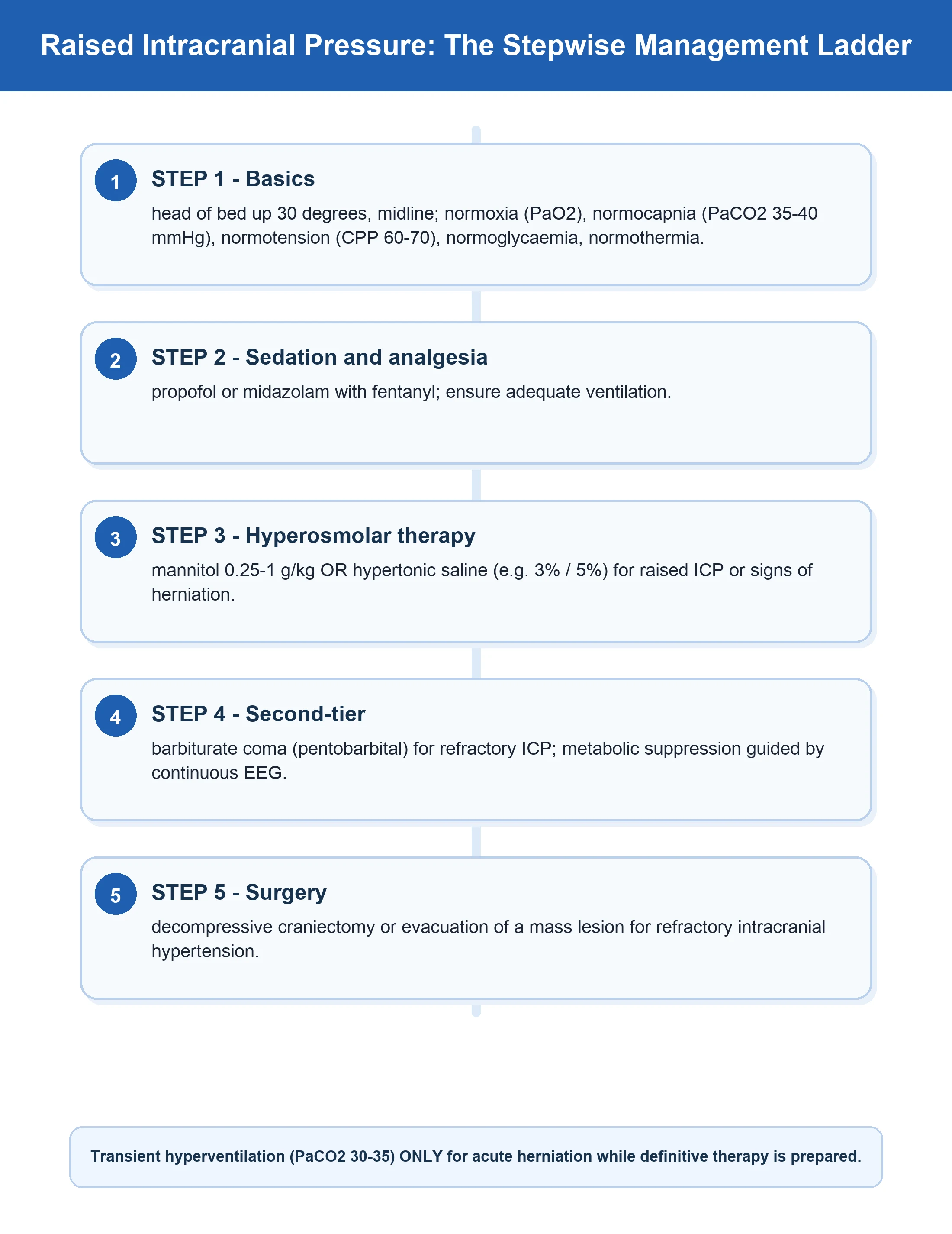
When the ICP spikes (an A-wave, or a sustained rise above 22 mmHg), the response is a graded staircase — escalate only as each step fails, and always look for a treatable surgical cause first.[1][1]
The acute ICP crisis — escalation staircase (first 30 minutes)
- RECOGNISE AND REMOVE THE TRIGGER. Check the airway (kinked tube?), breathing (hypoxia, hypercapnia — check the capnograph), circulation (hypotension dropping CPP), sedation/analgesia (pain, agitation), head position (rotation, tight collar, C-spine collar occluding venous outflow), temperature (fever, shivering), seizures (subclinical — check the cEEG), and abdominal pressure. Fix the obvious first — most 'refractory' spikes have a remediable trigger.
- SURGICAL TRIAGE — obtain a CT. Before medical escalation, exclude an evacuable lesion (an expanding extradural, subdural, or intracerebral haematoma; obstructive hydrocephalus; a blocked shunt). Osmotherapy is only a bridge; the cure may be the operating theatre.
- HEAD OF BED 30°, NEUTRAL, NORMOCAPNIA (PaCO₂ 35-40). Optimise venous outflow and avoid prophylactic hyperventilation. Transient mild hyperventilation (PaCO₂ 30-35) is acceptable only as a bridge during an acute herniation crisis, and never beyond 30 (ischaemia).
- SEDATION AND ANALGESIA. Deepen with propofol (first-line, titratable) ± fentanyl/morphine. Goal: a still, comfortable patient with a suppressed metabolic demand. Add a neuromuscular blocker only if coughing/bucking persists despite adequate sedation.
- CSF DRAINAGE (if EVD in place). Open the EVD, drain 5-10 mL aliquots to the prescribed pressure, and monitor the response. Re-clamp to measure the closing pressure.
- OSMOTHERAPY — mannitol 0.5-1 g/kg OR hypertonic saline 3-5% (bolus 250 mL of 3%, or 30 mL of 23.4%). Check serum osmolarity before and after mannitol (keep <320 mOsm/L — above, AKI). HTS preferred if hypovolaemic (expands volume, supports CPP). Re-assess the pupil size and GCS after 15-30 minutes.
- METABOLIC SUPPRESSION. If refractory, a propofol infusion to burst suppression, then a barbiturate coma (thiopental/pentobarbital titrated to burst suppression on cEEG). Monitor for myocardial depression and hypotension (have noradrenaline ready).
- DECOMPRESSIVE CRANIECTOMY. For refractory ICP >25 mmHg despite maximal medical therapy. RESCUEicp: reduces mortality but increases the rate of vegetative/severely-disabled survivors — a 'troubling trade-off' that must be discussed with the family before surgery.[2]
Key trials and evidence
Lundberg 1965 — Continuous Recording of the Ventricular-Fluid Pressure (PMID 5832775)
Source
Journal of Neurosurgery 1965;22(Suppl):1-193 — the foundational monograph defining continuous ventricular fluid pressure recording
What it established
Lundberg introduced the technique of **continuous ICP monitoring via an indwelling ventricular catheter** and classified the spontaneous pressure fluctuations into **A-waves (plateau waves, 50-100 mmHg, 5-20 min), B-waves (rhythmic, every 0.5-2 min), and C-waves (every 4-8 s)**. He showed that the A-wave is pathological and precedes neurological deterioration — the origin of every modern ICP waveform concept.
Key contribution
The single paper that turned intracranial pressure from a post-mortem concept into a continuously observable, clinically actionable variable. Every term in modern ICP waveform analysis (P1/P2/P3 components aside) traces to this work.
Clinical bottom line
If you are asked 'who defined the A, B, C waves?' the answer is Lundberg (1960, formalised 1965). The plateau wave he described is still the cardinal warning sign of critical loss of compliance.
Carney 2017 — Brain Trauma Foundation Guidelines, 4th Edition (PMID 27654000)
Source
Neurosurgery 2017;80(1):6-15 — the current global standard for severe TBI management
What it established
Revised the **ICP treatment threshold to >22 mmHg** (up from 20 in the 3rd edition); reaffirmed **CPP target 60-70 mmHg**; recommended ICP monitoring in severe TBI (GCS 3-8) with abnormal CT, or normal CT with ≥2 risk factors (age >40, motor posturing, SBP <90); downgraded prophylactic hyperventilation and hypothermia.
Key contribution
Codified the targets every ICU exam expects: **ICP <22, CPP 60-70**. Also explicitly integrated emerging multimodality monitoring (PbtO₂) and reaffirmed that steroids are contraindicated in TBI.
Clinical bottom line
The exam-defining reference — know the ICP threshold (22), the CPP target (60-70), and that the threshold moved from 20 to 22 between the 3rd and 4th editions.
Hutchinson 2016 — RESCUEicp: Decompressive Craniectomy for Refractory Intracranial Hypertension (PMID 27602507)
Source
New England Journal of Medicine 2016;375(12):1119-1130 — multinational RCT, 408 patients with severe TBI and refractory ICP >25 mmHg despite maximal medical therapy
What it found
Late decompressive craniectomy **REDUCED mortality** (26.9 per cent vs 48.9 per cent) but survivors were more often **vegetative or severely disabled** compared with the medical group.
Key contribution
Defined the 'troubling trade-off' of craniectomy for refractory ICP: it saves lives but at the cost of more dependent survivors. Craniectomy is appropriate ONLY for medically refractory ICP — never prophylactically (contrast DECRA 2011).
Clinical bottom line
For the ICP monitor that cannot be lowered medically, craniectomy saves life but changes the survivorship — the decision must involve explicit prognostic discussion with the family.
Chesnut 2012 — BEST:TRIP: ICP Monitoring in TBI (PMID 23234472)
Source
New England Journal of Medicine 2012;367(26):2471-2481 — RCT in 324 severe TBI patients comparing ICP-monitor-guided vs imaging-and-examination-guided therapy
What it found
No significant difference in the composite primary outcome between strategies. ICP-monitored patients received fewer days of intensive treatment but had similar outcomes.
Key contribution
Often misread as 'ICP monitoring doesn't matter' — the correct interpretation is that in well-resourced settings with serial imaging and exam, carefully protocolised care achieves equivalent outcomes; and that aggressive fluid/vasopressor loading to push CPP very high risks ARDS. Reinforced the **CPP 60-70 window**.
Clinical bottom line
ICP monitoring remains standard in severe TBI in well-resourced settings; BEST:TRIP tempers over-aggressive CPP-pushing and supports judicious, protocolised care matched to the patient.
Czosnyka 1996 — Monitoring of Cerebral Autoregulation in Head-Injured Patients (PMID 8841340)
Source
Stroke 1996;27(10):1829-1834 — the seminal description of the pressure reactivity index (PRx)
What it established
Introduced the **pressure reactivity index (PRx)** — the moving correlation between slow-wave fluctuations in MAP and ICP — as a bedside index of cerebral autoregulation. Showed that **PRx <0 indicates intact autoregulation (vasoconstriction to a pressure rise lowers ICP) and PRx >0 indicates pressure-passive failure**.
Key contribution
Made cerebral autoregulation — previously a laboratory concept — measurable continuously at the bedside using only an ICP monitor and an arterial line. Opened the path to **CPPopt** (individualised CPP targeting), which Czosnyka and Steiner subsequently linked to improved outcome.
Clinical bottom line
If asked 'what is PRx and what does it tell you?' — it is the bedside correlation of MAP and ICP slow waves; negative = autoregulating (good), positive = pressure-passive (bad); it can guide an individualised CPP target.
Stocchetti 2017 — CENTER-TBI Survey of ICP Management (PMID 28874206)
Source
Critical Care 2017;21:233 — survey of monitoring and treatment policies for intracranial hypertension across 66 neurotrauma centres participating in the CENTER-TBI study
What it found
Substantial **variation** in ICP monitoring uptake, device choice (intraparenchymal most common), treatment thresholds, osmotherapy agent preference (hypertonic saline increasingly first-line), and use of decompressive craniectomy across European centres.
Key contribution
Documented that real-world TBI care is heterogeneous and guideline-adherence is variable — motivating the CENTER-TBI cohort study and highlighting the shift toward hypertonic saline as first-line osmotherapy.
Clinical bottom line
Even experts vary — know the evidence (BTF 4th, RESCUEicp, BEST:TRIP) and apply it in a protocolised, patient-centred way; the intraparenchymal monitor is the most commonly placed device in real-world practice.
Exam practice
SAQ — Acute ICP crisis: the escalation staircase
10 minutes · 10 marks
A 35-year-old man is in the neurocritical care unit 36 hours after a severe traumatic brain injury (GCS 5 at the scene). He has an external ventricular drain in situ and an intraparenchymal ICP monitor. His ICP has been stable at 16 mmHg with a CPP of 65 mmHg. Suddenly the ICP alarm sounds — the ICP is now 45 mmHg and the CPP has dropped to 40 mmHg. The waveform shows a plateau pattern. His right pupil is dilated to 6 mm and unreactive.
SAQ — ICP monitoring devices, waveforms, and the pressure reactivity index
10 minutes · 10 marks
A 42-year-old woman with a severe diffuse TBI (GCS 4) has slit-like ventricles on her CT from the diffuse brain swelling. The neurosurgical team is debating whether to place an external ventricular drain or an intraparenchymal microsensor. You are asked for your recommendation and the rationale. Once monitored, her PRx is plotted and is consistently positive at 0.3-0.4 across a CPP range of 55-75 mmHg.
Clinical pearls — high-yield points for the exam
Additional red flags
Integration — the one-minute mental model
ICP monitoring answers two questions at once: how high is the pressure (the number) and how much reserve is left (the waveform). The EVD is the gold standard because it also treats (drains) and surveys (samples) — every other device is diagnostic only. Read the CPP (MAP − ICP, target 60-70) before the ICP, because a normal ICP with a low MAP still ischaemics the brain. Read the waveform: a P2 rising above P1 tells you the reserve is gone before the number climbs. Read the Lundberg waves: an A-wave is an emergency (a vasodilatory cascade), a B-wave is a warning, a C-wave is normal. Add PRx where you can — negative is good, positive is bad, and it can tell you the CPP your individual patient's brain wants. Prevent the complications (infection by prompt removal and the bundle; haemorrhage by correcting coagulopathy; over-drainage by careful levelling). Treat the trend and the trigger, not the number — and always look for a surgical lesion before you escalate the medicine.[1][1][5]
References
- [1]Carney N, Totten AM, O'Reilly C, Ullman JS, Hawryluk GWJ, Bell MJ, Bratton SL, Chesnut R, Harris OA, Kissoon N, Rubiano AM, Shutter L, Tasker RC, Vavilala MS, Wilberger J, Wright DW, Ghajar J. Guidelines for the Management of Severe Traumatic Brain Injury, Fourth Edition Neurosurgery, 2017.PMID 27654000
- [2]Hutchinson PJ, Kolias AG, Timofeev IS, Corteen EA, Czosnyka M, Timothy J, Anderson I, Bulters DO, Belli A, Eynon CA, Wadley J, Mendelow AD, Mitchell PM, Wilson MH, Critchley G, Sahuquillo J, Unterberg A, Servadei F, Teasdale GM, Pickard JD, Menon DK, Murray GD, Kirkpatrick PJ; RESCUEicp Trial Collaborators. Trial of Decompressive Craniectomy for Traumatic Intracranial Hypertension N Engl J Med, 2016.PMID 27602507
- [3]Chesnut RM, Temkin N, Carney N, Dikmen S, Rondina C, Videtta W, Petroni G, Lujan S, Pridgeon J, Barber J, Machamer J, Chaddock K, Celix JM, Cherner M, Hendrix T; Global Neurotrauma Research Group. A trial of intracranial-pressure monitoring in traumatic brain injury N Engl J Med, 2012.PMID 23234472
- [4]Chesnut RM, Marshall LF, Klauber MR, Blunt BA, Baldwin N, Eisenberg HM, Jane JA, Marmarou A, Foulkes MA. The role of secondary brain injury in determining outcome from severe head injury J Trauma, 1993.PMID 8459458
- [5]Lundberg N. Continuous recording of the ventricular-fluid pressure in patients with severe acute traumatic brain injury. A preliminary report J Neurosurg, 1965.PMID 5832775
- [6]Czosnyka M, Smielewski P, Kirkpatrick P, Laing RJ, Menon D, Pickard JD. Monitoring of cerebral autoregulation in head-injured patients Stroke, 1996.PMID 8841340
- [7]Czosnyka M, Brady K, Reinhard M, Smielewski P, Steiner LA. Monitoring of cerebrovascular autoregulation: facts, myths, and missing links Neurocrit Care, 2009.PMID 19127448
- [8]Stocchetti N, Carbonara M, Citerio G, Ercole A, Skrifvars MB, Smielewski P, Zoerle G, Menon DK, Czosnyka M, Maas AIR; CENTER-TBI participants and collaborators. Variation in monitoring and treatment policies for intracranial hypertension in traumatic brain injury: a survey in 66 neurotrauma centers participating in the CENTER-TBI study Crit Care, 2017.PMID 28874206