ICU · Neurocritical care / vascular
Intracerebral Haemorrhage — ICH Score, BP Control & Surgery
Also known as Intracerebral haemorrhage · ICH · Intraparenchymal haemorrhage · Hypertensive haemorrhage · Amyloid angiopathy · ICH score · Haematoma expansion · Spot sign · INTERACT2 · STICH II
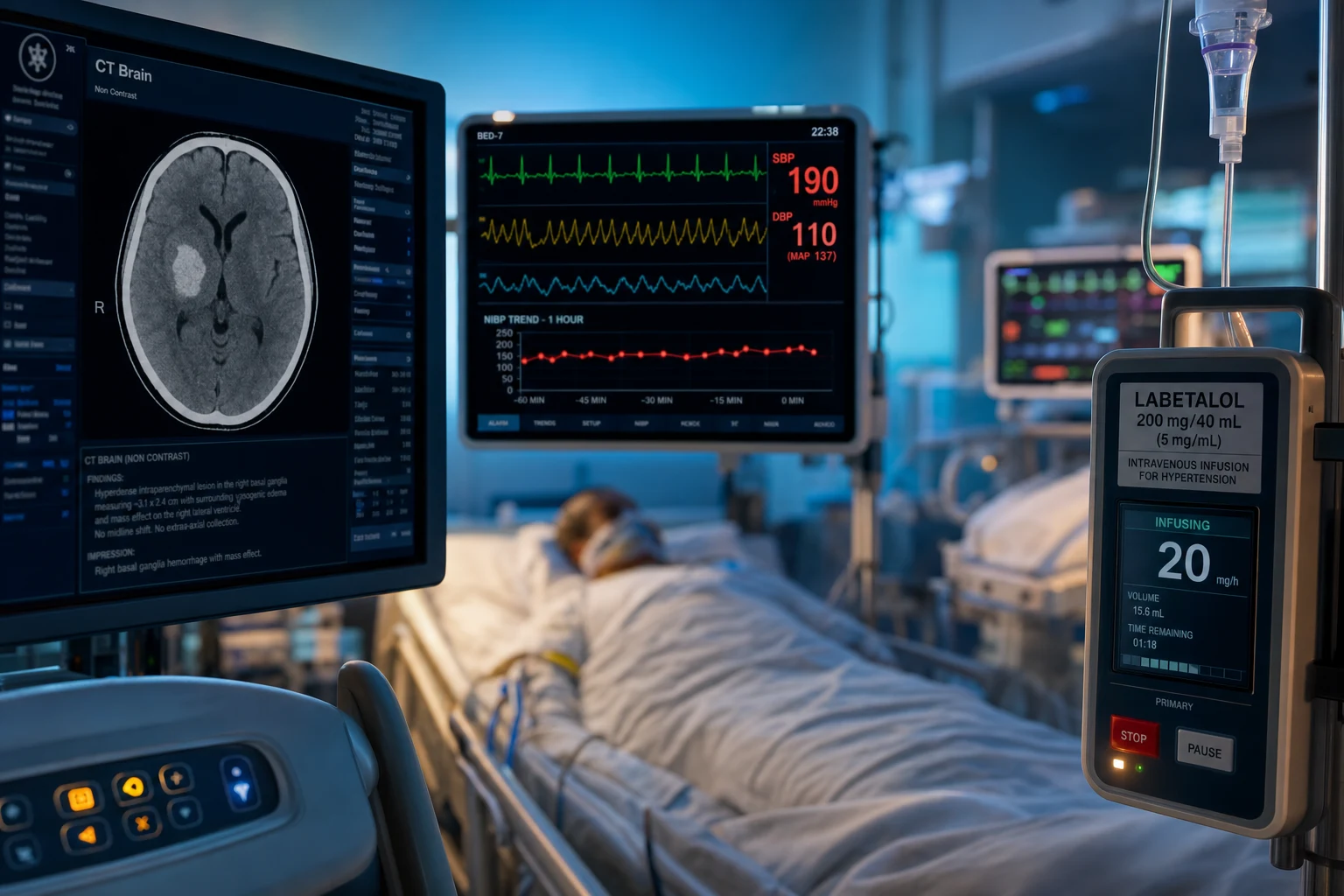
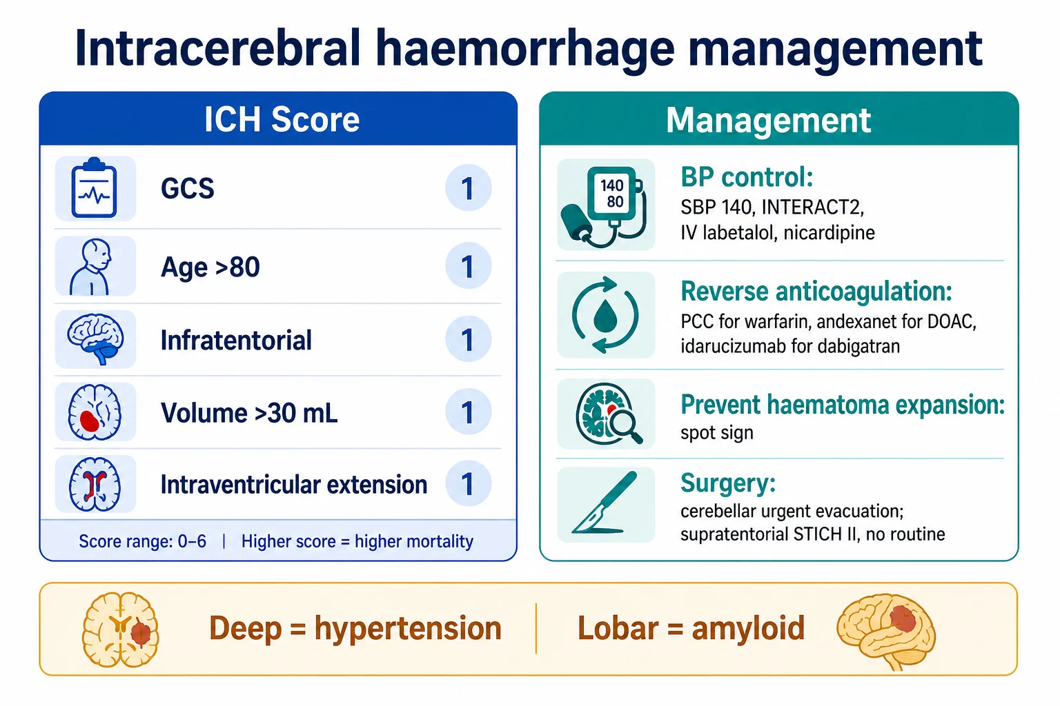
Intracerebral haemorrhage (ICH) is the bleeding into the brain parenchyma, most commonly from hypertension (the deep location — basal ganglia, thalamus, pons, cerebellum) or amyloid angiopathy (the lobar location in the elderly). The CT shows a hyperdense lesion. The ICH Score (the GCS, the age over 80, the infratentorial location, the volume over 30 mL, the intraventricular extension) predicts the 30-day mortality. The management: BP control to SBP 140 mmHg (the INTERACT2 trial — safe and improves the functional outcome; the IV labetalol or nicardipine), reverse the anticoagulation rapidly (the PCC for warfarin; the andexanet alfa for the DOAC; the idarucizumab for dabigatran), prevent the haematoma expansion (the spot sign on the CT angiogram predicts the expansion), and surgery (the cerebellar haemorrhage — the urgent evacuation; the supratentorial — the STICH II showed no routine benefit).
On this page & tools
Your progress
Saved locally on this device.
Target exams
Overview & definition
Intracerebral haemorrhage (ICH) is the bleeding into the brain parenchyma (distinct from the subarachnoid haemorrhage, which is the bleeding into the subarachnoid space). It presents with a sudden onset focal deficit and a decreased consciousness, often with a headache and a vomiting. The CT shows a hyperdense (bright) lesion. The outcome depends on the volume, the location, and the level of consciousness (the ICH Score). The two key modifiable factors are the BP control and the anticoagulation reversal.[1]
Causes and locations
- Hypertension (the commonest cause) — the deep location: the basal ganglia (the putamen, the thalamus), the internal capsule, the pons, and the cerebellum. The chronic hypertension weakens the small penetrating arteries (the Charcot-Bouchard microaneurysms), which rupture.[1]
- Cerebral amyloid angiopathy (CAA) — the lobar location (the cortex and the subcortical white matter), in the elderly. The amyloid deposits in the small and the medium vessels weaken the wall. The recurrent lobar haemorrhages in the elderly are the hallmark.[1]
- Arteriovenous malformation (AVM) — any location; typically in the younger patient. A CT angiogram or an MRI/MRA confirms.[1]
- Anticoagulation — the warfarin or the DOAC increases the risk and the volume of the ICH (and the haematoma expansion).[1]
- Sympathomimetic drugs (cocaine, amphetamines) — a sudden hypertensive surge rupturing a small vessel.[1]
- Trauma, tumour, haemorrhagic transformation of an infarct.[1]
Pathophysiology — why the vessel ruptures
The rupture of a small intraparenchymal vessel produces the haematoma. The mechanism depends on the underlying vasculopathy: [1]
- Hypertensive vasculopathy (lipohyalinosis + Charcot-Bouchard microaneurysms) — the chronic hypertension damages the small penetrating arteries (the lenticulostriate, the pontine, and the cerebellar perforators) through lipohyalinosis (the fibrinoid necrosis and the lipid deposition in the media) and the formation of the Charcot-Bouchard microaneurysms (the small fusiform dilatations at the bifurcations of the penetrating arterioles). The acute hypertensive surge ruptures the weakened wall.[1]
- Cerebral amyloid angiopathy (CAA) — the β-amyloid peptide deposits in the tunica media and the adventitia of the small and the medium leptomeningeal and cortical vessels, replacing the smooth muscle (the vasculopathic vessel). The wall becomes brittle and fissures, producing the lobar haemorrhage. The deposition is strictly lobar (it spares the deep grey matter, which is why the CAA never produces a basal-ganglia bleed). The modified Boston criteria (with the MRI) diagnose the probable CAA on the strictly lobar haemorrhages plus the cortico-subcortical microbleeds or the cortical superficial siderosis.[1]
- The surrounding oedema develops within hours and peaks at days 5-7, adding the mass effect and the midline shift and raising the ICP. The perihaematomal oedema is a mixture of the vasogenic (the blood-brain barrier disruption) and the cytotoxic (the direct compression) injury.
- The secondary injury — the haematoma breakdown products (the thrombin, the iron, the haemoglobin) drive the inflammation, the oedema, and the neuronal death in the surrounding tissue over the following days.
Hypertensive vs CAA — the location tells the cause
The CT location is the strongest clue to the aetiology. The deep location points to the hypertension; the lobar location in the elderly points to the amyloid angiopathy. [1]
| Feature | Hypertensive (deep) | Cerebral amyloid angiopathy (lobar) |
|---|---|---|
| Location | Putamen, thalamus, pons, cerebellum (the perforator territory) | Lobar — cortex + subcortical white matter (frontal, parietal, temporal, occipital) |
| Age | Typically 40-70 | Typically over 70 |
| Vessels | Small penetrating arterioles (lenticulostriate, pontine) | Leptomeningeal + cortical arteries |
| Lesion | Lipohyalinosis + Charcot-Bouchard microaneurysms | β-amyloid deposition (brittle wall) |
| Recurrence | Reduces with the BP control | Recurrent lobar bleeds (the hallmark); high recurrence |
| Imaging clue | Single deep bleed; left-ventricular hypertrophy | Strictly lobar bleed ± microbleeds ± superficial siderosis (MRI) |
| Diagnostic criteria | Clinical (HTN + deep location) | Modified Boston criteria (MRI) |
| Anticoagulant risk | Markedly increased | Markedly increased |
Deep (hypertensive)
Putamen / thalamus / pons / cerebellum
- Hypertension the cause in over 60 per cent; the Charcot-Bouchard microaneurysm ruptures
- Putaminal is the commonest: contralateral hemiparesis, conjugate gaze deviation toward the lesion
- Thalamic: prominent sensory loss; vertical gaze palsy; aphasia (dominant)
- Pontine: coma, pinpoint pupils, decerebrate posturing — devastating, high mortality
- Cerebellar: vertigo, ataxia, vomiting; urgent evacuation if over 3 cm
Lobar (CAA / amyloid)
Cortex + subcortical white matter
- Cerebral amyloid angiopathy in the elderly; the beta-amyloid weakens the leptomeningeal vessels
- Recurrent lobar haemorrhages are the hallmark
- MRI: strictly lobar microbleeds + cortical superficial siderosis (modified Boston criteria)
- Consider in any elderly lobar bleed; reconsider anticoagulation permanently
- Higher surgical candidacy (superficial, accessible) but higher recurrence
Other causes
AVM, anticoagulant, tumour, cocaine
- AVM — younger patient, any location; CTA/MRA confirms; the calcification or abnormal vessels on the CT
- Anticoagulant — markedly increased volume and expansion; reverse RAPIDLY
- Haemorrhagic transformation of an infarct — at the infarct territory, patchy, often after the thrombolysis
- Tumour — the underlying metastasis (melanoma, renal, lung, choriocarcinoma, hepatocellular) or the high-grade glioma
- Cocaine / amphetamines — a sudden hypertensive surge; often the ruptured AVM or aneurysm
Location-specific syndromes — what each bleed looks like
The ICH by location — the bedside recognition
Putaminal (commonest)
Contralateral hemiparesis and hemisensory loss; conjugate eye deviation toward the lesion (the eyes look at the haematoma); homonymous hemianopia; aphasia (dominant hemisphere) or neglect (non-dominant). Large bleeds compress the internal capsule and cause a dense hemiplegia and a reduced GCS.
Thalamic
Prominent contralateral sensory loss (all modalities); the vertical gaze palsy (the upgaze paresis); the convergence-retraction nystagmus; the lethargy from the intralaminar nuclear involvement; the aphasia (dominant). The thalamic bleed may extend into the third ventricle causing the acute hydrocephalus.
Pontine
Coma; the pinpoint pupils (the pontine sympathetic tract disruption); the absent oculocephalic reflexes; the decerebrate posturing; the rapid respiratory pattern (apneustic or ataxic); the hyperthermia. The mortality is high (over 60 per cent); the survivors often have the locked-in syndrome.
Cerebellar
Vertigo, vomiting, ataxia, occipital headache; the inability to walk (the truncal ataxia); the dysmetria on the finger-nose testing; the nystagmus. The deterioration is from the brainstem compression and the fourth-ventricle obstruction (the hydrocephalus). The URGENT evacuation (over 3 cm, or with the brainstem compression, or the hydrocephalus) is life-saving. Do NOT delay.
Lobar
The syndrome reflects the lobe: frontal (contralateral weakness, abulia), parietal (sensory loss, neglect), temporal (aphasia dominant, quadrantanopia), occipital (homonymous hemianopia). The CAA in the elderly; the recurrent bleeds. The seizures are commoner in the lobar than the deep bleeds (the cortical irritation).
The ICH Score
The ICH Score predicts the 30-day mortality:[1]
| Component | Points |
|---|---|
| GCS 3-4 (5), 5-12 (1), 13-15 (0) | 0, 1, or 2 |
| Age over 80 | 0 or 1 |
| Infratentorial origin | 0 or 1 |
| Volume above 30 mL | 0 or 1 |
| Intraventricular extension | 0 or 1 |
The total ICH Score (0-6) predicts the mortality (the score of 0 = about 0 per cent mortality; the score of 5+ = nearly 100 per cent mortality).[1]
The ICH Score (0-6) — the 30-day mortality by score (Hemphill 2001 derivation cohort):[5]
| ICH Score | 30-day mortality |
|---|---|
| 0 | 0 per cent |
| 1 | 13 per cent |
| 2 | 26 per cent |
| 3 | 72 per cent |
| 4 | 97 per cent |
| 5+ | ~100 per cent |
The ICH Score is the most widely used bedside prognostic score — five components (the GCS, the age, the location, the volume, the intraventricular extension), each weighted simply. The prospective validation confirmed the discrimination for the 12-month functional outcome.[5]
Haematoma volume — the ABC/2 formula
The haematoma volume is the key determinant of the prognosis and is one of the five ICH Score components (over 30 mL scores a point). The ABC/2 formula (the Kothari method) estimates the volume from the CT: [1]
Volume (mL) = (A × B × C) / 2 [1]
- A = the maximal haematoma diameter (in cm) on the axial slice with the largest area
- B = the diameter perpendicular to A on the same slice
- C = the number of 1-cm-thick CT slices in which the haematoma is visible (approximately, if the slices are thicker, adjust) [1]
Clinical thresholds: under 30 mL is generally a small bleed (favourable prognosis in the supratentorial); over 30 mL scores an ICH Score point; over 60 mL in the deep supratentorial is associated with a high mortality. The cerebellar haematoma over 3 cm (≈ 12 mL) or with the brainstem compression needs the urgent evacuation. The volume also drives the haematoma expansion — the larger the initial volume, the more likely the expansion. [1]
The spot sign — the marker of the active bleeding
The spot sign is the contrast extravasation (a tiny enhancing focus, 1-2 mm, within or at the edge of the haematoma) on the CT angiogram — it indicates the active bleeding. The spot sign (Wada 2007) predicts the haematoma expansion: the spot-sign-positive ICH has roughly a 2- to 3-fold higher risk of the expansion and a worse outcome.[6]
- Definition — the spot is a star-like or serpiginous enhancement, an attenuation over 120 HU, discontinuous with a vessel, seen on the CTA source images. The "spot sign score" (the number and the size of the spots) refines the prediction.
- Use — identify the high-risk bleeds (the spot-sign-positive) for the closer monitoring, the tighter BP control, and the trial enrolment (the haemostatic agents).
- Limitation — the spot sign requires the contrast (the CTA), which is not always done in the acute ICH (a non-contrast CT suffices for the diagnosis). The renal impairment and the contrast allergy may preclude the CTA.
- The rFVIIa (the recombinant activated factor VII) trials — the FAST trial (Mayer 2008) tried the rFVIIa to halt the expansion in the spot-sign-negative patients but found no benefit overall (and a thromboembolic risk).[10][9]
The spot sign is the most reliable predictor of the haematoma expansion, but it is not yet standard to base the treatment intensity on it — the BP control and the anticoagulant reversal apply to all the ICH patients regardless of the spot sign.[6]
Management
1. BP control — SBP to 140 mmHg
The INTERACT2 trial (NEJM 2013) showed that the rapid SBP reduction to under 140 mmHg (within 1 hour, using the IV labetalol or the nicardipine) was safe and improved the functional outcome (a trend toward a better modified Rankin Scale). The ATACH2 trial (NEJM 2016) showed that an even more aggressive reduction (to under 110-120 mmHg) was harmful (the renal adverse events and the trend toward a worse outcome) — the target is 140 mmHg, not lower.[1]
The drugs: the IV labetalol (10-20 mg boluses, or an infusion), the nicardipine (5-15 mg/h infusion), or the clevidipine. Avoid the nitroprusside and the GTN (the cerebral vasodilation increases the ICP).[1]
2. Reverse the anticoagulation — RAPIDLY
If the ICH is in a patient on warfarin or a DOAC, reverse the anticoagulation immediately:[1]
- Warfarin — stop the warfarin; give the vitamin K 10 mg IV (slowly) and the prothrombin complex concentrate (PCC) (25-50 units/kg — faster and more effective than the FFP; reverses the INR within minutes).[1]
- Dabigatran — the idarucizumab (5 g IV — the specific monoclonal antibody reversal agent).[1]
- Apixaban / rivaroxaban — the andexanet alfa (the specific reversal agent) or the PCC (if the andexanet is unavailable).[1]
The rapid reversal (within 1 hour) reduces the haematoma expansion — the bleeding continues (and the haematoma grows) in the anticoagulated patient unless the coagulopathy is corrected. Every minute of the delay increases the expansion.[1]
2a. The anticoagulant reversal — the agent-by-agent protocol
Every anticoagulated ICH patient has an ongoing haematoma expansion. The reversal must be immediate — the target is within 1 hour of the diagnosis. Do NOT wait for the confirmatory levels; treat empirically from the medication history. The 2022 AHA/ASA guideline (Greenberg) endorses the specific reversal agents (the idarucizumab for the dabigatran; the andexanet alfa for the apixaban/rivaroxaban) and the 4-factor PCC for the warfarin.[12]
| Anticoagulant | Mechanism | Reversal agent | Dose | Onset / Notes |
|---|---|---|---|---|
| Warfarin | Vitamin-K epoxide reductase inhibition | 4-factor PCC (Beriplex/Octaplex) + vitamin K | PCC 25-50 units/kg IV + vitamin K 10 mg IV slow | Reverses the INR within minutes; the PCC is faster and more effective than the FFP. The vitamin K prevents the re-anticoagulation (onset in 6-12 h). Avoid the FFP unless the PCC is unavailable (the FFP needs a large volume, the slow thaw, and the imperfect correction). |
| Dabigatran | Direct thrombin inhibitor | Idarucizumab (Praxbind) | 5 g IV (two 2.5 g vials) | The specific monoclonal antibody Fab fragment; binds the dabigatran with the 350-fold affinity; reverses immediately (REVERSE-AD).[8] |
| Apixaban / rivaroxaban / edoxaban | Factor Xa inhibitor (oral) | Andexanet alfa OR 4-factor PCC | Andexanet low-dose (400 mg IV bolus + 4 mg/min × 120 min) or high-dose (800 mg bolus + 8 mg/min) per the dose, the time since the last dose, and the drug; OR PCC 50 units/kg | Andexanet is the decoy receptor (the ANNEXA-4); the PCC is the alternative if the andexanet is unavailable (or for the cost). The thrombotic rebound risk after the andexanet.[7] |
| Unfractionated heparin | Antithrombin-mediated Xa/IIa inhibition | Protamine sulfate | 1 mg per 100 units of the heparin in the last 2-4 h (max 50 mg) | Neutralises the heparin; the full reversal if within 2 h, the partial if longer. The anaphylaxis risk. |
| LMWH (enoxaparin) | Factor Xa inhibition | Protamine (partial reversal) | 1 mg per 1 mg enoxaparin (within 8 h); 0.5 mg/mg at 8-12 h | Only partially reverses the LMWH (the ~60-75 per cent). The PCC if the protamine insufficient. |
| Antiplatelet (aspirin/clopidogrel) | Platelet aggregation | Platelet transfusion (controversial) | 1 pool — ONLY if the surgery is planned | The platelet transfusion for the spontaneous ICH on the antiplatelet is NOT recommended (the PATCH trial showed harm). The transfusion is reserved for the patient going to the surgery. |
3. Prevent the haematoma expansion
The haematoma expansion (the growth of the haematoma on the repeat CT in the first 24 hours) occurs in about 20-30 per cent and worsens the outcome. The predictors: a large initial volume, the anticoagulation, a high SBP (uncontrolled), and the spot sign on the CT angiogram (the contrast extravasation into the haematoma — the active bleeding). Prevent the expansion with the BP control (SBP under 140) and the rapid anticoagulation reversal.[1]
4. Surgery
- The cerebellar haemorrhage — the urgent surgical evacuation is life-saving (the cerebellar haematoma compresses the brainstem and obstructs the fourth ventricle — the hydrocephalus and the brainstem death). The evacuation is indicated for the cerebellar haematoma over 3 cm, or with the brainstem compression, or with the hydrocephalus.[1]
- The supratentorial haemorrhage — the STICH II trial (NEJM 2013) showed no clear benefit of the routine early evacuation for the supratentorial ICH. The surgery is considered for the deteriorating patient, the large superficial lobar haematoma, or the secondary deterioration from the mass effect. The minimally invasive surgery (the catheter evacuation with the thrombolysis — the MISTIE trial) is under investigation.[1]
5. ICP management and general care
- The ICP management (the haematoma and the surrounding oedema raise the ICP — the tiered protocol; the EVD for the hydrocephalus from the intraventricular extension).[1]
- The head elevation (30 degrees), the normoglycaemia, the normothermia, the DVT prophylaxis (start after 48 hours if the repeat CT shows the haematoma is stable).[1]
5a. The ICP management — the tiered protocol
The haematoma and the surrounding oedema (peak days 5-7) raise the ICP. The intraventricular extension obstructs the CSF and causes the hydrocephalus. The management follows the same tiered protocol as the other causes of the raised ICP: [1]
The ICP tiered protocol for the ICH
Tier 0 — resuscitation and the basics
Head of bed 30 degrees (unless the hypotension); the normoxia (PaO2), the normocapnia (avoid the prophylactic hyperventilation); the normoglycaemia, the normothermia; the normotension (SBP 140); the seizure prophylaxis if the lobar bleed. Avoid the hypo-osmolar fluids (the D5W); the sedation only as needed.
Tier 1 — the EVD and the monitoring
Place an external ventricular drain (EVD) if the intraventricular extension with the hydrocephalus. Consider the intraparenchymal ICP monitor (the Codman) if no hydrocephalus and the GCS under 8 or the exam unobtainable. The target: ICP under 22 mmHg, the CPP 60-70 mmHg.
Tier 2 — the hyperosmolar therapy
The 20 per cent mannitol 0.5-1 g/kg OR the 3 per cent hypertonic saline (the bolus or the infusion) for the ICP surges. Maintain the euvolaemia and the serum sodium 145-150. Alternate the mannitol and the HTS if needed. Watch the serum osmolality (under 320 for the mannitol) and the renal function.
Tier 3 — the deep sedation and the metabolic suppression
The propofol, the midazolam, the fentanyl; consider the pentobarbital or the thiopentone for the refractory ICP (the metabolic suppression). The continuous EEG to titrate the burst-suppression. The neuromuscular blockade only if the dyssynchrony drives the ICP.
Tier 4 — the surgery
The decompressive craniectomy (the RESCUEicp trade-off — the mortality reduction but more dependent survivors) for the refractory ICP. The cerebellar evacuation for the cerebellar bleed over 3 cm or the brainstem compression. The MISTIE (the minimally invasive catheter + the alteplase) for the selected deep supratentorial bleeds.
Tier 5 — the rescue
The mild hypothermia (35-36 deg C) and the aggressive metabolic suppression for the truly refractory ICP — the last-resort measures with the uncertain benefit.
5b. The intraventricular haemorrhage and the CLEAR III trial
The intraventricular extension of the ICH (the blood in the ventricles) causes the hydrocephalus and carries a worse prognosis. The CLEAR III trial (Hanley 2017) tested the intraventricular alteplase (1 mg q8h via the EVD, up to 12 doses) vs the saline placebo in the clot over 5 mL. The result: no significant difference in the primary outcome (mRS 0-3 at 180 days, 48 vs 45 per cent) but the lower mortality and the faster clot clearance. The post-hoc analyses suggested the benefit in the patients with the near-complete clot clearance — supporting the aggressive clearance but not the routine intraventricular alteplase.[11]
5c. The general ICU care
- Normoglycaemia — the hyperglycaemia worsens the outcome; the moderate control (the glucose under 10 mmol/L); avoid the hypoglycaemia.
- Normothermia — the paracetamol, the cooling if the fever; the fever worsens the outcome.
- Seizure prophylaxis — the levetiracetam if the lobar bleed (the higher seizure risk); NOT the routine prophylaxis for the deep bleeds. The continuous EEG if the GCS is low and the patient does not wake.
- DVT prophylaxis — start the enoxaparin after 48 hours if the repeat CT shows the haematoma is stable (the pre-48h pharmacological prophylaxis may worsen the expansion; the intermittent pneumatic compression from day 0).
- The swallow assessment before the oral intake (the aspiration risk); the early enteral nutrition.
- The early mobilisation once the haematoma is stable. [1]
BP control — INTERACT2 vs ATACH2
The two landmark BP trials together define the target: the SBP 140 mmHg, achieved within 1 hour, not lower. [1]
INTERACT2 (positive)
Anderson, NEJM 2013
- 2839 patients with the spontaneous ICH and the SBP 150-220; < 140 vs < 180
- No significant difference in the primary outcome (death or major disability, mRS 3-6) but the ORDINAL mRS shifted favourably (OR 0.87)
- Safe — no excess of the SAEs or the renal events
- Established the SBP 140 target as the standard of care
ATACH2 (negative for the aggressive arm)
Qureshi, NEJM 2016
- 1000 patients with the ICH and the SBP over 180; the intensive (SBP 110-139) vs the standard (140-179)
- No difference in the death or disability (38.7 vs 37.7 per cent)
- MORE renal adverse events in the intensive arm (5.6 vs 1.8 per cent)
- Concluded: the SBP 140 target is appropriate; going lower (the 110-139) is harmful
The synthesis
The current practice
- Target the SBP 130-140 mmHg, achieved within 1 hour
- Avoid the aggressive reduction (the 110-120) — the cerebral hypoperfusion and the renal events
- Use the IV labetalol, the nicardipine, or the clevidipine (NOT the GTN or the nitroprusside — the cerebral vasodilation raises the ICP)
- Avoid the sudden hypotension (the CPP = MAP - ICP; a low MAP compromises the perfusion)
Surgery — STICH, STICH II, MISTIE III
The surgical evidence for the supratentorial ICH is largely negative for the routine early evacuation. The exceptions are the cerebellar evacuation (life-saving) and the selected minimally invasive approaches. [1]
| Trial | Year | Design | Finding | Bottom line |
|---|---|---|---|---|
| STICH (Mendelow, Lancet 2005) | 2005 | 1033 patients with the supratentorial ICH — the early surgery vs the initial conservative | No overall benefit (favourable outcome 26 vs 24 per cent); a subgroup with the superficial lobar bleed (under 1 cm from the cortex) tended to benefit | The routine early evacuation is not justified; the superficial lobar bleeds may benefit |
| STICH II (Mendelow, Lancet 2013) | 2013 | 601 patients with the superficial lobar ICH (within 1 cm of the cortex, 10-100 mL, GCS over 8) | No clear benefit (favourable outcome 41 vs 39 per cent, p=0.41); a subgroup with the poor prognosis at baseline tended to benefit | The routine early evacuation of the superficial lobar bleed is not justified; consider only for the deteriorating patient |
| MISTIE III (Hanley, Lancet 2019) | 2019 | 506 patients with the deep/large ICH — the minimally invasive catheter + the alteplase (1 mg q8h) vs the medical | No significant difference in the good outcome (mRS 0-3, 45 vs 41 per cent); a per-protocol analysis with the adequate clot reduction (over 70 per cent) tended to benefit | The minimally invasive + the thrombolysis is not yet the standard; the trial refined the technique and the volume-reduction target for the future trials |
The decompressive craniectomy for the ICH
The decompressive craniectomy (the hemicraniectomy or the bifrontal) for the refractory ICP from the large ICH is a last-resort measure, informed by the RESCUEicp trial (Hutchinson 2016, in the TBI): it reduces the mortality but produces more dependent survivors. The decision must involve the explicit prognostic discussion with the family. The middle-cerebral-territory ICH in the younger patient with the refractory ICP is the most defensible indication. [1]
Evidence and landmark trials
INTERACT2
NEJM 2013
2839 patients with the spontaneous ICH and the SBP 150-220 — the intensive BP lowering (SBP under 140) vs the guideline (SBP under 180), within 1 h
Key finding
No significant difference in the primary outcome (death or major disability, 52 vs 55.6 per cent, p=0.06) but the ordinal mRS shifted favourably (OR 0.87); safe
Practice change
Established the SBP 140 target as the standard of care for the acute ICH
ATACH-2
NEJM 2016
1000 patients with the ICH and the SBP over 180 — the intensive (SBP 110-139) vs the standard (140-179)
Key finding
No difference in the death or disability (38.7 vs 37.7 per cent); MORE renal adverse events in the intensive arm (5.6 vs 1.8 per cent)
Practice change
Confirmed the SBP 140 target and warned against the aggressive reduction to 110-120 (the cerebral hypoperfusion and the renal harm)
STICH
Lancet 2005
1033 patients with the supratentorial ICH — the early surgery vs the initial conservative treatment
Key finding
No overall benefit (favourable outcome 26 vs 24 per cent); the superficial lobar subgroup tended to benefit
Practice change
The routine early evacuation of the supratentorial ICH is not justified
STICH II
Lancet 2013
601 patients with the superficial lobar ICH (within 1 cm of the cortex, 10-100 mL, GCS over 8) — the early surgery vs the initial conservative
Key finding
No clear benefit (favourable outcome 41 vs 39 per cent, p=0.41); the poor-prognosis subgroup tended to benefit
Practice change
The routine early evacuation of the superficial lobar bleed is not justified; consider for the deteriorating patient
MISTIE III
Lancet 2019
506 patients with the deep/large ICH — the minimally invasive catheter + the alteplase (1 mg q8h) vs the medical
Key finding
No significant difference in the good outcome (mRS 0-3, 45 vs 41 per cent); the adequate clot reduction (over 70 per cent) tended to benefit
Practice change
The minimally invasive + the thrombolysis is not yet the standard; refined the technique and the volume-reduction target
CLEAR III
Lancet 2017
500 patients with the IVH and a clot over 5 mL — the intraventricular alteplase (1 mg q8h, up to 12 doses) via the EVD vs the saline
Key finding
No significant difference in the primary outcome (mRS 0-3 at 180 d, 48 vs 45 per cent); lower mortality and faster clot clearance
Practice change
The routine intraventricular alteplase is not the standard; the near-complete clot clearance tended to benefit
Hemphill 2001 — the ICH Score (Stroke, PMID 11283388)
Study design
The retrospective cohort — 161 patients with the spontaneous ICH (the UCSF); a simple grading scale derived and validated
The five components
(1) GCS (3-4 = 2, 5-12 = 1, 13-15 = 0). (2) Age over 80 (1). (3) Infratentorial origin (1). (4) Volume over 30 mL (1). (5) Intraventricular extension (1)
The key finding
The total score (0-6) predicted the 30-day mortality: 0 = 0 per cent, 1 = 13 per cent, 2 = 26 per cent, 3 = 72 per cent, 4 = 97 per cent, 5+ = ~100 per cent
Clinical bottom line
The most widely used bedside prognostic score for the ICH — the five-component score, the 30-day mortality. The prospective validation (Hemphill 2009, Neurology) confirmed the discrimination for the 12-month functional outcome. The candidate must know the five components and the mortality curve.
Wada 2007 — the spot sign (Stroke, PMID 17322083)
Study design
The prospective cohort — 36 patients with the acute ICH; the CTA within 6 h, the repeat CT for the expansion
Key contribution
Defined the spot sign — the contrast extravasation (a 1-2 mm enhancing focus) within or at the edge of the haematoma on the CTA — as the marker of the active bleeding
Key finding
The spot sign predicted the haematoma expansion: the spot-positive patients had a roughly 2-3 fold higher risk of the expansion and a worse outcome. The expansion occurs in about 20-30 per cent of the ICH overall
Clinical bottom line
The spot sign is the most reliable predictor of the haematoma expansion. Identify the spot-positive bleeds for the closer monitoring and the tighter BP control. The rFVIIa to halt the expansion (the FAST trial) was negative — the haemostatic agent approach has not yet succeeded.
Connolly 2016 — the ANNEXA-4 andexanet alfa (NEJM, PMID 27573206)
Study design
The prospective, single-group, open-label study — 67 patients (initial report) with the acute major bleeding on a factor Xa inhibitor (apixaban or rivaroxaban)
Intervention
The andexanet alfa — a recombinant modified factor Xa decoy protein that binds and sequesters the factor Xa inhibitors
Key finding
The anti-factor Xa activity fell by over 90 per cent; the haemostasis rated good or excellent in 79 per cent at 12 h. The thrombotic events occurred in 18 per cent within 30 days (the rebound)
Clinical bottom line
The specific reversal agent for the apixaban and the rivaroxaban bleeding — the andexanet alfa. The 4-factor PCC (50 units/kg) is the alternative where the andexanet is unavailable. The thrombotic rebound is a real risk; resume the anticoagulation when safe.
Pollack 2017 — the REVERSE-AD idarucizumab (NEJM, PMID 28693366)
Study design
The prospective, single-group, open-label study — 503 patients (the full cohort) with the dabigatran-associated bleeding or the urgent procedure
Intervention
The idarucizumab (Praxbind) 5 g IV — the humanised monoclonal antibody Fab fragment that binds the dabigatran with a 350-fold higher affinity than the thrombin
Key finding
The dabigatran reversed immediately (the diluted thrombin time normalised in 88-93 per cent within 4 h); the haemostasis good or excellent in 81-93 per cent. The thrombotic events in 6-7 per cent within 90 days
Clinical bottom line
The specific, immediate reversal agent for the dabigatran — the idarucizumab. The 5 g IV dose reverses within minutes. The specific agent is preferred over the non-specific (the PCC, the activated PCC) for the dabigatran.
Mayer 2008 — the FAST trial rFVIIa (NEJM, PMID 18480205)
Study design
The multinational, double-blind, placebo-controlled RCT — 821 patients with the spontaneous ICH within 4 h — the rFVIIa (20 or 80 mcg/kg) vs the placebo
Intervention
The recombinant activated factor VII to halt the haematoma expansion in the non-anticoagulated ICH
Key finding
The rFVIIa reduced the haematoma growth but did NOT improve the clinical outcome (death or severe disability); the higher dose had more arterial thromboembolic events (5 vs 2 per cent)
Clinical bottom line
The rFVIIa is NOT recommended for the haematoma expansion in the non-anticoagulated ICH. The Mayer 2005 phase II trial was encouraging but the FAST phase III was negative — the haemostatic agent approach for the spontaneous ICH has not succeeded.
Greenberg 2022 — the AHA/ASA ICH Guideline (Stroke, PMID 35579034)
Document type
The comprehensive clinical practice guideline from the American Heart Association / American Stroke Association — the 2022 update
Key recommendations
(1) The SBP 130-140 within 1 h (INTERACT2, ATACH2). (2) The specific reversal agents for the anticoagulants (the idarucizumab, the andexanet, the 4F-PCC for the warfarin). (3) No routine platelet transfusion for the antiplatelet ICH (the PATCH). (4) No routine early surgery for the supratentorial ICH (the STICH II); the cerebellar over 3 cm needs the urgent evacuation. (5) Against the early withdrawal before the 48-72 h (the self-fulfilling prophecy). (6) The minimally invasive surgery still investigational (the MISTIE III)
Clinical bottom line
The definitive reference for the modern ICH management. Cite for the BP target, the reversal, the surgery, and the prognostication. The candidate should frame the ICH management around the modifiable factors (the BP, the anticoagulation) and the location-specific surgery.
Exam practice
SAQ — Hypertensive basal-ganglia haemorrhage with mass effect
10 minutes · 10 marks
A 68-year-old hypertensive man is brought to the ED with sudden left-sided weakness, vomiting and a GCS of 10 (E3 V3 M4). CT shows a 45-mL right putaminal haematoma with intraventricular extension and early midline shift. BP 198/112. He takes apixaban for atrial fibrillation.
SAQ — Lobar haemorrhage in the elderly (cerebral amyloid angiopathy)
10 minutes · 10 marks
A 78-year-old woman on warfarin for atrial fibrillation presents with a sudden headache, confusion and a left homonymous hemianopia. CT shows an acute right occipital (lobar) haematoma of 25 mL with a small fluid level; the INR is 3.8. An MRI from 2 years ago showed scattered strictly lobar microbleeds.
Red flags
High-yield clinical pearls
AHA/ASA 2022 Guideline — the practical summary
| Domain | Recommendation | Evidence |
|---|---|---|
| BP control | SBP 130-140 within 1 h | INTERACT2, ATACH2 |
| Warfarin | 4F-PCC + vitamin K; avoid the FFP | AHA/ASA 2022 |
| Dabigatran | Idarucizumab 5 g IV | REVERSE-AD |
| Apixaban/rivaroxaban | Andexanet alfa OR 4F-PCC | ANNEXA-4 |
| Antiplatelet | NO routine platelet transfusion (unless surgery) | PATCH |
| Supratentorial surgery | NOT routine (the STICH II, MISTIE III negative) | STICH II, MISTIE III |
| Cerebellar surgery | URGENT evacuation if over 3 cm / brainstem compression / hydrocephalus | Standard of care |
| Intraventricular alteplase | NOT routine; the CLEAR III negative overall | CLEAR III |
| rFVIIa | NOT recommended (the FAST negative) | FAST |
| Prognostication | NO withdrawal before the 48-72 h | AHA/ASA 2022 |
| DVT prophylaxis | IPC from day 0; enoxaparin at 48 h if stable | AHA/ASA 2022 |
| Seizure prophylaxis | Levetiracetam if lobar; NOT routine for deep | AHA/ASA 2022 |
References
- [1]Anderson CS, Heeley E, Huang Y, et al Rapid blood-pressure lowering in patients with acute intracerebral hemorrhage N Engl J Med, 2013.PMID 23713578
- [2]Qureshi AI, Palesch YY, Barsan WG, et al Intensive Blood-Pressure Lowering in Patients with Acute Cerebral Hemorrhage N Engl J Med, 2016.PMID 27276234
- [3]Mendelow AD, Gregson BA, Rowan EN, et al Early surgery versus initial conservative treatment in patients with spontaneous supratentorial lobar intracerebral haematomas (STICH II): a randomised trial Lancet, 2013.PMID 23726393
- [4]Hanley DF, Thompson RE, Rosenblum M, et al Efficacy and safety of minimally invasive surgery with thrombolysis in intracerebral haemorrhage evacuation (MISTIE III): a randomised, controlled, open-label, blinded endpoint phase 3 trial Lancet, 2019.PMID 30739747
- [5]Hemphill JC 3rd, Bonovich DC, Besmertis L, et al The ICH score: a simple, reliable grading scale for intracerebral hemorrhage Stroke, 2001.PMID 11283388
- [6]Wada R, Aviv RI, Fox AJ, et al CT angiography spot sign predicts hematoma expansion in acute intracerebral hemorrhage Stroke, 2007.PMID 17322083
- [7]Connolly SJ, Milling TJ Jr, Eikelboom JW, et al Andexanet Alfa for Acute Major Bleeding Associated with Factor Xa Inhibitors N Engl J Med, 2016.PMID 27573206
- [8]Pollack CV Jr, Reilly PA, van Ryn J, et al Idarucizumab for Dabigatran Reversal - Full Cohort Analysis N Engl J Med, 2017.PMID 28693366
- [9]Mayer SA, Brun NC, Begtrup K, et al Recombinant activated factor VII for acute intracerebral hemorrhage N Engl J Med, 2005.PMID 15728810
- [10]Mayer SA, Brun NC, Begtrup K, et al Efficacy and safety of recombinant activated factor VII for acute intracerebral hemorrhage N Engl J Med, 2008.PMID 18480205
- [11]Hanley DF, Lane K, McBee N, et al Thrombolytic removal of intraventricular haemorrhage in treatment of severe stroke: results of the randomised, multicentre, multiregion, placebo-controlled CLEAR III trial Lancet, 2017.PMID 28081952
- [12]Greenberg SM, Ziai WC, Cordonnier C, et al 2022 Guideline for the Management of Patients With Spontaneous Intracerebral Hemorrhage: A Guideline From the American Heart Association/American Stroke Association Stroke, 2022.PMID 35579034
- [13]Mendelow AD, Gregson BA, Fernandes HM, et al Early surgery versus initial conservative treatment in patients with spontaneous supratentorial intracerebral haematomas in the International Surgical Trial in Intracerebral Haemorrhage (STICH): a randomised trial Lancet, 2005.PMID 15680453