ICU · Neurocritical care / neuromuscular
Motor Neuron Disease & Neuromuscular Weakness in ICU — ALS, NIV & End-of-Life
Also known as Motor neuron disease · MND · Amyotrophic lateral sclerosis · ALS · Neuromuscular respiratory failure · Riluzole · Non-invasive ventilation · El Escorial criteria · Fasciculation · Critical illness polyneuromyopathy · Multifocal motor neuropathy · Bulbar weakness
Motor neuron disease (MND), or the amyotrophic lateral sclerosis (ALS), is a progressive degeneration of the upper and the lower motor neurons, causing a mixed UMN and LMN signs (the muscle wasting, the fasciculations, and the brisk reflexes) in the SAME limb, with the sensation and the sphincters SPARED. It is relentlessly progressive and fatal — the typical terminal event is the respiratory failure from the diaphragm weakness. In the ICU, the MND presents with a type 2 respiratory failure or an aspiration from the bulbar weakness. The management: the riluzole (extends the survival by about 3 months); the non-invasive ventilation (the standard for the symptomatic respiratory weakness — the FVC under 50 per cent or the orthopnoea; improves the survival and the quality of life); the PEG nutrition before the weight loss; and the advance care planning (the decision to ventilate is ethically complex). The neuromuscular weakness in the ICU has a broad differential — the GBS, the myasthenia, the MND, the critical-illness polyneuromyopathy, the electrolyte disorders, and the cord lesion.
On this page & tools
Your progress
Saved locally on this device.
8 MCQs with explanations
Target exams
Overview & definition
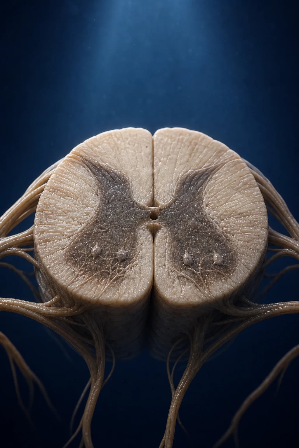
Motor neuron disease (MND) — the amyotrophic lateral sclerosis (ALS) is the commonest form — is a progressive degeneration of the upper and the lower motor neurons. The hallmark is the mixed UMN and LMN signs in the same limb: the muscle wasting, the weakness, and the fasciculations (the LMN) alongside the brisk reflexes and the spasticity (the UMN). The sensation and the sphincters are spared (early) — a key discriminator from the spinal cord lesion and the peripheral neuropathy. The course is relentlessly progressive and fatal — the median survival is 3 to 5 years, with the death typically from the respiratory failure (the diaphragm weakness) or the aspiration.[1]
In the ICU, the MND presents with the type 2 respiratory failure (the ventilatory failure from the diaphragm weakness) or the aspiration pneumonia (the bulbar weakness). The management is the symptomatic support, the disease modification (the riluzole), the non-invasive ventilation, and the advance care planning.[1]
The clinical signature
- A progressive, asymmetric weakness — the limbs, the bulbar (the dysarthria, the dysphagia), and eventually the respiratory muscles.[1]
- The mixed UMN and LMN signs in the same limb — the muscle wasting and the fasciculations (the LMN) plus the brisk reflexes and the spasticity (the UMN). This combination is the characteristic signature of the MND.[1]
- The bulbar involvement — the tongue wasting and the fasciculations, the dysarthria, the dysphagia.[1]
- The sensation and the eye movements are spared. The sphincters are spared early.[1]
- The cognitive change — the frontotemporal dementia in about 15 per cent.[1]
- The no pain, no sensory loss — a useful discriminator.[1]
The diagnosis
- The clinical diagnosis (the El Escorial criteria) — the combination of the UMN and the LMN signs in multiple regions, with the progression, and the exclusion of the mimics.[1]
- The EMG — the denervation: the fibrillations, the positive sharp waves, the fasciculations, and the chronic reinnervation (the large polyphasic units). The sensory conduction is normal.[1]
- Exclude the mimics:[1]
- The multifocal motor neuropathy with the conduction block (MMN) — a treatable mimic (the IVIG-responsive); the LMN-only, the conduction block on the NCS.[1]
- The cervical and the lumbar radiculomyelopathy — the cord and the root compression on the MRI.[1]
- The Kennedy disease (the X-linked bulbar and spinal muscular atrophy) — the LMN-only, the sensory involvement, the gynaecomastia.[1]
The ICU presentation
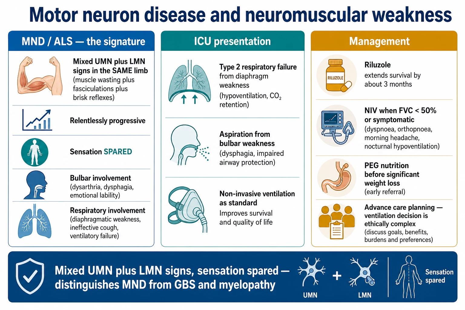
- The type 2 respiratory failure — the ventilatory failure from the diaphragm weakness. The patient may present with the orthopnoea, the morning headaches (the nocturnal hypoventilation), the daytime somnolence, or an overt hypercapnic respiratory failure. This may be the presenting feature of an undiagnosed MND.[1]
- The aspiration pneumonia — from the bulbar weakness and the impaired swallow.[1]
- The acute deterioration — often a precipitant (the infection, the aspiration, a sedative).[1]
The management
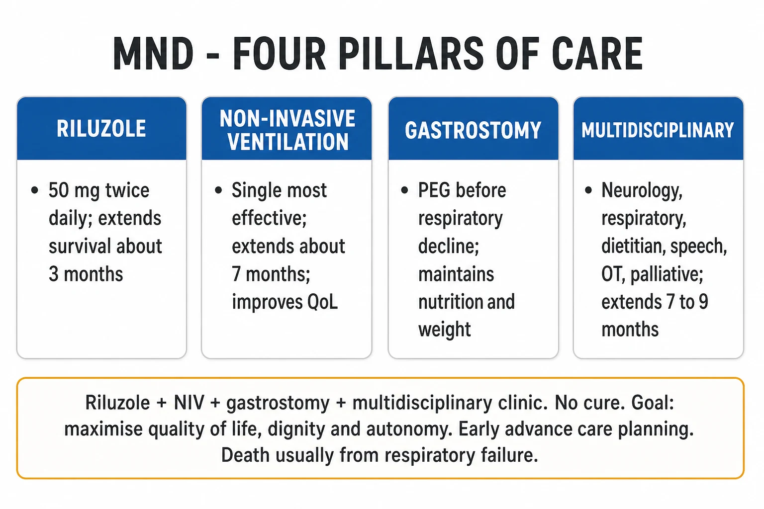
- The riluzole — the glutamate antagonist; the disease-modifying drug that extends the survival by about 3 months. Started early in the disease.[1]
- The non-invasive ventilation (the NIV) — the standard for the symptomatic respiratory weakness. The NIV improves the survival (especially in the patients without a severe bulbar weakness), the quality of life, and the sleep. The NIV is started when the FVC is under 50 per cent of the predicted, or when the patient is symptomatic (the orthopnoea, the morning headache, the daytime somnolence), or the SNIP or the MIP is low. The NIV is preferred over the invasive ventilation for the symptomatic support.[1]
- The invasive ventilation — the tracheostomy and the long-term ventilation. Offered to a selected minority; a major life decision that the patient must make with the full information. Many MND patients have an advance directive declining the invasive ventilation.[1]
- The nutrition — the PEG or the RIG tube placed BEFORE the significant weight loss (and ideally when the FVC is over 50 per cent, for the safe sedation and the insertion).[1]
- The symptom control — the sialorrhoea (the glycopyrrolate, the amitriptyline), the thick secretions, the pain, the depression, the constipation.[1]
- The advance care planning and the palliative care — the discussion of the ventilation decision, the ceiling of the care, and the end-of-life preferences is a core part of the management.[1]
- Avoid the drugs that worsen the weakness (the aminoglycosides, the magnesium, the prolonged neuromuscular blockers).[1]
The neuromuscular weakness in the ICU — the differential
When a weak patient arrives in the ICU, the broad differential of the neuromuscular respiratory failure:[1]
- The acute — the Guillain-Barre, the myasthenia gravis (the crisis), the electrolyte disorders (the hypokalaemia, the hypophosphataemia, the hypomagnesaemia), the spinal cord lesion (the compression, the transverse myelitis), the tick paralysis, the botulism, the organophosphate poisoning.[1]
- The chronic — the MND, the muscular dystrophy, the myotonic dystrophy, the chronic inflammatory demyelinating polyneuropathy (the CIDP).[1]
- The critical-illness polyneuromyopathy (CIPNM) — the acquired weakness of the prolonged ICU stay (the sepsis, the multi-organ failure); the sensorimotor axonal neuropathy and the myopathy; the difficulty weaning from the ventilation.[1]
Red flags
The clinical features in depth — the regional onset and the progression
The MND begins in one region and spreads — the limb onset (about 70 per cent), the bulbar onset (about 25 per cent), and the respiratory onset (rare, under 5 per cent). The bulbar onset carries the worse prognosis (the median survival about 2 to 3 years from the diagnosis, the older, the female). The progression is the contiguous spread (the limb to the bulbar to the respiratory). The sensation, the eye movements (until the very late), the sphincters, and the bowel and the bladder function are spared — the key discriminator.[1]
| Feature | The UMN sign (the corticospinal tract) | The LMN sign (the anterior horn cell) |
|---|---|---|
| The tone | The spasticity, the clonus | The flaccidity, the reduced tone |
| The reflexes | The brisk, the hyperreflexia, the Babinski | The reduced or the absent (the late — the UMN may keep them brisk despite the wasting — the key paradox) |
| The bulk | The normal (the no wasting) | The wasting, the atrophy, the fasciculations |
| The strength | The weakness (the pyramidal distribution — the extensors in the legs, the flexors in the arms) | The weakness (the segmental, the wasting-matched) |
| The hallmark in the MND | The brisk reflex in a wasted, the fasciculating limb | The wasting and the fasciculations in the limb with the brisk reflex |
| The site of the lesion | The motor cortex, the corticospinal tract | The anterior horn cell, the cranial nerve nucleus |
The bulbar features — the dysarthria (the spastic-flaccid mix; the slow, the laboured, the nasal), the dysphagia (the liquids worse than the solids — the opposite of the mechanical obstruction), the tongue wasting and the fasciculations (the pathognomonic), the sialorrhoea (the poor swallow of the saliva, NOT the overproduction), the pseudobulbar affect (the emotional lability — the inappropriate laughter and crying), the brisk jaw jerk and the gag (the UMN). The pseudobulbar affect responds to the dextromethorphan-quinidine (the Nuedexta).[1]
The respiratory features — the dyspnoea on exertion, then at rest, then the orthopnoea (the diaphragm fails in the supine — the orthopnoea is the early, the sensitive sign of the diaphragm weakness). The morning headache and the daytime somnolence (the nocturnal hypoventilation). The poor cough (the expiratory muscle weakness — the secretion retention). The recurrent chest infections and the aspiration (the bulbar). The overt hypercapnic respiratory failure is the late sign.[1]
The cognitive and the behavioural change — the frontotemporal dysfunction in up to the half (the mild executive impairment); the frontotemporal dementia (the FTD) in about 15 per cent. The C9orf72 hexanucleotide repeat expansion is the commonest genetic cause and links the ALS to the FTD.[1]
The respiratory involvement — the monitoring
The respiratory muscle weakness is the universal and the terminal event. The monitoring from the diagnosis:[1]
- The FVC (the forced vital capacity, the seated AND the supine — a drop over 25 per cent on lying is the early diaphragm weakness even when the seated FVC is normal).[1]
- The SNIP (the sniff nasal inspiratory pressure) and the MIP (the maximum inspiratory pressure) — the more sensitive than the FVC in the bulbar and the early disease (the under 40 cmH2O for the SNIP, the under 60 cmH2O for the MIP as the trigger thresholds).[1]
- The peak cough flow — under 270 L/min (or under 160 L/min for the severe) predicts the poor secretion clearance and the cough-assist need.[1]
- The overnight oximetry (and the transcutaneous CO₂) — the nocturnal hypoventilation precedes the daytime failure.[1]
- The symptoms — the orthopnoea is the single best predictor of the respiratory failure.[1]
| Parameter | The normal | The consider the NIV | The start the NIV (the consensus) |
|---|---|---|---|
| FVC (the seated) | over 80% predicted | 60 to 80% | under 50% predicted (or under 80% with the symptoms) |
| FVC (the supine drop) | under 10% | 10 to 25% | over 25% drop on lying |
| SNIP | over 70 cmH2O | 40 to 70 | under 40 cmH2O |
| MIP (the PImax) | over 60 cmH2O (negative) | 40 to 60 | under 60 cmH2O |
| PaCO₂ | 35 to 45 mmHg | 45 to 50 | over 45 to 50 mmHg (the daytime hypercapnia — the late) |
| Nocturnal SpO₂ | over 90% | dips to 88 to 90% | under 90% for over 5 minutes (the nocturnal desaturation) |
| Symptoms | none | the orthopnoea, the morning headache | the symptomatic (the orthopnoea, the EDS, the unrefreshing sleep) |
The ICU admission — the causes and the structured assessment
The three principal ICU presentations of the MND:[1]
- The acute type 2 respiratory failure — the ventilatory exhaustion from the diaphragm weakness. The precipitant may be the chest infection, the aspiration, the sedation, or the natural progression. This may be the presenting feature of the undiagnosed MND — the clues are the wasting, the tongue fasciculations, the brisk reflexes with the preserved sensation.[1]
- The aspiration pneumonia — the bulbar weakness with the impaired swallow and the cough. The recurrent aspiration predicts the rapid decline and the poor NIV tolerance.[1]
- The elective NIV initiation (or the rare tracheostomy) — the planned admission for the respiratory support set-up, the mask fitting, and the patient and the family education.[1]
The new MND admission to the ICU — the structured assessment
- The airway, the breathing, the circulation. The IV access, the oxygen (the titrated — the CO₂ retainer caution), the cardiac monitoring, the capnography.[1]
- The reason for the deterioration. The three commonest: (a) the acute on chronic type 2 respiratory failure (the ventilatory exhaustion); (b) the aspiration pneumonia (the bulbar weakness); (c) the intercurrent infection (the chest, the urinary, the line). The sedative or the opioid (the oversedation of the chronic respiratory failure) is the iatrogenic precipitant.[1]
- The baseline and the trajectory. The diagnosis, the time from the diagnosis, the prior FVC, the ALSFRS-R score, the advance directive (the NIV accepted, the invasive ventilation declined, the DNAR). The family and the MND specialist nurse.[1]
- The respiratory support decision. The NIV if the FVC under 50% or the symptomatic and the patient accepts; the invasive ventilation ONLY if the patient has explicitly opted for the long-term ventilation (rare).[1]
- The reversible precipitant. The treat the infection, the aspiration, the electrolyte disorder, the secretions (the humidification, the suction, the cough assist).[1]
- The goals of the care and the ceiling. The early discussion with the patient (if the capacity), the family, and the palliative care team. The escalation plan in the chart.[1]
The respiratory management — the NIV and the tracheostomy controversy
The non-invasive ventilation (the NIV)
The NIV is the standard of the care for the symptomatic respiratory weakness in the MND. The NIV improves the survival (the median survival gain of about 205 days in the Bourke trial; the larger gain in the patients without the severe bulbar weakness), the sleep, the quality of life, and the cognitive function (the reversal of the nocturnal-hypoventilation encephalopathy).[1]
The initiation:[1]
- The bilevel (the BiPAP) — the IPAP titrated from 8 to 10 cmH2O up to 12 to 18 cmH2O (the tidal volume and the symptom driven); the EPAP 3 to 5 cmH2O (the higher in the obesity or the OSA overlap).[1]
- The start the night-only, then the daytime as the weakness progresses; the ramp, the humidification, and the interface (the nasal, the full face, the total face — the bulbar prefers the oronasal).[1]
- The secretion management — the cough assist (the mechanical insufflation-exsufflation), the suction, the humidification. The peak cough flow under 270 L/min mandates the cough assist.[1]
The caveats:[1]
- The severe bulbar weakness — the NIV is less effective (the mask leak, the aspiration, the poor tolerance), the survival benefit is attenuated, but the symptom control (the orthopnoea) is still useful.[1]
- The cognitive impairment (the FTD) — the adherence is poor; the family support and the simple interface.[1]
- The CO₂ retention — the EPAP too low for the IPAP (the inadequate pressure support); re-titrate.[1]
The tracheostomy and the long-term ventilation — the controversy
The tracheostomy with the long-term invasive ventilation is the most ethically complex decision in the MND. It prolongs the survival dramatically (the median survival from the tracheostomy of 12 to 24 months, sometimes the years; the longest in the Japanese cohorts), but it does NOT halt the disease progression — the limb paralysis becomes complete, the communication is lost (the bulbar progression), and the patient may enter the "locked-in" state with the preserved cognition.[1]
The considerations:[1]
- The patient must make the decision before the respiratory crisis (the capacity, the full information, the time to deliberate).[1]
- The advance directive declining the invasive ventilation is the norm in the most Western countries; the uptake is the higher in the Japan (the cultural, the infrastructure).[1]
- The family burden (the 24-hour care, the suctioning, the tracheostomy emergencies).[1]
- The withdrawal of the ventilation is the legal and the ethical right once it no longer meets the patient's goals; it must be supported by the palliative and the symptom-focused care.[1]
| Feature | The NIV (the non-invasive) | The tracheostomy (the invasive) |
|---|---|---|
| The survival benefit | The modest (the ~7 months; the larger in the non-bulbar) | The large (the months to the years) |
| The disease modification | The none | The none (the disease continues) |
| The bulbar disease | The less effective, the symptom relief only | The effective (the bypassed airway) |
| The secretion clearance | The poor (the cough assist needed) | The good (the direct suction) |
| The communication | The preserved (the speech with the NIV off) | The lost or the impaired (the speaking valve, the ventilation timing) |
| The comfort and the QoL | The high (the home, the family) | The lower (the care burden, the isolation) |
| The interface | The mask (the leak, the skin breakdown) | The tracheostomy tube (the stoma care, the infection) |
| The withdrawal | The easy (the ethically straightforward — the end-of-life) | The difficult (the ethically and the emotionally loaded) |
| The patient preference | The accepted by the most | The declined by the most (the advance directive) |
The disease-modifying therapy — the riluzole, the edaravone, the tofersen
The riluzole
The riluzole (the Rilutek, the Teglutik) is the glutamate antagonist — it reduces the presynaptic glutamate release (the inhibition of the voltage-gated sodium channels, the enhancement of the astrocytic glutamate reuptake). The Cochrane meta-analysis (the Bensimon 1994 trial and the subsequent) confirms the modest survival benefit of about 2 to 3 months.[1][1]
The practical use:[1]
- The dose — 50 mg twice daily (the orally; the liquid formulation for the dysphagic). Started at the diagnosis.[1]
- The monitoring — the ALT and the AST every month for the first 3 months, then every 3 months. Stop if the transaminases rise over 5 times the upper limit of the normal.[1]
- The side effects — the fatigue, the nausea, the diarrhoea, the hepatic dysfunction (the 5 per cent), the (rare) pneumonitis. Reduce the dose in the hepatic impairment.[1]
The edaravone
The edaravone (the Radicava) is the free-radical scavenger and the antioxidant. The MCI186-16 and the MCI186-19 trials (the Japanese) showed the slowing of the ALSFRS-R decline in the selected, the early-onset, the mild-respiratory patients.[1]
The practical use:[1]
- The regimen — the IV or the oral; the cycle of the daily dosing for 14 days, then the 14-day off, then the 10 days on in the second cycle, then the off; the maintenance cycles.[1]
- The selection — the early disease (the duration under 2 years, the FVC over 80%, the ALSFRS-R over 2 points per item); the modest benefit in the selected.[1]
- The side effects — the bruising, the gait disturbance, the headache, the (rare) hypersensitivity.[1]
The tofersen — the SOD1-ALS gene-directed therapy
The tofersen (the Qalsody) is the antisense oligonucleotide targeting the SOD1 mRNA for the degradation — the first gene-directed therapy, the intrathecal, for the SOD1-mutant ALS (about 2 per cent of the ALS). The VALOR trial (the Cudkowicz et al, the NEJM, 2022) and the open-label extension showed the reduction of the neurofilament light chain (the NfL — the biomarker of the axonal injury) and the slowing of the clinical decline.[1]
The practical use:[1]
- The selection — the confirmed SOD1 mutation (the genetic testing of the all the familial and the early-onset ALS). The A4V variant is the aggressive; the some SOD1 variants are the very slowly progressive.[1]
- The administration — the intrathecal (the lumbar puncture, the 100 mg monthly after the loading). The Ommaya reservoir or the intrathecal port for the long-term.[1]
- The side effects — the lumbar puncture headache, the procedural pain, the (rare) myelitis, the neurological.[1]
The nutritional support — the PEG and the RIG
The weight loss and the malnutrition are the independent predictors of the mortality (the 5 to 10% weight loss worsens the survival). The mechanisms: the dysphagia (the bulbar), the hypermetabolism, the arm weakness (the cannot self-feed). The PEG (the percutaneous endoscopic gastrostomy) or the RIG (the radiologically-inserted gastrostomy) is placed BEFORE the significant weight loss (the BMI under 18.5, the over 10% weight loss).[1]
The timing — the ideal is when the FVC is over 50% of the predicted (the safer sedation and the procedure; the lower risk of the respiratory depression and the aspiration under the sedation). The risk of the procedural respiratory failure rises sharply below the 50%.[1]
The PEG decision and the procedure in the MND
- The screen — the weight loss over 10%, the BMI under 18.5, the dysphagia, the FVC trend. The dietitian and the speech pathologist.[1]
- The respiratory assessment — the FVC and the SNIP. The FVC over 50% predicted is the safe. The FVC under 50% mandates the NIV during the procedure (the BiPAP-supported PEG; the avoidance of the deep sedation; the propofol sparing; the local and the midazolam).[1]
- The choice — the PEG (the endoscopic, the larger bore, the endoscopy risk) vs the RIG (the radiological, the smaller, the often no sedation). The RIG is the preferred in the bulbar disease and the lower FVC.[1]
- The post-procedure — the NIV continued, the monitoring in the HDU, the gradual feed introduction (the 24-hour post-procedure), the medication conversion (the riluzole via the tube).[1]
- The follow-up — the weight, the nutrition, the stoma care, the fluid, the medication.[1]
The prognosis
The median survival from the diagnosis is 3 to 5 years (the 5-year survival about 20 per cent; the 10-year survival about 10 per cent). The death is typically from the respiratory failure, the aspiration, or the pulmonary embolism (the immobility).[1]
| Factor | The favourable (the slower progression) | The unfavourable (the faster progression) |
|---|---|---|
| The site of the onset | The limb onset | The bulbar onset (the survival ~2 years) |
| The age at the onset | The younger (under 50) | The older (over 65) |
| The diagnostic delay | The long (the indolent) | The short (the rapid progression recognised) |
| The ALSFRS-R slope | The shallow (the under 0.5/month) | The steep (the over 1/month) |
| The FVC at the diagnosis | The over 80% predicted | The under 50% predicted |
| The nutrition | The normal weight | The weight loss, the low BMI |
| The cognitive | The normal cognition | The FTD (the poor adherence, the worse survival) |
| The gene | The none (the sporadic) | The C9orf72, the SOD1 A4V (the aggressive) |
| The time to the NIV | The early NIV | The delayed or the bulbar-poor NIV tolerance |
The end-of-life decisions and the advance care planning
The advance care planning is the core, not the afterthought, of the MND management. The decisions:[1][1]
- The ventilation decision — the NIV (the accepted by the most), the invasive ventilation (the declined by the most, the accepted by the few). The decision must be made early and revisited (the disease progresses; the goals may change).[1]
- The resuscitation decision — the DNAR (the do-not-attempt-resuscitation) is the appropriate for the most advanced MND (the CPR is the futile and the survivorship is the poor). The documented and the discussed.[1]
- The escalation ceiling — the ICU, the HDU, the ward, the palliative. The written plan.[1]
- The symptom-focused care — the dyspnoea (the low-dose opioid — the morphine 2.5 to 5 mg orally or the 1 to 2 mg subcutaneously; the benzodiazepine for the anxiety), the sialorrhoea (the glycopyrrolate, the hyoscine, the amitriptyline; the Botox to the salivary glands), the thick secretions (the hydration, the mucolytics — the avoid the thickening), the pain (the NSAIDs, the neuropathic agents, the opioids), the depression, the pseudobulbar affect (the dextromethorphan-quinidine), the insomnia, the constipation.[1]
- The palliative care involvement — the early, the parallel with the disease-modifying therapy, NOT only the terminal. The hospice and the home.[1]
The withdrawal of the ventilation
The withdrawal of the long-term ventilation is the ethically and the legally supported when the patient decides the burden outweighs the benefit (the "locked-in", the loss of the communication, the suffering). The protocol:[1]
- The decision and the consent (the patient if the capacity; the advance directive; the family and the surrogate).[1]
- The palliative and the symptom-focused preparation — the opioid and the benzodiazepine premedication, the sedation if requested (the proportional; the terminal sedation in the refractory).[1]
- The withdrawal — the gradual reduction of the pressure support over the 30 to 60 minutes (the preferred over the abrupt), the family present, the symptom control.[1]
- The death typically within the hours (the deep sedation, the symptom-free).[1]
The mimics — the expanded differential
| Feature | The MND (the ALS) | The GBS | The CIDP | The MMN | The cervical myelopathy | The Kennedy disease |
|---|---|---|---|---|---|---|
| The onset | The progressive, the asymmetric | The acute, the ascending | The chronic, the relapsing | The progressive, the asymmetric, the multifocal | The progressive, the arm then the leg | The slow, the proximal, the bulbar |
| The UMN signs | The yes | The no | The no | The no | The yes (the leg) | The no |
| The LMN signs | The yes (the mixed in the same limb) | The yes (the areflexia, the diffuse) | The yes (the areflexia) | The yes (the multifocal, the conduction block) | The yes (the arm at the level) | The yes (the bulbar, the proximal) |
| The sensation | The spared | The impaired | The impaired | The spared | The impaired (the level) | The impaired (the sensory neuropathy) |
| The sphincters | The spared | The spared | The spared | The spared | The involved (the late) | The spared |
| The CSF protein | The normal | The raised (the cytoalbuminologic dissociation) | The raised | The normal | The normal | The normal |
| The NCS | The denervation, the normal sensory | The demyelination (the conduction block, the slow) | The demyelination (the chronic) | The conduction block (the multifocal motor) | The normal or the radiculopathy | The sensory motor axonal |
| The treatment | The riluzole, the supportive | The IVIG, the PLEX | The steroids, the IVIG | The IVIG | The surgery (the decompression) | The supportive (the testosterone) |
| The course | The progressive, the fatal | The monophasic, the recovery | The relapsing or the progressive, the treatable | The slowly progressive, the treatable | The variable (the surgical) | The very slow, the near-normal lifespan |
The key trials
Clinical evidence
- The trial: The non-invasive ventilation in the MND (the randomised trial of the NIV vs the standard care)
- The authors: Bourke SC, Tomlinson M, Williams TL, Bullock RE, Shaw PJ, Gibson GJ
- The journal: The Lancet Neurology, 2006
- The finding: The NIV improved the survival (the median survival gain of about 205 days overall; the larger gain of about 9 months in the patients without the severe bulbar weakness), the quality of life, and the cognitive function. The NIV became the standard of the care.[1]
Clinical evidence
- The trial: The riluzole in the ALS (the pivotal randomised trial)
- The authors: Bensimon G, Lacomblez L, Meininger V (the ALS/Riluzole Study Group)
- The journal: The New England Journal of Medicine, 1994
- The finding: The riluzole 100 mg daily extended the survival (especially in the bulbar-onset), the reduced the tracheostomy-free mortality, the modest benefit of about 3 months. The first disease-modifying therapy.[1]
Clinical evidence
- The trial: The Cochrane meta-analysis of the riluzole in the ALS
- The authors: Miller RG, Mitchell JD, Moore DH
- The journal: The Cochrane Database of Systematic Reviews, 2012
- The finding: The pooled analysis of the trials confirmed the modest survival benefit (about 2 to 3 months at 12 months), the acceptable safety. The riluzole remains the first-line.[1]
Clinical evidence
- The trial: The edaravone in the ALS (the MCI186-16 phase 3)
- The authors: The Edaravone (MCI-186) ALS 16 Study Group
- The journal: The Lancet Neurology, 2017
- The finding: The edaravone slowed the ALSFRS-R decline in the selected, the early, the mild-respiratory patients (the disease duration under 2 years, the FVC over 80%, the ALSFRS-R over 2 per item). The benefit modest; the selection strict.[1]
Clinical evidence
- The trial: The tofersen in the SOD1-ALS (the VALOR phase 3)
- The authors: Cudkowicz ME, van den Berg LH, Shefner JM, et al. (the VALOR and OLE Investigators)
- The journal: The New England Journal of Medicine, 2022
- The finding: The tofersen (the intrathecal antisense oligonucleotide) reduced the plasma NfL and the CSF SOD1 protein, the modest (the non-significant primary) but the favourable-trend clinical effect in the SOD1-ALS; the open-label extension showed the clearer clinical benefit. The first gene-directed therapy.[1]
Clinical evidence
- The trial: The EFNS guidelines on the clinical management of the ALS (the MND)
- The authors: Andersen PM, Abrahams S, Borasio GD, et al. (the EFNS Task Force)
- The journal: The European Journal of Neurology, 2012
- The finding: The consensus on the riluzole (the early), the NIV (the FVC under 50% or the symptomatic), the PEG (the early, the FVC over 50%), the palliative care (the early and the parallel), and the symptom management.[1]
Exam practice
SAQ — Type 2 respiratory failure as the presenting feature of MND
10 minutes · 10 marks
A 64-year-old man is admitted to the ICU with a 3-day history of increasing breathlessness and confusion. His arterial blood gas on room air shows pH 7.28, PaCO2 82 mmHg, PaO2 52 mmHg, HCO3 34 mmol/L. He has no prior respiratory diagnosis. On examination he has marked muscle wasting in the hands, fasciculations in the tongue and the deltoids, brisk reflexes in all four limbs, but normal sensation. His FVC is 38 per cent of the predicted and drops by 30 per cent when supine.
SAQ — NIV vs tracheostomy: the ethical decision and the advance care planning
10 minutes · 10 marks
A 58-year-old woman with a 3-year history of bulbar-onset MND is admitted with an acute exacerbation of her type 2 respiratory failure precipitated by an aspiration pneumonia. She is currently on NIV (BiPAP IPAP 14, EPAP 4) overnight. She is now drowsy (GCS 13), her PaCO2 has risen to 75 mmHg despite the NIV, and the team is considering whether to intubate. Her husband reports she has an advance directive stating she does not want invasive ventilation or cardiopulmonary resuscitation.
The clinical pearls
[1] [1] [1] [1] [1] [1] [1] [1] [1] [1] [1] [1] [1] [1] [1] [1] [1] [1]The additional red flags
[1]References
- [1]van Es MA, Hardiman O, Chio A, Al-Chalabi A, Pasterkamp RJ, Veldink JH, van den Berg LH Amyotrophic lateral sclerosis. Lancet, 2017.PMID 28552366
- [2]Bourke SC, Tomlinson M, Williams TL, Bullock RE, Shaw PJ, Gibson GJ Effects of non-invasive ventilation on survival and quality of life in patients with amyotrophic lateral sclerosis: a randomised controlled trial. Lancet Neurology, 2006.PMID 16426990
- [3]Bensimon G, Lacomblez L, Meininger V (ALS/Riluzole Study Group) A controlled trial of riluzole in amyotrophic lateral sclerosis. New England Journal of Medicine, 1994.PMID 8302340
- [4]Miller RG, Jackson CE, Kasarskis EJ, et al. (AAN ALS Practice Parameters Task Force) Practice parameter update: the care of the patient with amyotrophic lateral sclerosis: multidisciplinary care, symptom management, and cognitive/behavioral impairment (an evidence-based review): report of the Quality Standards Subcommittee of the American Academy of Neurology. Neurology, 2009.PMID 19822873
- [5]Writing Group; Edaravone (MCI-186) ALS 16 Study Group Safety and efficacy of edaravone in well defined patients with amyotrophic lateral sclerosis: a randomised, double-blind, placebo-controlled trial. Lancet Neurology, 2017.PMID 28522181
- [6]Cudkowicz ME, van den Berg LH, Shefner JM, et al. (VALOR and OLE Investigators) Trial of Antisense Oligonucleotide Tofersen for SOD1 ALS. New England Journal of Medicine, 2022.PMID 36129998
- [7]Miller RG, Mitchell JD, Moore DH Riluzole for amyotrophic lateral sclerosis (ALS)/motor neuron disease (MND). Cochrane Database of Systematic Reviews, 2012.PMID 22419278
- [8]Andersen PM, Abrahams S, Borasio GD, et al. (EFNS Task Force on the Management of Amyotrophic Lateral Sclerosis) EFNS guidelines on the clinical management of amyotrophic lateral sclerosis (MALS)--revised report of an EFNS task force. European Journal of Neurology, 2012.PMID 21914052
- [9]Chiò A, Logroscino G, Hardiman O, et al. Prognostic factors in ALS: A critical review. Amyotrophic Lateral Sclerosis, 2009.PMID 19922118