ICU · Neurocritical care / neuromuscular
Myasthenia Gravis & Myasthenic Crisis — NMJ Weakness, PLEX/IVIG & Steroid Exacerbation
Also known as Myasthenia gravis · MG · Myasthenic crisis · Cholinergic crisis · Acetylcholine receptor antibody · AChR antibody · MuSK antibody · Pyridostigmine · Plasma exchange · Thymoma · Neuromuscular junction · Fatigable weakness
Myasthenia gravis (MG) is an autoimmune disease of the post-synaptic acetylcholine receptor at the neuromuscular junction — the acetylcholine receptor antibodies damage the receptors, causing a fatigable weakness (worse with the activity, better with the rest). The ocular (the ptosis, the diplopia), the bulbar, and the limb muscles are affected; the reflexes and the sensation are PRESERVED (a key discriminator from the Guillain-Barre). The myasthenic crisis is the life-threatening respiratory or the bulbar failure, precipitated by the infection, the surgery, or the drugs (the aminoglycosides, the beta-blockers, the magnesium). The management: intubate early (the NIF under 30 cmH2O or the FVC under 20 mL/kg); the plasma exchange OR the IVIG (equivalent); the corticosteroids for the long-term immunosuppression — but the steroids WORSEN the MG in the first 1 to 2 weeks (the steroid-induced exacerbation), so a monitored, low-dose start. The cholinergic crisis (the excess pyridostigmine) causes the weakness PLUS the cholinergic overdrive (the miosis, the salivation, the bradycardia, the sweating) — wet vs the dry myasthenic crisis. Screen for the thymoma (the chest CT); the thymectomy if the thymoma.
On this page & tools
Your progress
Saved locally on this device.
8 MCQs with explanations
Target exams
Overview & definition
Myasthenia gravis (MG) is an autoimmune disease of the post-synaptic acetylcholine receptor at the neuromuscular junction. The acetylcholine receptor antibodies (the AChR antibodies) damage and reduce the receptor density, so the muscle receives a weaker signal — producing a fatigable weakness (worse with the repeated activity, better with the rest). The classic presentation is the ocular weakness (the ptosis, the diplopia) that fatigues as the day progresses, often spreading to the bulbar and the limb muscles.[1]
The myasthenic crisis is the life-threatening weakness of the respiratory or the bulbar muscles requiring the ventilatory support. The two ICU priorities: the early intubation (before the respiratory failure) and the immunomodulation (the plasma exchange or the IVIG).[1]
The pathophysiology and the antibodies
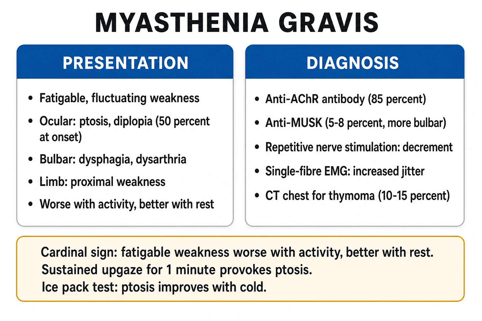
- The acetylcholine receptor (AChR) antibodies — positive in about 85 per cent of the generalised MG. The hallmark.[1]
- The MuSK antibodies (the muscle-specific tyrosine kinase) — present in many of the AChR-negative cases. A different phenotype: the bulbar and the respiratory predominance, the facial and the tongue atrophy, a poorer response to the acetylcholinesterase inhibitors, and a better response to the plasma exchange and the rituximab.[1]
- The thymus — the thymic hyperplasia in about 65 per cent, and the thymoma in about 15 per cent (a malignancy that must be resected). The chest CT screens for the thymoma.[1]
The presentation
- A fatigable weakness — the hallmark. The ocular (the ptosis, the diplopia) is often the first and may remain isolated (the ocular MG) or generalise. The bulbar (the dysphagia, the dysarthria, the facial weakness, the chewing fatigue). The limb (the proximal more than the distal). The respiratory (the diaphragm).[1]
- The weakness worsens with the activity and improves with the rest; worse at the end of the day, in the heat, with the infection, and with the precipitating drugs.[1]
- The reflexes and the sensation are PRESERVED — a key discriminator from the Guillain-Barre (the areflexia) and the spinal cord lesion.[1]
The precipitants of the crisis
The myasthenic crisis is precipitated by:[1]
- An infection (the commonest).
- A surgery (including the thymectomy).
- The pregnancy and the postpartum.
- The drugs — the aminoglycosides, the beta-blockers, the magnesium, the neuromuscular blockers, the macrolides, the fluoroquinolones, the iodinated contrast.
- The tapering of the immunosuppression.
- An aspiration.[1]
The diagnosis
- The AChR antibodies (and the MuSK if the AChR is negative).[1]
- The nerve conduction studies — a decremental response to the repetitive stimulation at 3 Hz (the amplitude drops with the repeated stimuli). The single-fibre EMG shows the increased jitter (the most sensitive test).[1]
- The bedside tests — the ice pack test (the cold improves the ptosis by reducing the acetylcholinesterase activity); the edrophonium (the Tensilon) test (rarely used now — the edrophonium is a short-acting acetylcholinesterase inhibitor that briefly improves the weakness; the risk of the bradycardia and the bronchospasm).[1]
- The chest CT — to screen for the thymoma.[1]
The myasthenic vs the cholinergic crisis
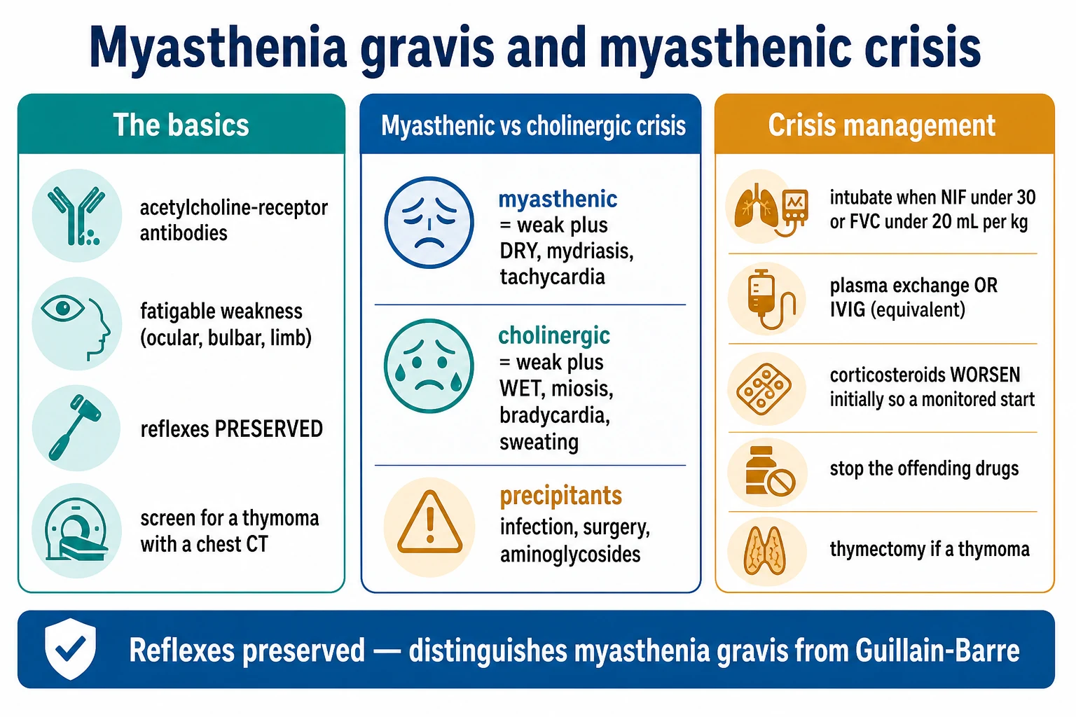
A critical distinction at the bedside:[1]
| Feature | The myasthenic crisis | The cholinergic crisis |
|---|---|---|
| Cause | The worsening MG (too little acetylcholine signalling) | The excess pyridostigmine (too much acetylcholine) |
| Secretions | Dry | Wet (the salivation, the lacrimation, the bronchorrhoea) |
| Pupils | Mydriasis | Miosis |
| Heart rate | Tachycardia | Bradycardia |
| GI | Dry | The cramping, the diarrhoea |
| Skin | Dry | Sweating |
In the crisis, the two may be indistinguishable — the safest approach is to hold the pyridostigmine, ventilate, and treat (the pyridostigmine increases the secretions and worsens the ventilation).[1]
The crisis management
- The airway and the ventilation — intubate early.[1] The thresholds (similar to the Guillain-Barre): the NIF under 30 cmH2O, the FVC under 20 mL/kg, the bulbar weakness (the aspiration), or the rapid progression. Intubate electively — do NOT wait for the respiratory arrest. The non-invasive ventilation (the BiPAP) may avert the intubation in the early or the mild crisis (the fatigue-reversal pattern).[1]
- The immunomodulation — the plasma exchange OR the IVIG (equivalent; choose one). The plasma exchange (5 sessions over 1 to 2 weeks) works faster (preferred for the severe or the rapidly progressive crisis). The IVIG (2 g/kg over 5 days) is an alternative. Both take days to work; neither is immediately effective.[1]
- The corticosteroids for the long-term immunosuppression — but the steroids WORSEN the MG initially.[1] The prednisolone is the mainstay of the long-term treatment, but a high starting dose causes the steroid-induced exacerbation in the first 1 to 2 weeks (the steroid un masks and worsens the weakness as it begins to work). Start the prednisolone at a low dose (10 to 20 mg) and titrate up in a monitored setting (especially with the bulbar or the respiratory weakness), and only after the airway is secured in the crisis.[1]
- Stop the offending drugs (the aminoglycosides, the magnesium, the beta-blockers). Treat the precipitant (the infection).[1]
- The pyridostigmine — held in the crisis (the secretions worsen the ventilation, and the cholinergic crisis is a risk). Restarted once the patient is improving and the secretions are manageable.[1]
- The long-term — the prednisolone plus a steroid-sparing agent (the azathioprine, the mycophenolate, the rituximab); the thymectomy if the thymoma, or in the young AChR-positive generalised MG (improves the long-term outcome).[1]
The pathophysiology in detail — the autoantibodies and the complement
The myasthenia gravis is the prototypical antibody-mediated autoimmune disease — the pathogenic autoantibodies target the proteins of the post-synaptic membrane of the neuromuscular junction (the NMJ), and the result is a reduced safety factor for the neuromuscular transmission. The acetylcholine is released normally from the presynaptic nerve terminal, but the post-synaptic receptor density is too low to reliably depolarise the muscle fibre — so a fraction of the nerve impulses fail to generate a muscle action potential. The clinically apparent weakness is the summation of these transmission failures across millions of motor units, made worse by the sustained activity (the progressive receptor occupancy depletion and the depletion of the presynaptic vesicle pool).[1]
| Antibody | The frequency | The target | The phenotype | The special notes |
|---|---|---|---|---|
| AChR (the binding) | ~85% of the generalised MG | The post-synaptic acetylcholine receptor | The classic ocular-bulbar-limb, the fatigable | The hallmark; the complement-mediated lysis of the junctional fold |
| MuSK | 5 to 8% (up to 40% of the AChR-negative) | The muscle-specific tyrosine kinase (a post-synaptic scaffolding protein) | The bulbar and the respiratory predominance; the facial and the tongue atrophy; the sparing of the limbs | The females; the onset younger (the fourth decade); the acetylcholinesterase inhibitors poorly tolerated; the thymus rarely abnormal; the rituximab highly effective |
| LRP4 | 2 to 5% (the AChR- and MuSK-negative) | The LDL-receptor-related protein 4 (an agrin receptor required for the AChR clustering) | A milder phenotype, the ocular predominance, occasionally the limb | Often a co-existing AChR antibody; the diagnosis of exclusion |
| Agrin / ColQ / cortactin | Rare | The other post-synaptic and the synaptic-cleft proteins | Variable | Suspect in the seronegative MG with the typical electrodiagnostic findings |
| Striated muscle (the anti-titin, the anti-Kv1.4) | ~40% of the thymoma-associated MG, ~30% of the late-onset AChR-MG | The titin and the ryanodine receptor | A more severe disease, the late onset, the more steroid-dependent | A marker of the thymoma — screen the chest CT |
| Seronegative | 5 to 10% | Unknown (likely the cell-mediated, or the low-affinity antibodies not detected) | Variable | The diagnosis rests on the electrodiagnostic criteria and the exclusion of the congenital myasthenic syndromes |
The AChR-antibody-positive MG — the classic phenotype
The AChR antibodies are the IgG1 and the IgG3 subclass — they are directly pathogenic through three mechanisms: (1) the complement-mediated destruction of the post-synaptic junctional folds (the simplified fold architecture reduces the receptor surface area); (2) the antigenic modulation (the cross-linking and the accelerated internalisation and the degradation of the receptors); and (3) the functional blockade (the antibody occupies the acetylcholine binding site). The combined effect is a reduction in the functional receptor density to under 30 per cent of the normal — the threshold below which the safety factor fails and the fatigable weakness emerges.[1]
The two clinical subgroups within the AChR-MG:
- The early-onset (under the age of 50) — the thymic hyperplasia in 65 per cent, the female predominance, the HLA-B8 and DR3 association. The most responsive to the thymectomy.
- The late-onset (over the age of 50) — the thymic atrophy, the male predominance, the more severe disease, the higher anti-titin antibody rate. The less responsive to the thymectomy; the more steroid-dependent. [1]
The MuSK-antibody-positive MG — a distinct entity
The MuSK antibodies are the IgG4 subclass — they do NOT fix the complement (the IgG4 cannot). Instead, they disrupt the agrin–LRP4–MuSK signalling pathway that maintains the AChR clustering at the junctional fold, so the post-synaptic membrane loses its dense receptor organisation. The clinical phenotype is distinct:
- A bulbar and respiratory predominance (the dysarthria, the dysphagia, the tongue weakness, the facial atrophy, the early respiratory crisis).
- A relative sparing of the limbs — the ocular and the bulbar weakness is often disproportionate to the limb power.
- The facial and the tongue atrophy (a unique feature not seen in the AChR-MG) — from the chronic denervation.
- The acetylcholinesterase inhibitors are poorly tolerated and often worsen (the fasciculations, the cramps) — many MuSK patients are misdiagnosed as the cholinergic crisis. Use them cautiously; many MuSK patients are managed without them.
- The thymus is rarely abnormal — the thymectomy is NOT recommended in the MuSK-MG.
- The females, the fourth decade, the ethnic variation (more common in the Mediterranean and the Asian populations).
- The rituximab is highly effective and is now the preferred steroid-sparing agent; the plasma exchange works well and faster than the IVIG.[1]
The thymus and the thymoma
The thymus is the central tolerance organ, and the thymic germinal centres are the site of the anti-AChR antibody production in the AChR-MG. The thymic pathology in the MG:
- The thymic hyperplasia in about 65 per cent of the early-onset AChR-MG (the lymphoid follicular hyperplasia with the germinal centres).
- The thymoma in about 10 to 15 per cent (a neoplasm of the thymic epithelial cells — the WHO classification A, AB, B1, B2, B3 — all capable of producing the MG). The thymoma must be surgically resected (the malignancy indication, independent of the MG).
- The atrophic thymus in the late-onset AChR-MG (no role for the thymectomy).
- The normal thymus in the MuSK-MG (no role for the thymectomy). [1]
Every newly diagnosed MG patient requires a contrast-enhanced chest CT (or the MRI) to screen for the thymoma — a missed thymoma is a missed malignancy. [1]
The bedside clinical assessment — the fatigability
The myasthenic weakness is fatigable — it worsens with the sustained activity and improves with the rest. The bedside examination tests this fatigability: [1]
The bedside fatigability assessment in the suspected MG
- The sustained upgaze for 60 seconds — the progressive ptosis (the levator palpebrae fatigues). The most sensitive ocular test. Stop when the ptosis appears and observe for the recovery with the rest.
- The sustained lateral gaze — the diplopia or the adventitious squint develops as the lateral rectus fatigues.
- The arm abduction held for 5 minutes (the outstretched arm) — the slow drift downwards. Time the descent (the seconds before the drift) for a quantifiable bedside metric.
- The straight leg raise held against the gravity — the lower limb fatigability.
- The vital capacity measured at rest and after 1 minute of the sustained arm exercise — the drop of over 15 per cent confirms the respiratory fatigability.
- The counting aloud on a single breath — under 20 (the normal over 30) suggests the respiratory reserve depletion. The single-breath count test correlates with the FVC and the NIF and requires no equipment.
- The bulbar assessment — the palatal lift (the symmetrical, the fatigue with the repeated "ah"), the gag (the bilaterally preserved but the fatigable), the tongue protrusion (the side-to-side, the fatigable), the swallow assessment (the sips of water, the cough or the wet voice).
The respiratory monitoring in the impending crisis
- The FVC every 4 to 6 hours (the seated, the best of three) — the trend matters more than the absolute. A falling trend warns of the impending failure even when the absolute is above the intubation threshold.
- The NIF (the maximum inspiratory force) every 4 to 6 hours — a value under -30 cmH2O (the under 30, the negative convention) is the intubation threshold.
- The single-breath count test every shift — a bedside surrogate that requires no spirometry.
- The trend of the FVC, the NIF, and the single-breath count together — the decline across all three is the ominous sign.
- The arterial blood gas — the rising PaCO₂ (the hypercapnia is a LATE sign; the normocapnia does NOT exclude the impending failure), the falling PaO₂, the respiratory acidosis. The hypercapnia is the sign to intubate — but waiting for the hypercapnia is the late intubation.
- The bulbar assessment each shift — the aspiration risk is itself an intubation indication, even with a normal FVC.
The myasthenic crisis — the thresholds and the timing
| Parameter | The normal | The warning | The intubation threshold |
|---|---|---|---|
| FVC | 60 to 70 mL/kg | under 25 mL/kg | under 20 mL/kg (some use under 15 mL/kg for the severe crisis) |
| NIF | under -60 cmH2O (the under 60) | -40 to -30 cmH2O | under -30 cmH2O (the under 30) |
| PEF (the peak expiratory, the cough strength) | over 300 L/min | under 200 L/min | under 150 L/min (the poor cough — the secretion clearance) |
| The single-breath count | over 30 | 15 to 25 | under 10 |
| The PaCO₂ | 35 to 45 mmHg | the rising trend | over 45 to 50 mmHg (the hypercapnia — a LATE sign) |
| The bulbar weakness | the normal swallow, the clear voice | the dysarthria | the aspiration, the inability to clear the secretions — intubate regardless of the FVC |
The absolute numbers are guides — the trend matters more. A patient whose FVC falls from 35 to 22 mL/kg over 12 hours is intubated earlier than the threshold, because the trajectory predicts the failure before the next shift. The bulbar weakness is itself an indication — a patient who cannot clear the secretions or protect the airway is intubated regardless of the spirometry. [1]
The plasma exchange vs the IVIG — the head-to-head
| Feature | The plasma exchange (PLEX) | The intravenous immunoglobulin (IVIG) |
|---|---|---|
| The mechanism | The mechanical removal of the circulating antibodies (the AChR, the MuSK) | The immunomodulation (the Fc-receptor blockade, the anti-idiotype, the complement inhibition) |
| The dose and the regimen | 5 sessions over 1 to 2 weeks (3 to 5 L per session, or the 1 to 1.5 plasma volumes) | 2 g/kg over 5 days (the 0.4 g/kg per day) |
| The onset | The faster (the improvement within the first week; the peak at 2 to 3 weeks) | The slower (the improvement over 1 to 2 weeks) |
| The efficacy in the MG | The equivalent to the IVIG (the randomised trials show no difference) | The equivalent to the PLEX |
| The preferred use | The severe and the rapidly progressive crisis; the MuSK-MG (the better response); the pre-thymectomy optimisation | The mild to the moderate crisis; the central-line avoidance; the easier logistics |
| The access | The central venous access (the plasma separation needs a large bore) | The peripheral IV (a Y-port) |
| The vascular considerations | The vascath; the bleeding (the citrate anticoagulation, the fibrinogen depletion) | The volume (the 4 to 5 L over 5 days); the thrombosis risk |
| The contraindications | The severe sepsis, the unstable haemodynamics, the severe coagulopathy | The IgA deficiency (the anaphylaxis), the renal failure (the osmotic nephrosis), the hyperviscosity, the thrombosis history |
| The cost and the logistics | The expensive, the plasma, the specialist staff | The expensive, the limited supply |
Both take days to a week to work — neither is immediately effective. The airway and the ventilation are the immediate priorities. In the refractory crisis (the no response by the end of the second week), switch to the other agent (the PLEX to the IVIG, or the IVIG to the PLEX), or add the rituximab (especially in the MuSK-MG).[1]
The long-term immunosuppression — the agents
| Agent | The onset of action | The dose | The monitoring | The role |
|---|---|---|---|---|
| The prednisolone | 2 to 4 weeks (the clinical); the WORSENS in the first 1 to 2 weeks | 10 to 20 mg, titrate up to 0.75 to 1 mg/kg, then taper to the minimum effective | The glucose, the bone density, the BP, the weight, the infection | The first-line; the bridge to the steroid-sparing |
| The azathioprine | 6 to 12 months (the slow) | 1 to 3 mg/kg/day | The FBC and the LFTs (the first 2 months every 2 weeks, then monthly); the TPMT genotype before the start (the deficiency → the fatal pancytopenia) | The first-line steroid-sparing; the cheapest; the slowest |
| The mycophenolate mofetil | 3 to 6 months | 1 to 2 g/day | The FBC (the leucopenia) | The first-line steroid-sparing; the faster than the azathioprine |
| The methotrexate | 3 to 6 months | 7.5 to 15 mg/week | The FBC, the LFTs, the renal; the folic acid | The alternative; the cheap; the pulmonary fibrosis |
| The tacrolimus / the cyclosporine | 2 to 3 months | The tacrolimus 3 to 5 mg/day; the cyclosporine 2.5 to 5 mg/kg/day | The levels (the tacrolimus), the renal, the BP | The second-line; the renal toxicity; the calcineurin inhibitors |
| The rituximab | 1 to 3 months | 1 g × 2 (the days 1 and 15) or 375 mg/m² × 4 weekly | The CD19 / the CD20 (the B-cell depletion); the immunoglobulins (the hypogammaglobulinaemia) | The preferred in the MuSK-MG; the refractory AChR-MG; the highly effective |
| The eculizumab / the ravulizumab | The weeks | The eculizumab 1200 mg every 2 weeks | The meningococcal vaccination (the encapsulated organisms); the meningococcal prophylaxis | The refractory AChR-MG; the complement inhibitor; the anti-C5; the expensive |
The pyridostigmine — the symptomatic treatment
The pyridostigmine (the Mestinon) is the acetylcholinesterase inhibitor — it increases the synaptic acetylcholine concentration and partially overcomes the receptor loss. The pharmacology:[1]
- The onset is 30 to 60 minutes; the peak at 1 to 2 hours; the duration 4 to 6 hours. The dosing is the 60 mg four times daily (the daytime), titrated up to 120 mg every 4 hours.
- The maximum daily dose is around 1200 mg (the higher doses risk the cholinergic crisis).
- The side effects — the muscarinic overdrive (the salivation, the lacrimation, the sweating, the abdominal cramps, the diarrhoea, the bradycardia, the miosis) — the cholinergic signs. The nicotinic overdrive (the fasciculations, the cramps) at the higher doses.
- The slower-release preparation (the Mestinon Timespan 180 mg) at the bedtime helps the overnight and the morning weakness.
- The IV equivalent — the neostigmine is 1 mg for every 60 mg of the pyridostigmine (the 1:30 ratio; the neostigmine methylsulfate). Used in the nil-by-mouth or the perioperative.
- The pyridostigmine is HELD in the crisis (the secretions worsen the ventilation; the cholinergic crisis is a risk). Restarted once the patient is improving and the secretions are manageable.
- The MuSK-MG — the acetylcholinesterase inhibitors are often poorly tolerated and may worsen; many MuSK patients are managed without them.
The thymectomy — the MGTX trial and the indications
The thymectomy has two distinct indications:
- The thymoma (the malignancy) — every thymoma is resected (the WHO A, AB, B1, B2, B3 — all resected; the surgery, occasionally the adjuvant therapy for the B2/B3 and the invasive). Independent of the MG.
- The non-thymomatous AChR-MG (the immunomodulatory indication) — the thymectomy improves the long-term outcome in the selected patients. [1]
Clinical evidence
- The trial: The Thymectomy Trial in Non-Thymomatous MG (the MGTX)
- The authors: Wolfe JF, Kaminski HJ, Aban IB, et al.
- The journal: The New England Journal of Medicine, 2016
- The design: The randomised, the rater-blinded, the controlled trial. 126 patients (the 18 to 65 years, the AChR-antibody-positive, the non-thymomatous, the generalised MG, the disease duration under 5 years).
- The intervention: The extended transsternal thymectomy plus the prednisolone vs the prednisolone alone.
- The primary endpoint: The time-weighted MGFA quantitative myasthenia gravis score over 3 years (the lower the better); plus the secondary endpoints of the steroid dose and the need for the azathioprine.
- The result: The thymectomy group had a lower time-weighted QMG score (the 6.1 vs the 8.5; p under 0.001), a lower prednisolone dose (the 44 mg vs the 60 mg at 18 months; p under 0.001), and a lower azathioprine requirement. The clinical favourability improved.
- The conclusion: The extended transsternal thymectomy improved the clinical outcome and reduced the immunosuppression requirement in the non-thymomatous AChR-MG. The basis for the modern recommendation.
The modern indications for the thymectomy in the non-thymomatous MG:
- The AChR-antibody-positive, the generalised MG, the age 18 to 65, the disease duration under 5 years.
- NOT the MuSK-MG (the thymus is rarely abnormal; no benefit).
- NOT the ocular MG (the surgery is not indicated for the ocular-only disease).
- NOT the late-onset AChR-MG over the age of 60 (the atrophic thymus; no benefit).
- The video-assisted thoracoscopic thymectomy (the VATS) is the modern approach (the less invasive than the transsternal; the equivalent extent of the resection). [1]
Clinical evidence
- The trial: The plasma exchange vs the IVIG in the myasthenic crisis (the Gajdos trial)
- The authors: Gajdos P, Chevret S, Clair B, et al.
- The journal: The Lancet, 1997
- The design: The randomised, the controlled trial. 87 patients in the myasthenic crisis.
- The intervention: The plasma exchange (the 5 sessions) vs the IVIG (the 0.4 g/kg per day for 5 days, the 2 g/kg total).
- The result: No significant difference in the improvement at 15 days (the 60% vs the 50%; the p=NS). The PLEX group had more side effects (the line-related, the haemodynamic).
- The conclusion: The plasma exchange and the IVIG are EQUIVALENT in the myasthenic crisis. The basis for the modern "the PLEX or the IVIG — choose one" approach. The Neonimus trial (the 2008) and the meta-analyses confirmed the equivalence.
Clinical evidence
- The trial: The eculizumab in the refractory AChR-MG (the REGAIN trial)
- The authors: Howard JF Jr, Utsugisawa K, Benatar M, et al.
- The journal: The Lancet Neurology, 2017
- The design: The randomised, the double-blind, the placebo-controlled, the phase-3 trial. 62 patients with the refractory AChR-MG.
- The intervention: The eculizumab (the anti-C5 monoclonal) vs the placebo over 26 weeks.
- The result: The eculizumab group had a greater improvement in the MG-ADL (the responder analysis significant; the primary endpoint nominally non-significant). The 6-minute walk improved. The meningococcal infection in 2 patients (the vaccination-preventable).
- The conclusion: The eculizumab is effective in the refractory AChR-MG (the complement-mediated). The expensive; the meningococcal vaccination mandatory. NOT effective in the MuSK-MG (the IgG4 does not fix the complement).
The MuSK-MG vs the AChR-MG — the head-to-head
| Feature | The AChR-MG | The MuSK-MG |
|---|---|---|
| The antibody | The anti-AChR (the IgG1, IgG3) | The anti-MuSK (the IgG4) |
| The frequency | 85% of the generalised MG | 5 to 8% (the 40% of the AChR-negative) |
| The sex and the age | The bimodal (the young females, the older males) | The females, the fourth decade |
| The phenotype | The ocular to the bulbar to the limb (the proximal) | The bulbar and the respiratory predominance; the sparing of the limbs |
| The facial and the tongue atrophy | Rare | Common and the characteristic |
| The thymus | The hyperplasia (65%), the thymoma (15%) | The normal; the thymoma rare |
| The response to the acetylcholinesterase inhibitors | Good | Poor; often worsens (the fasciculations, the cramps) |
| The response to the plasma exchange | Good | Good (the faster; the preferred in the crisis) |
| The response to the IVIG | Good | Variable; some patients respond poorly |
| The response to the rituximab | The second-line | The preferred steroid-sparing; the highly effective |
| The thymectomy | Indicated in the AChR-positive generalised MG (the MGTX) | NOT indicated (no benefit; the normal thymus) |
| The crisis pattern | The respiratory failure in the advanced disease | The early and the severe respiratory crisis (the bulbar and the diaphragm) |
The drugs to AVOID in the myasthenia gravis
| Drug class | The examples | The mechanism | The risk |
|---|---|---|---|
| The aminoglycosides | The gentamicin, the tobramycin, the amikacin, the neomycin | The presynaptic calcium blockade (the reduced acetylcholine release) | The high — avoid |
| The fluoroquinolones | The ciprofloxacin, the levofloxacin, the moxifloxacin | The impaired NMJ function | The high — avoid if the alternatives |
| The macrolides | The azithromycin, the clarithromycin, the erythromycin | The presynaptic effect | The moderate — use with the caution |
| The beta-blockers | The propranolol, the metoprolol, the labetalol, the eye drops (the timolol) | The reduced ACh release (the presynaptic) | The high — avoid; the eye drops are SYSTEMIC |
| The calcium channel blockers | The verapamil, the diltiazem | The presynaptic calcium entry | The moderate |
| The magnesium | The IV magnesium (the pre-eclampsia), the antacids | The presynaptic ACh release blockade | The high — avoid the IV magnesium; the oral cautiously |
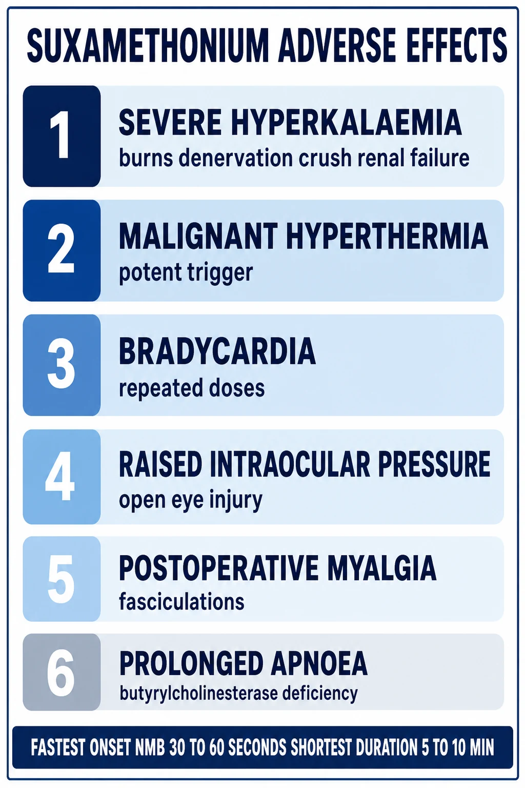
| The neuromuscular blockers | The rocuronium, the vecuronium, the suxamethonium | The direct receptor blockade (the prolonged paralysis — the MG patients are extremely sensitive to the non-depolarisers; the resistance to the suxamethonium) | The avoid; if the RSI, halve the rocuronium dose (the 0.6 to 1 mg/kg); the suxamethonium may be ineffective |
| The iodinated contrast | The CT contrast, the angiography | The unknown | The moderate — use with the caution; can precipitate a crisis |
| The penicillamine | The D-penicillamine (the Wilson, the RA) | The drug-induced MG (the antibody induction) | The absolute contraindication |
| The botulinum toxin | The cosmetic, the dystonia | The presynaptic ACh release blockade | The avoid |
| The steroids | The prednisolone (the high starting dose) | The steroid myopathy and the initial exacerbation | The use with the caution (the low-dose start, the titrate up) |
The crises — the rapid sequence of the management
The crisis management sequence
- The airway, the breathing, the circulation. The IV access, the oxygen, the cardiac monitoring.
- The respiratory assessment. The FVC, the NIF, the single-breath count, the bulbar assessment, the arterial blood gas.
- The early intubation if the FVC under 20 mL/kg, the NIF under -30 cmH2O, the bulbar weakness (the aspiration), or the rapid progression. Use the modified RSI (the halved rocuronium 0.6 to 1 mg/kg; the suxamethonium may be ineffective — the MG patients are resistant to the suxamethonium and sensitive to the non-depolarisers).
- Hold the pyridostigmine. The secretions worsen the ventilation; the cholinergic crisis is a risk. Hold until the patient is improving.
- Stop the offending drugs. The aminoglycosides, the beta-blockers, the magnesium, the fluoroquinolones, the macrolides. Switch the antibiotics (the cephalosporin, the carbapenem).
- Search for and treat the precipitant. The infection (the sputum, the urine, the blood cultures), the aspiration, the recent surgery, the pregnancy.
- The immunomodulation. The plasma exchange (the 5 sessions over 1 to 2 weeks, preferred in the severe and the MuSK) OR the IVIG (the 2 g/kg over 5 days, preferred in the moderate and the line-avoidance). Choose one — both are equivalent. Do NOT give both.
- The corticosteroids — the low-dose start. The prednisolone 10 to 20 mg, titrate up by 5 mg every 2 to 3 days to the target 0.75 to 1 mg/kg, ONLY after the airway is secured. The high-dose start worsens the MG.
- The thymoma screen. The contrast chest CT.
- The long-term plan. The prednisolone plus a steroid-sparing agent (the azathioprine or the mycophenolate in the AChR-MG; the rituximab in the MuSK-MG); the thymectomy if the thymoma, or the non-thymomatous AChR-MG (the MGTX criteria).
- The extubation. Only when the FVC over 20 mL/kg, the NIF under -40 cmH2O, the bulbar strength recovered, the secretions manageable, and the immunomodulation has taken effect (the 2 to 3 weeks). A tracheostomy is considered if the prolonged ventilation (over 2 to 3 weeks) is anticipated.
The Clinical Pearls — the high-yield exam points
[1] [1] [1] [1] [1] [1] [1] [1] [1] [1] [1] [1] [1] [1] [1] [1] [1] [1]Exam practice
SAQ — Myasthenic crisis with impending respiratory failure
10 minutes · 10 marks
A 38-year-old woman with known AChR-antibody-positive myasthenia gravis presents with worsening ptosis, diplopia, dysphagia and limb weakness over 3 days, precipitated by a chest infection. She has slurred speech, a weak cough and a nasal voice. FVC has fallen from 35 to 17 mL/kg (best of three), NIF −28 cmH2O, single-breath count 12. She takes pyridostigmine 90 mg five times daily.
SAQ — Plasma exchange vs IVIG and the drug precipitants
10 minutes · 10 marks
A 55-year-old man with AChR-positive myasthenia gravis is intubated for myasthenic crisis. The team is deciding between plasma exchange and IVIG. He has a history of difficult venous access and mild chronic kidney disease.
Red flags
References
- [1]Gilhus NE Myasthenia gravis: subgroup classification and therapeutic strategies. Lancet Neurology, 2015.PMID 26376969