ICU · neurocritical-care
Myasthenia Gravis Crisis — Comprehensive ICU Management
Also known as Myasthenic crisis · Myasthenia gravis exacerbation · Cholinergic crisis · Neuromuscular respiratory failure · AChR myasthenia gravis · MuSK myasthenia gravis · Acetylcholine receptor antibody disease · Fatigable weakness · Plasmapheresis for myasthenia · IVIG for myasthenia
Myasthenic crisis is an acute life-threatening exacerbation of myasthenia gravis (MG) severe enough to compromise ventilation (impending or actual respiratory failure) or airway protection (severe bulbar weakness). MG is a T-cell-dependent, antibody-mediated autoimmune disorder of the POSTSYNAPTIC neuromuscular junction (NMJ): pathogenic IgG autoantibodies attack the muscle membrane and either destroy acetylcholine receptors (AChR) or disrupt related proteins (MuSK, LRP4), producing FATIGABLE (fluctuating, use-dependent) weakness. Crisis is triggered in ~70% of cases by an identifiable precipitant — infection is 1 (pneumonia, aspiration, upper respiratory tract infection), followed by surgery, pregnancy/peripartum, tapering of immunosuppression, and drugs that impair NMJ transmission (aminoglycosides, beta-blockers, magnesium, fluoroquinolones/macrolides, neuromuscular blockers, iodinated contrast). Two antibody phenotypes behave differently in crisis: AChR-MG (limb + ocular + bulbar, thymoma-associated, responds to acetylcholinesterase inhibitors and thymectomy) versus MuSK-MG (selective bulbar/respiratory/facial weakness, SPARES the eyes and limbs, relative resistance to pyridostigmine, more frequent and more severe crises). The intensivist's priorities are: (1) recognise fatigable weakness + impending respiratory failure EARLY; (2) monitor bedside respiratory function every 4-6 h — FVC, NIF (MIP), single breath count — using the SAME thresholds as Guillain-Barre syndrome; (3) intubate electively (not as an emergency) when FVC <15 mL/kg, NIF < -30 cmH2O, or bulbar weakness threatens the airway; (4) distinguish myasthenic crisis (UNDER-treated, 'dry') from cholinergic crisis (OVER-treated with anticholinesterase, 'wet' with SLUDGE); (5) treat with IVIG 0.4 g/kg/day x 5 days OR plasma exchange (PLEX, 5 sessions over 1-2 weeks) — equally effective — plus identify and remove the trigger and hold/limit pyridostigmine; (6) plan thymectomy for thymoma (always) or AChR-positive non-thymomatous generalised MG; (7) rigorously avoid MG-worsening drugs. Mortality has fallen from 30-40% (1960s) to 4-8% with modern ICU care.
On this page & tools
Your progress
Saved locally on this device.
Target exams
Red flags
Overview
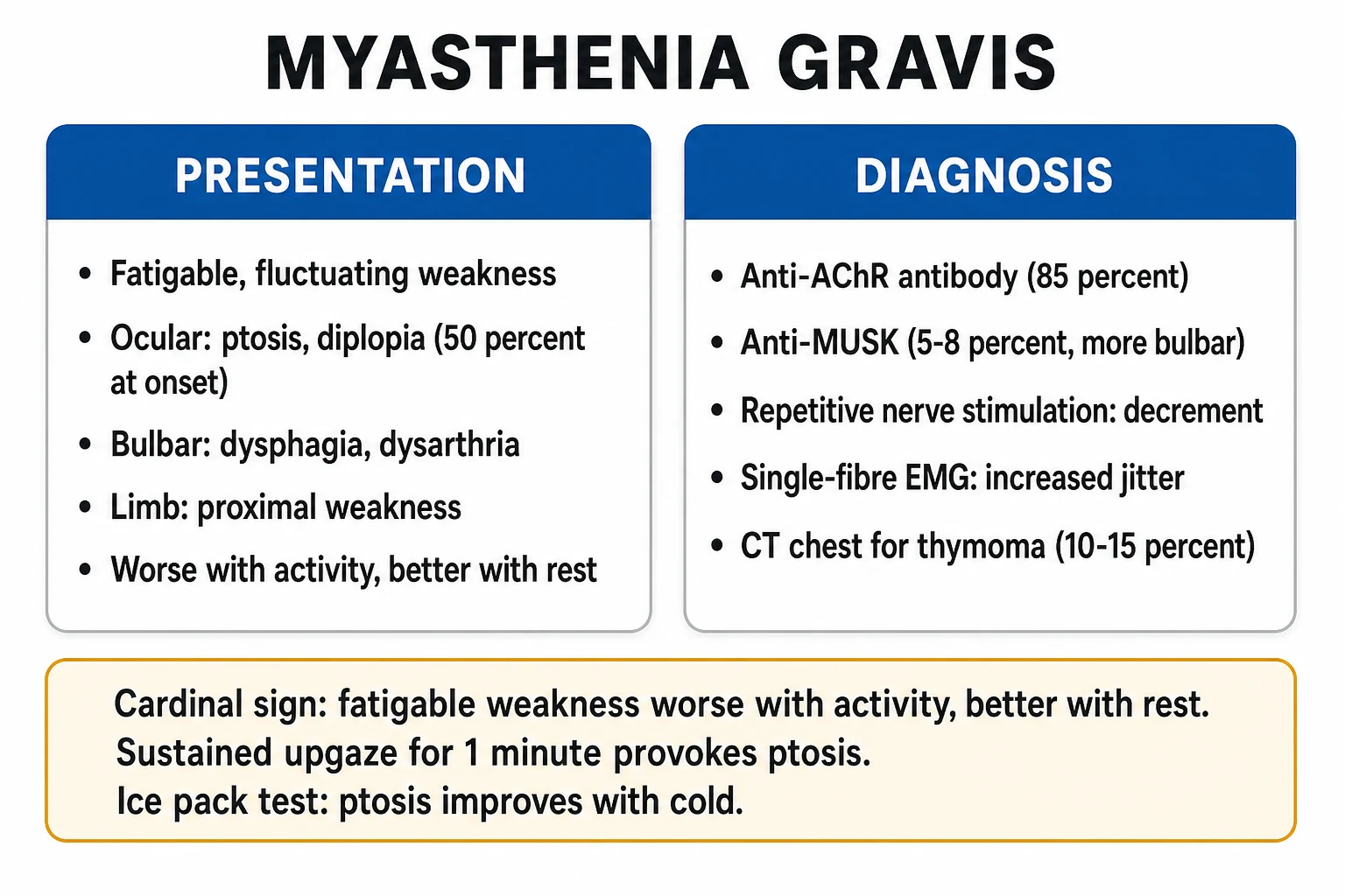
Myasthenia gravis (MG) is the most common primary disorder of neuromuscular transmission, with a prevalence of ~20 per 100,000 and a bimodal age distribution (young women, older men). The defining clinical feature is fatigable (use-dependent) weakness — weakness that worsens with sustained activity and improves with rest. Between 15% and 20% of MG patients will experience at least one myasthenic crisis in their lifetime; the incidence has fallen with modern immunotherapy but crisis remains a true neurocritical-care emergency that tests the intensivist's ability to (1) recognise an evolving neuromuscular respiratory failure, (2) time intubation correctly, (3) deploy short-term immunomodulation, and (4) avoid the long list of drugs that worsen NMJ transmission. The intensivist encounters MG in three scenarios: the known MG patient deteriorating on the ward (the commonest — identify the trigger, monitor respiratory function, treat), the undiagnosed patient presenting with respiratory failure of unclear cause (think MG when weakness is fatigable, bulbar, or ocular; check for ptosis, diplopia, facial weakness, and a decremental response on repetitive nerve stimulation), and the intubated MG patient (prolonged ventilation, weaning dictated by disease response to IVIG/PLEX, thymectomy planning).[2][3]
A critical conceptual point that distinguishes MG crisis from Guillain-Barre syndrome (GBS): in GBS the weakness is progressive and monophasic (it will keep getting worse, then recover), whereas in MG the weakness is fatigable and fluctuating — it varies with activity, time of day (worse in the evening), infection, and drugs. This means MG crisis can sometimes be rapidly reversed (remove the offending drug, rest, treat the infection, give IVIG/PLEX) in a way that GBS cannot. But it also means a MG patient can decompensate quickly and recover quickly, so thresholds and trends matter.[4]
Pathophysiology — AChR versus MuSK phenotypes
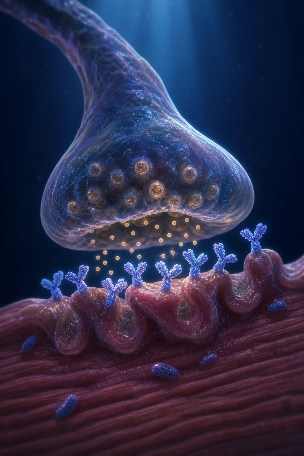
MG is a T-cell-dependent, B-cell-mediated, antibody-mediated autoimmune disease of the postsynaptic muscle membrane at the NMJ. The autoantibodies are IgG, they cross the placenta (transient neonatal MG), and their target determines the clinical phenotype. [1]
AChR-MG versus MuSK-MG — the two phenotypes that matter in crisis
| Feature | AChR-MG (acetylcholine receptor antibodies) | MuSK-MG (muscle-specific tyrosine kinase antibodies) |
|---|---|---|
| Frequency | ~85% of generalised MG | 5-8% of generalised MG (up to 30-40% of AChR-negative MG) |
| Antibody target | Postsynaptic nicotinic ACh receptor (destroys/speeds turnover of receptors + activates complement → simplified postsynaptic folds + reduced receptor density) | MuSK protein (the receptor tyrosine kinase that clusters AChRs at the junction during development and maintenance) — NO complement-mediated destruction (hence relatively preserved junctional architecture) |
| Typical phenotype | OCULAR onset (ptosis, diplopia) → generalises to LIMB, BULBAR, RESPIRATORY. Fatigable, fluctuating. Thymic abnormalities common (thymoma 10-15%, thymic hyperplasia 65%) | SELECTIVE BULBAR + RESPIRATORY + FACIAL weakness (dysarthria, dysphagia, tongue/facial weakness, respiratory failure) with RELATIVE SPARING of ocular and limb muscles |
| Respiratory failure pattern | Limb weakness often parallels respiratory decline — FVC falls gradually | Respiratory failure may be DISPROPORTIONATE to limb weakness — patients look "too well" to be in crisis then crash. Bulbar crisis is the rule |
| Pyridostigmine response | GOOD (symptomatic relief) | POOR or even worsens (cholinergic excess can predominate) — MuSK patients often cannot tolerate pyridostigmine |
| Thymus | Thymoma / hyperplasia common — thymectomy beneficial | Thymus usually NORMAL — thymectomy NOT beneficial |
| Crisis frequency | ~15-20% lifetime | HIGHER — more frequent and more severe crises, disproportionate bulbar/respiratory involvement |
| Best acute therapy | IVIG or PLEX (equally effective) | PLEX often preferred; rituximab increasingly used for long-term control |
| Sex / age | Bimodal: young women (20-30s), older men (60-70s) | Predominantly women, age 30-50 |
A third, rarer antibody — anti-LRP4 — accounts for a minority of double-seronegative cases and behaves more like mild AChR-MG. "Seronegative" MG (no detectable antibody on standard assays) may represent low-titre AChR-MG detectable only on cell-based assays. The intensivist should confirm antibody status whenever possible because it dictates both acute management (MuSK → favour PLEX, de-escalate pyridostigmine) and long-term strategy (AChR + thymoma → surgery; MuSK → rituximab).[5]
Triggers of myasthenic crisis
A precipitant can be identified in ~70% of crises. Identifying and removing it is as important as immunomodulation — crisis will often not resolve while the trigger persists. [1]
Precipitants of myasthenic crisis
| Trigger | Relative frequency | Notes |
|---|---|---|
| Infection | #1 (~30-40% of crises) | Pneumonia (including aspiration), upper respiratory tract infection, urosepsis. Infection both increases metabolic demand and provokes immune activation. Treat promptly with MG-SAFE antibiotics. |
| Aspiration | Common and vicious cycle | Bulbar weakness → aspiration → pneumonia → worsened crisis → more bulbar weakness. Break the cycle by protecting the airway early. |
| Surgery / anaesthesia | Major precipitant | Especially thymectomy itself (paradoxically), cardiothoracic, upper abdominal. Stress, anaesthetic agents, NMBAs, opioids, and post-op infection all converge. Crisis may present 24-72 h post-op. |
| Drugs that impair NMJ transmission | ~20% | Aminoglycosides, beta-blockers (including topical timolol eye drops!), magnesium, fluoroquinolones, macrolides, calcium channel blockers, neuromuscular blockers, iodinated contrast, telithromycin, botulinum toxin, lithium, phenytoin. |
| Pregnancy / peripartum / puerperium | Risk periods | Worsening common in first trimester and postpartum; neonatal MG from transplacental IgG (transient, resolves in weeks). Magnesium for pre-eclampsia/eclampsia is DANGEROUS — use alternative tocolytic/anticonvulsant. |
| Tapering / non-adherence to immunosuppression | Common | Sudden steroid taper, missed mycophenolate/azathioprine, rituximab wearing off in MuSK-MG. |
| Emotional / physical stress, hyper-/hypothyroidism | Contributory | Thyroid disease coexists in 5-10% of MG — check TSH. |
| Immune checkpoint inhibitors (ICIs) | Emerging, severe | ICI-induced MG is often severe, bulbar, and overlaps with myositis — high crisis rate, consider early IVIG/PLEX + steroid + drug cessation. |
Clinical features and diagnosis
Clinical presentation — fatigable weakness
The hallmark of MG is fatigable weakness that improves with rest. The distribution is characteristic and helps distinguish MG from GBS, MND, and myopathies: [1]
- Ocular (>50% present here; ~15% stay purely ocular): ptosis (worse with sustained upgaze), diplopia, asymmetric. The "ice pack test" (cold reduces AChE activity — 2 minutes of ice on a ptotic eyelid improves ptosis) is a useful bedside screen. "Peek sign" (sclera visible above the limbus on gentle eye closure — weak orbicularis oculi).
- Bulbar: dysarthria (nasal voice, fading with prolonged speech), dysphagia, weak cough, nasal regurgitation of fluids, pooled secretions, fatigable chew. Bulbar weakness is the airway/airway-protection emergency.
- Facial: expressionless face, snarl, weakness of eye closure.
- Limb: proximal > distal; arms > legs typically. Fatigable on repeated testing (e.g., repeated shoulder abduction, rising from a chair).
- Respiratory: diaphragm and intercostal weakness → breathlessness, particularly on lying flat (orthopnoea from diaphragm load) or speaking full sentences; weak cough → inability to clear secretions.
- SPARED: sensation (normal), deep tendon reflexes (preserved — a key discriminator from GBS), bowel/bladder, pupillary responses. [1]
MG crisis versus its neuromuscular mimics in the ICU
| Feature | MG crisis | GBS (AIDP) | Motor neuron disease (ALS) | Critical illness polyneuromyopathy | Organophosphate poisoning |
|---|---|---|---|---|---|
| Onset/progression | Hours-days, fluctuating, fatigable | Days-4 weeks, progressive, monophasic ascending | Months-years, progressive, mixed UMN + LMN | Days-weeks, plateau in critical illness | Minutes-hours after exposure |
| Distribution | Ocular + bulbar + limb + respiratory (variable); MuSK = bulbar/respiratory | Ascending (legs → arms → cranial) | Asymmetric, bulbar, tongue fasciculations, hyperreflexia | Limb-predominant, distal, flaccid | Cholinergic flooding + weakness |
| Reflexes | Preserved | Absent (early) | Variable (often brisk = UMN) | Reduced/absent | Variable |
| Sensation | Normal | Often affected (paraesthesia) | Normal | Variable | Normal |
| Antibody | AChR / MuSK / LRP4 | Anti-ganglioside (GM1, GQ1b) | None | None | — |
| CSF | Normal | Albuminocytologic dissociation | Normal | Normal | — |
| Pupils | Normal | Normal | Normal | Normal | Miosis |
| Secretions | Variable (bulbar) | Variable | Variable | Minimal | Profuse (SLUDGE) |
| Key discriminator | Fatigable + reflexes preserved + AChR/MuSK antibody | Areflexia + ascending + CSF protein | UMN signs + tongue fasciculations + progressive | Critical illness context | Exposure history + miosis + low cholinesterase |
Diagnostic workup in crisis
In the intubated or deteriorating patient, treatment (airway + IVIG/PLEX) should never be delayed for confirmatory tests — MG crisis is a clinical diagnosis in a patient with known MG, or strongly suspected in a new presentation with fatigable weakness and preserved reflexes. The workup can proceed in parallel. [1]
Diagnostic workup for suspected myasthenic crisis
- BEDSIDE CLINICAL — (a) Known MG with worsening fatigable weakness + a new trigger → clinical diagnosis of crisis. (b) Unknown patient: look for fatigable ptosis/diplopia, bulbar weakness, preserved reflexes, normal sensation. Perform the ice pack test (improvement of ptosis after 2 min cold). Check for a "peek sign." Ask about diplopia worsening toward evening.
- BEDSIDE RESPIRATORY FUNCTION (every 4-6 h) — FVC (forced vital capacity), NIF/MIP (negative inspiratory force / maximum inspiratory pressure), single breath count (count out loud on one breath — normal >30). Trends more important than single values. (See monitoring section for thresholds.)
- ANTIBODY TESTING — anti-AChR (binding, blocking, modulating — ~85% sensitive in generalised MG) and anti-MuSK (in AChR-negative cases). Anti-LRP4 if both negative. NB: results take days — do NOT delay treatment. MuSK status changes management (de-escalate pyridostigmine, favour PLEX).
- ELECTRODIAGNOSIS (if diagnostic uncertainty / patient not in extremis) — (a) Repetitive nerve stimulation (RNS) at 3 Hz: a decremental response (>10% amplitude decline between 1st and 4th/5th stimulus) in a weak muscle supports MG. (b) Single-fibre EMG (sfEMG): markedly increased jitter and blocking — highest sensitivity (>95%) but low specificity and technically demanding.
- EDROPHONIUM (Tensilon) TEST — Largely HISTORICAL and now avoided in crisis: edrophonium (short-acting AChE inhibitor) should transiently improve myasthenic weakness. Risky (bradycardia, bronchospasm, cholinergic worsening, arrhythmia) — only with atropine drawn up and full monitoring. Replaced by antibody testing and ice pack test. Do NOT perform in an unstable patient.
- SEARCH FOR THE TRIGGER — chest X-ray/CT (pneumonia, aspiration, thymoma), blood/urine/sputum cultures, CBC, CRP, electrolytes (hypokalaemia/hypomagnesaemia/hypocalcaemia all worsen weakness), TSH (coexisting thyroid disease), pregnancy test in women. Review the medication list line-by-line for MG-worsening drugs.
- CT/MRI THORAX — for ALL patients (even known MG) to identify or re-stage thymoma, which changes management (surgical resection after stabilisation).
Myasthenic versus cholinergic crisis — "dry" versus "wet"
This distinction is a classic CICM/FFICM examination point and changes management fundamentally. Both produce weakness; the autonomic/cholinergic stigmata separate them. [1]
Myasthenic crisis (under-treated) versus cholinergic crisis (over-treated)
| Feature | Myasthenic crisis (insufficient NMJ transmission — TOO LITTLE acetylcholine effect) | Cholinergic crisis (excess acetylcholine — TOO MUCH, from excess anticholinesterase / pyridostigmine-neostigmine) |
|---|---|---|
| Mechanism | Insufficient ACh effect at postsynaptic receptor (autoimmune destruction + antibodies) — under-treated or triggered MG | Excess ACh at muscarinic + nicotinic receptors from anticholinesterase overdose — over-treated MG |
| "Dry versus wet" | "DRY" — anticholinergic autonomic picture | "WET" — cholinergic autonomic flooding (SLUDGE) |
| Secretions/sweat | Dry mouth, dry skin, dry eyes | Salivation, sweating, lacrimation, bronchial secretions |
| Pupils | Normal or dilated (mydriasis) | Constricted (miosis) |
| Heart rate | Normal or tachycardia | Bradycardia |
| Gut/bladder | Constipation, urinary retention | Diarrhoea, abdominal cramps, urination, emesis (SLUDGE) |
| Muscle fasciculations | Absent | Present (nicotinic overstimulation — muscle twitches, then flaccid paralysis from depolarising block) |
| Pallor | Dry, warm skin | Flushed, wet skin |
| Trigger | Infection, surgery, drug (aminoglycoside, etc.), taper | Overzealous pyridostigmine/neostigmine — usually >120 mg pyridostigmine every 3 h or IV neostigmine error |
| Response to edrophonium | IMPROVES (more ACh helps) | WORSENS (more ACh worsens the excess) — dangerous to test |
| Management | IVIG or PLEX + treat trigger + (may continue/adjust pyridostigmine) | STOP anticholinesterase immediately + supportive care + atropine for muscarinic effects; the patient may still need intubation because nicotinic depolarising block causes paralysis |
Respiratory monitoring — when to intubate
Respiratory failure in MG is due to weakness of the diaphragm, intercostal muscles, and accessory muscles (plus loss of an effective cough from bulbar weakness). The intensivist's central skill is timing elective intubation before the patient arrests. Bedside pulmonary function (spirometry / hand-held manometer) is far more reliable than arterial blood gases — hypercapnia is a LATE and ominous sign in neuromuscular respiratory failure (the patient is fatiguing to arrest). Do not wait for the PaCO2 to rise.[2][3]
The thresholds are the SAME as for Guillain-Barre syndrome — this is a deliberate, exam-testable parallel. Memorise one set of numbers and apply to both. [1]
Respiratory function monitoring in MG crisis — thresholds for intubation (same as GBS)
| Parameter | Normal | Concerning | ACTION THRESHOLD | Intubate |
|---|---|---|---|---|
| FVC (forced vital capacity) | 60-70 mL/kg | 25-35 mL/kg | <20 mL/kg = start preparing for intubation | <15 mL/kg = intubate |
| NIF / MIP (negative inspiratory force / maximum inspiratory pressure) | < -50 (more negative = stronger) | -30 to -40 | -25 to -30 = concerning | < -30 cmH2O (i.e., -20, -15) = intubate |
| MEP / PEF (maximum/peak expiratory force) | >80 cmH2O | 40-60 | <40 = weak cough | Intubate if cough ineffective (can't clear secretions) |
| Single breath count (count aloud on one breath) | >30 | 15-20 | <10 | Prepare / intubate (surrogate for FVC) |
| Bulbar weakness | Normal swallow/cough | Dysphagia, nasal voice, weak cough, pooled secretions | Any bulbar weakness + declining FVC = VERY HIGH RISK | Intubate early (aspiration risk overrides the number) |
| Arterial blood gas | Normal | — | Hypercapnia (PaCO2 rising) is LATE | Hypercapnia = patient is tiring — intubate immediately |
| Monitoring frequency | — | — | Every 4-6 hours (more often if declining) | Trend monitoring; trajectory beats absolute value |
NIV — a bridge, not a substitute, for intubation
Non-invasive ventilation (BiPAP) can be used as a short-term bridge in carefully selected patients (cooperative, able to protect airway, NO severe bulbar weakness, FVC in the 15-20 mL/kg "pre-intubation" zone). NIV rests fatigued respiratory muscles and may avert intubation in a minority. But NIV is contraindicated in significant bulbar weakness — the patient cannot manage secretions, and positive pressure will force air into a poorly protected airway, accelerating aspiration. Do not use NIV to delay inevitable intubation; if the trend is downward or bulbar weakness is present, intubate.[3]
Intubation technique — RSI in MG
Rapid sequence intubation in myasthenic crisis
- Paralytic choice — ROCURONIUM, not succinylcholine. MG patients have a reduced margin of safety at the NMJ and altered receptor populations: they are resistant to succinylcholine (fewer functioning AChRs to depolarise → need higher doses → unpredictable, prolonged phase II block and delayed recovery) and sensitive to non-depolarising NMBAs (fewer receptors to block → reduced dose required). Use rocuronium 1.0-1.2 mg/kg (reversible with sugammadex if needed). Avoid succinylcholine entirely.
- Reduce non-depolarising NMBA doses. MG patients need roughly half the usual intubating dose of rocuronium/vecuronium. Many intensivists use the FULL RSI dose to guarantee intubating conditions, accepting prolonged blockade in an already ventilated patient — but be aware recovery will be slow; have sugammadex available.
- Induction agent. Propofol, ketamine, etomidate, and thiopental are all NMJ-safe in standard doses; choose by haemodynamic profile. Beware cardiovascular depression in the volume-depleted septic patient.
- Ventilation. Most MG patients have normal lungs — ventilation is straightforward. Volume-control or pressure-support, PEEP 5, target SpO2 94-98%, PaCO2 35-45 mmHg (or permissive hypercapnia if ARDS coexists). Avoid auto-PEEP. Wean as NMJ function recovers (IVIG/PLEX take 1-2 weeks to work).
- Sedation/analgesia. Propofol or midazolam infusions; fentanyl for analgesia. Avoid aminoglycoside-containing regimens and high-dose magnesium-containing antacid/laxative protocols. Bowel regimen: avoid magnesium-containing products — use lactulose/PEG.
- Post-intubation. Once intubated, HOLD pyridostigmine/neostigmine (excess secretions, cholinergic risk, no additional benefit while ventilated and on immunomodulation). Secure the airway, confirm placement, start IVIG or PLEX, treat the trigger, and plan thymectomy if indicated.
Treatment — IVIG versus plasma exchange
Short-term immunomodulation is the disease-modifying therapy for crisis. The two options are equally effective — no trial has demonstrated superiority of IVIG over PLEX (or vice versa) for myasthenic crisis.[1][1]
IVIG versus plasma exchange for myasthenic crisis — equally effective
| Feature | IVIG | Plasma exchange (PLEX / plasmapheresis) |
|---|---|---|
| Efficacy in crisis | EQUIVALENT to PLEX (Mandawat comparative analysis — no significant outcome difference; clinical practice confirms equivalence) | EQUIVALENT to IVIG — historically the older, "gold standard" acute therapy |
| Dose / regimen | 0.4 g/kg/day IV x 5 days (total 2 g/kg) | 5 exchanges over 1-2 weeks (~1-1.5 plasma volumes per session, ~200-250 mL/kg total) — replace with albumin ± FFP |
| Onset | 1-2 weeks (slightly slower onset than PLEX in some series) | Often faster onset (days) — physically removes antibody |
| Mechanism | Neutralises/blockades autoantibodies, modulates complement, saturates Fc receptors (FcRn), immunomodulation | Physically removes circulating AChR/MuSK antibodies and complement from plasma |
| Advantages | Simple peripheral IV access, widely available, no central line, fewer procedural complications, no plasma donor exposure | Faster antibody clearance (favour for MuSK-MG and very severe/rapid crisis), avoids transfusion-related risks, no hyperviscosity |
| Disadvantages | Cost, thrombosis (hyperviscosity), AKI (sucrose-containing products), aseptic meningitis, headache, flu-like illness, rare anaphylaxis (IgA deficiency) | Requires central venous catheter (infection, thrombosis, pneumothorax), bleeding (citrate/anticoagulation, removal of clotting factors), hypotension/cramps from volume/calcium shifts, more nursing effort |
| MuSK-MG preference | Effective but MuSK patients sometimes respond less robustly | Often preferred for MuSK-MG (more complete antibody clearance) |
| Which to choose? | First choice in most centres (simpler, no central line, equivalent efficacy). Prefer if coagulopathy, difficult venous access preference, sepsis | Prefer if very severe/rapid crisis, MuSK-MG, IVIG contraindicated (IgA deficiency, severe renal failure, hyperviscosity), or IVIG unavailable |
| Corticosteroids | NOT acute monotherapy — may transiently WORSEN MG in first 5-10 days. Useful long-term immunosuppression; if used, co-administer with IVIG/PLEX and monitor | Same caveat |
Mandawat et al. — comparative analysis of IVIG versus PLEX in myasthenia gravis (PMID 21105079)
Study design
Retrospective comparative analysis — Nationwide Inpatient Sample (large US administrative database)
Population
Hospitalised adults with myasthenia gravis treated with IVIG or plasma exchange
Comparison
IVIG versus plasma exchange for acute MG exacerbation/crisis
Key finding
Both therapies effective; no consistent, clinically significant superiority of one over the other across outcomes. Selection bias likely (sicker patients triaged to PLEX)
Clinical bottom line
IVIG and PLEX are equally effective for myasthenic crisis — choose by availability, comorbidity, and phenotype (MuSK, severity), not by perceived superiority
Comprehensive ICU management protocol
Myasthenic crisis — comprehensive ICU management protocol
-
AIRWAY & BREATHING (first priority):
- Serial FVC + NIF + single breath count every 4-6 h (more often if declining). Bulbar assessment every shift.
- Intubate when FVC <15-20 mL/kg, NIF < -30 cmH2O, single breath count <10, or bulbar weakness threatens the airway. Elective > emergency.
- RSI with rocuronium 1.0-1.2 mg/kg (NOT succinylcholine); reduced-dose non-depolarising NMBA (MG patients sensitive). Sugammadex available.
- Standard lung-protective ventilation (most MG patients have normal lungs). PEEP 5, SpO2 94-98%, PaCO2 35-45 mmHg.
- HOLD pyridostigmine/neostigmine once intubated (excess secretions, cholinergic risk, no added benefit on IVIG/PLEX). [1]
-
SHORT-TERM IMMUNOMODULATION (disease-modifying):
- IVIG 0.4 g/kg/day x 5 days OR PLEX 5 sessions over 1-2 weeks — equally effective.
- Choose by availability, comorbidity, phenotype (MuSK → favour PLEX; severe/rapid → PLEX; straightforward AChR → IVIG).
- Screen IVIG: check IgA level (anaphylaxis in IgA deficiency), renal function (AKI risk), viscosity/thrombosis risk (hydrate, consider prophylactic anticoagulation).
- PLEX: central venous access, coagulation monitoring, calcium for citrate-induced hypocalcaemia, volume management.
- Onset: 1-2 weeks — set expectations with family; the patient may need prolonged ventilation. [1]
-
IDENTIFY AND TREAT THE TRIGGER:
- Infection (pneumonia, aspiration, URTI, UTI) — cultures, MG-SAFE antibiotics (cephalosporins, penicillins, carbapenems, vancomycin, linezolid, macrolide-free regimens). Avoid aminoglycosides, fluoroquinolones, macrolides.
- Review the medication chart line-by-line — STOP beta-blockers (switch to rate-control alternative), magnesium-containing products, offending antibiotics, iodinated contrast.
- Surgery — stabilise first; defer elective surgery.
- Pregnancy — involve obstetrics + neurology; avoid magnesium (use alternative for eclampsia prophylaxis).
- Check and correct electrolytes (K+, Mg2+, Ca2+ all affect NMJ), TSH. [1]
-
CORTICOSTEROIDS & LONG-TERM IMMUNOSUPPRESSION:
- Corticosteroids are NOT acute monotherapy — prednisolone 1-1.5 mg/kg/day can transiently WORSEN MG in the first 5-10 days (steroid-induced worsening). If introducing, do so WITH IVIG/PLEX cover and monitor closely (consider in ICU).
- Continue pre-existing long-term immunosuppression (azathioprine, mycophenolate, methotrexate, tacrolimus). Note these are slow-acting (months) and do not treat the acute crisis.
- Rituximab increasingly used for refractory disease and MuSK-MG (often dramatic, durable response). [1]
-
PYRIDOSTIGMINE STRATEGY:
- Hold while intubated/on IVIG-PLEX (see above).
- For non-intubated mild-moderate cases: continue or cautiously adjust oral pyridostigmine (60 mg q6h is typical). Do not escalate aggressively (>120 mg q3h risks cholinergic crisis).
- IV neostigmine = 1/30th the oral pyridostigmine dose (a common and dangerous dosing error).
- MuSK-MG: pyridostigmine often poorly tolerated — de-escalate; rely on immunotherapy. [1]
-
GENERAL ICU CARE:
- VTE prophylaxis (LMWH — MG patients are immobile; balance against PLEX anticoagulation).
- Stress ulcer prophylaxis (avoid magnesium-containing antacids — use PPI).
- Nutrition (early enteral; bulbar weakness → NG/NJ; assess aspiration risk).
- Bowel care: lactulose/PEG, NOT magnesium-containing laxatives.
- DVT, pressure-area care, physiotherapy, early mobilisation as able.
- Speech-language and dietetics for dysphagia rehabilitation as the patient improves. [1]
-
THYMECTOMY PLANNING (after stabilisation — see dedicated section). [1]
-
WEANING & EXTUBATION:
- Wean as NMJ function recovers (IVIG/PLEX effect, typically 1-2 weeks).
- Extubation criteria: FVC >15 mL/kg, NIF < -30 cmH2O (stronger = more negative), MEP >40 cmH2O, adequate cough, minimal secretions, intact bulbar function, passing SBT.
- Tracheostomy if prolonged ventilation (>10-14 days) — does not improve outcomes but improves comfort and weaning. Consider early tracheostomy discussion in MuSK-MG (often prolonged). [1]
-
MEDICATION SAFETY — DRUGS TO AVOID (see dedicated section). Every new prescription checked against the MG-safe list.
Thymectomy indications
Thymectomy in myasthenia gravis — who and when
| Indication | Recommendation | Timing |
|---|---|---|
| Thymoma (10-15% of AChR-MG) | Always resect (oncological indication — even if MG is mild; thymoma can be malignant) | After crisis stabilisation (surgery itself can precipitate/worsen crisis). Not during acute crisis. |
| AChR-positive, non-thymomatous generalised MG, age 18-65 | Recommended (MGTX trial — improved outcomes over 3-5 years: reduced QMG score, reduced immunosuppression requirement, lower hospitalisation rate) | Within 1-2 years of diagnosis ideally; after any acute crisis resolved. Steroid-sparing benefit accrues over years, not weeks. |
| MuSK-MG | NOT recommended (thymus usually normal; no benefit; MGTX did not include MuSK patients) | — |
| Pure ocular MG | Generally not recommended (limited evidence) | — |
| Age >65 or <18 | Individualise (less benefit, higher operative risk in elderly; juvenile MG special considerations) | — |
Drugs to avoid in MG — the essential list
This is one of the highest-yield exam topics and a frequent source of iatrogenic crisis. Memorise by mechanism and know the safe alternatives.[2][4]
Drugs that worsen MG — and safe alternatives
| Drug class | Examples (AVOID) | Mechanism | Safe alternative |
|---|---|---|---|
| Aminoglycosides | Gentamicin, tobramycin, amikacin, neomycin | Inhibit presynaptic Ca2+-dependent ACh release | Cephalosporins, penicillins, carbapenems, vancomycin |
| Fluoroquinolones | Ciprofloxacin, levofloxacin, moxifloxacin | Direct NMJ toxicity (black-box MG warning) | As above; doxycycline, linezolid |
| Macrolides | Azithromycin, clarithromycin, erythromycin | Decrease presynaptic ACh release | As above |
| Beta-blockers | Propranolol, metoprolol, timolol eye drops, labetalol | Blocks postsynaptic AChR; reduces ACh release | Rate-control with diltiazem/digoxin (use CCB cautiously); avoid ophthalmic beta-blockers |
| Magnesium | MgSO4 (eclampsia, arrhythmia, laxatives/antacids) | Inhibits presynaptic ACh release; antagonises Ca2+ | For eclampsia seizures: alternative agents, early delivery; for hypomagnesaemia correct cautiously |
| Calcium channel blockers | Verapamil, diltiazem (high dose) | Inhibit presynaptic Ca2+ entry → less ACh release | Beta-blocker-free rate control; use lowest effective dose if essential |
| Neuromuscular blockers | Rocuronium, vecuronium, succinylcholine, cisatracurium | Direct NMJ blockade (MG patients sensitive to ND-NMBAs; resistant/unpredictable to sux) | Minimise/avoid; if essential, reduced dose rocuronium + sugammadex reversal |
| Iodinated contrast | High-osmolar contrast | Historically worsens MG (varies by agent) | Use lowest necessary dose; pre-medicate; consider non-ionic low-osmolar |
| Others | Penicillamine (induces MG), telithromycin (black-box), lithium, phenytoin, chloroquine/H-CQ, procainamide, botulinum toxin | Various | Avoid; monitor if essential |
| Immune checkpoint inhibitors | Pembrolizumab, nivolumab, ipilimumab | Induce immune-mediated MG (often severe, bulbar, overlaps myositis) | Cautious use only; high index of suspicion; early IVIG/PLEX + steroid |
Clinical pearls
Key trials and evidence
MGTX trial — thymectomy in non-thymomatous AChR-MG (NEJM 2016)
Study design
Randomised, rater-blinded trial — 126 patients, 36 months (extended to 5 y)
Population
Adults 18-65 y, AChR-antibody-positive, non-thymomatous, generalised MG of <5 years duration (excluded MuSK-MG)
Arms
Extended transsternal thymectomy + prednisone versus prednisone alone
Primary outcome
Time-weighted quantitative myasthenia (QMG) score over 3 years — favoured thymectomy
Key findings
Lower QMG score, lower prednisone dose, fewer immunosuppressant requirements, fewer hospitalisations in thymectomy group — benefit sustained at 5 years
Clinical bottom line
Extended thymectomy + prednisone superior to prednisone alone for AChR-positive non-thymomatous generalised MG of recent onset. Does NOT apply to MuSK-MG, ocular-only MG, or elderly patients
Mandawat et al. — IVIG versus PLEX comparative analysis (PMID 21105079)
Study design
Retrospective comparative analysis — large US administrative database (Nationwide Inpatient Sample)
Population
Hospitalised adults with myasthenia gravis treated acutely with IVIG or plasma exchange
Comparison
IVIG versus plasma exchange for acute exacerbation/crisis
Key finding
Both effective; no consistent superiority of one over the other across outcomes. Selection bias (sicker patients triaged to PLEX) acknowledged
Clinical bottom line
IVIG and PLEX are EQUALLY EFFECTIVE for myasthenic crisis — choose by availability, comorbidity, and phenotype, not by perceived superiority
Sanders 2016 — international consensus guidance for MG (PMID 27358333)
Source
MGFA Task Force — 15 international experts, RAND/UCLA appropriateness methodology
Scope
Formal consensus guidance for symptomatic and immunosuppressive therapy, IVIG/PLEX, impending and manifest crisis, thymectomy, juvenile MG, MuSK-MG, MG in pregnancy
Crisis recommendations
Define impending vs manifest crisis; ICU for respiratory monitoring; IVIG (2 g/kg over 5 days) or PLEX (5 sessions) equally effective; identify/treat trigger; hold/avoid offending drugs
Clinical bottom line
The international reference standard for MG management — endorse both IVIG and PLEX as equally effective for crisis, with phenotype-tailoured choices
Exam practice — SAQs
SAQ — Myasthenic crisis with impending respiratory failure
10 minutes · 10 marks
A 38-year-old woman with known AChR-antibody-positive myasthenia gravis presents with worsening ptosis, diplopia, dysphagia and limb weakness over 3 days after a chest infection. FVC has fallen from 35 to 17 mL/kg, NIF −28 cmH2O, single-breath count 12. She takes pyridostigmine 90 mg five times daily.
Red flags
Prognosis
Myasthenic crisis — outcomes and timelines
| Outcome | Proportion / value | Notes |
|---|---|---|
| Mortality | 4-8% (down from 30-40% in the 1960s) | Modern ICU care, early intubation, IVIG/PLEX. Worse with: older age, pre-existing lung disease, aspiration pneumonia, delay to immunomodulation, MuSK-MG |
| Require intubation | Most patients in florid crisis | Elective intubation mortality far lower than emergency |
| Duration of ventilation | Median ~10-14 days | Reflects IVIG/PLEX onset (1-2 weeks). MuSK-MG often longer |
| Tracheostomy rate | 10-30% | Consider if ventilated >10-14 days; does not improve outcomes but improves comfort and weaning |
| Hospital length of stay | 2-4 weeks | Crisis is a multi-week illness |
| Full recovery to baseline | Most patients (weeks-months) | With immunotherapy + (where indicated) thymectomy |
| Recurrence of crisis | 10-40% | Higher if trigger not addressed, immunosuppression inadequate, or MuSK-MG |
| Long-term immunosuppression | Most patients | Azathioprine, mycophenolate, methotrexate, tacrolimus, rituximab (MuSK), eculizumab/razaclurzumab/FcRn inhibitors (refractory AChR) |
Prognostic principles: (1) Early elective intubation + prompt IVIG/PLEX + trigger control is the formula for survival. (2) The decline in mortality from 30-40% to 4-8% over six decades reflects ICU respiratory support more than any single drug — the intensivist's airway and ventilation management IS the disease-modifying therapy. (3) Older patients and those with MuSK-MG or intercurrent aspiration pneumonia have the worst outcomes. (4) Most survivors return to baseline or near-baseline function within weeks to months with adequate long-term immunotherapy and (where indicated) thymectomy.[2][3][5]
Related topics: Guillain-Barre syndrome, Non-invasive ventilation, Therapeutic plasma exchange, Critical illness polyneuromyopathy, ICU-acquired weakness (CIP/CIM). [1]
References
- [1]Sanders DB, Wolfe GI, Benatar M, et al. International consensus guidance for management of myasthenia gravis: Executive summary Neurology, 2016.PMID 27358333
- [2]Bershad EM, Feen ES, Suarez JI. Myasthenia gravis crisis South Med J, 2008.PMID 18176295
- [3]Lacomis D. Myasthenic crisis Neurocrit Care, 2005.PMID 16377829
- [4]Chaudhuri A, Behan PO. Myasthenic crisis QJM, 2009.PMID 19060020
- [5]Gilhus NE. Myasthenia Gravis N Engl J Med, 2016.PMID 28029925