ICU · Neurocritical care / post-arrest
Neuro-Prognostication After Cardiac Arrest — The 72-Hour Multimodal Rule
Also known as Neuro-prognostication · Post-anoxic coma · Post-cardiac-arrest prognostication · Anoxic brain injury · 72-hour rule · Somatosensory evoked potentials · SSEP · N20 · NSE · Self-fulfilling prophecy · TTM2 · Multimodal prognostication · ERC/ESICM
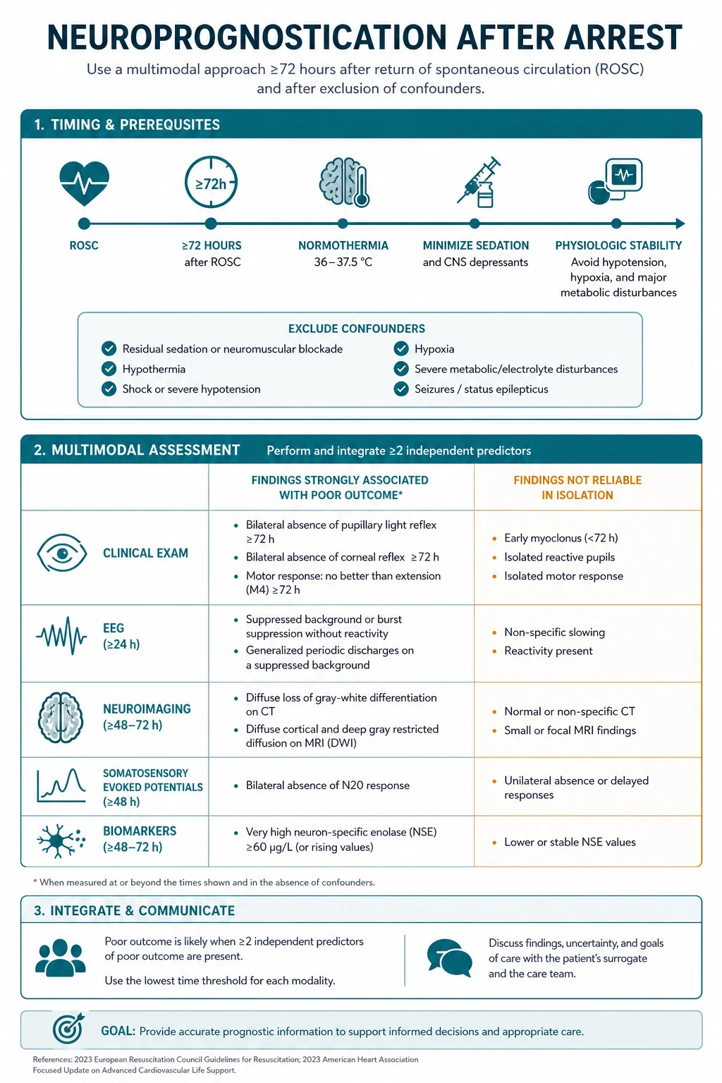
The neuro-prognostication after a cardiac arrest is deferred until at least 72 hours after the return of the spontaneous circulation, with the patient normothermic and off the sedation. The principle is the MULTIMODAL assessment — no single predictor determines the prognosis, because the falsely pessimistic prediction creates the self-fulfilling prophecy (the early withdrawal of the care). The predictors (examined together): the absent brainstem reflexes (the pupillary, the corneal, the vestibulo-ocular), the absent motor response to pain, the bilateral absent N20 on the somatosensory evoked potentials (the most reliable single test), the suppressed or the burst-suppression EEG, the NSE over 60, and the MRI diffusion anoxic pattern. The TTM trial (33 vs 36, no difference) and the TTM2 trial (33 vs the normothermia, no benefit) shifted the practice toward the fever avoidance rather than the therapeutic hypothermia. The sedatives confound the examination (the slower metabolism and the washout under the hypothermia).
On this page & tools
Your progress
Saved locally on this device.
8 MCQs with explanations
Target exams
Overview & definition
The neuro-prognostication after a cardiac arrest predicts the neurological outcome of the patient who remains comatose after the return of the spontaneous circulation (the ROSC). The outcome ranges from the full recovery to the persistent vegetative state and the death. The prognostication is the most ethically charged judgement in the intensive care — the falsely pessimistic prediction leads to the early withdrawal of the life-sustaining therapy (the WLST), which causes the death — the self-fulfilling prophecy. The two principles: wait until at least 72 hours (with the normothermia and off the sedation), and the multimodal assessment (no single predictor).[1]
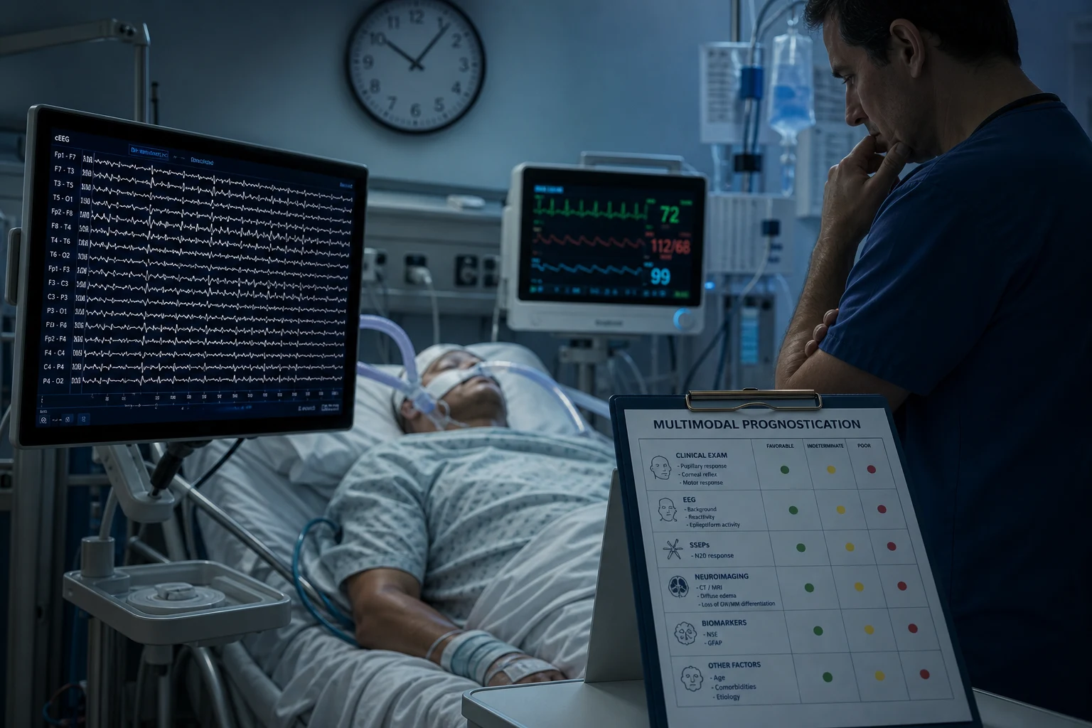
The timing and the principle
- Wait until at least 72 hours after the ROSC. The earlier prediction is unreliable (the sedatives, the hypothermia, the metabolic disturbance, and the delayed recovery confound the examination).[1]
- The normothermia (avoid the fever — the fever worsens the anoxic injury; the TTM2 trial shifted the practice toward the fever avoidance rather than the hypothermia).[1]
- Off the sedation — ensure the adequate washout before the prognostication. The sedatives (especially the slower metabolism and the washout under the hypothermia and in the renal or the hepatic impairment) confound the clinical examination. Wait at least 24 hours after the cessation of the sedation if the examination is pessimistic.[1]
- The multimodal assessment — examine the multiple predictors together (the clinical, the SSEP, the EEG, the biomarkers, the imaging). NO single predictor should determine the prognosis (the risk of the falsely pessimistic prediction).[1]
- Avoid the self-fulfilling prophecy — the falsely pessimistic prediction leads to the early WLST, which causes the death and "confirms" the prediction. The bias is the central pitfall. The conservative, the multimodal, the repeatable approach is essential.[1]
The TTM context
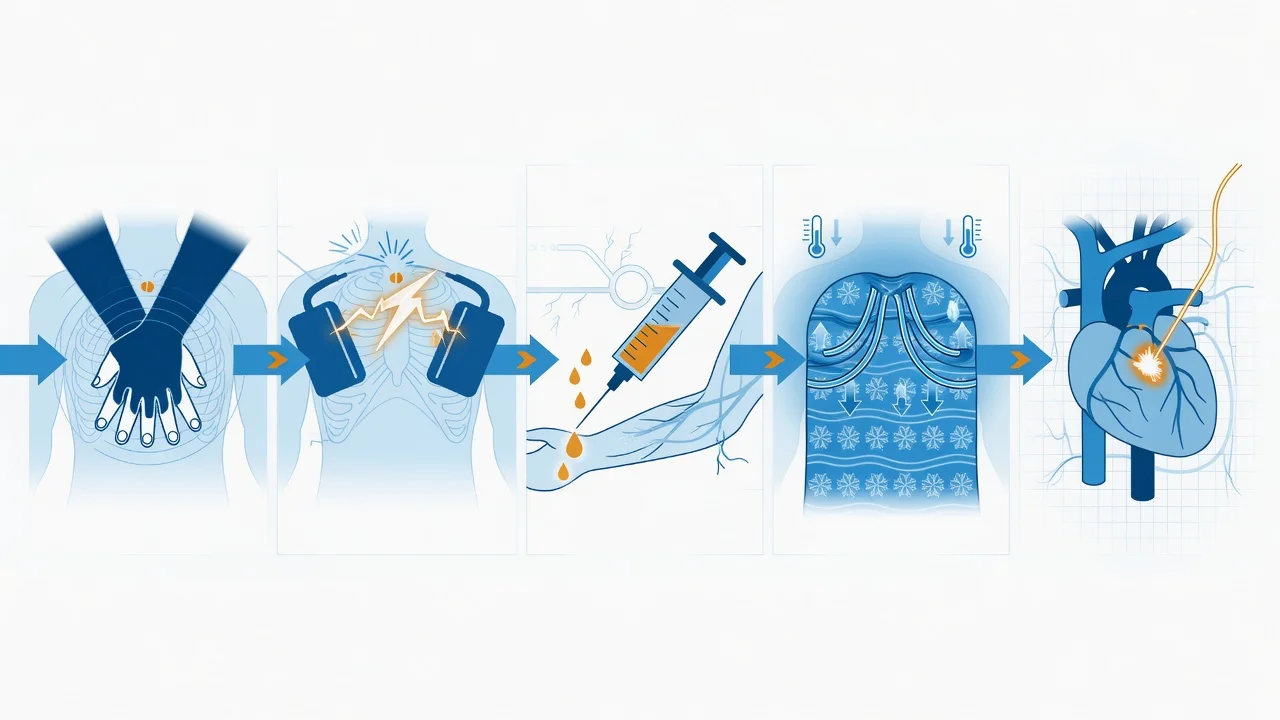
- The TTM trial (NEJM 2013) — the 33 degrees C vs the 36 degrees C showed no difference in the mortality or the neurological outcome. The routine cooling to 33 degrees was abandoned in favour of the targeted temperature management at 36 degrees.[1]
- The TTM2 trial (NEJM 2021) — the 33 degrees C vs the normothermia (the fever avoidance, about 37.8 degrees) showed NO benefit of the hypothermia. The practice shifted to the normothermia with the active fever avoidance rather than the routine hypothermia. The hypothermia (the 33 degrees) is still used by some centres for the specific indications, but the fever avoidance is the standard.[1]
- The implication for the prognostication: under the hypothermia, the sedative metabolism and the washout are slower (the propofol, the fentanyl, the midazolam accumulate) — the examination is confounded for longer. Wait longer after the hypothermia before the prognostication.[1]
The predictors (examined together)
1. The clinical examination
- The absent brainstem reflexes — the pupillary (the fixed and dilated), the corneal, the cough, and the vestibulo-ocular (the caloric) reflexes. The absent pupillary and the absent corneal at 72 hours (off the sedation, normothermic) are the poor prognostic signs (the high specificity).[1]
- The absent motor response to the pain (no withdrawal, no extensor, no flexor) at 72 hours — a poor sign, but the low specificity (the sedatives, the metabolic disturbance confound it).[1]
- The myoclonus — the myoclonic status epilepticus (the continuous, the generalized, the within 72 hours) is a poor prognostic sign. Distinguish from the transient myoclonus (the benign). The status myoclonus is NOT used alone.[1]
2. The somatosensory evoked potentials (SSEP) — the most reliable single test
- The bilateral absent N20 (the cortical response to the median nerve stimulation) at 24 to 72 hours is the most reliable single predictor of the poor outcome — the high specificity, the low false-positive rate. The N20 is resistant to the sedatives (unlike the EEG and the clinical examination). The bilateral absent N20 (with the median nerve stimulation) is a robust poor-prognosis sign.[1]
- The present N20 does NOT predict the good outcome (the low sensitivity).[1]
3. The EEG
- The highly malignant patterns — the suppressed background (with the periodic discharges), the burst-suppression, the status epilepticus, the unreactive background. These are the poor prognostic signs but the LOW specificity (the sedatives, the metabolic disturbance, the seizures confound it). The EEG is never used alone for the prognostication.[1]
- The benign patterns — the reactive background, the normal voltage — predict the better outcome.[1]
- The continuous EEG (cEEG) detects the non-convulsive seizures and the status epilepticus (the treatable, the reversible).[1]
4. The biomarkers
- The NSE (the neuron-specific enolase) — a high level (over 60 µg/L at 24 to 72 hours) is a poor prognostic sign. The haemolysis falsely raises the NSE (the caveat). The sensitivity and the specificity are lower than the SSEP.[1]
- The neurofilament light chain (NfL) — a newer, a more sensitive biomarker (the plasma and the CSF); emerging.[1]
5. The imaging
- The brain MRI (the DWI) — the diffuse the cortical, the basal-ganglia, and the thalamic the diffusion restriction (the anoxic pattern) is a poor prognostic sign. The brain CT may show the sulcal effacement and the loss of the grey-white differentiation (a late sign).[1]
Red flags
The ERC-ESICM 2021 algorithm — the multimodal, deferral-first approach
The 2021 ERC-ESICM guidelines on post-resuscitation care (Nolan, Sandroni, et al.) consolidated the modern approach: prognostication is a physician-led, multidisciplinary, multimodal process, deferred to at least 72 hours after ROSC, performed only after the exclusion of confounders (residual sedation/analgesia, hypothermia, metabolic derangement, severe electrolyte disturbance, neuromuscular blockade, hypotension/hypoxaemia), and never relies on a single predictor. The algorithm explicitly tolerates uncertainty: when findings are equivocal or discordant, the recommendation is to wait, re-evaluate, and repeat rather than to conclude.[1][2]
The five-step ERC-ESICM 2021 algorithm
ERC-ESICM 2021 multimodal prognostication algorithm — comatose survivor of cardiac arrest
Step 1 — Establish eligibility (≥72h after ROSC)
Confirm the patient remains comatose (GCS <8, not following commands) at ≥72h after ROSC. CRITICAL pre-conditions BEFORE any prognostication: (1) patient is NORMOTHERMIC (core temperature >36°C for ≥72h, no active fever); (2) sedation and analgesia have been OFF for adequate washout — at least 5 half-lives, ≥24h off midazolam/fentanyl/propofol, longer if hepatic/renal impairment or if TTM was used (slower metabolism under hypothermia); (3) NO residual neuromuscular blockade (train-of-four ≥4/4, or sufficient time since last dose); (4) exclude metabolic, endocrine, infectious, toxicological confounders — check Na+, Ca2+, Mg2+, glucose, ammonia, thyroid function, septic shock. If ANY confounder is present → DELAY prognostication, correct, and re-assess daily. The guideline explicitly states: prognostication before 72h or with active confounders is INVALID.
Step 2 — Multimodal clinical examination
A standardised neurological examination by an experienced clinician: (a) Brainstem reflexes — pupillary light reflex (use magnification or pupillometry; fixed dilated pupils >4 mm and unreactive is ominous), corneal reflex (cotton wisp), gag and cough reflex (suction catheter), oculocephalic (Doll’s eyes — ONLY if cervical spine cleared) and/or vestibulo-ocular (cold caloric — 50 mL ice water into each ear). (b) Motor response to painful stimulus ( supraorbital pressure, nailbed) — document best response. (c) Myoclonus — presence, distribution, duration; distinguish myoclonus status epilepticus (within 72h, generalized, continuous — POOR prognosis) from Lance-Adams myoclonus (later, in AWAKENED patients, action myoclonus — does NOT predict poor outcome). ABSENT pupillary AND corneal reflexes at ≥72h (normothermic, off sedation) = POOR prognosis with very high specificity (false-positive rate approaching zero).
Step 3 — Objective electrophysiology (SSEP ± EEG)
Order SSEP (median nerve stimulation, bilateral, cortical N20) at 24–72h (or later) — BILATERAL ABSENT N20 is the most reliable single predictor of poor outcome (false-positive rate ~0–3%, sedative-resistant). If SSEP is technically possible, this is the cornerstone test. Order EEG (preferably continuous cEEG for ≥24h) — the ACNS-standardised terminology classifies patterns; HIGHLY MALIGNANT patterns (suppression with periodic discharges, burst-suppression with identical bursts, status epilepticus, unreactive discontinuous background) are associated with poor outcome but LOW specificity in isolation — confounded by sedatives, seizures (treatable/reversible), and metabolic disturbance. EEG is NEVER used alone to declare poor prognosis. A reactive, continuous, normal-voltage background is REASSURING (high negative predictive value).
Step 4 — Biomarkers and imaging
NSE (neuron-specific enolase) at 48–72h: a level >60 µg/L supports poor prognosis (cut-off varies by assay — many centres use serially rising trend >60–80 µg/L). Caveats: haemolysis falsely elevates NSE (erythrocytes contain NSE) — check plasma-free haemoglobin/colour, draw carefully, process promptly; extracranial malignancies (small-cell lung cancer, neuroendocrine tumours) also elevate NSE. Emerging biomarkers: NfL (neurofilament light chain) — higher sensitivity and specificity in pooled analyses, plasma now available; S100B — less used. IMAGING: brain MRI with DWI/ADC (the most sensitive modality) — diffuse cortical, basal ganglia, thalamic, and hippocampal diffusion restriction indicates severe anoxic injury; brain CT may show sulcal effacement, loss of grey-white differentiation, and reversal/haze of the denser cerebellum/brainstem relative to the oedematous cortex (the “reversal sign” — very late). Imaging supports but rarely adjudicates the prognosis alone.
Step 5 — Synthesise — multimodal, conservative, repeatable
Integrate ALL modalities. NO single predictor (not even bilateral absent N20) determines the prognosis in isolation. Three prognostic categories: (a) LIKELY POOR outcome — concordant multiple predictors (absent brainstem reflexes + bilateral absent N20 + highly malignant EEG + NSE >60 + diffuse MRI diffusion restriction) at ≥72h, normothermic, off sedation. (b) LIKELY GOOD outcome — early awakening, reactive EEG, present N20, low NSE, normal MRI. (c) UNCERTAIN — discordant or incomplete predictors. The guideline is explicit: UNCERTAINTY mandates WAITING and REPEATING (re-examine at 96h, 120h, even 7 days; recheck NSE trend; repeat SSEP; re-image). The conservative approach protects against the SELF-FULFILLING PROPHECY. Engage the family EARLY, document the reasoning, and involve a second senior clinician. The decision to withdraw life-sustaining therapy (WLST) is multidisciplinary and never rests on a single test.
Timing in detail — why 72 hours, and what deferral actually requires
The 72-hour threshold is the empirical floor, not a ceiling. The original rationale traces to pre-TTM cohort studies (Zandbergen 1998, the seminal Lancet pooled analysis) showing that the false-positive rate of individual predictors fell to acceptable levels only after 72h, and that delayed awakening (patients waking up after 72h) became uncommon.[1] Two things changed after 2013:
- The TTM era (33°C) prolonged drug clearance. Hypothermia reduces hepatic and renal clearance by ~25–30% and prolongs the half-life of propofol, midazolam, fentanyl, and neuromuscular blockers. In the original 36°C trials, sedation washout after 72h was usually adequate; in patients cooled to 33°C, sedation was still detectable at 72h. The pragmatic response was to defer prognostication to at least 72h after RETURN TO NORMOTHERMIA, not 72h after ROSC — a deferral that may push formal assessment to 4–5 days post-arrest.[4]
- TTM2 (2021) normalised the timeline. Because TTM2 demonstrated no benefit of 33°C over normothermia with active fever avoidance, most centres now maintain patients at ~37°C (with aggressive antipyretic control), so sedation metabolism is faster and the 72h-from-ROSC threshold is again reasonable.[3]
Delayed awakening — the common clinical reality
Up to 20–30% of patients awaken after 72h, and ~10% awaken after 7 days (Paul et al., Parisian registry). Predictors of delayed awakening include: longer CPR duration, initial non-shockable rhythm, higher lactate, older age, renal/hepatic dysfunction, and use of TTM at 33°C. The practical implication: an unreactive patient at 72h is NOT necessarily destined for poor outcome — proceed to multimodal assessment, and if discordant, wait.[11]
The confounders checklist (must be cleared BEFORE prognostication)
Confounders that invalidate the post-arrest neurological examination — exclude ALL before prognostication
| Confounder | Mechanism of confounding | How to exclude |
|---|---|---|
| Residual sedation (propofol, midazolam, fentanyl, ketamine) | Suppresses EEG background, abolishes motor response, blunts brainstem reflexes | Stop infusion; wait ≥5 half-lives (≥24h for most agents; longer post-hypothermia, in hepatic/renal failure). Document time off. |
| Neuromuscular blockade | Abolishes motor response, blunts gag/cough, may mimic brain death | Train-of-four ≥4/4; or ≥24h since last dose (rocuronium/succinylcholine/vecuronium) |
| Hypothermia (<36°C) | Slows drug clearance; itself depresses neurological function | Normothermic (>36°C) for ≥72h before prognostication |
| Hypotension (MAP <65) | Reduces cerebral perfusion, confounds EEG and exam | MAP ≥65 mmHg, no high-dose vasopressors; lactate normalising |
| Hypoxaemia / hypercarbia / hypocarbia | Cerebral injury, vasoconstriction (low PaCO2) | PaO2 >80 mmHg, PaCO2 35–45 mmHg |
| Metabolic (Na+, Ca2+, Mg2+, glucose, ammonia) | Each can produce or mimic coma | Normalise all; check ammonia and thyroid function |
| Seizures / status epilepticus | Post-ictal state, ongoing non-convulsive status (cEEG to exclude) | Continuous EEG; treat seizures before prognosticating |
| Sepsis / infection | Encephalopathy, hypotension | Cultures, source control, antibiotics |
Clinical examination in detail — what each finding means
Brainstem reflexes — the most specific clinical findings
The absent pupillary light reflex at ≥72h (normothermic, off sedation) is the most specific clinical predictor of poor outcome (false-positive rate approaching 0%). Specificity of absent corneal reflex is similarly high. The combination of absent pupillary AND absent corneal reflexes at 72h has a positive predictive value for poor outcome of ~95–100%. Caveats: [1]
- Pupil size and reactivity are confounded by drugs — atropine (dilates), opioids (miosis), anticholinergics, topical mydriatics. Use automated pupillometry (Neuroptics NPi) when available — quantifies the constriction amplitude and velocity, more reproducible than the bedside flashlight assessment, and the Neurological Pupil index (NPi <3 is abnormal) is increasingly used in prognostication algorithms.
- Direct ocular trauma during CPR (peri-orbital haematoma, corneal abrasion) can confound the local findings — examine both sides.
- The vestibulo-ocular (cold caloric) reflex is the most robust of the brainstem reflexes: 50 mL ice water irrigated into each external auditory canal (after confirming intact tympanic membrane) should produce tonic eye deviation toward the irrigated ear. Absence bilaterally, after confirmation of normothermia, is highly specific for severe brainstem injury. [1]
Motor response — the least specific clinical finding
An absent or extensor (decerebrate) motor response to pain at ≥72h is associated with poor outcome, but the false-positive rate is unacceptably high (~10–25%) because it is exquisitely sensitive to sedation, neuromuscular blockade, and metabolic disturbance. Motor response is therefore NEVER used in isolation. A patient who withdraws or localises at 72h is, however, more likely to have a favourable outcome. [1]
Myoclonus — distinguish status from Lance-Adams
| Pattern | Onset | Characteristics | Prognosis |
|---|---|---|---|
| Myoclonus status epilepticus | Within 72h of ROSC | Continuous, generalised, often facial/shoulder; synchronous with EEG spikes or burst-suppression | POOR (specificity ~95%, but treat seizures first; never the sole criterion) |
| Chronic post-anoxic myoclonus (Lance-Adams) | Days–weeks, in AWAKENED patients | Action/intention myoclonus, cerebellar ataxia, triggered by movement | Does NOT predict poor outcome — patients may have good cognitive recovery |
The exam trap: a patient who has awakened and then develops action myoclonus has Lance-Adams syndrome, NOT myoclonus status — the distinction is clinical (awake vs comatose) and prognostically critical.[1]
EEG in detail — the ACNS terminology and what is malignant
Continuous EEG (cEEG) for at least 24h is the standard in modern post-arrest care — it detects non-convulsive seizures and status epilepticus (present in ~20–30% of comatose post-arrest patients), both of which are treatable and must not be mistaken for a terminal anoxic pattern. [1]
The American Clinical Neurophysiology Society (ACNS) standardised terminology classifies post-arrest EEGs into benign and malignant categories. The highly malignant EEG (per Sandroni/Sideris meta-analyses and the ERC-ESICM 2021 synthesis) carries a poor prognosis BUT with low specificity in isolation: [1]
EEG patterns after cardiac arrest — malignant vs benign (ACNS-standardised terminology)
| Category | Specific pattern | Implication | Specificity for poor outcome |
|---|---|---|---|
| Highly malignant | Suppression (<10 µV) with continuous or periodic discharges | Severe diffuse cortical injury | High (but confounded by sedation) |
| Highly malignant | Burst-suppression with identical bursts (highly periodic) | Cortical network destruction | High — more specific than non-identical bursts |
| Highly malignant | Status epilepticus (electrographic, after myoclonus or independently) | Poor, but treat the SE first; re-prognosticate | Moderate — sedation, hypoxia confound |
| Highly malignant | Unreactive discontinuous background with periodic discharges | Severe injury | Moderate |
| Malignant | Low-voltage unreactive background without periodic discharges | Concerning but less specific | Lower |
| Benign / REASSURING | Continuous, reactive, normal-voltage background with sleep architecture | Strong negative predictor — favours good outcome | n/a (high NPV) |
The reactivity test — and its limits
A reactive EEG (background amplitude/frequency changes with external stimulation — noise, pain, passive eye opening) has a strong negative predictive value — most reactive EEG patients awaken. An unreactive background is concerning but confounded by sedation, and inter-rater agreement on reactivity is only moderate. The most robust single EEG feature for poor prognosis is suppression with periodic discharges or burst-suppression with identical bursts.[7]
Treating seizures does not always save the prognosis
Post-anoxic status epilepticus is frequently refractory. The STEPP study and the systematic review by Eertmans et al. (2022) show that even with aggressive antiseizure therapy (levetiracetam, valproate, lacosamide, propofol, ketamine, midazolam infusions, occasionally barbiturates), the outcome remains poor in the majority — but a meaningful minority (~15–20%) achieve good neurological outcome. The implication: treat the seizures, complete the multimodal assessment, and do not equate post-anoxic SE with futility.[12]
SSEP (N20) in detail — the most reliable single test
The median nerve somatosensory evoked potential records the cortical N20 response (over the contralateral parietal cortex, referenced to Fz or the contralateral ear) to electrical stimulation of the median nerve at the wrist. The bilateral absence of N20 (with preserved brachial plexus and cervical cord potentials to confirm the stimulus was delivered) at 24–72h or later is the single most reliable predictor of poor outcome after cardiac arrest: [1]
- False-positive rate ~0–3% in pooled analyses — i.e., the rare patient with bilateral absent N20 who nevertheless awakens.
- Sedative-resistant — N20 is preserved under anaesthetic doses of propofol, midazolam, and even barbiturates. This makes SSEP the test of choice when the clinical examination and EEG are confounded by residual sedation.[5]
- Resistant to hypothermia — Leithner et al. confirmed that bilateral absent N20 retains its predictive value even when recorded during hypothermia (33°C), although the guideline still recommends recording at normothermia to maximise waveform quality.
- Low sensitivity — a PRESENT N20 does not predict a good outcome (many patients with present N20 still have poor outcome). SSEP is therefore a high-specificity/low-sensitivity test — useful for confirming poor prognosis, not for ruling it out.
- Pitfalls — pre-existing peripheral neuropathy (diabetic, alcoholic, Guillain-Barré), severe limb oedema, electrical interference, and improper electrode placement can abolish N20 spuriously. Always confirm the peripheral (Erb’s point) and cervical cord (N13) potentials are intact — if these are absent, the test is non-diagnostic.
Biomarkers in detail — NSE, NfL, S100B
Biomarkers of anoxic brain injury after cardiac arrest — NSE, NfL, S100B
| Biomarker | Source | Optimal timing | Cut-off for poor prognosis | Pros | Cons / caveats |
|---|---|---|---|---|---|
| NSE (neuron-specific enolase) | Plasma (serum also, but plasma preferred) | 48–72h | >60 µg/L (assay-dependent; many use rising trend >60–80) | Widely available; well-validated | Falsely elevated by haemolysis (erythrocytes contain NSE) and extracranial small-cell/neuroendocrine tumours; assay variability |
| NfL (neurofilament light chain) | Plasma (CSF also) | 48–72h (and later) | Cut-off assay-dependent (~>80–400 pg/mL on Simoa) | Higher sensitivity AND specificity than NSE in pooled TTM/TTM2 analyses; not confounded by haemolysis | Less widely available; emerging — cut-offs still being standardised |
| S100B | Plasma | 24–72h | Higher than assay-specific cut-off | Marker of astroglial injury | Less specific than NSE/NfL; extracranial sources (melanoma, adipose); largely supplanted |
The pooled analysis by Streitberger et al. (2017, 4824 patients) established NSE >60 µg/L as the widely cited cut-off, with sensitivity ~50% and specificity ~95% for poor outcome at 48–72h.[6] The Moseby-Knappe TTM/TTM2 analysis (2021) showed that NfL outperformed NSE and may become the biomarker of choice as assays become standardised.[13]
The haemolysis pitfall — the single most tested NSE caveat
NSE is present in erythrocytes. A poorly drawn or processed sample (small needle, traumatic tap, delayed centrifugation, haemolysed sample) releases erythrocytic NSE and falsely elevates the result — sometimes dramatically. Always: draw from a large-bore fresh venepuncture, transport on ice, centrifuge within 60 minutes, and inspect the plasma for haemolysis. A haemolysed NSE sample is uninterpretable — do not act on it. This is a classic exam point.[6]
Imaging in detail — CT, MRI DWI, and the patterns
Brain CT — usually normal early
The initial CT (within 24–48h) is most often performed to exclude a primary intracranial cause of arrest (intracerebral haemorrhage, subarachnoid haemorrhage, mass). In anoxic injury, the early CT is usually normal or shows only subtle loss of grey-white differentiation. Late CT findings (after 48–72h) include: sulcal effacement, loss of grey-white differentiation, decreased attenuation of the basal ganglia and thalami, and the reversal sign (cerebellum and brainstem appear relatively dense compared with the hypodense, oedematous cortex — a very late and very poor-prognosis finding). [1]
Brain MRI (DWI/ADC) — the most sensitive modality
MRI with diffusion-weighted imaging (DWI) and the apparent diffusion coefficient (ADC) map is the most sensitive neuroimaging modality for anoxic injury. Cytotoxic oedema (restricted diffusion — bright on DWI, dark on ADC) appears within hours and peaks at 3–5 days. The classical anoxic pattern involves: [1]
- Diffuse cortical diffusion restriction (“bright cortex”) — laminar necrosis pattern
- Basal ganglia (caudate, putamen, globus pallidus)
- Thalami
- Hippocampi (CA1 sector — selectively vulnerable)
- Cerebellar Purkinje cells (also selectively vulnerable)
- Watershed zones (anterior-middle cerebral artery, posterior-middle cerebral artery) — especially in prolonged hypotension rather than primary cardiac arrest [1]
A diffuse, severe DWI/ADC pattern involving cortex AND deep grey matter is highly associated with poor outcome, but MRI is used to corroborate the multimodal picture, not to adjudicate alone. The absence of MRI abnormalities does not exclude significant injury (MRI sensitivity is high but not 100%, and early imaging may miss evolving injury).[9]
The role of advanced imaging
MR spectroscopy (elevated lactate, reduced N-acetylaspartate), perfusion CT/MRI, and susceptibility-weighted imaging (for microhaemorrhages in the corpus callosum and deep white matter — the typical pattern of traumatic axonal injury rather than anoxic) are research tools in most centres and not part of the routine prognostication algorithm. [1]
The self-fulfilling prophecy — the central ethical pitfall
The self-fulfilling prophecy is the single most important concept in post-arrest prognostication. It is a bias in which: [1]
- A clinician predicts poor outcome based on (often early, often confounded) findings.
- The prediction leads to early withdrawal of life-sustaining therapy (WLST).
- The patient dies — which appears to “confirm” the prediction. [1]
The bias is pernicious because it is invisible in retrospective outcome data: the patients deemed poor were withdrawn, so they died, so the predictor appears “accurate” — but it may have been falsely pessimistic. The landmark studies by Grossestreuer et al. (2018) quantified the substantial variability in WLST rates between centres and clinicians, even after adjustment for patient factors — strong indirect evidence that prognosis and WLST decisions are partly a function of clinician/centre practice, not just biology.[10]
The defences against the self-fulfilling prophecy: [1]
- Defer to ≥72h, normothermic, off sedation, confounders excluded.
- Multimodal — no single predictor determines prognosis.
- Conservative and repeatable — when discordant or uncertain, wait and re-test.
- Acknowledge uncertainty — prognostication is probabilistic, not deterministic. Tell the family what you do and do not know.
- Multidisciplinary — involve a second senior clinician (intensivist + neurologist), nursing, and allied health; document the reasoning.
- Separate prognosis from the WLST decision — prognosis informs, but does not dictate, the WLST decision. The WLST decision is values-based and family-centred, made WITH the family/surrogate, weighing the patient’s previously expressed wishes, quality of life, and the prognostic information.
- Be aware of the “early WLST” bias — patients withdrawn early are systematically excluded from outcome analyses, biasing the literature toward pessimism.[1][10]
The TTM impact on prognostication timing — a special case
The TTM trials reshaped prognostication timing in two ways: [1]
- TTM (Nielsen 2013) showed 33°C = 36°C, but in either arm the practice of TTM at 33°C prolonged sedation washout. Cohort studies of patients prognosticated under 33°C TTM reported higher false-positive rates for clinical examination and EEG — because residual sedation confounded the exam. The response was to defer prognostication to ≥72h after RETURN TO NORMOTHERMIA, not just 72h after ROSC.[4]
- TTM2 (Dankiewicz 2021) removed routine hypothermia in favour of normothermia with fever avoidance — restoring drug clearance to normal rates and allowing the 72h-from-ROSC threshold to be used again (provided fever is controlled and sedation is off).[3]
The practical rule in 2026: if the patient was cooled to 33°C at any point, defer prognostication to at least 72h after they returned to ≥36°C AND sedation has been off for ≥24–48h. If maintained normothermic throughout, the standard 72h-from-ROSC threshold applies. Either way, the multimodal, conservative, repeatable approach is unchanged. [1]
Prognostication timeline — pre-TTM era, TTM (33°C) era, and post-TTM2 (normothermia) era
| Era | Default temperature | Sedation clearance | Recommended earliest prognostication | Comment |
|---|---|---|---|---|
| Pre-2002 (no TTM) | Normothermia | Normal | 72h from ROSC | Zandbergen 1998 pooled analysis |
| 2002–2013 (HACA, 32–34°C) | 32–34°C | Prolonged by hypothermia | ≥72h from ROSC, longer if still hypothermic | HACA-era; high false-positive risk if prognosticated too early |
| 2013–2021 (TTM, 33 or 36°C) | 33 or 36°C | Prolonged if 33°C | ≥72h after return to normothermia + sedation off ≥24h | TTM trial |
| 2021–present (TTM2, normothermia + fever avoidance) | ~37°C, antipyretics | Normal | 72h from ROSC (if sedation off ≥24h, normothermic) | TTM2 trial; TTM at 33°C now reserved for selected cases |
Compare — specificity and false-positive rates of the individual predictors
Predictors of poor outcome after cardiac arrest — specificity, false-positive rate, and confounders (ERC-ESICM 2021 synthesis)
| Predictor | Timing | Specificity | False-positive rate | Major confounders |
|---|---|---|---|---|
| Bilateral absent N20 (SSEP) | 24–72h+ | ~97–100% | ~0–3% | Peripheral neuropathy, technical error; SEDATIVE-RESISTANT |
| Absent pupillary light reflex | ≥72h | ~95–100% | ~0–4% | Atropine, anticholinergics, topical mydriatics, ocular trauma |
| Absent corneal reflex | ≥72h | ~95–100% | ~0–4% | Facial/oedema, neuromuscular blockade |
| NSE >60 µg/L (48–72h, no haemolysis) | 48–72h | ~92–96% | ~4–8% | Haemolysis, extracranial small-cell/neuroendocrine tumour, assay variability |
| Status epilepticus / myoclonus status on EEG | 24–72h+ | ~90–98% | Variable | Treat the SE first; many with good outcome after aggressive treatment |
| Highly malignant EEG (suppression + discharges, burst-suppression with identical bursts) | 24–72h+ | ~90–96% | Variable | Sedation, hypoxia, metabolic disturbance |
| Absent motor response (extensor or none) | ≥72h | ~70–85% | ~10–25% (UNACCEPTABLE alone) | Sedation, neuromuscular blockade, metabolic disturbance |
| Diffuse MRI DWI/ADC anoxic pattern | 48h–5d | ~85–95% | Less well quantified | Timing-dependent; early MRI may miss evolving injury |
| NfL (plasma, emerging) | 48–72h+ | Potentially > NSE | Being standardised | Less widely available |
Landmark trials
Zandbergen 1998 (Lancet) — the seminal systematic review of anoxic-ischaemic coma prognosis
Design
Systematic review of 33 studies (1914 patients) of poor-outcome prediction in anoxic-ischaemic coma — the pre-TTM era evidence base from which the 72h threshold and the SSEP N20 dominance derive.
Key findings
Only a few individual findings had a false-positive rate of ZERO: (1) bilateral absent cortical N20 SSEP; (2) absent pupillary light reflex at day 3; (3) absent motor response (extensor or none) at day 3 had FPR ~5% (NOT zero, despite common misquotation); (4) myoclonus status epilepticus within 24h. EEG burst-suppression and status epilepticus had unacceptably high false-positive rates.
Significance
Established bilateral absent N20 as the most reliable single predictor and entrenched the 72h-from-ROSC threshold. The conceptual foundation for all subsequent ERC-ESICM advisory statements.
TTM trial — Nielsen 2013 (NEJM) — 33°C vs 36°C and the impact on prognostication timing
Design
Multinational RCT (36 ICUs, 939 comatose adults after OHCA, ALL rhythms). The largest hypothermia trial at the time.
Intervention
Target 33°C vs 36°C for 24h, then rewarming 0.5°C/h to normothermia, maintained to 72h. Strict temperature control and protocolised fever avoidance in BOTH arms.
Outcome
No difference in all-cause mortality (50% vs 48%) or poor neurological outcome (CPC 3–5) at 180 days. Rewrote post-arrest care.
Prognostication impact
Showed that hypothermia at 33°C prolongs sedation clearance — cohort substudies found higher false-positive rates for clinical exam and EEG in 33°C patients prognosticated at 72h. Led to the recommendation: defer to ≥72h after RETURN TO NORMOTHERMIA in patients cooled to 33°C.
TTM2 — Dankiewicz 2021 (NEJM) — hypothermia vs normothermia with early fever treatment
Design
Multinational RCT (14 countries, 61 centres, 1858 comatose adults after OHCA, ALL rhythms). The largest TTM trial ever conducted.
Intervention
33°C for 28h (hypothermia) vs 37.5°C (normothermia) with EARLY, AGGRESSIVE fever treatment (cooling device triggered at 37.8°C, antipyretics). Identical protocolised care in both arms.
Outcome
No difference in all-cause mortality at 180 days (50% vs 48%). No subgroup benefit — including shockable vs non-shockable rhythm. TTM at 33°C conferred no benefit over normothermia with fever avoidance.
Prognostication impact
Normothermia restores normal drug clearance — the 72h-from-ROSC threshold is again reasonable (provided sedation off ≥24h). Prognostication substudies (Moseby-Knappe 2021) refined NSE and NfL cut-offs in this cohort.
ERC-ESICM 2021 guidelines (Nolan, Sandroni) — the contemporary multimodal algorithm
Source
Nolan JP, Sandroni C, Böttiger BW, et al. European Resuscitation Council and European Society of Intensive Care Medicine Guidelines 2021: Post-resuscitation care. Resuscitation.
Key recommendations
(1) Defer prognostication to ≥72h after ROSC, only after normothermia, sedation off, confounders excluded. (2) Multimodal assessment — no single predictor. (3) Bilateral absent N20 SSEP is the most reliable single predictor. (4) Absent pupillary AND corneal reflexes at ≥72h are highly specific. (5) NSE >60 µg/L (no haemolysis) supports poor prognosis. (6) When discordant or uncertain, WAIT and REPEAT. (7) The WLST decision is multidisciplinary and family-centred.
Significance
The current international standard adopted by ILCOR, ERC, ESICM, AHA, and ANZCOR — the algorithm examiners expect candidates to reproduce.
ProNeCA — Sandroni 2022 (pooled meta-analysis) — prognostic performance of individual predictors in the modern era
Design
Updated systematic review and meta-analysis (post-TTM era) of the false-positive rates of individual predictors — pooled across 30+ studies.
Key findings
Bilateral absent N20 retains FPR ~0–3% even in the TTM era. Absent pupillary and corneal reflexes at ≥72h retain FPR ~0–4%. EEG highly malignant patterns have higher FPR than in the pre-TTM era (sedation confounding). NSE >60 µg/L FPR ~4–8% (haemolysis-sensitive).
Significance
Validated the ERC-ESICM 2021 algorithm in the TTM era and confirmed the dominance of bilateral absent N20 as the single most reliable predictor.
High-yield clinical pearls — what the examiner tests
Red flags — expanded
Exam practice
SAQ — Multimodal prognostication after out-of-hospital cardiac arrest
10 minutes · 10 marks
A 58-year-old man suffers an out-of-hospital VF cardiac arrest with immediate bystander CPR. ROSC is achieved after 25 minutes. He is intubated, sedated, and cooled to 33 degrees C per the local protocol. At 78 hours post-ROSC he remains comatose (GCS M4), has been normothermic for 24 hours, and sedation has been off for 30 hours. The ICU consultant asks you to prognosticate.
SAQ — SSEP and EEG interpretation after cardiac arrest
10 minutes · 10 marks
A 65-year-old woman is admitted post-asystolic cardiac arrest (ROSC after 30 minutes). At 72 hours she is comatose, normothermic, off sedation for 36 hours. Bilateral median nerve SSEP shows absent cortical N20 with preserved brachial plexus and cervical potentials. Continuous EEG demonstrates burst-suppression with identical bursts and absent reactivity. NSE is 78 micrograms per litre (the sample is not haemolysed).
References
- [1]Nolan JP, Sandroni C, Böttiger BW, et al. Oligopeptides from Jinhua ham prevent alcohol-induced liver damage by regulating intestinal homeostasis and oxidative stress in mice Food Funct, 2021.PMID 34515716
- [2]Sandroni C, Cariou A, Cavallaro F, et al. Ciliary gene RPGRIP1L is required for hypothalamic arcuate neuron development JCI Insight, 2019.PMID 30728336
- [3]Dankiewicz J, Cronberg T, Lilja G, et al. Response to Comment on Tynjälä et al. Arterial Stiffness Predicts Mortality in Individuals With Type 1 Diabetes. Diabetes Care 2020;43:2266-2271 Diabetes Care, 2021.PMID 33741700
- [4]Nielsen N, Wetterslev J, Cronberg T, et al. Universal aptamer-based real-time monitoring of enzymatic RNA synthesis J Am Chem Soc, 2013.PMID 23991672
- [5]Leithner C, Ploner CJ, Hasper D, Storm C. Genistein, a soy isoflavone, up-regulates expression of antioxidant genes: involvement of estrogen receptors, ERK1/2, and NFkappaB FASEB J, 2006.PMID 16966488
- [6]Streitberger HJ, Leithner C, Welschehold S, et al. Robust Fusion of Diffusion MRI Data for Template Construction Sci Rep, 2017.PMID 29021588
- [7]Rossetti AO, Carrera E, Oddo M. Are cystic fibrosis mutation carriers a potentially highly vulnerable group to COVID-19? J Cell Mol Med, 2020.PMID 33009727
- [8]Sekhon MS, Hiebert B, Gooderham P, et al. Novel approach to engage left main coronary artery in aneurysmally dilated aortic root BMJ Case Rep, 2021.PMID 33602771
- [9]Cronberg T, Greer DM, Lilja G, et al. The Effect of 30-Second Sprints During Prolonged Exercise on Gross Efficiency, Electromyography, and Pedaling Technique in Elite Cyclists Int J Sports Physiol Perform, 2020.PMID 31693997
- [10]Grossestreuer AV, Gaieski DF, Abella BS, et al. Validation of PI-RADS Version 2 in Transition Zone Lesions for the Detection of Prostate Cancer Radiology, 2018.PMID 29786491
- [11]Paul M, Bougouin W, Geri G, et al. The Kinetochore-Microtubule Coupling Machinery Is Repurposed in Sensory Nervous System Morphogenesis Dev Cell, 2019.PMID 30827898
- [12]Eertmans A, Genbrugge E, Haentjens P, et al. Comparison of PHQ-9 and PHQ-2 as Screening Tools for Depression and School Related Stress in Inner City Adolescents J Prim Care Community Health, 2021.PMID 34905994
- [13]Moseby-Knappe M, Mattsson-Carlgren N, Stammet N, et al. A human voice, but not human visual image makes people perceive food to taste better and to eat more: Social facilitation of eating in a digital media Appetite, 2021.PMID 34416287