ICU · Neurocritical Care
Post-cardiac arrest prognostication
Also known as Post-arrest prognostication · Neurological outcome prediction · Somatosensory evoked potentials (SSEP) · Neuron-specific enolase (NSE)
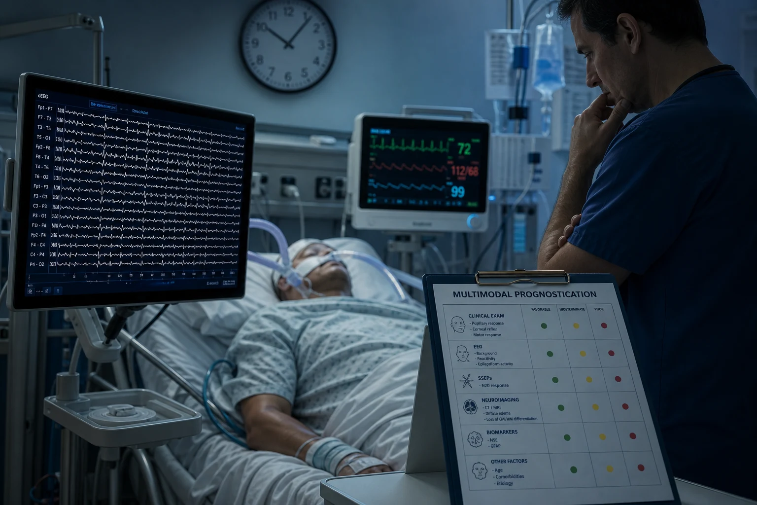
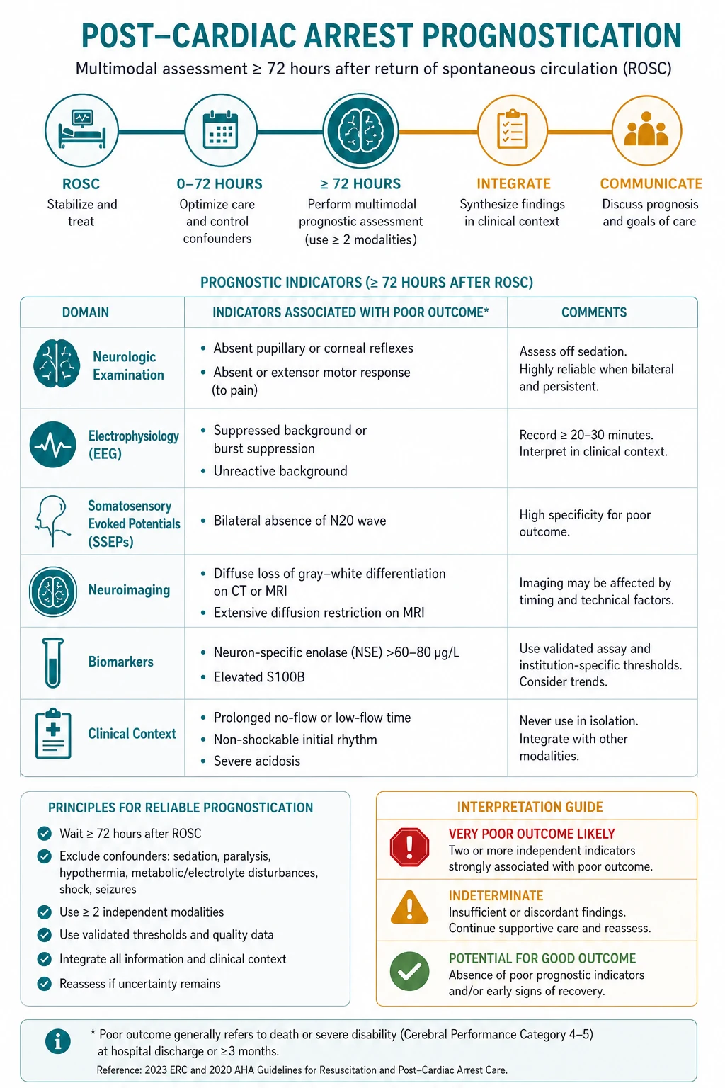
Prognostication after cardiac arrest predicts neurological outcome to guide continuation or withdrawal of life-sustaining therapy. The key principle: use a MULTIMODAL approach — no single test is 100% reliable. Timing: do NOT prognosticate before 72h post-arrest. Must be: normothermic (no fever for 72h), off sedatives for adequate washout, off NMBAs, no severe metabolic derangement. Assessment tools: (1) Clinical examination (brainstem reflexes, motor response, myoclonus), (2) Electrophysiology (SSEP N20, EEG), (3) Biomarkers (NSE), (4) Imaging (CT/MRI brain). The most reliable single indicator: bilateral absence of N20 response on SSEP (positive predictive value for poor outcome 95%). Goal: accurate, honest, compassionate prognostication for families.
On this page & tools
Your progress
Saved locally on this device.
Target exams
Red flags
Timeline and prerequisites
Prognostication timing
Days 0-2: NO prognostication
Do NOT attempt prognostication in first 48-72h. Rationale: (1) Sedatives (propofol, midazolam) have accumulated — need 2-5 half-lives to washout (propofol 2h x 5 = 10h, midazolam 3h x 5 = 15h, fentanyl 4h x 5 = 20h). (2) Organ dysfunction (renal, hepatic) may slow drug metabolism. (3) TTM (hypothermia) affects drug clearance. (4) Early neurological signs are unreliable. Focus: TTM, organ support, prevent complications.
Day 3+: Begin assessment (if prerequisites met)
Prerequisites for reliable assessment: (1) Normothermic (no fever, <37.5C, for >=72h). (2) Off sedatives for adequate washout (>=5 half-lives: propofol >10h, midazolam >15h, fentanyl >20h). (3) Off NMBAs (train-of-four 4/4). (4) Normoglycaemic (glucose 6-10). (5) Normocapnic (PaCO2 35-45). (6) No severe metabolic derangement (Na, Ca, Mg normal). (7) Haemodynamically stable. If any prerequisite not met: delay assessment.
Multimodal assessment
Perform MULTIPLE assessments simultaneously: (1) CLINICAL: GCS (especially motor response), brainstem reflexes (pupils, corneal, gag, cough), myoclonus. (2) SSEP: N20 response (median nerve stimulation — cortical response). (3) EEG: continuous (if available) or intermittent. (4) BIOMARKER: NSE (neuron-specific enolase) at 48-72h. (5) IMAGING: CT brain (early — structural injury) or MRI brain (later — diffusion changes in cortex/basal ganglia). Combine ALL results for overall prognostic impression.
Assessment tools
Clinical examination
Bedside, repeatable
- Motor response: extensor (M3) or absent (M1) at >=72h = poor prognosis (but affected by sedatives — must be off)
- Brainstem reflexes: absent pupillary light reflex at >=72h = poor prognosis (high specificity, moderate sensitivity)
- Myoclonus: status myoclonus (continuous/intermittent myoclonus for >=30 min) at <72h = poor prognosis
- Limitation: affected by sedatives, metabolic disturbance, hypothermia. MUST combine with other tests.
SSEP (N20)
Most reliable single test
- Somatosensory evoked potentials: stimulate median nerve, measure cortical response (N20 wave)
- BILATERAL ABSENCE of N20 = most reliable predictor of poor outcome (PPV for poor outcome >95%)
- Advantages: not affected by sedatives, metabolic disturbance, hypothermia. Highly reproducible.
- Timing: perform at >=24h (normothermia) or >=72h (post-hypothermia). Validated extensively.
- Limitation: presence of N20 does NOT predict GOOD outcome (some with present N20 still have poor outcome).
EEG
Seizure detection + prognostication
- Malignant EEG patterns: generalized suppression (<10 uV), burst-suppression, status epilepticus, absent reactivity
- Reactive EEG (responds to external stimuli) = better prognosis
- Continuous EEG: detects non-convulsive status epilepticus (common post-arrest, 10-30%)
- Limitations: affected by sedatives, hypothermia. Inter-rater variability for some patterns.
Biomarkers (NSE)
Blood test
- Neuron-specific enolase: released from damaged neurons. Elevated at 48-72h = neuronal injury.
- NSE >60 ug/L at 48-72h = poor prognosis (but cut-off varies by assay and study)
- Advantages: simple blood test, quantitative, repeatable.
- Limitations: also released from erythrocytes (haemolysis elevates NSE — check for haemolysis). Cut-off varies. Not universally available.
SAQ — Multimodal neurological prognostication at 72h post-arrest
10 minutes · 10 marks
A 58-year-old man is admitted to ICU after an out-of-hospital VF cardiac arrest (bystander CPR, defibrillated at 12 min, ROSC at 18 min). He received Targeted Temperature Management at 36°C for 24h and is now normothermic; sedation (propofol + fentanyl) has been off for 36h. At 72h post-ROSC he is intubated, GCS E4VTM5, no purposeful response to pain, brainstem reflexes intact, with intermittent right-arm myoclonus. The family ask about his neurological prognosis.
SAQ — SSEP N20 interpretation in post-arrest prognostication
10 minutes · 10 marks
A 65-year-old woman is day 3 post in-hospital cardiac arrest (PEA, ROSC at 25 min). She remains comatose (GCS M4), intubated and ventilated, normothermic (36.8°C), with propofol and fentanyl discontinued 24h ago. Median nerve somatosensory evoked potentials (SSEP) are requested to assist prognostication.
Clinical pearls
Red flags
Pathophysiology of post-arrest brain injury (why timing matters)
Global cerebral ischaemia-reperfusion after cardiac arrest produces a stereotyped cascade: excitotoxicity (glutamate release, intracellular calcium influx), mitochondrial failure with energy depletion, free-radical and protease-mediated injury, peri-infarct depolarisations, and delayed apoptotic and necrotic neuronal death. Neuronal loss is concentrated in the selectively vulnerable zones — hippocampal CA1, cortical layers 3 and 5, basal ganglia (particularly the putamen and thalamus), cerebellar Purkinje cells, and the brainstem reticular activating system. This secondary, delayed injury matures over days, which is precisely why early examination is unreliable and why prognostication must be DEFERRED: the lesion is still evolving. It also explains why a single time-point test can mislead — the brain is being examined while it is still changing. The multimodal, delayed, concordant approach exists to wait out this evolution and avoid the irreversible error of premature withdrawal of life-sustaining therapy (WLST). [1]
Multimodal prognostication algorithm (the 2021 ERC/ESICM framework)
The principle that unifies every contemporary guideline (ERC/ESICM 2014 advisory and 2021 guideline, the American Academy of Neurology, and the Neurocritical Care Society) is that no single predictor has a zero false-positive rate in routine practice, and therefore the WLST decision must never rest on one modality. The decision is built from concordance — two or more major predictors of poor outcome, with confounders excluded, examined at the correct time, ideally by two clinicians, with neurology/neurophysiology input. Bilateral absent N20 on SSEP comes closest to a zero false-positive rate, but even it must be corroborated. [1]
The 2021 ERC/ESICM prognostication algorithm — step by step
Step 0 — Satisfy the prerequisites
Delay until >=72h after ROSC AND >=72h after sedatives were stopped (whichever is LATER). For TTM at 33C: start the clock at the END of rewarming, not at ROSC. Confirm: normothermia (no fever >=37.8C for 24h), off NMBAs (train-of-four 4/4), normoglycaemia, normocapnia, no severe metabolic or haemodynamic derangement. If any confounder persists, postpone the assessment.
Step 1 — Clinical examination
Two independent observers document: (a) motor response to pain (M1-M5); (b) brainstem reflexes — pupil, corneal, gag, cough, oculocephalic. Bilaterally absent pupil AND corneal at >=72h is the strongest clinical signal. An absent or extensor motor response (M1-M2) at >=72h is suggestive but heavily confounded by residual sedation — never use in isolation.
Step 2 — Somatosensory evoked potentials (SSEP)
Bilateral median-nerve SSEP. Bilaterally absent N20 cortical response (with a preserved N9 brachial-plexus potential to confirm the stimulus was delivered) has a false-positive rate of ~1% — the single most reliable predictor of poor outcome. Unilateral absence is NOT sufficient. Record at >=24h if always normothermic, or >=72h post-rewarming if TTM at 33C was used.
Step 3 — EEG
Continuous (preferred) or repeat 30-minute epochs, reported in ACNS standardised terminology. A highly malignant pattern (background suppression, suppression with periodic discharges, or burst-suppression) plus absent reactivity is strongly predictive. A reactive background favours good outcome. Detect and treat non-convulsive status epilepticus (present in 10-30%).
Step 4 — Biomarkers and imaging
NSE at 48h and 72h — a rising trend across both, with no haemolysis, is more reliable than a single value. CT brain: diffuse oedema, sulcal effacement, reduced grey-white ratio. MRI brain DWI: extensive cortical and basal-ganglia diffusion restriction is the most sensitive structural marker of anoxic injury.
Step 5 — Require CONCORDANCE before WLST
Integrate ALL modalities. WLST is justified only when >=2 CONCORDANT major predictors of poor outcome are present, with confounders excluded. A single abnormal predictor — even bilaterally absent N20 — must NOT trigger WLST in isolation: obtain a second confirmatory modality and a second clinical opinion. If results are discordant, continue observation and serially re-assess rather than withdraw.
Predictors ranked by false-positive rate (FPR)
The lower the FPR, the more weight a predictor carries in isolation — but every predictor must still be combined. [1]
Absent pupil (bilateral)
FPR ~0%
- Most specific clinical sign — false-positive rate approaching 0% at >=72h off sedation
- Insensitive (~20% of poor-outcome patients show it)
- Exclude ocular trauma, topical mydriatics (atropine, tropicamide), and severe anisocoria
Absent N20 (bilateral SSEP)
FPR ~1%
- The single most robust predictor across the entire literature (PPV for poor outcome >95%, FPR ~1%)
- Unaffected by sedatives, hypothermia, or metabolic disturbance — the key advantage over clinical exam
- Insensitive (~45%); a present N20 does NOT guarantee a good outcome
- Confirm N9 (plexus) was present, otherwise the stimulus failed and the reading is uninterpretable
Highly malignant EEG
FPR ~0%
- Suppression / suppression with periodic discharges / burst-suppression: specificity ~100% (Westhall 2016)
- Sensitive in only ~50% of poor-outcome patients
- Must be reported in ACNS standardised terminology to be reliable
Status myoclonus
FPR low
- Continuous myoclonus >=30 min within 72h of ROSC: highly specific when strictly defined
- Must be distinguished from treatable non-convulsive status epilepticus (cEEG)
Absent corneal (bilateral)
FPR ~1-4%
- High specificity; combine with absent pupil for the strongest clinical signal
- Slightly less specific than pupil (more sedation-sensitive)
NSE >60 ug/L
FPR 5-15%
- Historic 60 ug/L cut-off is assay-dependent; haemolysis falsely elevates NSE
- Trend 48h then 72h; modern cut-offs (~80-100 ug/L) drive FPR toward 0
Motor M1-M2
FPR up to 5-10%
- Extensor (M2) or absent (M1) at >=72h — suggestive but the LEAST reliable individual predictor
- Heavily confounded by residual sedation, NMBAs, peripheral injury — never use alone
MRI DWI restriction
FPR low
- Extensive cortical + basal-ganglia diffusion restriction: high specificity for poor outcome
- Most sensitive structural marker; quantitative ADC volumetry improves reproducibility
CT grey-white ratio
FPR moderate
- Ratio <1.18 at basal ganglia level + sulcal effacement + cisternal effacement
- Cruder and later (>24h) than MRI; subjective
The single-modality rule and concordance
The most important concept for the exam and for safe practice: WLST must NEVER be based on a single modality. Even bilateral absence of the N20 SSEP response — the closest thing to a perfect predictor — is to be corroborated, not acted upon in isolation. The decision to withdraw life-sustaining therapy is built from concordance: two or more major predictors of poor outcome, recorded at the correct time with confounders excluded, documented by two clinicians, and reviewed by a neurologist or neurophysiologist. Where any single result is discordant, the correct response is continued observation and serial re-assessment, not premature withdrawal. The procedural safeguard matters as much as the data: a structured, time-locked, documented assessment is what protects the patient from the self-fulfilling prophecy. [1]
Clinical examination — what each sign is worth
Brainstem reflexes
Highest-yield clinical domain
- Bilaterally absent PUPILLARY light reflex at >=72h: specificity ~100%, FPR ~0%
- Bilaterally absent CORNEAL reflex at >=72h: specificity ~96-99%
- Absent gag, cough, oculocephalic reflexes: supportive but operator- and sedation-dependent
- Examine for at least 5-10 min; magnify pupils; exclude eye trauma and topical drugs
Motor response
Confounded by sedation
- Absent (M1) or extensor (M2) to pain at >=72h: suggestive BUT FPR historically up to 5-10%
- Never interpret motor response in isolation — too easily depressed by residual sedation
- Withdrawal (M4) or better: no reliable prediction either way
- Best motor response can be absent due to NMBAs, severe peripheral injury, or limb restraint
Myoclonus
Status myoclonus = ominous
- STATUS MYOCLONUS = sustained, continuous or intermittent jerks >=30 min within 72h of ROSC
- High specificity for poor outcome when strictly defined
- Must distinguish from non-convulsive status epilepticus — request cEEG and treat seizures
- Myoclonus confined to a single limb or face, or that resolves, does NOT by itself imply poor outcome
Somatosensory evoked potentials (SSEP / N20)
SSEPs are recorded by stimulating the median nerve at the wrist and measuring the cortical response over the contralateral parietal cortex. The N20 is the first negative cortical deflection at ~20 ms. The key finding is bilateral absence of N20, which — provided the peripheral N9 (brachial plexus) potential is present to confirm the stimulus was delivered — carries a false-positive rate of only ~1% and is the single most reliable predictor of poor outcome. SSEPs are largely unaffected by sedatives, hypothermia, and metabolic disturbance, which is their central advantage over the clinical examination and over EEG. Their limitation is sensitivity: only about 45% of patients with a poor outcome have bilaterally absent N20, and a present N20 does not predict a good outcome (some patients with present N20 still do poorly). Unilateral absence is not sufficient — both sides must be absent. Timing: record at >=24h if the patient was always normothermic, or >=72h after rewarming if TTM at 33C was used.[1][8]
EEG — ACNS standardised terminology
EEG serves two roles after arrest: detecting and guiding treatment of seizures (including non-convulsive status epilepticus, present in 10-30% of comatose survivors), and prognostication. For prognostication, EEG must be reported in the American Clinical Neurophysiology Society (ACNS) standardised terminology, which categorises patterns as highly malignant, malignant, or benign and dramatically reduces inter-rater variability compared with free-text reporting. [1]
Highly malignant
Predicts poor outcome
- Background suppression (<10 uV)
- Suppression with periodic discharges
- Burst-suppression (with or without identical bursts)
- Specificity ~100%, sensitivity ~50% — the patterns on which WLST can rest when concordant
Malignant
Suspicious; NOT alone enough
- Periodic or rhythmic patterns (GPDs, LRDA, SIRPDs)
- Spike-wave patterns
- Pathological or non-reactive background
- Isolated single feature: specificity only ~48% — require >=2 malignant features (specificity ~96%)
Benign
Favours good outcome
- No malignant features; normal voltage and reactivity
- Reactive background (responds to external stimulation)
- Only ~1% of patients with a benign pattern had a poor outcome — reassuring
Westhall 2016 — Standardised EEG predicts prognosis after cardiac arrest (Neurology; PMID 27016587)
Design
Prospective cohort within the TTM trial — 103 comatose survivors with standardised ACNS EEG reporting after rewarming
Highly malignant patterns
Suppression, suppression with periodic discharges, or burst-suppression: 100% specific for poor outcome (sensitivity ~50%)
Malignant patterns
An isolated single malignant feature was unreliable (specificity ~48%); two or more malignant features raised specificity to ~96%
Benign patterns
Only ~1% of patients with a benign EEG had a poor outcome
Clinical bottom line
Use ACNS terminology; only a highly malignant (or >=2 malignant features) pattern predicts poor outcome. EEG must be combined with other modalities and never used alone for WLST
Biomarkers (NSE and S100B)
NSE
Neuron-specific enolase
- Sampled at 48h and 72h; a rising trend across both is more reliable than a single value
- Cut-off ~60 ug/L historically associated with poor outcome, but assay-dependent FPR up to 5-15%
- Modern trend: higher cut-offs (~80-100 ug/L) to drive the FPR toward 0
- HAEMOLYSIS falsely elevates NSE (erythrocytes are rich in NSE) — always check the haemolysis index before acting
S100B
Astrocytic calcium-binding protein
- Released from injured astrocytes; rises within hours of anoxic injury
- Less affected by haemolysis than NSE
- Limited availability and less evidence than NSE — not a standalone test
Neuroimaging (CT and MRI)
CT brain
Early, available, lower sensitivity
- Diffuse cerebral oedema with sulcal effacement
- Loss or reversal of grey-white matter differentiation (ratio <1.18 at basal-ganglia level)
- Effacement of the basal cisterns — a grave sign
- Changes usually evident only after >24h; earliest and most specific structural modality that is universally available
MRI brain
Later, most sensitive
- Diffusion restriction (high DWI, low ADC) in cortex and basal ganglia — the hallmark of anoxic injury
- A watershed-zone infarction pattern suggests a hypoperfusion (prolonged low-flow) component
- Thalamic and hippocampal involvement are common and characteristic
- Quantitative whole-brain ADC volumetry improves reproducibility; extensive restriction carries high specificity for poor outcome
Targeted temperature management and the timing of prognostication
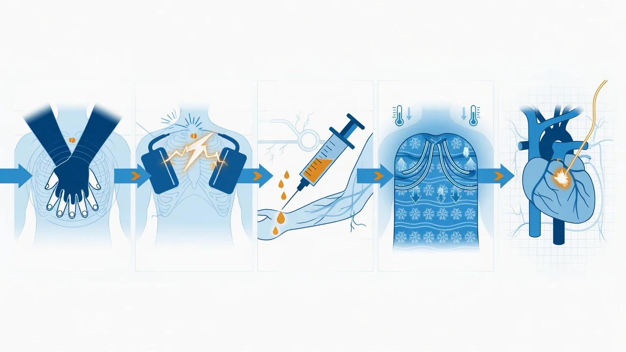
TTM directly shapes WHEN and HOW you prognosticate. Hypothermia (33C) slows hepatic and renal drug clearance, prolonging sedative half-lives and forcing a longer washout; it also delays the maturation of the anoxic lesion. Historically (post-HACA, post-TTM-trial 33C era), the rule was to wait >=72h after ROSC and >=24-48h after reaching normothermia. The 2021 TTM2 trial established targeted normothermia (fever prevention, 37.5C) as the standard for OHCA, which simplifies the timeline — the 72h clock runs from ROSC rather than from the end of rewarming, because there is no prolonged hypothermic phase to wash out. [1]
TTM2 — Hypothermia vs Normothermia after OHCA (Dankiewicz 2021, NEJM; PMID 34133859)
Design
International multicentre RCT — 1900 comatose adults after out-of-hospital cardiac arrest
Intervention
Targeted hypothermia at 33C for 28h with controlled rewarming vs targeted normothermia at 37.5C with active fever treatment
Primary outcome
All-cause mortality at 6 months: 50% hypothermia vs 48% normothermia (RR 1.04, 95% CI 0.94-1.14, p=0.37) — NO difference
Prognostication relevance
More arrhythmias with hypothermia; normothermia is now the standard, shortening and simplifying the prognostication timeline
Clinical bottom line
33C hypothermia confers no benefit over fever prevention — modern post-arrest care is targeted NORMOTHERMIA, so the >=72h clock starts from ROSC rather than from the end of rewarming
TTM — 33C vs 36C after OHCA (Nielsen 2013, NEJM; PMID 24237052)
Design
International multicentre RCT — 950 comatose OHCA survivors
Intervention
33C vs 36C for 36h
Outcome
No difference in mortality or neurological outcome — shifted practice away from routine 33C
Prognostication relevance
Patients managed at 36C clear sedatives faster, so the 72h prognostication window is more reliably met
HYPERION — Hypothermia for non-shockable-rhythm arrest (Lascarrou 2019, NEJM; PMID 31491141)
Design
Multicentre RCT — 584 comatose survivors of non-shockable-rhythm (PEA/asystole) arrest
Intervention
33C for 24h vs targeted normothermia at 37C
Primary outcome
Favourable outcome (CPC 1-2) at 90 days: 10.2% hypothermia vs 5.7% normothermia (p=0.04) — small absolute benefit
Prognostication relevance
Non-shockable arrests have a much worse baseline prognosis; even with hypothermia only ~1 in 10 recover consciousness, so false-positive vigilance matters even more
PARAMEDIC2 — Adrenaline vs placebo in OHCA (Perkins 2018, NEJM; PMID 29741852)
Design
Double-blind RCT — 8014 OHCA patients; adrenaline vs placebo
Outcome
Improved 30-day survival (3.2% vs 2.4%) but more survivors in severe neurological impairment (CPC 3-4)
Prognostication relevance
Adrenaline increases the number of patients arriving comatose in ICU, with a higher proportion on a poor-outcome trajectory — reinforcing the need for meticulous, delayed, multimodal prognostication to avoid premature WLST
Confounders and the false-positive trap
A predictor is only valid when confounders are excluded. The cardinal confounders are residual sedation, neuromuscular blockade, hypothermia, metabolic derangement, and ongoing seizures. Each can convert an otherwise reliable predictor into a false positive. [1]
Residual sedation
Most common confounder
- Propofol, midazolam, fentanyl, ketamine, and volatile anaesthetics all depress the exam and the EEG
- Allow >=5 half-lives; consider drug levels and ammonia/lactate; an exam performed under sedation is uninterpretable
- TTM at 33C roughly halves drug clearance — recompute the washout interval
Neuromuscular blockade
Confounds the motor exam
- Confirm train-of-four 4/4 before trusting the motor response or EEG reactivity
- SSEP is preserved (it tests the afferent sensory pathway, not the motor unit) — a key advantage over the motor exam
Metabolic and temperature
Reversible depressants
- Hypothermia, hypoglycaemia, hyponatraemia, hepatic or uraemic encephalopathy, and severe hypoxaemia can all mimic brain injury
- Normalise temperature, glucose, sodium, PaCO2, and PaO2 before prognosticating
- Rewarm fully and observe 24-48h of normothermia
Seizures
Treatable and misleading
- Non-convulsive status epilepticus is common (10-30%) and causes coma independent of anoxic injury
- Continuous EEG is mandatory for unexplained coma; treat seizures and then reassess
- Status epilepticus is itself a poor-prognosis marker but must not be the sole basis for WLST
Organ dysfunction
Pharmacokinetic
- Acute kidney injury and hepatopathy prolong sedative half-lives, so sedation accumulates disproportionately
- Adjust washout expectations and account for the effect of renal replacement therapy on drug clearance
Multimodal prognostication pearls (Part 2)
Prognostication red flags — traps that cause irreversible harm
Exam practice — viva-style questions
Q. A comatose arrest survivor at 72h after ROSC (normothermic, off sedation for 36h) has bilaterally absent N20 on SSEP but a normal NSE and a reactive EEG. What do you do? A. This is a DISCORDANT picture. Bilaterally absent N20 is the most reliable single predictor, but the reactive EEG and normal NSE argue against a uniformly poor outcome. The correct action is NOT to withdraw. Re-examine the SSEP for technical adequacy (was N9 present bilaterally?), confirm the timing (off sedation, normothermic, no NMBAs), treat any seizures, and continue observation with serial re-assessment. WLST requires concordance of >=2 major predictors; a single predictor — even absent N20 — is insufficient.[1][8]
Q. Why is motor response the least reliable individual predictor after arrest? A. It is the most susceptible to confounding by residual sedation, neuromuscular blockade, peripheral limb injury, and restraint. Its historical false-positive rate (up to 5-10%) is the highest among the major predictors, so it must never be used in isolation.[1]
Q. How does TTM change the prognostication timeline? A. Hypothermia slows drug clearance and delays lesion maturation. Under 33C TTM, start the 72h clock at the END of rewarming and allow extra washout. Since TTM2 (2021), the standard for OHCA is targeted normothermia (fever prevention), so the 72h clock typically runs from ROSC.[3][4]
Q. Which predictor has the lowest false-positive rate? A. Bilateral absence of the N20 cortical SSEP response (FPR ~1%), provided the peripheral N9 is present. Bilaterally absent pupillary reflexes share an FPR approaching 0% but are far less sensitive.[1][8]
Q. What is the ACNS highly malignant EEG pattern, and what is its prognostic value? A. Background suppression, suppression with periodic discharges, or burst-suppression. Per Westhall 2016, it is ~100% specific for poor outcome but only ~50% sensitive — so it predicts poor outcome when present but must still be combined with other modalities and never used alone for WLST.[7]
References
- [1]Sandroni C, et al. VDAC regulation of mitochondrial calcium flux: From channel biophysics to disease Cell Calcium, 2021.PMID 33529977
- [2]Friberg H, et al. Notum palmitoleoyl-protein carboxylesterase regulates Fas cell surface death receptor-mediated apoptosis via the Wnt signaling pathway in colon adenocarcinoma Bioengineered, 2021.PMID 34402722
- [3]Dankiewicz J, et al. Hypothermia versus Normothermia after Out-of-Hospital Cardiac Arrest N Engl J Med, 2021.PMID 34133859
- [4]Nielsen N, et al. Impaired hepatic and intestinal ATP-binding cassette transporter G5/8 was associated with high exposure of β-sitosterol and the potential risks to blood-brain barrier integrity in diabetic rats J Pharm Pharmacol, 2014.PMID 24237052
- [5]Lascarrou JB, et al. Photon Pair Condensation by Engineered Dissipation Phys Rev Lett, 2019.PMID 31491141
- [6]Perkins GD, et al. [Identification and phytotoxic activity of fungus QTYC-51 from the gut of Pantala flavescens larvae] Wei Sheng Wu Xue Bao, 2016.PMID 29741852
- [7]Westhall E, et al. Isolation of individual cellular components from lung tissues of patients with lymphangioleiomyomatosis Am J Physiol Lung Cell Mol Physiol, 2016.PMID 27016587
- [8]Sandroni C, et al. A new algorithm for automatic vascular mapping of DCE-MRI of the breast: Clinical application of a potential new biomarker Comput Methods Programs Biomed, 2014.PMID 25262335