ICU · Neurocritical Care
Raised ICP and traumatic brain injury management
Also known as Raised intracranial pressure (ICP) · Traumatic brain injury (TBI) · Intracranial hypertension · Cerebral perfusion pressure (CPP) · Decompressive craniectomy · Seattle ICP algorithm
Raised ICP is a life-threatening complication of severe TBI requiring urgent ICP monitoring and stepwise management. The Seattle International Consensus Conference (SICC) algorithm guides tiered therapy: Tier 0 (basics: head elevation, normocapnia, normoglycaemia, normothermia), Tier 1 (CSF drainage, hyperosmolar therapy — hypertonic saline or mannitol), Tier 2 (metabolic suppression with barbiturates, decompressive craniectomy). CPP target 60-70 mmHg. RESCUEicp (2016): decompressive craniectomy reduced mortality but increased vegetative/severe disability. POLAR (2018): prophylactic hypothermia does NOT improve outcomes.
On this page & tools
Your progress
Saved locally on this device.
Target exams
Red flags

Pathophysiology: the Monro-Kellie doctrine and cerebral autoregulation
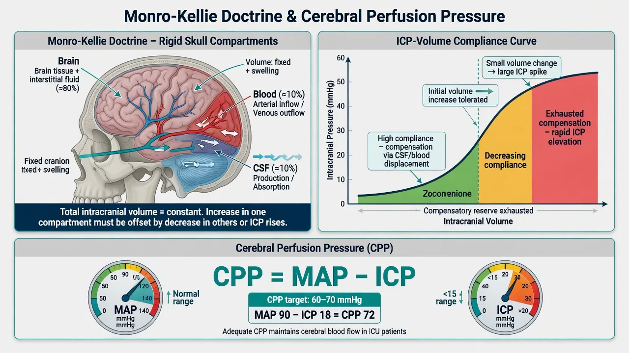
Monro-Kellie doctrine
The skull is a rigid, incompressible container holding three components:[7]
- Brain tissue (~80% of intracranial volume)
- Blood (~10%)
- CSF (~10%)
The total volume is constant. If one component increases (e.g., oedema, haematoma), the others must decrease to compensate. Initially, CSF is displaced into the spinal canal and venous blood is displaced into the systemic circulation. Once this compensatory reserve is exhausted, small increases in volume cause exponential increases in ICP (the steep part of the pressure-volume curve). [1]
Cerebral perfusion pressure (CPP)
CPP = MAP - ICP [1]
- Normal CPP: 70-90 mmHg
- Target in TBI: 60-70 mmHg[1][2]
- CPP <60: risk of cerebral ischaemia
- CPP >70: risk of ARDS (from fluid overload/vasopressors to achieve high MAP) and did NOT improve outcomes in the SELF/Phase II trial
Cerebral autoregulation
Normally, cerebral blood flow (CBF) is maintained constant across a wide range of MAP (50-150 mmHg) by autoregulation — arterioles dilate when BP falls and constrict when BP rises. [1]
In TBI, autoregulation is impaired or lost in many patients. CBF becomes pressure-passive — falls in MAP directly reduce CBF, causing ischaemia. This is why maintaining CPP is critical. Assessment of autoregulation (pressure reactivity index, PRx) can guide individualised CPP targets.[8]
ICP monitoring
Indications
ICP monitoring is recommended for:[1][2]
- Severe TBI (GCS 3-8) with:
- Abnormal CT scan (haematoma, contusion, swelling, herniation, compressed basal cisterns), OR
- Normal CT scan if TWO or more of: age >40, motor posturing, SBP <90 [1]
Monitoring methods
External ventricular drain (EVD)
Gold standard
- Placed into lateral ventricle via frontal approach (Kocher point)
- Measures global ICP AND allows CSF drainage (therapeutic)
- Most accurate method
- Risk: infection (~5-10%), haemorrhage (~1-2%)
- Can be zeroed and recalibrated in situ
Intraparenchymal probe
Most common in practice
- Placed into brain parenchyma (usually right frontal)
- Easy insertion, low complication rate
- Measures local pressure (may miss global gradients)
- Cannot be recalibrated once inserted (zero drift)
- Camino, Codman, Raumedic are common brands
Non-invasive
Adjunctive only
- Optic nerve sheath diameter (ONSD) on ultrasound
- Transcranial Doppler (for pulsatility index)
- NOT a substitute for invasive monitoring
- Useful for screening and trend monitoring
ICP waveform
Lundberg A (plateau) waves: sustained ICP >50 mmHg for 5-20 minutes — indicate critically low intracranial compliance and imminent herniation risk.[7]
Lundberg B waves: rhythmic oscillations 20-50 mmHg every 1-2 minutes — indicate impaired compliance. [1]
Lundberg C waves: rapid oscillations — less clinically significant. [1]
The Seattle International Consensus Conference (SICC) algorithm
The SICC (2019) provides a tiered management algorithm for raised ICP:[1][2]
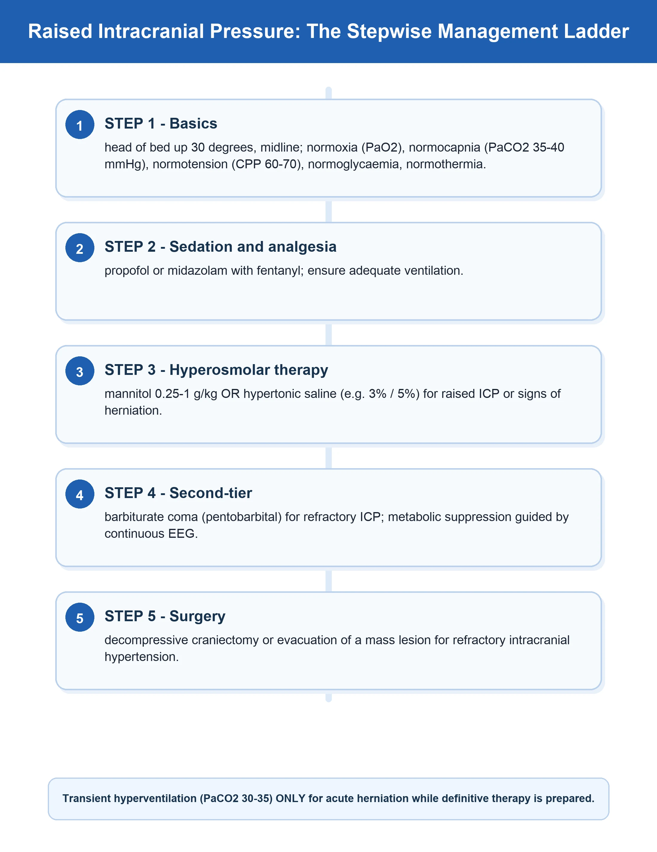
Seattle ICP management algorithm — Tier 0 (baseline measures)
Head elevation 30-45 degrees
Reduces ICP by promoting venous drainage and CSF return. Keep head midline (avoid neck vein compression from tight tapes/collar).
Normocapnia (PaCO2 35-40 mmHg)
Hypocapnia causes cerebral vasoconstriction → reduces ICP but risks ischaemia. Target PaCO2 35-40. PROPHYLACTIC hyperventilation (PaCO2 <30) is HARMFUL — use only as a temporary bridge in imminent herniation.
Normoxaemia (PaO2 >60, SpO2 >=94%)
Hypoxia causes cerebral vasodilation → increases ICP and causes secondary injury. Each hypoxic episode doubles mortality in severe TBI.
Normotension (SBP >=110)
Hypotension (SBP <90) doubles mortality in TBI. Use noradrenaline to maintain SBP >=110 or MAP >=80 (for CPP). Avoid excessive hypertension.
Normoglycaemia (6-10 mmol/L)
Both hypoglycaemia (neuronal injury) and hyperglycaemia (>10, worsens oedema) are harmful. Avoid intensive insulin therapy (NICE-SUGAR: increased hypoglycaemic episodes).
Normothermia (36-37C)
Fever increases cerebral metabolic rate and ICP. Treat with paracetamol and cooling blankets. Target normothermia. POLAR trial: prophylactic hypothermia does NOT improve outcomes.
Seizure prophylaxis
Phenytoin or levetiracetam for 7 days in patients with GCS <10, haematoma, depressed skull fracture, penetrating injury, or seizure on imaging.
Sedation and analgesia
Propofol (reduces cerebral metabolic rate and ICP) + fentanyl. Avoid ketamine in traditional teaching (may increase ICP), but evidence suggests it is safe with controlled ventilation.
Tier 1 — ICP >22 mmHg despite Tier 0
- CSF drainage via EVD (if in place) — drain 5-10 mL aliquots
- Hyperosmolar therapy:[9]
- 3% hypertonic saline bolus 250 mL over 10-15 minutes (preferred first-line — sustained effect, less rebound)
- Mannitol 0.25-1 g/kg bolus over 15 minutes (osmotic diuresis — monitor serum osmolar gap and urine output; stop if osmolarity >320 or Na >155)
- Optimise CPP: ensure MAP is adequate for CPP 60-70; use noradrenaline to increase MAP
- Repeat CT scan: to identify surgical lesions (haematoma enlargement, new haematoma)
Tier 2 — ICP refractory to Tier 1
Decompressive craniectomy
RESCUEicp evidence
- RESCUEicp (NEJM 2016): reduced mortality (26.5% vs 48.9%)
- BUT: increased rates of vegetative state and severe disability
- Reserved for refractory ICP not responsive to Tier 0 + Tier 1
- Must be discussed with family re functional outcomes
- Bilateral craniectomy for diffuse swelling; unilateral for lateral lesion
Metabolic suppression (barbiturates)
Last resort
- Thiopent loading 5-15 mg/kg, infusion 1-5 mg/kg/hr
- Suppresses cerebral metabolic rate → reduces CBF → reduces ICP
- Monitor with continuous EEG (burst suppression target)
- Significant complications: hypotension, immunosuppression, ileus
- Not associated with improved survival in clinical trials
Hyperventilation (temporary)
Bridge only
- Reduce PaCO2 to 30-35 for SHORT periods only
- Causes cerebral vasoconstriction → reduces ICP
- Risk: cerebral ischaemia from prolonged vasoconstriction
- Use ONLY as a bridge to definitive treatment (surgery, hyperosmolar)
- Never prophylactic or sustained
ICP-guided versus imaging/clinical-guided therapy
A central debate in severe TBI management is whether routine invasive ICP monitoring improves outcomes, or whether serial neuroimaging and clinical examination is sufficient. The Brain Trauma Foundation 4th Edition guidelines recommend ICP monitoring in salvageable severe TBI patients with abnormal CT (or normal CT with two or more risk factors), but the strength of evidence is variable.[15]
The BEST-TRIP trial
The landmark BEST-TRIP (Benchmark Evidence from South American Trials: Treatment of Intracranial Pressure) trial by Chesnut et al (NEJM 2012) randomised 324 patients with severe TBI to either ICP-monitor-guided therapy (treatment triggered by ICP >20 mmHg) or imaging-clinical examination-guided therapy (treatment triggered by CT signs of compression or neurological deterioration).[13]
- Primary outcome: No difference in composite neurological outcome (mortality, impaired consciousness, neuropsychological testing) at 6 months
- ICP group had shorter ICU stay and fewer days on ventilation in survivors
- Imaging-clinical group received more hyperosmolar therapy, hyperventilation, barbiturates and craniectomies (because clinical/CT changes are a late marker, treatments triggered later are more aggressive)
- Crucially: the trial did NOT test "no monitoring" vs "monitoring" — it compared two monitoring strategies, and the imaging-clinical arm used aggressive tier-2 escalation rather than the structured SICC algorithm [1]
Interpretation: BEST-TRIP does NOT show that ICP monitoring is futile. It shows that a well-structured imaging-clinical protocol can yield equivalent outcomes in centres without ICP monitoring capability. In well-resourced settings, ICP monitoring remains the standard of care because it provides earlier detection, allows individualised CPP targets, and reduces unnecessary escalation. [1]
ICP-guided therapy
Standard of care in most ICUs
- Continuous numeric data — early detection of intracranial hypertension
- Allows calculation of CPP and individualised autoregulation targets (PRx)
- Triggered Tier 1/Tier 2 escalation per Seattle algorithm
- Reduced unnecessary hyperosmolar/barbiturate use in BEST-TRIP survivors
- Complications: infection, haemorrhage, malposition (~5-10%)
- Recommended by Brain Trauma Foundation 4th Edition (level IIb)
Imaging-clinical guided
Resource-limited settings
- Serial CT scans + neurological examination (GCS, pupils)
- Non-invasive, no procedural risk
- Late marker — by the time CT shows shift or pupil dilates, injury is established
- Triggered MORE aggressive therapy in BEST-TRIP (more barbiturates, more craniectomies)
- Acceptable ONLY if ICP monitoring unavailable (BEST-TRIP equipoise)
CPP-guided versus ICP-guided therapy
The Rosner and Lund concepts represent two historical schools of thought on cerebral perfusion in TBI: [1]
Rosner (CPP-guided)
Volume expansion, high MAP
- Maintain CPP 60-70 mmHg (or up to 70 in older concept)
- Avoid hypotension aggressively with fluids and vasopressors
- Theory: cerebral vasoconstriction in response to raised ICP causes ischaemia — high MAP overcomes this
- Risk: fluid overload, ARDS (especially with CPP >70)
- Modern interpretation: CPP 60-70 is the target
Lund (ICP-guided)
Reduce intracranial volume
- Lower CPP target (~50-60), accept lower perfusion to minimise oedema
- Maintain colloid osmotic pressure, avoid fluid overload
- Reduce cerebral blood volume via venous drainage, head elevation, sedation
- Theory: high CPP worsens vasogenic oedema across disrupted blood-brain barrier
- Less popular in modern practice — risk of ischaemia
Autoregulation-guided
Individualised CPP
- Use PRx (pressure reactivity index) to define optimal CPP per patient
- Patients with intact autoregulation tolerate higher CPP
- Patients with impaired autoregulation need lower CPP to avoid hyperaemia and oedema
- COGITATE trial (2020) suggested feasibility of individualised CPP targets
- Increasingly available on multimodal neuromonitoring platforms
Practical synthesis: Target CPP 60-70 mmHg for all patients. Avoid both hypotension (CPP <60 — ischaemia) and aggressive hypertension (CPP >70 — ARDS, fluid overload, no outcome benefit). The Brain Trauma Foundation recommends 60-70 mmHg.[15]
Hyperosmolar therapy: mannitol versus hypertonic saline
The choice of osmotic agent for ICP control is one of the most debated topics in neurocritical care. The Brain Trauma Foundation 4th Edition guidelines state mannitol is effective for ICP control but consider hypertonic saline an equivalent option; neither has been shown to improve long-term outcome.[15]
Mannitol 20%
0.25-1 g/kg bolus
- Mechanism: osmotic gradient draws water from brain to plasma; also reduces blood viscosity → cerebral vasoconstriction (counter-regulation) → reduces cerebral blood volume
- Onset: 15-30 minutes, duration 4-6 hours
- Dose: 0.25-1 g/kg IV bolus (typical 0.5 g/kg)
- Diuresis: significant — insert urinary catheter, monitor urine output
- Monitoring: serum osmolarity (stop if >320 mOsm/L), osmolar gap (>20)
- Caution: hypovolaemia, hypotension, electrolyte disturbance, acute kidney injury
- Contraindicated in hypotension/hypovolaemia
Hypertonic saline 3%
250 mL bolus or infusion
- Mechanism: osmotic gradient + Na-driven water shift + restores intravascular volume
- Onset: similar to mannitol; more sustained effect, less rebound
- Dose: 3% NaCl 250 mL over 10-15 min (bolus), or infusion 20-50 mL/hr
- No diuresis — preferred when hypovolaemic or hypotensive
- Monitoring: serum Na (target 145-150, stop if >160), serum osmolarity
- Preferred first-line in many centres; meta-analyses suggest superior ICP control vs mannitol
- Preferred in shock/hypotension (volume-expanding)
- Caution: hypernatraemia, hyperchloraemic acidosis, central line for concentrated solutions
Practical approach
Hyperosmolar therapy cascade
First-line: 3% NaCl 250 mL bolus
Over 10-15 minutes. Preferred first-line in most centres — sustained effect, no diuresis, useful in shock. Recheck Na and osmolarity after 30 minutes.
Alternative: mannitol 0.5 g/kg bolus
Over 15 minutes. Use if hypertonic saline unavailable or if Na already elevated. Insert urinary catheter, monitor urine output and serum osmolarity (<320 mOsm/L).
Reassess ICP and re-dose as needed
If ICP remains >22 after 30-60 minutes, alternate agents (mannitol after HTS, or vice versa). Maintain Na 145-150, osmolarity <320.
Continuous HTS infusion if recurrent
2-3% NaCl infusion at 20-50 mL/hr to maintain Na 145-150. Avoid rapid swings; rapid cessation can cause rebound.
Escalate to Tier 2 if refractory
ICP >22 despite optimised hyperosmolar therapy, sedation, CSF drainage and CPP optimisation — proceed to decompressive craniectomy or barbiturate coma.
The Froelich et al (2022) comparison of hypertonic saline versus mannitol found both agents significantly reduce ICP, but the effect is more sustained with hypertonic saline.[9]
Decompressive craniectomy: timing and evidence
Two pivotal RCTs inform decompressive craniectomy practice. They differ fundamentally in timing (early vs late) and patient population. [1]
DECRA (NEJM 2011) — early bifrontal decompressive craniectomy14
Cooper et al randomised 155 patients with severe diffuse TBI and early refractory intracranial hypertension (ICP >20 mmHg for >15 min within 72 hours) to early bifrontotemporoparietal decompressive craniectomy vs standard medical care. [1]
- Primary outcome: Worse Glasgow Outcome Score-Extended at 6 months in the craniectomy group (OR 1.70 unfavourable)
- Mortality: lower with craniectomy (19% vs 33%)
- BUT: more patients in surgery group had bilateral fixed pupils pre-randomisation (selection imbalance)
- Caveat: used bifrontal craniectomy; lower ICP threshold (>20) than RESCUEicp (>25) [1]
RESCUEicp (NEJM 2016) — late decompressive craniectomy for refractory ICP3
Hutchinson et al randomised 408 patients with refractory ICP >25 mmHg (above medical management) to decompressive craniectomy (unilateral or bilateral) vs continued medical therapy (including barbiturates). [1]
- Mortality reduced: 26.5% (craniectomy) vs 48.9% (medical)
- BUT increased severe disability: vegetative 8.5% vs 2.1%; lower severe disability 21.9% vs 14.5%
- More upper severe disability survivors (dependent for daily care)
- Critical message: craniectomy converts death into survival-with-disability. Decision requires explicit family discussion of acceptable functional outcomes. [1]
DECRA (early)
ICP >20 within 72h, bifrontal
- Cooper 2011, n=155
- Earlier intervention (within 72h of ICP rise)
- Worse functional outcome at 6 months in surgery group
- Selection imbalance confounds interpretation
- Cautious use of early craniectomy for ICP control
RESCUEicp (late)
Refractory ICP >25
- Hutchinson 2016, n=408
- Late intervention (refractory ICP after Tier 0 + Tier 1)
- Reduced mortality (26.5% vs 48.9%)
- Increased vegetative state and severe disability
- Last-resort therapy with explicit family counselling
Practice synthesis: Decompressive craniectomy is NOT a "fix" for raised ICP. It is a life-saving intervention with significant risk of severe disability. Use in the context of: (1) refractory ICP despite Tier 0 + Tier 1 + barbiturates; (2) salvageable neurological injury (no devastating primary destruction); (3) explicit family discussion about functional outcomes. [1]
Barbiturate coma and metabolic suppression
Barbiturates (thiopental, pentobarbital) suppress cerebral metabolic rate of oxygen (CMRO2), reducing CBF and cerebral blood volume and thereby reducing ICP. They are used as a Tier 2 last-resort for refractory ICP. [1]
Thiopent coma protocol
Pre-requisites
Refractory ICP >22 despite Tier 0 + Tier 1 (hyperosmolar, CSF drainage) and decompressive craniectomy (if performed or declined). Continue invasive BP monitoring, central venous access, vasopressor ready.
Loading: thiopental 5-10 mg/kg over 30 min
Expect significant hypotension — preload with fluid, ready noradrenaline infusion. Monitor EEG continuously.
Maintenance infusion: 1-5 mg/kg/hr
Titrate to burst suppression (2-5 bursts/min) on continuous EEG, OR to ICP <22. Avoid over-suppression which prolongs recovery.
Monitor for complications
Hypotension (vasoplegia), immunosuppression (nosocomial infection), paralytic ileus, skin breakdown, hepatic dysfunction, hypothermia, prolonged sedation. Daily review of ongoing need.
Weaning
Once ICP controlled for 24-48h, wean by 1 mg/kg/hr every 6-12 hours. Expect prolonged elimination (half-life 20-60h after prolonged infusion). Watch for rebound ICP and seizures.
Important: The Cochrane review and multiple trials have NOT shown that barbiturates improve survival in severe TBI. They reduce ICP but at the cost of significant complications (hypotension, infection, prolonged coma). Reserved for refractory cases where decompressive craniectomy is not feasible or has failed.[15]
Multimodal neuromonitoring
Beyond ICP and CPP, modern neurocritical care increasingly uses multimodal monitoring to detect secondary brain injury and individualise therapy.[18]
Brain tissue oxygen (PbtO2)
Direct regional oxygenation
- Clark electrode placed into parenchyma
- Target PbtO2 >15-20 mmHg (normal 25-40)
- Detects regional ischaemia missed by systemic PaO2
- BOOST-2 trial (2020): PbtO2-guided therapy showed signal of benefit
- Threshold for treatment: <15 mmHg warrants intervention
Cerebral microdialysis
Brain biochemistry
- Measures lactate, pyruvate, glucose, glutamate, glycerol
- Lactate:pyruvate ratio >40 = metabolic distress
- Low glucose (<0.7 mmol/L) = energy failure
- High glycerol = cell membrane breakdown
- Detects ischaemia/metabolic crisis before clinical change
Continuous EEG (cEEG)
Detects NCS and seizures
- Up to 20% of comatose TBI patients have non-convulsive seizures
- Non-convulsive status epilepticus worsens outcome
- Recommended for any comatose patient with unexplained ICP rise
- Also used to titrate barbiturate coma to burst suppression
Jugular venous oximetry (SjvO2)
Global oxygen balance
- Fibreoptic catheter in jugular bulb
- SjvO2 <50% = global cerebral ischaemia
- SjvO2 >75% = hyperaemia or AV shunting
- Largely superseded by PbtO2 and microdialysis
The Pressure Reactivity Index (PRx) — a correlation coefficient between MAP and ICP over time — provides a continuous measure of cerebral autoregulation. PRx <0 indicates intact autoregulation; PRx >0.3 indicates impaired autoregulation and is associated with worse outcomes. Individualised optimal CPP (CPPopt) can be derived from PRx curves.[8]
Multimodal prognostication in severe TBI
Prognostication in severe TBI is multimodal and probabilistic, never deterministic. No single test predicts outcome; a recent international consensus emphasised avoiding early prognostic pessimism and using multiple data points over time.[19]
Prognostication timeline and approach
Days 0-3: avoid prognostic statements
Sedation, hypothermia, organ failure, alcohol intoxication confound early assessment. Do NOT prognosticate in the first 72 hours. The Ethicus and similar studies show early WLST (withdrawal of life-sustaining therapy) introduces self-fulfilling prophecies.
Days 3-7: structural assessment
Repeat CT for evolving lesions (delayed haematoma, infarct, hydrocephalus). MRI (diffusion, susceptibility) for diffuse axonal injury grade. Initial pupil examination at 72h.
Days 5-7: multimodal testing
Somatosensory evoked potentials (N20 bilateral absence = poor prognosis, high specificity). Continuous EEG for NCS. Motor response trend. Pupil reactivity.
Days 7-14: integrated prognostication
Combine: (1) clinical (age, GCS motor, pupils at 72h), (2) structural (CT Marshall grade, MRI DAI), (3) electrophysiology (SSEP, cEEG), (4) biomarkers (NSE, S100B, GFAP — emerging). Probabilistic model, not binary.
Discuss with family
Frame prognosis in terms of probabilities, not certainties. Acknowledge uncertainty. Discuss functional outcomes (independence, cognition, dependency) and quality of life, not just survival. Involve palliative care and rehabilitation teams early.
Established prognostic factors
- Age: mortality and poor outcome increase with age; >60 particularly poor
- Initial GCS motor: M1-2 worst prognosis; M6 best
- Pupil reactivity at 72h: bilateral fixed dilated = poor prognosis (specificity ~90%)
- Hypotension/hypoxia episodes: each doubles mortality
- CT Marshall classification: grade V-VI (compression of basal cisterns, midline shift >5mm, evacuated/non-evacuated mass) predicts poor outcome
- ICP trend: refractory ICP >20 mmHg for >48h predicts poor outcome
- Glucose: persistent hyperglycaemia (>10 mmol/L) worsens outcome
- C-reactive protein, NSE, S100B, GFAP: emerging biomarkers (not routine) [1]
Cautions
Avoid harmful therapies
Several historical or proposed therapies have been definitively shown NOT to improve outcome in severe TBI and may cause harm:[15][17]
Corticosteroids
CRASH trial — harmful
- CRASH trial (Lancet 2004, n=10008): high-dose methylprednisolone INCREASED mortality at 14 days (risk ratio 1.18)
- Risk of death increased regardless of injury severity
- Corticosteroids are CONTRAINDICATED in severe TBI
- Exception: if there is documented adrenal insufficiency or co-existing spinal cord injury (NASCIS guidelines)
Prophylactic hypothermia
POLAR, Eurotherm — not helpful
- POLAR (JAMA 2018): prophylactic hypothermia 33-35C for 72h did not improve 6-month outcome; more pneumonia
- Eurotherm3235 (NEJM 2015): cooling for ICP control actually worsened outcomes (terminated early)
- Use hypothermia only for concurrent cardiac arrest (TTM2 — even there, normothermia preferred)
Pharmacological neuroprotection
All failed
- Progesterone (SYNAPSE, ProTECT III): no benefit
- Erythropoietin: no benefit on outcome
- Magnesium, statins, dexanabinol, nimodipine, pegorgotein: all negative
- No drug has yet shown neuroprotective benefit in severe TBI phase III trials
Albumin fluid resuscitation
SAFE-TBI — harmful
- SAFE-TBI substudy (NEJM 2007): albumin resuscitation associated with HIGHER mortality than saline at 24 months (33.2% vs 20.4%)
- Use crystalloid (0.9% saline or balanced solution) for fluid resuscitation in TBI
- Avoid albumin in acute severe TBI
Prophylactic hyperventilation
Harmful
- Reduces CBF by cerebral vasoconstriction → ischaemia
- Muizelaar 1991: prophylactic hyperventilation to PaCO2 25 worsened outcomes at 3 and 6 months
- Use only as temporary bridge in imminent herniation
Evidence and landmark trials
RESCUEicp
NEJM 2016
408 pts with refractory ICP >25 — decompressive craniectomy vs medical management
Key finding
Mortality reduced: 26.5% craniectomy vs 48.9% medical. BUT: more vegetative state (8.5% vs 2.1%) and severe disability (21.9% vs 14.5%)
Practice change
Craniectomy reduces mortality but increases severe disability — decision must involve family
POLAR
JAMA 2018
511 pts severe TBI — prophylactic hypothermia 33-35C for 72h vs normothermia
Key finding
No difference in favourable outcomes at 6 months (48.8% vs 49.1%). More pneumonia in hypothermia group.
Practice change
Prophylactic hypothermia is NOT recommended in severe TBI
SYNAPSE
NEJM 2015
1195 pts severe TBI — progesterone vs placebo
Key finding
No benefit in outcomes at 3 or 6 months. Another negative neuroprotection trial.
Practice change
No pharmacological neuroprotective agent has shown benefit in TBI
SICC Algorithm
ICM 2019
42 international experts — consensus on ICP management algorithm
Key finding
Tiered approach: Tier 0 (basics) → Tier 1 (hyperosmolar, CSF drainage) → Tier 2 (craniectomy, barbiturates). CPP target 60-70.
Practice change
Standardised algorithm adopted worldwide for ICP management
BEST-TRIP
NEJM 2012
324 pts severe TBI — ICP-monitor-guided vs imaging/clinical-guided therapy
Key finding
No difference in 6-month composite outcome. ICP group had shorter ICU stay but imaging arm used more Tier 2 therapy. Did NOT prove monitoring futile — compared two monitoring strategies.
Practice change
ICP monitoring remains standard of care in well-resourced ICUs; imaging-clinical acceptable only where monitoring unavailable
DECRA
NEJM 2011
155 pts with early diffuse TBI — bifrontal decompressive craniectomy vs standard care
Key finding
Worse functional outcome (GOS-E) at 6 months in surgery group; mortality lower (19% vs 33%). Selection imbalance confounds. Used ICP >20 threshold and bifrontal approach.
Practice change
Caution against EARLY craniectomy for ICP >20 — RESCUEicp more influential
BTF 4th Edition
Neurosurgery 2017
Systematic review of evidence for severe TBI management
Key finding
ICP monitoring recommended (level IIb); CPP 60-70; avoid prophylactic hyperventilation, steroids, hypothermia; mannitol or HTS for hyperosmolar therapy.
Practice change
Updated international guidelines — replaces 3rd edition
SAFE-TBI
NEJM 2007
Substudy of SAFE trial — 460 TBI pts resuscitated with albumin vs saline
Key finding
Albumin associated with HIGHER mortality at 24 months (33.2% vs 20.4%) vs saline.
Practice change
Avoid albumin for fluid resuscitation in severe TBI — use crystalloid
CRASH
Lancet 2004
10008 adults with clinically significant head injury — methylprednisolone vs placebo within 8h
Key finding
Corticosteroids INCREASED mortality at 14 days (RR 1.18) and 6 months. Harm regardless of severity.
Practice change
Corticosteroids are CONTRAINDICATED in severe TBI
Eurotherm3235
NEJM 2015
387 pts TBI with ICP >20 — prophylactic hypothermia 32-35C vs standard care
Key finding
Worse neurological outcomes at 6 months in hypothermia group; trial terminated early for harm.
Practice change
Therapeutic hypothermia for ICP control is HARMFUL in severe TBI — reinforced by POLAR
Monitoring targets
| Parameter | Target | Rationale |
|---|---|---|
| ICP | <22 mmHg | Above this, treatment is indicated. Sustained ICP >22 associated with worse outcomes.[1] |
| CPP | 60-70 mmHg | CPP = MAP - ICP. Below 60: ischaemia risk. Above 70: ARDS risk without outcome benefit. |
| PaCO2 | 35-40 mmHg | Normocapnia. Hypocapnia causes vasoconstriction (temporary bridge only). Hypercapnia causes vasodilation → raised ICP. |
| SBP | ≥110 mmHg | Avoid hypotension — each SBP <90 episode doubles mortality. Use noradrenaline if needed. |
| PaO2 | ≥60 mmHg | Avoid hypoxia — each PaO2 <60 episode doubles mortality. |
| Glucose | 6-10 mmol/L | Both hypo and hyper harmful. Avoid intensive insulin (hypoglycaemic episodes). |
| Temperature | 36-37C | Fever raises cerebral metabolic rate and ICP. Normothermia only — prophylactic hypothermia does NOT help (POLAR). |
| Sodium | 140-145 mmol/L | Mild hypernatraemia (145-150) may reduce cerebral oedema. Avoid rapid correction of chronic hypernatraemia. |
| Haemoglobin | ≥70 g/L (or 90 if ischaemic) | Transfusion threshold individualised. Avoid anaemia (reduces O2 delivery to injured brain). |
Prognosis
Severe TBI outcomes
- Prognostic factors: age, initial GCS, pupil reactivity, CT findings (Marshall classification, cistern compression, midline shift), ICP trend, hypotensive episodes, glucose level
- Long-term: cognitive impairment (memory, executive function), personality change, epilepsy, pituitary dysfunction, emotional lability [1]
Exam practice
SAQ — Severe TBI with raised ICP
10 minutes · 10 marks
A 25-year-old man is brought to ICU after a motor vehicle accident. GCS 6 (E1V1M4). CT shows a right acute subdural haematoma (10mm thick, 4mm midline shift) which has been evacuated. Post-op CT shows diffuse cerebral oedema with compressed basal cisterns. An intraparenchymal ICP monitor shows ICP 28 mmHg. MAP 75. Temperature 37.8C. PaCO2 38. ABG: pH 7.32, lactate 2.8, Na 138. Sedated with propofol and fentanyl.
SAQ — Severe TBI with refractory intracranial hypertension and multimodal monitoring
10 minutes · 10 marks
A 42-year-old man is admitted to ICU 48 hours after a 6-metre fall. Initial GCS 4 (E1V1M2). CT brain shows diffuse traumatic axonal injury with compressed basal cisterns and no surgical mass lesion (Marshall grade III). He is intubated and ventilated, sedated with propofol 200 mg/hr and fentanyl 100 mcg/hr, on 3% NaCl infusion at 30 mL/hr (serum Na 152, osmolarity 316). An intraparenchymal ICP monitor and a brain tissue oxygen (PbtO2) probe are in situ. Current readings: ICP 26 mmHg, MAP 85, PbtO2 12 mmHg, PRx +0.4. Continuous EEG shows no seizures.
SAQ — Decompressive craniectomy: DECRA, RESCUEicp and goals-of-care
10 minutes · 10 marks
A 28-year-old woman sustained a severe closed head injury in a motorcycle crash (no helmet). GCS 5 (E1V1M3) on scene. CT shows diffuse bihemispheric swelling with compressed basal cisterns and a small right temporal contusion — no operable mass. She is intubated and on Day 3 of ICU care. Despite optimised Tier 0 measures, 3% NaCl infusion (serum Na 153, osmolarity 318), CSF drainage via an external ventricular drain, deep propofol/midazolam/fentanyl sedation and noradrenaline to MAP 90 (CPP 60), her ICP has remained 30-34 mmHg for the last 4 hours. The neurosurgical team is considering decompressive craniectomy. Her parents are at the bedside and are asking what the operation would mean for her future.
Clinical pearls
Red flags
References
- [1]Robba C, Czosnyka M, Carniero A, et al. A management algorithm for patients with intracranial pressure monitoring: the Seattle International Severe Traumatic Brain Injury Consensus Conference (SIBICC) Intensive Care Med, 2019.PMID 31659383
- [2]Hawryluk GWJ, Aguilera S, Buki A, et al. Guidelines for the Management of Severe Traumatic Brain Injury: 2020 Update of the Decompressive Craniectomy Recommendations Neurosurgery, 2020.PMID 32761068
- [3]Hutchinson PJ, Kolias AG, Timofeev IS, et al. Trial of Decompressive Craniectomy for Traumatic Intracranial Hypertension N Engl J Med, 2016.PMID 27602507
- [4]Cooper DJ, Nichol AD, Bailey M, et al. Effect of Early Sustained Prophylactic Hypothermia on Neurologic Outcomes Among Patients With Severe Traumatic Brain Injury: The POLAR Randomized Clinical Trial JAMA, 2018.PMID 30357266
- [5]Skolnick BE, Maas AI, Narayan RK, et al. Progesterone in traumatic brain injury N Engl J Med, 2015.PMID 25932489
- [6]Citerio G, Oddo M, Taccone FS, et al. Consensus-Based Management Protocol (CREVICE Protocol) for the Treatment of Severe Traumatic Brain Injury Based on Imaging and Clinical Examination for Use When Intracranial Pressure Monitoring Is Not Employed J Neurotrauma, 2020.PMID 32013721
- [7]Tsitsopoulos PP, Kourbeti D, Valadakis A, et al. An overview of management of intracranial hypertension in the intensive care unit J Anesth, 2020.PMID 32440802
- [8]Bhatti T, Czosnyka M, Smielewski P, et al. Beyond intracranial pressure: monitoring cerebral perfusion and autoregulation in severe traumatic brain injury Curr Opin Crit Care, 2023.PMID 36762674
- [9]Froelich M, Ni Q, Wessell A, et al. Comparison of Intracranial Pressure Measurements Before and After Hypertonic Saline or Mannitol Treatment in Children With Severe Traumatic Brain Injury JAMA Netw Open, 2022.PMID 35267036
- [10]Robba C, Goffi A, Padayachy L, et al. Using Optic Nerve Sheath Diameter for Intracranial Pressure (ICP) Monitoring in Traumatic Brain Injury: A Scoping Review Neurocrit Care, 2024.PMID 38114797
- [11]Bukhari S, Hussain R, Yonas H, et al. Efficacy and safety of erythropoietin for traumatic brain injury BMC Neurol, 2020.PMID 33138778
- [12]Schauer SG, Marques NR, Shackelford SA, et al. Hypertonic Saline for Severe Traumatic Brain Injury With Herniation: A Military Prehospital Case Report J Spec Oper Med, 2022.PMID 35862837
- [13]Chesnut RM, Temkin N, Carney N, et al. A trial of intracranial-pressure monitoring in traumatic brain injury N Engl J Med, 2012.PMID 23234472
- [14]Cooper DJ, Rosenfeld JV, Murray L, et al. Decompressive craniectomy in diffuse traumatic brain injury N Engl J Med, 2011.PMID 21434843
- [15]Carney N, Totten AM, O'Reilly C, et al. Guidelines for the Management of Severe Traumatic Brain Injury, Fourth Edition Neurosurgery, 2017.PMID 27654000
- [16]Myburgh J, Cooper DJ, Finfer S, et al. Saline or albumin for fluid resuscitation in patients with traumatic brain injury N Engl J Med, 2007.PMID 17761591
- [17]Roberts I, Yates D, Sandercock P, et al. Effect of intravenous corticosteroids on death within 14 days in 10008 adults with clinically significant head injury (MRC CRASH trial): randomised placebo-controlled trial Lancet, 2004.PMID 15474134
- [18]Le Roux P, Menon DK, Citerio G, et al. The International Multidisciplinary Consensus Conference on Multimodality Monitoring in Neurocritical Care: a list of recommendations and additional conclusions: a statement for healthcare professionals from the Neurocritical Care Society and the European Society of Intensive Care Medicine Neurocrit Care, 2014.PMID 25501689
- [19]van Veen E, Citerio G, Cnossen M, et al. Occurrence and timing of withdrawal of life-sustaining measures in traumatic brain injury patients: a CENTER-TBI study Intensive Care Med, 2021.PMID 34351445