ICU · neurocritical care
Acute Raised Intracranial Pressure and Traumatic Brain Injury — Comprehensive Neurocritical Care
Also known as Raised intracranial pressure (ICP) · Intracranial hypertension · Traumatic brain injury (TBI) · Severe head injury · Cerebral perfusion pressure (CPP) · Decompressive craniectomy · Monro-Kellie doctrine · Brain herniation · Osmotherapy (mannitol, hypertonic saline) · Brain Trauma Foundation guidelines · RESCUEicp · Secondary brain injury
Raised intracranial pressure (ICP >22 mmHg) after traumatic brain injury (TBI) is the most preventable cause of secondary brain damage and the central target of neurocritical care. The MONRO-KELLIE DOCTRINE is the foundation: the skull is a rigid box whose fixed volume holds BRAIN (~80%) + BLOOD (~10%) + CSF (~10%); an increase in one component REQUIRES an equal decrease in the others or ICP rises. CAUSES of raised ICP: trauma (extradural/subdural/intracerebral haematoma, contusions, cerebral oedema), tumour (mass effect + peritumoural oedema), infection (abscess, meningitis/encephalitis), hydrocephalus (CSF obstruction), and hepatic encephalopathy (cytotoxic oedema). CLINICAL FEATURES: headache, vomiting, progressive alteration of consciousness, and CUSHING'S TRIAD (hypertension with widened pulse pressure + bradycardia + irregular respiration) which is a LATE, pre-terminal sign signalling brainstem herniation is imminent. ICP MONITORING: external ventricular drain (EVD) is the GOLD STANDARD (measures ICP AND drains CSF therapeutically) but carries higher infection/haemorrhage risk; intraparenchymal monitor (Camino/Codman) is most commonly used, has lower infection risk but cannot drain. TARGETS (Brain Trauma Foundation 4th edition, 2017): ICP <22 mmHg — every mmHg above 22 increases mortality; CPP 60-70 mmHg (CPP = MAP − ICP). MANAGEMENT STAIRCASE (escalate sequentially): (1) HEAD OF BED 30° NEUTRAL + NORMOCAPNIA (PaCO2 35-40 — AVOID prophylactic hyperventilation, especially the first 24 h); (2) SEDATION + ANALGESIA (propofol first-line); (3) CSF DRAINAGE via EVD; (4) OSMOTHERAPY — mannitol 0.5-1 g/kg OR hypertonic saline 3-5% (target Na 145-155 mmol/L); (5) METABOLIC SUPPRESSION — propofol infusion, then barbiturate coma (thiopental/pentobarbital titrated to burst suppression on EEG) if refractory; (6) DECOMPRESSIVE CRANIECTOMY — RESCUEicp trial (2016) showed bifrontal decompressive craniectomy for refractory ICP >25 after TBI REDUCED mortality but INCREASED the rate of vegetative/severely-disabled survival (a 'troubling trade-off' requiring family discussion). CPP 60-70 mmHg: avoid <60 (cerebral ischaemia) and >70 (ARDS risk per the BEST:TRIP trial). SECONDARY INSULTS must be prevented: hypotension (SBP <110 DOUBLES mortality) and hypoxia (SpO2 <90 TRIPLES mortality) — even a SINGLE episode worsens outcome. Steroids are CONTRAINDICATED in TBI (CRASH trial — increased mortality).
On this page & tools
Your progress
Saved locally on this device.
3 MCQs with explanations
Target exams
Red flags
Overview

Severe traumatic brain injury (TBI) is the signature disease of neurocritical care and one of the highest-yield topics in the CICM/FFICM/EDIC examinations. The intensivist's job is NOT to undo the primary impact injury — that is fixed at the moment of collision — but to prevent secondary brain injury: the cascade of ischaemia, oedema, herniation and cell death that unfolds over hours to days and is driven by raised ICP, hypotension, hypoxia, fever, hyperglycaemia and seizures. Every intervention in the protocol — from head positioning to decompressive craniectomy — exists to keep the injured brain perfused and oxygenated while it recovers. The conceptual spine is the CPP equation (CPP = MAP − ICP): you can improve brain perfusion either by lowering ICP (osmotherapy, CSF drainage, craniectomy) or by raising MAP (noradrenaline) — and the correct answer to almost every TBI scenario is to do BOTH simultaneously while searching for a surgical lesion.[1][6]
Pathophysiology — the Monro-Kellie doctrine and the intracranial compliance curve
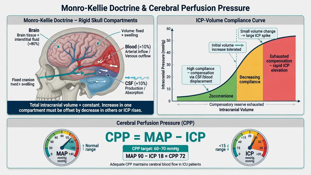
The Monro-Kellie doctrine (named for Alexander Monro, 1783, and George Kellie) states that because the skull is a rigid, unyielding container of fixed volume, the sum of its three contents — brain tissue (~80%), intracranial blood (~10%, mostly venous), and CSF (~10%) — must remain constant. Therefore any ADDITION of volume (a haematoma, a tumour, oedematous brain, extra blood, or obstructed CSF) can only be tolerated if an EQUAL volume of one of the other components is DISPLACED out of the cranium. The body achieves this by, in sequence: (1) shunting CSF from the cranial subarachnoid space into the spinal thecal sac, and (2) displacing venous blood out through the jugular veins into the extracranial circulation. These two mechanisms constitute intracranial compensation.[1]
The crucial clinical implication is the intracranial volume–pressure (compliance) curve. While compensation is active, ICP stays nearly flat despite a growing mass — the patient may harbour a large subdural haematoma yet have a normal ICP and a normal examination. This is the compensated (flat) phase. Once the displacement reserves are exhausted, however, the system reaches the critical volume, the knee of the curve: from this point, a TINY further increase in volume produces a LARGE, near-exponential rise in ICP. This is decompensation — and it explains why a TBI patient can appear stable for hours then deteriorate catastrophically over minutes (a 'lucid interval' followed by sudden herniation). The take-home: a normal early GCS does NOT exclude a dangerous intracranial mass; serial neurological observation and a low threshold for CT are mandatory.[1]
Cerebral autoregulation and why CPP matters
In the healthy brain, cerebral autoregulation keeps cerebral blood flow (CBF) constant across a wide range of mean arterial pressures (MAP ~50-150 mmHg) by adjusting arteriolar calibre: when MAP falls, arterioles dilate (increasing cerebral blood volume, CBV); when MAP rises, they constrict (reducing CBV). After TBI this autoregulation is frequently impaired or lost — CBF becomes passively pressure-passive, so hypotension directly causes cerebral ischaemia and hypertension can worsen oedema. This is why a defined CPP target (60-70 mmHg) is critical: it is the narrow window where the injured brain is perfused without being over-pressured. CPP = MAP − ICP, so when ICP is high you MUST raise MAP proportionally (with noradrenaline) to defend CPP, while simultaneously working to lower ICP.[1][3]
Causes of raised ICP — by intracranial compartment
| Compartment (Monro-Kellie) | Mechanism of raised ICP | Examples | Key management implication |
|---|---|---|---|
| BRAIN (tissue) ↑ | Cerebral oedema (cytotoxic / vasogenic) or mass | TBI contusions & diffuse oedema; tumour + peritumoural oedema; anoxic encephalopathy; hepatic encephalopathy (cytotoxic, ammonia-driven); meningitis/encephalitis | Osmotherapy; treat cause (e.g. lactulose/ritonavir for hepatic); steroids work for vasogenic tumour oedema but are CONTRAINDICATED in TBI |
| BLOOD ↑ | Haemorrhage (intra- or extra-axial) or venous congestion | Extradural (lens, fracture); subdural (crescent); intracerebral haematoma; SAH; venous sinus thrombosis | SURGICAL evacuation if mass lesion (the definitive treatment) — osmotherapy is only a bridge |
| CSF ↑ | Hydrocephalus (obstructive or communicating) | Intraventricular haemorrhage; posterior fossa mass occluding 4th ventricle; post-SAH; meningitis adhesions | EVD drainage is both diagnostic and therapeutic |
| BLOOD (flow) ↑ | Loss of autoregulation / hyperaemia | Hypercapnia (vasodilation); fever; seizures; reperfusion | Normocapnia; treat fever; antiseizure; sedation |
| Systemic worsening | Secondary insults superimposed | Hypotension, hypoxia, hyponatraemia, hyperglycaemia | Prevent aggressively — these convert a survivable injury into a lethal one |
Cerebral oedema subtypes — mechanisms and relevance to raised ICP
| Type | Mechanism | Blood-brain barrier | Typical setting | Response to osmotherapy |
|---|---|---|---|---|
| Cytotoxic | Na+/K+ pump failure → intracellular swelling | Intact | TBI (ischaemic cascade), anoxia, hepatic encephalopathy, infarction | Moderate (water moves out of cells along osmotic gradient) |
| Vasogenic | BBB breakdown → protein-rich fluid leaks into extracellular space | Disrupted | Tumour, abscess, inflammation, contusion margins | Poor (osmotic agents cross disrupted BBB → rebound); steroids responsive (tumour) |
| Interstitial | CSF trans-ependymal flow | Intact | Hydrocephalus | Treat hydrocephalus (EVD/shunt), not osmotherapy |
| Osmotic | Rapid fall in serum osmolality → water shifts into brain | Intact | Rapid correction of hypernatraemia/DKA | Prevent by slow osmolar correction |
Note: in severe TBI a MIX of cytotoxic and vasogenic oedema is typical — this is why mannitol rebound (the agent crossing a disrupted BBB and paradoxically worsening oedema) is a real concern, and why monitoring serum osmolarity and avoiding repeated blind dosing is essential.[1]
Clinical features — recognising raised ICP before it herniates
The clinical features of raised ICP evolve along a continuum and the intensivist's task is to intervene well before the terminal signs. Early features are non-specific: headache (often worse in the morning or on lying flat, from raised intracranial venous pressure), nausea and vomiting (pressure on the vomiting centre in the medulla), drowsiness / falling GCS (compression of the reticular activating system), and confusion or behavioural change. As ICP climbs, focal signs appear that localise the lesion and the impending herniation: unilateral pupillary dilatation (CN III compression from uncal herniation — a neurosurgical emergency), hemiparesis (corticospinal tract compression), papilloedema (a chronic sign, usually absent in acute trauma), and seizures.[1]
Cushing's triad — the late, pre-terminal warning
Cushing's triad is the classic but LATE sign of raised ICP and indicates that brainstem herniation is imminent or already occurring. The triad is: (1) HYPERTENSION with a widened pulse pressure (high systolic, normal/low diastolic), (2) BRADYCARDIA, and (3) IRREGULAR RESPIRATION (Cheyne-Stokes or ataxic breathing). Mechanism: rising ICP compresses the brainstem → ischaemia → a sympathetic catecholamine surge drives hypertension in a desperate attempt to maintain cerebral perfusion pressure; the hypertension then triggers baroreceptor-mediated reflex bradycardia; and direct medullary compression disrupts the respiratory centre. Clinical bottom line: Cushing's triad is NOT a cue to start treatment — by the time it appears you are already in a crisis. Treatment must begin far earlier, guided by ICP monitoring, GCS trends and pupillary signs. If Cushing's triad IS present, treat it as an impending-herniation emergency: immediate temporary hyperventilation (PaCO2 30-35 as a bridge), osmotherapy, and urgent neurosurgery/CT.[1][6]
Brain herniation syndromes — recognise the pattern
| Syndrome | Mechanism | Classic signs | Urgency |
|---|---|---|---|
| Uncal (lateral) | Medial temporal lobe (uncus) herniates through tentorial notch, compresses CN III + cerebral peduncle | Ipsilateral fixed dilated pupil (CN III), contralateral hemiparesis; later bilateral fixed pupils + decerebrate posturing | Neurosurgical emergency — often from a lateral supratentorial mass (SDH/EDH) |
| Central (transtentorial) | Downward displacement of diencephalon/midbrain through tentorial notch | Bilateral small/reactive → midposition fixed pupils; impaired upgaze (Parinaud); progressive coma; diabetes insipidus | Emergency — diffuse bilateral swelling or central mass |
| Subfalcine (cingulate) | Cingulate gyrus slips under the falx, compresses the anterior cerebral artery | Contralateral leg weakness; often silent, seen on CT as midline shift | Urgent — indicates significant lateral mass effect |
| Tonsillar (downward) | Cerebellar tonsils herniate through the foramen magnum, compressing the medulla | Sudden respiratory arrest, cardiac irregularity, coma — rapidly fatal | Catastrophic — posterior fossa mass or extreme global swelling |
| Upward (rare) | Posterior fossa content pushed upward through tentorial notch | Coma, upgaze palsy | Emergency — posterior fossa mass |
The practical rule: any new unilateral dilated pupil in a TBI patient is uncal herniation until proven otherwise — give immediate osmotherapy, hyperventilate as a bridge, and obtain/ repeat CT for an evacuable lesion.[1]
ICP monitoring — who, when, and with what
ICP monitoring converts the management of severe TBI from guesswork into a target-driven process. The Brain Trauma Foundation 4th edition (2017) recommends placing an ICP monitor in: (a) all patients with severe TBI (GCS 3-8 after resuscitation) AND an abnormal head CT (haematomas, contusions, swelling, herniation, compressed basal cisterns); and (b) severe TBI with a NORMAL CT if two or more of the following are present: age >40, unilateral or bilateral motor posturing, or systolic BP <90 mmHg. These features identify patients at >60% risk of intracranial hypertension.[1]
ICP monitoring devices — choose the right one
| Device | What it measures | Can drain CSF? | Accuracy | Infection risk | Haemorrhage risk | Use |
|---|---|---|---|---|---|---|
| External ventricular drain (EVD) — catheter in lateral ventricle | True global ICP (CSF pressure) | YES (therapeutic) | GOLD STANDARD | Higher (~5-10% ventriculitis) — rises with duration in situ | Higher (~1-2%, esp. coagulopathy) | When ventricles are accessible and drainage is desired; allows CSF sampling and intracranial compliance testing |
| Intraparenchymal (Camino fibre-optic, Codman microsensor) | Local tissue pressure | No | High (±1-2 mmHg) but measures LOCAL pressure, not global | Lowest (~1%) — closed system | Low | Most commonly used; easiest to place; preferred when ventricles are slit/compressed (severe swelling) |
| Subdural/epidural | Surface pressure | No | Lower (less reliable) | Low | Low | Largely historical; less accurate, used mainly intraoperatively |
| Lumbar drain | CSF pressure (spinal) | Yes | Only valid if basilar cisterns open | Low (post-LP) | Low (but spinal haematoma risk) | CONTRAINDICATED in raised ICP with mass effect — risk of tonsillar herniation; only for communicating hydrocephalus |
The EVD is the gold standard because it measures true ventricular (global) pressure AND allows therapeutic CSF drainage — the only monitor that is both diagnostic and a treatment. However, it is harder to place when ventricles are effaced (slit-like) by severe swelling, carries a higher infection (ventriculitis) and haemorrhage risk, and can become blocked by debris. The intraparenchymal monitor is therefore the workhorse in most ICUs: easy bolt-placement at the bedside, low infection rate, but it samples only local pressure and cannot drain CSF. Best practice: place an EVD if the ventricles are visible and you anticipate needing CSF drainage; use an intraparenchymal bolt if the ventricles are slit or drainage is unlikely to be needed — many centres place BOTH.[1][5]
The targets — ICP and CPP
| Target | Value | Source / rationale | Consequence if violated |
|---|---|---|---|
| ICP | <22 mmHg (treat above this) | Brain Trauma Foundation 4th ed (2017); each mmHg above 22 independently increases mortality | Cerebral ischaemia (low CPP), herniation, death |
| CPP | 60-70 mmHg | CPP = MAP − ICP; BTF 4th ed. The narrow perfusion window for the injured brain | <60 → cerebral ischaemia (infarction); >70 → ARDS / fluid overload risk (BEST:TRIP, 2012) |
| MAP | Sufficient to keep CPP 60-70 (commonly MAP 80-100) | Raised with noradrenaline (α-vasoconstriction) when ICP is high | Hypotension (SBP <110) DOUBLES mortality |
| PaCO2 | 35-40 mmHg (normocapnia) | Avoid vasodilation (>40 raises CBV/ICP) and vasoconstriction (<35 causes ischaemia) | Hypocapnia → ischaemia; hypercapnia → raised ICP |
| SpO2 / PaO2 | SpO2 ≥94%, PaO2 >60 mmHg | Prevent hypoxic secondary injury | Hypoxia (SpO2 <90) TRIPLES mortality |
| Serum sodium | 145-155 mmol/L (if using hypertonic saline) | Maintains osmotic gradient pulling water out of brain | Hyponatraemia worsens cerebral oedema |
| Temperature | Normothermia (36-37°C); avoid fever | Fever raises cerebral metabolic demand and ICP | Hyperthermia worsens secondary injury; therapeutic hypothermia is NOT routine (Eurotherm — harm) |
| Glucose | 6-10 mmol/L | Avoid both hypo- and hyperglycaemia | Both extremes worsen neurological outcome |
The secondary insult — the preventable killer
The single most important concept in TBI management is the secondary brain injury: damage that occurs AFTER the primary impact, caused by systemic physiological insults to an already vulnerable brain. Chesnut's landmark 1993 Traumatic Coma Data Bank analysis showed that a SINGLE episode of hypotension (SBP <90 mmHg) DOUBLED mortality (from 27% to 55%), and hypoxia (apnoea/cyanosis in the field) similarly worsened outcome — and the combination was near-universally fatal. This is why every element of the management staircase ultimately serves to prevent secondary insults: maintain blood pressure, oxygenation, normocapnia, normothermia, normoglycaemia, and control ICP/seizures.[4]
The secondary insults — and how each is prevented
| Secondary insult | Threshold / effect on outcome | Prevention strategy |
|---|---|---|
| Hypotension | SBP <90 (or <110) → DOUBLES mortality (Chesnut 1993) | Avoid hypotonic fluids; use isotonic crystalloid/blood; noradrenaline to maintain MAP/CPP; treat haemorrhage |
| Hypoxia | SpO2 <90 → TRIPLES mortality | Early intubation (GCS <8), oxygen, PEEP, ventilate to normocapnia; treat pneumothorax/haemothorax |
| Raised ICP | >22 mmHg → each mmHg increases mortality | The entire osmotherapy / CSF drainage / craniectomy staircase |
| Hypocapnia (iatrogenic) | PaCO2 <35 sustained → cerebral ischaemia | AVOID prophylactic hyperventilation; target PaCO2 35-40 |
| Hypercapnia | PaCO2 >40 → vasodilation raises ICP | Normoventilate; check tube/ventilator; sedate |
| Fever | Raises metabolic demand + ICP | Paracetamol, surface/intravascular cooling; treat infection |
| Hyperglycaemia / hypoglycaemia | Both worsen outcome; tight control risks hypoglycaemia | Target 6-10 mmol/L; insulin infusion if high |
| Hyponatraemia | Worsens cerebral oedema | Use hypertonic saline if needed; diagnose SIADH vs cerebral salt-wasting |
| Seizures | Raise metabolic demand + ICP; early post-traumatic seizures common | Levetiracetam or phenytoin for 7 days prophylaxis (severe TBI) |
| Anaemia | Reduces oxygen delivery | Transfusion threshold individualised (generally Hb >70-80 g/L); avoid over-transfusion |
Management — the staircase (escalate only as each step fails)
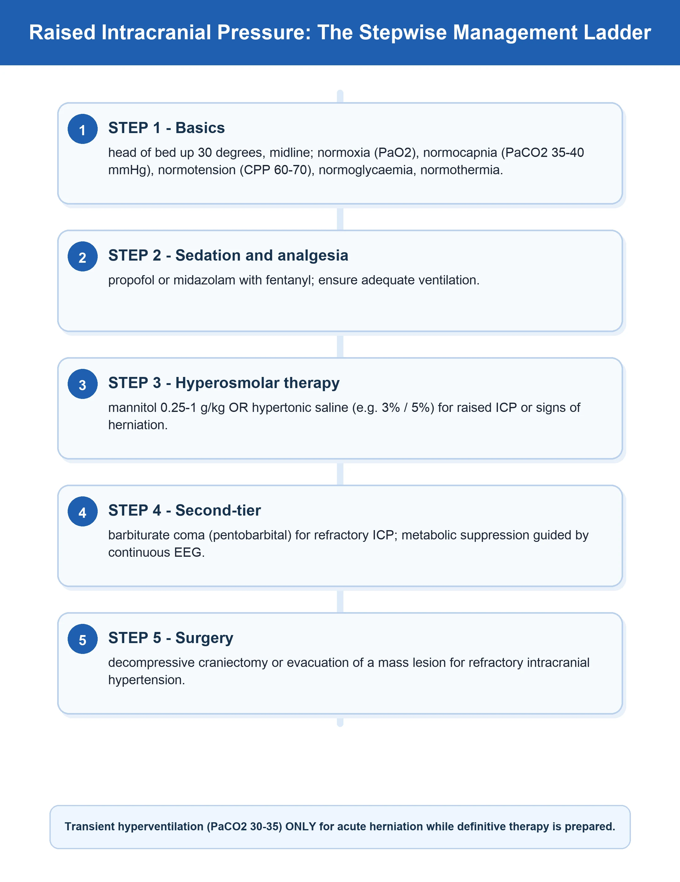
TBI management is a stepwise escalation: start with simple physiological measures, add interventions as ICP remains above 22 mmHg, and reserve the most aggressive (barbiturates, craniectomy) for refractory intracranial hypertension. Throughout, the non-negotiable priorities are: defend CPP (60-70), prevent secondary insults, and never stop looking for a surgical lesion. The order below mirrors the Brain Trauma Foundation 4th edition algorithm and the way every CICM/FFICM answer must flow.[1]
Stepwise management of raised ICP in severe TBI — the escalation staircase
-
AIRWAY, BREATHING, CIRCULATION + SURGICAL TRIAGE FIRST. Intubate and ventilate any patient with GCS ≤8 (cannot protect airway) or with hypoxia/hypercapnia — use rapid sequence intubation with a neuro-friendly induction (propofol or thiopental; avoid dropping the BP — have noradrenaline ready). Maintain SBP ≥110, SpO2 ≥94% from the moment of first contact — a single hypotensive or hypoxic episode doubles/triples mortality. Obtain an immediate non-contrast CT head to find a surgical lesion (extradural, subdural, intracerebral haematoma, depressed fracture). Evacuate any significant mass lesion — this is the definitive treatment; osmotherapy is only a bridge to surgery. Give tranexamic acid 1 g IV within 3 h of injury (CRASH-3 — reduces mortality from intracranial haemorrhage). Do NOT give steroids (CRASH — increased mortality).[4]
-
BASIC PHYSIOLOGICAL MEASURES (FIRST-TIER, ALL PATIENTS) — the foundation. (a) HEAD OF BED 30° NEUTRAL: promotes jugular venous drainage and lowers ICP; ensure the head is midline (a turned head kinks the jugular and obstructs venous outflow); remove tight cervical collars/ties that compress the neck. (b) NORMOCAPNIA — PaCO2 35-40 mmHg: avoid hypoventilation (hypercapnia → cerebral vasodilation → raised ICP) and avoid prophylactic hyperventilation (hypocapnia → cerebral vasoconstriction → ischaemia, especially in the first 24 h when the brain is most vulnerable). (c) NORMOTHERMIA: treat fever aggressively (paracetamol, surface/intravascular cooling) — do NOT use therapeutic hypothermia for ICP (Eurotherm — harm). (d) NORMOGLYCAEMIA (6-10 mmol/L). (e) SEIZURE PROPHYLAXIS — levetiracetam (preferred) or phenytoin for 7 days in severe TBI (haematoma, depressed fracture, penetrating injury). (f) Maintain serum Na >140 (avoid hyponatraemia). (g) Maintain CPP 60-70 with noradrenaline if needed.[1][6]
-
SEDATION + ANALGESIA — reduce metabolic demand and ICP. Deepen sedation to reduce cerebral metabolic rate (CMRO2) → autoregulatory vasoconstriction → reduced cerebral blood volume → lower ICP, and to prevent coughing/bucking/fighting the ventilator (each raises intrathoracic pressure and ICP). Propofol is first-line (rapidly titratable, reduces CMRO2, short half-life allowing neurological examination); fentanyl/morphine for analgesia. Avoid agents that drop the BP (excessive propofol can cause hypotension — watch CPP; propofol infusion syndrome at high doses >4 mg/kg/h for >48 h). Midazolam is an alternative but accumulates. Ensure adequate paralysis only if coughing/fighting persists despite sedation (cisatracurium) — but continuous paralysis masks seizures, so monitor with continuous EEG.[1]
-
CSF DRAINAGE (if an EVD is in situ). Drain 5-10 mL of CSF per episode for ICP >22 mmHg — immediate and effective. The EVD is both diagnostic and therapeutic. If only an intraparenchymal monitor is in place, CSF drainage is not possible — consider converting to/adding an EVD if ventricles are visible and drainage is repeatedly needed.[1]
-
OSMOTHERAPY — mannitol OR hypertonic saline (ICP >22 despite steps 1-4). (a) MANNITOL 0.5-1 g/kg IV bolus over 10-15 min (20% solution): osmotic gradient pulls water from brain into the intravascular space → reduces brain water and ICP; also improves rheology (lowers blood viscosity → autoregulatory vasoconstriction → lower ICP). Onset minutes, duration 2-6 h. MANDATORY monitoring: serum osmolarity (keep <320 mOsm/L — above → AKI), serum sodium (rises), urine output (diuretic effect — replace fluid to avoid hypovolaemia → maintain CPP), and ensure the patient is not hypovolaemic before giving (mannitol initially expands then depletes volume). AVOID repeated blind dosing — risk of rebound (mannitol crosses a disrupted BBB and paradoxically worsens oedema). (b) HYPERTONIC SALINE (3%, 5%, or 23.4%): osmotic gradient + volume expansion (maintains BP/CPP — useful in shocked TBI) + less rebound than mannitol. 3% NaCl 250 mL bolus or a continuous infusion (30-50 mL/h) titrated to serum Na 145-155 mmol/L; 23.4% (30-60 mL via central line) for acute crisis. Increasingly preferred in many centres because it preserves intravascular volume. Both agents are effective — the choice often depends on volume status (shocked → HTS; volume-overloaded → mannitol) and local protocol.[1]
-
SHORT-TERM HYPERVENTILATION — ONLY AS A BRIDGE for ACUTE HERNIATION. If the patient is actively herniating (Cushing's triad, unilateral fixed pupil, sudden GCS drop), transient hyperventilation to PaCO2 30-35 mmHg is acceptable as a TEMPORARY measure to lower ICP (vasoconstriction → reduced CBV) WHILE preparing definitive therapy (osmotherapy, surgery). It is NOT for sustained use — prolonged hypocapnia causes cerebral ischaemia. NEVER use prophylactic hyperventilation, and never sustain PaCO2 <30-35 beyond the acute crisis. This is the most overused and dangerous 'treatment' for raised ICP.[1]
-
REFRACTORY ICP (persists >22 despite osmotherapy + first-tier measures) — METABOLIC SUPPRESSION. (a) PROPOFOL infusion — titrate up (2-3 mg/kg/h, cautiously) to reduce CMRO2; monitor for hypotension and propofol infusion syndrome. (b) BARBITURATE COMA — if ICP still refractory: thiopental (5-10 mg/kg load, then 0.5-3 mg/kg/h) or pentobarbital (10 mg/kg load, then 1-4 mg/kg/h) titrated to burst suppression on continuous EEG (0-5 bursts/20 s). Barbiturates profoundly suppress CMRO2 → autoregulatory vasoconstriction → reduced CBV → lower ICP. They DO reduce ICP but show NO mortality benefit (meta-analyses), and carry major side effects: hypotension (nearly universal — requires vasopressors, which can paradoxically threaten CPP), immunosuppression, prolonged coma, paralytic ileus, hypothermia. Reserve for genuinely refractory ICP as a bridge to craniectomy or recovery. (c) NEUROMUSCULAR BLOCKADE (cisatracurium) — reduces coughing/intrathoracic pressure but masks seizures (requires cEEG) and is adjunctive only.[1]
-
DECOMPRESSIVE CRANIECTOMY — the last tier (refractory ICP). Remove a large bone flap (frontotemporoparietal, or bifrontal) ± duraplasty to allow the swollen brain to expand OUTWARDS, relieving intracranial pressure. Indications: refractory ICP (>20-25 mmHg persisting despite maximal medical therapy for >1-12 h) after severe TBI. RESCUEicp (2016, NEJM): late decompressive craniectomy for refractory ICP >25 after TBI REDUCED mortality (26.9% vs 48.9%) BUT increased the rate of vegetative and severely-disabled survival — a 'troubling trade-off' that must be openly discussed with the family. DECRA (2011, NEJM): EARLY (prophylactic, before medical failure) craniectomy was HARMFUL (worse outcomes) — do not craniectomise early; reserve it for medically refractory cases. Craniectomy is also indicated for malignant MCA infarct (swelling kills — DESTINY/HAMLET/DECIMAL) in younger patients.[2]
-
MULTIMODALITY / ADVANCED MONITORING (selected centres). Where available, brain tissue oxygenation (PbtO2) monitoring (target >15-20 mmHg) in addition to ICP/CPP: ICP and CPP can be 'at target' yet the brain still hypoxic (microvascular dysfunction). BOOST-2 (2019) suggested PbtO2-guided therapy improves favourable outcomes (emerging). Also consider cerebral microdialysis (lactate/pyruvate ratio for ischaemia) and jugular venous oximetry (SjvO2, target 55-75%) in selected severe cases. These are adjuncts to, not replacements for, ICP/CPP-guided care.[1]
Mannitol vs hypertonic saline — the osmotherapy choice
| Feature | Mannitol 20% | Hypertonic saline (3%, 5%, 23.4%) |
|---|---|---|
| Mechanism | Osmotic gradient (pulls water from brain) + rheology (lowers viscosity → autoregulatory vasoconstriction) + free-radical scavenging | Osmotic gradient (NaCl draws water out) + volume expansion + vasoregulatory + anti-inflammatory |
| Dose | 0.5-1 g/kg IV bolus over 10-15 min | 3% NaCl 250 mL bolus or 30-50 mL/h infusion; 23.4% 30-60 mL (central line) |
| Onset / duration | Minutes / 2-6 h | Minutes / 4-12 h |
| Volume effect | Diuresis → may cause hypovolaemia (replace fluid) | Volume expansion (Na retains water — beneficial if shocked) |
| Monitoring | Serum osmolarity <320 mOsm/L (above → AKI); serum Na; urine output | Serum Na 145-155 (>160 → risk); osmolarity |
| Rebound oedema | Yes (crosses disrupted BBB → reverse pull) — avoid repeated blind dosing | Less rebound (NaCl crosses BBB poorly) |
| Renal toxicity | Risk of AKI (if osmolarity >320 or hypovolaemia) | Lower (but monitor Na) |
| Best for | Acute ICP crisis with adequate volume; bolus therapy | TBI with shock/hypotension (volume expansion); continuous infusion; increasingly preferred |
| Key danger | Hypovolaemia → CPP falls → ischaemia; osmolarity >320 → AKI | Hypernatraemia (>160); central line needed for 23.4% |
Practical rule: if the TBI patient is hypotensive/volume-depleted, choose hypertonic saline (it expands volume and supports CPP); if volume-overloaded or in pulmonary oedema, mannitol may be preferred (but watch for the diuresis-induced hypovolaemia). Always check serum osmolarity before each mannitol dose and keep it <320.[1]
What NOT to do — the dangerous and contraindicated
Common TBI errors and why they harm
| Error | Why it is wrong | Evidence |
|---|---|---|
| Prophylactic hyperventilation (PaCO2 <35) | Cerebral vasoconstriction → ischaemia; most harmful in first 24 h | BTF guidelines; reserve for acute herniation bridge only |
| Corticosteroids (dexamethasone) | Increased mortality in TBI — no benefit, real harm (infection, hyperglycaemia) | CRASH trial (2004, Lancet) — steroids CONTRAINDICATED in TBI |
| Therapeutic hypothermia for raised ICP | Worse outcome (infection, coagulopathy, electrolyte shifts, rebound on rewarming) | Eurotherm (2015, NEJM) — harm; maintain normothermia |
| Hypotonic / glucose-containing fluids | Worsen cerebral oedema (free water enters brain); glucose worsens outcome | Use isotonic crystalloid (0.9% saline / balanced); avoid dextrose |
| Letting SBP fall <110 | A single hypotensive episode doubles mortality | Chesnut 1993 (Traumatic Coma Data Bank) |
| EARLY (prophylactic) craniectomy | Worse outcomes than medical management | DECRA (2011, NEJM) — only craniectomise when refractory |
| Repeated blind mannitol without osmolarity | Rebound oedema + AKI (osmolarity >320) | BTF guidelines — check osmolarity each dose |
| Sustained high CPP (>70) | ARDS / fluid overload / adult respiratory distress | BEST:TRIP (2012, NEJM) — keep CPP 60-70 |
| Forgetting to look for a surgical lesion | Osmotherapy is only a bridge — the cure is evacuation | Any expanding haematoma needs the operating theatre |
Clinical pearls
Key trials and evidence
Carney 2017 — Brain Trauma Foundation Guidelines for Severe TBI, 4th Edition (PMID 27654000)
Source
Neurosurgery 2017;80(1):6-15 — the current global standard for severe TBI management, updating the 3rd edition (Bratton 2007)
What it established
Revised the **ICP treatment threshold to >22 mmHg** (up from 20 in the 3rd edition, based on the SIGN trial and outcome data — each mmHg above 22 increases mortality); reaffirmed **CPP target 60-70 mmHg**; recommended ICP monitoring in severe TBI (GCS 3-8) with abnormal CT OR normal CT with ≥2 risk factors (age >40, motor posturing, SBP <90); downgraded early/prophylactic hypothermia and prophylactic hyperventilation
Key contribution
Codified the escalation staircase (head elevation → sedation → CSF drainage → osmotherapy → barbiturates → craniectomy), explicitly recommended against steroids (CRASH), and integrated emerging multimodality monitoring (PbtO2). The exam-defining reference for every TBI question.
Clinical bottom line
The single most important document in neurotrauma — know the ICP threshold (22), the CPP target (60-70), and the staircase. Every modern TBI guideline builds on this.
Hutchinson 2016 — RESCUEicp: Decompressive Craniectomy for Traumatic Intracranial Hypertension (NEJM) (PMID 27602507)
Source
New England Journal of Medicine 2016;375(12):1119-1130 — multinational RCT, 408 patients with severe TBI and refractory ICP >25 mmHg despite maximal medical therapy, randomised to decompressive craniectomy (bifrontal or large unilateral) vs continued medical care (including barbiturates)
What it found
Late decompressive craniectomy **REDUCED mortality** (26.9% vs 48.9%) — a 22-percentage-point absolute mortality reduction. BUT survivors were more likely to be **vegetative or severely disabled** (lower upper, upper severe, lower severe disability on the GOS-E) compared with the medical group.
Key contribution
Defined the 'troubling trade-off' of craniectomy for refractory ICP: it saves lives but at the cost of more dependent survivors. Established that craniectomy is appropriate ONLY for medically refractory ICP — never prophylactically (contrast DECRA 2011, where early craniectomy was harmful).
Clinical bottom line
Craniectomy for refractory ICP >25 after TBI saves life but increases vegetative/severe-disability survival — the decision MUST be made with the family after explicit prognostic discussion.
Chesnut 2012 — BEST:TRIP: ICP Monitoring in TBI (NEJM) (PMID 23234472)
Source
New England Journal of Medicine 2012;367(26):2471-2481 — RCT in 324 severe TBI patients in Bolivia/Ecuador, comparing ICP-monitor-guided therapy vs imaging-and-examination-guided therapy
What it found
No significant difference in the composite primary outcome (survival time, impaired consciousness, functional status at 3/6 months, neuropsychological function) between the two strategies. ICP-monitored patients received fewer days of intensive treatment but had similar outcomes.
Key contribution
Often misread as 'ICP monitoring doesn't matter' — the correct interpretation is that in well-resourced settings with serial imaging and exam, carefully protocolised care can achieve equivalent outcomes; and that aggressive fluid/vasopressor loading to push CPP very high risks ARDS. Reinforced the CPP 60-70 window and the principle that treatment intensity should match the patient, not a single number.
Clinical bottom line
ICP monitoring remains standard in severe TBI in well-resourced settings (BTF recommends it); BEST:TRIP tempers over-aggressive CPP-pushing and supports judicious, protocolised care.
Chesnut 1993 — Secondary Brain Injury (Traumatic Coma Data Bank) (PMID 8459458)
Source
Journal of Trauma 1993;34(2):216-222 — landmark analysis of the Traumatic Coma Data Bank (714 severe head-injury patients) examining systemic secondary insults
What it found
A SINGLE episode of **hypotension (SBP <90 mmHg)** was associated with **DOUBLE the mortality** (from ~27% to 55%) and worse outcomes in survivors; **hypoxia** independently worsened outcome; the combination of hypotension and hypoxia was nearly universally fatal. These were the strongest modifiable predictors of bad outcome.
Key contribution
Provided the foundational evidence that **secondary brain injury is the preventable killer** — and that preventing hypotension and hypoxia from the moment of first contact is the single most impactful intervention in TBI. Every element of prehospital and early hospital TBI care (avoiding hypotonic fluids, early intubation, maintaining SBP/MAP) traces back to this paper.
Clinical bottom line
The 'why' behind the entire resuscitative priority in TBI — never let a head-injured patient become hypotensive or hypoxic; a single episode can convert a survivable injury into a lethal one.
Cnossen / Stocchetti 2017 — CENTER-TBI Survey of ICP Management (PMID 28874206)
Source
Critical Care 2017;21:233 — survey of monitoring and treatment policies for intracranial hypertension across 66 neurotrauma centres participating in the CENTER-TBI study (including Stocchetti N, Maas AIR, Menon D, Citerio G)
What it found
Substantial VARIATION in ICP monitoring uptake, device choice (intraparenchymal most common), treatment thresholds, osmotherapy agent preference (hypertonic saline increasingly first-line), and use of decompressive craniectomy across European centres — reflecting genuine clinical uncertainty where evidence is incomplete
Key contribution
Documented that real-world TBI care is heterogenous and guideline-adherence is variable, motivating the CENTER-TBI cohort study to generate the evidence needed to standardise care; highlighted the shift toward hypertonic saline as first-line osmotherapy and the controversy over craniectomy after RESCUEicp
Clinical bottom line
Even the experts vary — know the evidence (BTF 4th edition, RESCUEicp, BEST:TRIP) and apply it in a protocolised, patient-centred way; the field is still refining who benefits most from aggressive ICP-directed therapy.
Bratton / Brain Trauma Foundation 2007 — Severe TBI Guidelines, 3rd Edition (PMID 17511549)
Source
Journal of Neurotrauma 2007;24 Suppl 1:S1-106 — the 3rd edition of the BTF guidelines (preceded the 2017 4th edition); this citation is Chapter I (Blood pressure and oxygenation)
What it established
Set the earlier **ICP treatment threshold at 20 mmHg** (revised upward to 22 in the 4th edition); CPP target 60-70; the blood-pressure-and-oxygenation chapter formalised the avoidance of hypotension (SBP <90) and hypoxia (PaO2 <60) as Level II/III recommendations, building on the Chesnut 1993 data
Key contribution
The reference that governed neurotrauma care for a decade (2007-2017) and that many older studies and local protocols were built upon — know it because exam questions and older literature still cite the 20 mmHg threshold and its recommendations
Clinical bottom line
The 3rd edition is the historical baseline; the 4th edition (2017) supersedes it for current thresholds (ICP 22). Know the transition and why the threshold moved.
Red flags
Prognosis
Severe TBI outcomes and prognostic factors
| Factor | Outcome impact | Notes |
|---|---|---|
| Severe TBI overall mortality | 20-40% with optimal neurocritical care | Worse with older age, comorbidity, polytrauma, delayed presentation |
| ICP control | Each mmHg >22 increases mortality | ICP that cannot be controlled medically predicts poor outcome; refractory ICP prompts craniectomy decision |
| Hypotension (SBP <90) | DOUBLES mortality (27% → 55%) | Chesnut 1993 — a single episode; prevention is paramount |
| Hypoxia (SpO2 <90) | TRIPLES mortality | Chesnut 1993 — early intubation/oxygenation |
| Age | Mortality rises steeply >60 | Older brains have less compliance and more comorbidity |
| Initial GCS (motor) | Lower motor score → worse outcome | GCS 3 (motor 1) at 72 h is a grave sign (but not alone sufficient to withdraw) |
| Pupillary response | Bilateral fixed dilated → very poor | Bilateral absent pupillary + corneal reflex at 72 h strongly predicts death/vegetative |
| CT findings | Compressed/absent basal cisterns, midline shift >5 mm, traumatic SAH → worse | Marshall CT classification correlates with outcome |
| Decompressive craniectomy (RESCUEicp) | Reduces mortality (27% vs 49%) but more vegetative/severe disability | The trade-off must be discussed with family |
| Best:TRIP ICP-monitoring strategy | No outcome difference vs imaging/exam-guided | Supports protocolised care; tempers over-aggressive CPP pushing |
| Multimodality (PbtO2-guided) | BOOST-2 — trend to favourable outcome | Emerging; not yet standard |
| Functional recovery | Often months-years; many survivors have cognitive/behavioural sequelae | Rehabilitation is essential; young motivated patients do best |
Exam practice — SAQ
SAQ — Refractory ICP after severe TBI
10 minutes · 10 marks
A 28-year-old after RTA has GCS 6, intubated, ICP monitor reading 28 mmHg, MAP 78 mmHg, PaCO2 32 mmHg, Na 138. Pupils equal and reactive. CT shows diffuse swelling without surgical mass.
Integration — the one-minute mental model
When asked to manage a severe TBI in the exam or at the bedside, run this sequence every time: (1) ABC + defend SBP/SpO2 from the scene + RSI for GCS ≤8 + CT for a surgical lesion (evacuate if present) + TXA within 3 h. (2) Place an ICP monitor (EVD gold standard / intraparenchymal) for severe TBI. (3) Set targets: ICP <22, CPP 60-70. (4) First-tier: HOB 30° neutral, normocapnia (35-40, NO prophylactic hyperventilation), normothermia, normoglycaemia, sedation, seizure prophylaxis (levetiracetam 7 days), NO steroids. (5) Escalate for ICP >22: CSF drainage → osmotherapy (mannitol 0.5-1 g/kg with osmolarity check, OR HTS to Na 145-155) → barbiturate coma (burst suppression) if refractory → decompressive craniectomy (RESCUEicp — with family discussion). (6) Defend CPP with noradrenaline throughout; never let it fall. (7) Look for and prevent every secondary insult. Master this sequence and you have mastered the topic.[1][4][2]
References
- [1]Carney N, Totten AM, O'Reilly C, Ullman JS, Hawryluk GW, Bell MJ, Bratton SL, Chesnut R, Harris OA, Kissoon N, Rubiano AM, Shutter L, Tasker RC, Vavilala MS, Wilberger J, Wright DW, Ghajar J. Guidelines for the Management of Severe Traumatic Brain Injury, Fourth Edition Neurosurgery, 2017.PMID 27654000
- [2]Hutchinson PJ, Kolias AG, Timofeev IS, Corteen EA, Czosnyka M, Timothy J, Anderson I, Bulters DO, Belli A, Eynon CA, Wadley J, Mendelow AD, Mitchell PM, Wilson MH, Critchley G, Sahuquillo J, Unterberg A, Servadei F, Teasdale GM, Pickard JD, Menon DK, Murray GD, Kirkpatrick PJ; RESCUEicp Trial Collaborators. Trial of Decompressive Craniectomy for Traumatic Intracranial Hypertension N Engl J Med, 2016.PMID 27602507
- [3]Chesnut RM, Temkin N, Carney N, Dikmen S, Rondina C, Videtta W, Petroni G, Lujan S, Pridgeon J, Barber J, Machamer J, Chaddock K, Celix JM, Cherner M, Hendrix T; Global Neurotrauma Research Group. A trial of intracranial-pressure monitoring in traumatic brain injury N Engl J Med, 2012.PMID 23234472
- [4]Chesnut RM, Marshall LF, Klauber MR, Blunt BA, Baldwin N, Eisenberg HM, Jane JA, Marmarou A, Foulkes MA. The role of secondary brain injury in determining outcome from severe head injury J Trauma, 1993.PMID 8459458
- [5]Cnossen MC, Huijben JA, van der Jagt M, Volovici V, van Essen T, Polinder S, Nelson D, Ercole A, Stocchetti N, Citerio G, Peul WC, Maas AIR, Menon D, Steyerberg EW, Lingsma HF; CENTER-TBI investigators. Variation in monitoring and treatment policies for intracranial hypertension in traumatic brain injury: a survey in 66 neurotrauma centers participating in the CENTER-TBI study Crit Care, 2017.PMID 28874206
- [6]Brain Trauma Foundation, American Association of Neurological Surgeons, Congress of Neurological Surgeons, Joint Section on Neurotrauma and Critical Care, AANS/CNS, Bratton SL, Chestnut RM, Ghajar J, McConnell Hammond FF, Harris OA, Hartl R, Manley GT, Nemecek A, Newell DW, Rosenthal G, Schouten J, Shutter L, Timmons SD, Ullman JS, Videtta W, Wilberger JE, Wright DW. Guidelines for the management of severe traumatic brain injury. I. Blood pressure and oxygenation J Neurotrauma, 2007.PMID 17511549