ICU · Seizures & weakness
Status Epilepticus, Guillain-Barré and Myasthenia Gravis
Also known as Status epilepticus · Seizures · Guillain-Barre syndrome · Myasthenia gravis · Neuromuscular respiratory failure · Non-convulsive status
Status epilepticus is the prolonged or the recurrent seizure that becomes self-sustaining — a neurological emergency with a mortality of 10 to 20 per cent. Guillain-Barre and myasthenia gravis are the two neuromuscular diseases that most commonly cause the respiratory failure needing the ICU. This topic builds the examiner's framework on the SE management (the benzodiazepine, the fosphenytoin/levetiracetam, the RAMPART and the ESETT evidence), the GBS (the IVIG, the plasma exchange, the respiratory monitoring), and the myasthenia (the IVIG, the plasma exchange, the crisis).
On this page & tools
Your progress
Saved locally on this device.
8 MCQs with explanations
Target exams
Overview & definition
This domain covers three conditions that bring the patient to the ICU for a neurological reason — the status epilepticus (the prolonged or the recurrent seizure), the Guillain-Barre syndrome (the acute, the ascending paralysis), and the myasthenia gravis (the fatigable weakness). Each can cause the respiratory failure requiring the ICU, and each has a specific, the evidence-based management.[1][1]
Status epilepticus: definition and the phases
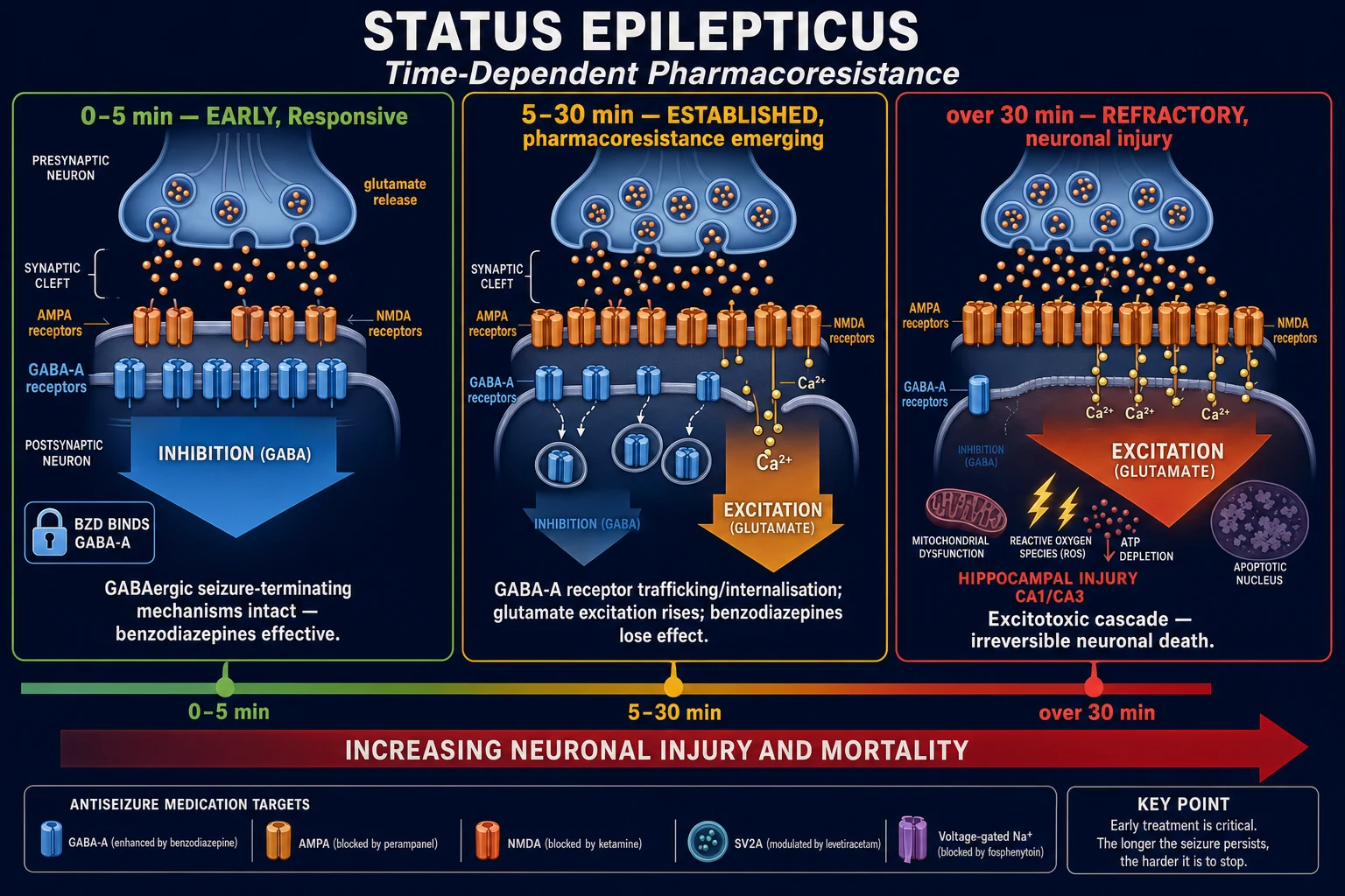
The status epilepticus is the seizure lasting over 5 minutes (continuous), or the recurrent seizures without the recovery of the consciousness in between. The operational ILAE definition (Trinka 2015) introduces two time-points: t1 — the threshold beyond which the seizure should be treated (5 minutes for the generalised convulsive SE, 10 minutes for the focal SE with the impaired consciousness); and t2 — the time beyond which the long-term consequences (the neuronal injury, the pharmacoresistance, the network plasticity) become likely (30 minutes for the GCSE). The operational implication: treat at t1, do not wait for t2.[4][1]
The refractory SE (the failure of the first-line benzodiazepine and the second-line antiepileptic drug — typically at the 40-to-60-minute mark) and the super-refractory SE (the persistence or the recurrence beyond 24 hours despite the anaesthetic infusion) carry a rising mortality — the refractory SE mortality approaches 30 to 40 per cent, the super-refractory 35 to 60 per cent in the adult series. The super-refractory SE affects roughly 10 to 15 per cent of the refractory cases.[1][12]
The four phases of the SE management
The phases of the SE management drive the staged drug ladder. The receptor pharmacology underpins the urgency — as the seizure persists, the GABA-A receptors internalise (the benzodiazepines lose efficacy) and the NMDA receptors externalise (the glutamatergic drive strengthens). The early ladder is therefore GABAergic; the late ladder adds the NMDA antagonist (the ketamine).[1][5]
| Phase | Time window | Drug target | The drug |
|---|---|---|---|
| The early SE | 5 to 20 min | The GABA-A receptor (still surface-expressed) | The IV lorazepam 4 mg or the IM midazolam 10 mg |
| The established SE | 20 to 40 min | The synaptic membrane and the ion channels | The IV levetiracetam 60 mg/kg, the fosphenytoin 20 mg PE/kg, or the valproate 40 mg/kg |
| The refractory SE | 40 to 60 min | The anaesthetic GABAergic / NMDA receptor | The propofol, the midazolam, the thiopentone, the ketamine — intubate and ventilate |
| The super-refractory SE | Beyond 24 h of the anaesthetic | The immune, the metabolic, the surgical | The ketamine, the ketogenic diet, the immunotherapy, the surgery, the hypothermia |
The causes — the aetiology drives the prognosis
The cause is the dominant prognostic factor in the SE — more so than the drug choice. The five cause-groups: [1]
- The acute symptomatic — the stroke, the trauma, the tumour, the intracranial haemorrhage, the CNS infection, the anoxia (after the cardiac arrest), the alcohol withdrawal, the drug toxicity, the metabolic (the hyponatraemia, the hypoglycaemia, the hepatic and the renal encephalopathy).
- The remote symptomatic — the prior stroke, the prior trauma, the malformation of the cortical development.
- The progressive — the tumour, the degenerative, the autoimmune (the anti-NMDA receptor, the anti-LGI1, the anti-GABA-A receptor encephalitis), the paraneoplastic.
- The cryptogenic / the NORSE — the new-onset refractory SE of unknown cause after the exhaustive work-up; the FIRES subtype follows a febrile prodrome.
- The known epilepsy with the subtherapeutic level — the non-adherence or the interaction-driven drop in the antiepileptic level. [1]
The NORSE and the FIRES are the cryptogenic syndromes that account for a disproportionate share of the super-refractory SE — the Hirsch consensus definitions (2018) standardise the diagnostic criteria and mandate the autoimmune and the infectious work-up.[9][5]
The staged management of the status epilepticus — the time-critical ladder
0 to 5 min — Stabilise and assess
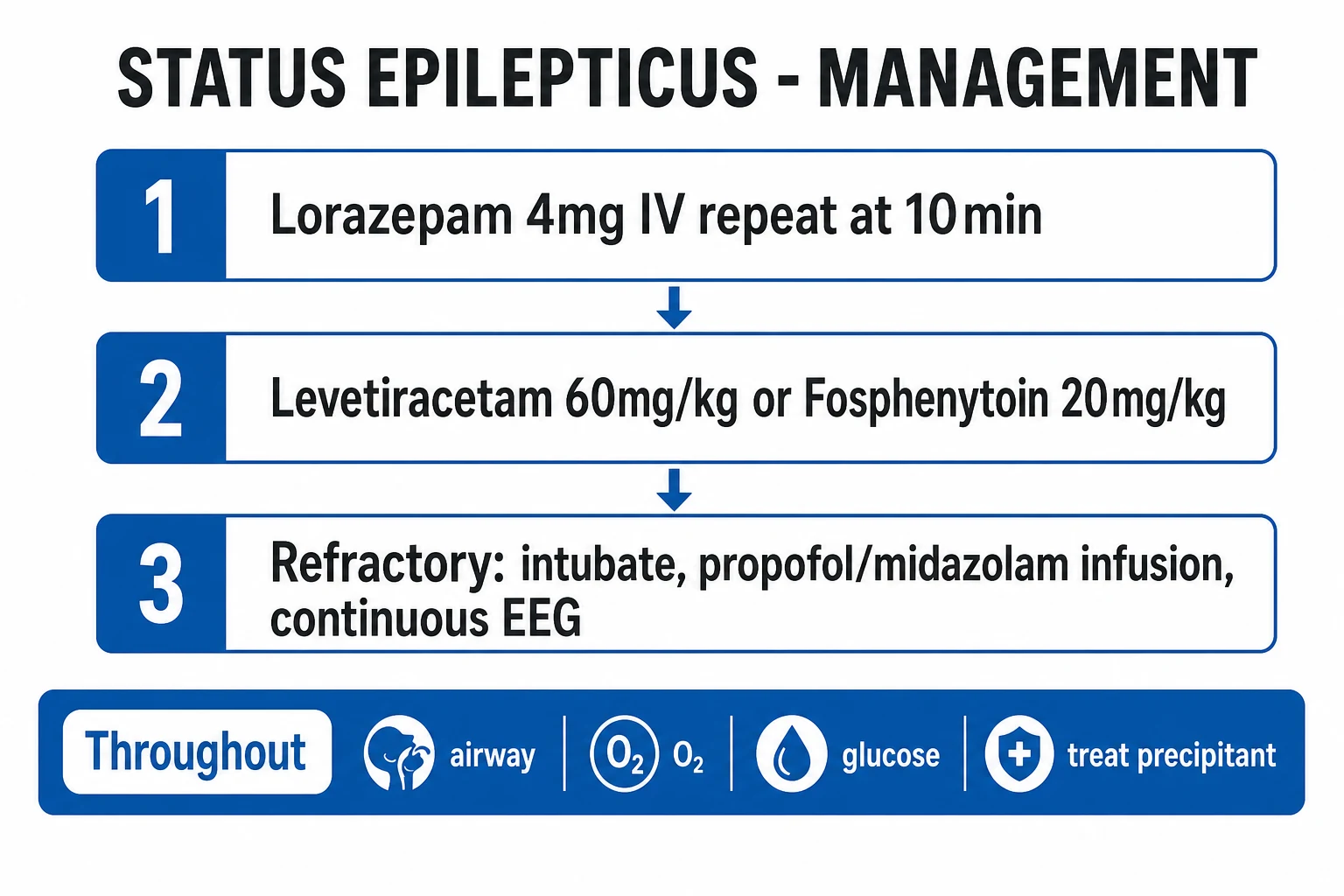
Airway (recovery position, suction, oxygen 15 L/min via the non-rebreather), Breathing (the saturation, the intubate if the airway or the ventilation fails), Circulation (the IV access, the blood pressure), Disability (the capillary glucose immediately — the hypoglycaemia is the single most reversible cause; give 50 mL of the 50 per cent dextrose with the thiamine 100 mg IV in the suspected alcohol misuse), check the temperature. Do not delay the benzodiazepine.
5 to 10 min — First line: the benzodiazepine
Lorazepam 4 mg IV (0.1 mg/kg), repeated once after 5 to 10 minutes if still seizing. If no IV access: midazolam 10 mg IM (RAMPART — superior to the IV lorazepam because the IV access is often delayed). Alternatives: diazepam 10 mg IV (redistributes fast, recurs) or clonazepam 1 mg IV. The benzodiazepines are the most effective drugs but only in the first 10 to 20 minutes — beyond that the GABA-A receptor internalisation reduces their efficacy.
20 to 40 min — Second line: the IV antiepileptic
Give regardless of whether the benzodiazepine worked, to prevent the recurrence. Levetiracetam 60 mg/kg IV (max 4.5 g), OR fosphenytoin 20 mg PE/kg IV (max 1.5 g PE, at 150 mg PE/min), OR valproate 40 mg/kg IV (max 3 g). ESETT showed all three equivalent (about half stopped seizing within 10 minutes). Valproate is avoided in the woman of the child-bearing age and the hepatic failure; the fosphenytoin in the severe cardiac disease.
40 to 60 min — The refractory SE: the anaesthetic intubation
If still seizing after the benzodiazepine and the second-line agent, the SE is refractory. Induce, intubate, and ventilate. Start a continuous anaesthetic infusion titrated to the burst suppression on the continuous EEG: propofol OR midazolam first-line, the ketamine or the thiopentone as the alternatives. Admit to the ICU. The cEEG is the only way to confirm the seizure has stopped in the paralysed, sedated patient.
After 24 h — The super-refractory SE
If the seizure continues or recurs for more than 24 hours after the anaesthetic (including on weaning), the SE is super-refractory. Maintain the anaesthesia, add the ketamine infusion, the empirical immunotherapy (the methylprednisolone, the IVIG, the plasma exchange) for the suspected autoimmune cause, the ketogenic diet, the surgery (the resection of the epileptic focus), and the targeted temperature management. Prolonged ICU stay, high morbidity, ~40 to 60 per cent mortality.
The SE management: the evidence
The benzodiazepine first. The benzodiazepines are the most effective drugs in the early SE (the first 10 to 20 minutes), acting on the GABA-A receptor to potentiate the chloride influx and the neuronal hyperpolarisation. Three landmark trials anchor the evidence.[1][5]
- The VA Cooperative Study (Treiman, NEJM 1998) — the open-label randomised trial of the first-line therapy in the overt and the subtle GCSE (n = 518; 384 overt). The IV lorazepam was the most effective monotherapy in the overt SE (the seizure stopped in 65 per cent), superior to the phenytoin (58 per cent), the phenobarbital (56 per cent), and the diazepam (42 per cent). In the subtle SE all four were poor (10 to 25 per cent). The lorazepam became the first-line IV agent.[3]
- The RAMPART trial (Silbergleit, NEJM 2012) — the double-blind non-inferiority RCT of the intramuscular midazolam 10 mg versus the intravenous lorazepam 4 mg for the prehospital SE (n = 893). The IM midazolam was actually superior — the seizure terminated before the arrival in 63 per cent versus 53 per cent — because the IV access was difficult and delayed (no IV access in 23 per cent). The fewer recurrences and the fewer intubations with the IM midazolam. The practice change: the IM midazolam is the first-line prehospital drug when the IV access is not available.[1]
- The Misra 2016 trial — the four-drug Indian comparison (the lorazepam, the levetiracetam, the valproate, the lacosamide) in the established SE — found no significant difference between the drugs, supporting the ESETT-equivalence conclusion in a different population.[8]
The takeaway: a benzodiazepine at five minutes is the single most effective intervention in the SE; the route (the IV lorazepam if the access is present, the IM midazolam if not) is less important than the timing.[5]
RAMPART
NEJM 2012
Double-blind non-inferiority RCT; prehospital SE; IM midazolam 10 mg (adult over 40 kg, 5 mg if 13 to 40 kg) vs IV lorazepam 4 mg; n=893 (892 adults)
Key finding
IM midazolam was superior to IV lorazepam — seizure terminated before arrival in 63% vs 53%. No IV access in 23% of the IV arm. Fewer recurrences and fewer intubations with the IM midazolam.
Practice change
IM midazolam is the first-line prehospital therapy when IV access is not yet established — give it early, do not struggle for a line. RSI for the refractory is delayed if the IV access is awaited.
VA Cooperative Study
NEJM 1998
Open-label randomised trial; overt and subtle GCSE; lorazepam vs phenytoin vs phenobarbital vs diazepam; n=518 (384 overt)
Key finding
In the overt GCSE, lorazepam was most effective (stopped in 65%) and superior to diazepam (42%); in the subtle SE all four were poor (10-25%).
Practice change
Lorazepam became the preferred first-line IV benzodiazepine for the established SE; phenobarbital and phenytoin are not superior first-line agents. The poor response in the subtle SE drove the staged ladder upward.
Second-line therapy — the IV antiepileptics (ESETT)
The ESETT (Kapur, NEJM 2019) is the definitive second-line trial. It randomised the patients with the established SE (the benzodiazepine-refractory convulsive SE within 60 minutes) to levetiracetam 60 mg/kg, fosphenytoin 20 mg PE/kg, or valproate 40 mg/kg. The trial was stopped early at the interim analysis for the futility — there was no difference between the three drugs. Each stopped the seizure in about half of the patients within 10 minutes (levetiracetam 47 per cent, fosphenytoin 45 per cent, valproate 46 per cent). The takeaway: any of the three is acceptable, the choice is driven by the patient, and all three are inadequate alone in roughly half.[2]
ESETT
NEJM 2019
Double-blind RCT; convulsive SE refractory to benzodiazepines; levetiracetam 60 mg/kg vs fosphenytoin 20 mg PE/kg vs valproate 40 mg/kg; n=384
Key finding
No difference: seizure stopped within 10 min in 47% (levetiracetam), 45% (fosphenytoin), 46% (valproate). Stopped early for futility. All three equivalent and all inadequate alone in roughly half.
Practice change
Any of the three is acceptable second line; choose by the patient (valproate avoided in the pregnancy and the hepatic failure; fosphenytoin in the cardiac disease; levetiracetam preferred if the cause or the organ function is unknown).
Levetiracetam
60 mg/kg IV (max 4.5 g)
- The broad-spectrum, the favoured default — no hepatic metabolism, no cardiac, no interaction
- Commonest choice when the cause and the organ function are unknown
- Side effects: the sedation, the agitation, the thrombocytopenia (rare)
- Loading over 5 minutes; well tolerated
Fosphenytoin
20 mg PE/kg IV (max 1.5 g PE)
- The water-soluble prodrug of the phenytoin — can be given faster (150 mg PE/min) and via the IM route
- Less tissue necrosis and the purple-glove than the phenytoin
- Monitor the ECG (the PR prolongation, the QRS widening, the hypotension) — give via a large vein, stop if the arrhythmia
- Avoid in the heart block and the severe cardiac disease
Valproate
40 mg/kg IV (max 3 g)
- Effective across the broad spectrum — the generalised, the focal, the absence
- Avoid in the woman of the child-bearing age (the teratogenicity) and the hepatic failure (the hepatotoxicity)
- Monitor the ammonia (the hyperammonaemic encephalopathy)
- Platelet inhibitor — caution with the bleeding and the thrombocytopenia
Lacosamide
400 to 800 mg IV
- The novel sodium-channel modulator (the slow inactivation); emerging fourth option in the ESETT-failed SE
- Less hypotension than the fosphenytoin; the renal excretion (no hepatic load)
- Off-label for the established SE; the evidence is the case series and the small trials
- The PR prolongation — avoid in the second- or third-degree heart block
Refractory SE — the anaesthetic infusions and the continuous EEG
When the seizure continues despite the benzodiazepine and the second-line agent, the SE is refractory and the patient requires the anaesthetic intubation, the ventilation, and the continuous infusion of an anaesthetic agent titrated to the burst suppression on the continuous EEG. The goal is the burst suppression (the bursts of the activity alternating with the flat suppression, the inter-burst interval of 5 to 10 seconds) maintained for 24 to 48 hours before the weaning and the re-assessment.[1][5]
The Brophy (Neurocritical Care Society 2012) guideline mandates the anaesthetic infusion and the cEEG in the refractory SE, the burst-suppression target for 24 to 48 hours, and the systematic work-up of the underlying cause.[5]
Propofol
GABA agonist
- Fast onset, fast offset (the easy titration and the rapid wean) — the most-used first-line anaesthetic for the RSE
- Bolus 1 to 2 mg/kg, infusion 30 to 200 mcg/kg/min
- Risk: the propofol infusion syndrome (PRIS) — the metabolic acidosis, the rhabdomyolysis, the bradycardia, the cardiac arrest, especially over 4 mg/kg/h for over 48 hours
- Monitor the lactate, the CK, the triglycerides, and the ECG; cap the dose in the child
Midazolam
GABA agonist
- Fast onset, water-soluble; the reliable continuous infusion
- Bolus 0.2 mg/kg, infusion 0.05 to 2 mg/kg/h
- Risk: the tachyphylaxis (the rapid receptor tolerance — the dose escalates), the hypotension
- The seizure often recurs on the weaning — the slow taper required
Thiopentone / Pentobarbital
GABA agonist (barbiturate)
- The most potent and the longest-acting — reserved for the super-refractory after the propofol, the midazolam, and the ketamine fail
- Bolus 5 mg/kg, infusion 1 to 5 mg/kg/h (or the pentobarbital 1 to 3 mg/kg/h)
- Profound burst suppression but the severe side effects: the hypotension, the ileus, the immunosuppression, the pneumonia, the prolonged coma
- Long half-life — the slow wean over days; the drug accumulates and the level is hard to interpret
Ketamine
NMDA antagonist
- The NMDA-receptor antagonist — the rationale strengthens as the seizure persists (the NMDA receptors externalise)
- Bolus 1 to 2 mg/kg, infusion 0.5 to 15 mg/kg/h
- Preserves the BP and the sympathetic tone (the inotropic — avoids the hypotension of the other anaesthetics)
- Evidence: the Rosati 2012 series found it stopped or reduced the seizures in roughly two-thirds of the refractory SE, with a favourable haemodynamic profile
- Side effects: the hallucination (irrelevant in the intubated), the hypertension, the sialorrhoea, the theoretical raised ICP
The continuous EEG — the mandatory monitor in the refractory SE
In the refractory and the super-refractory SE, the EEG is the only way to know whether the seizure has stopped, because the patient is paralysed and sedated and the clinical signs are abolished. The continuous EEG (the cEEG) serves three purposes:[6]
- To confirm the seizure termination — the ictal rhythm resolves and the background returns, or (in the anaesthetised patient) the burst suppression is achieved.
- To titrate the anaesthetic — the infusion is adjusted to maintain the burst suppression (the inter-burst interval of 5 to 10 seconds), maintained for 24 to 48 hours before the weaning. The depth targets: the burst suppression, the suppression-burst (a deeper level with longer suppression), or the complete suppression (the isoelectric EEG — reserved for the most refractory).
- To detect the non-convulsive SE — the patient who fails to wake up after the convulsive SE may be in the NCSE (the continuing epileptiform activity without the motor convulsions). The cEEG detects it; the bedside examination cannot. [1]
The Claassen (Neurology 2004) study of the continuous EEG in the critically ill patient detected the non-convulsive seizures in 8 per cent and the NCSE in 18 per cent of the comatose patients monitored — many with no clinical sign of the ongoing seizure. The Towne (Neurology 2000) study found the 8 per cent prevalence of the NCSE in the comatose ICU patients without the clinical seizure activity, and recommended the cEEG for any comatose patient with the unexplained depressed consciousness.[6][7]
The weaning of the anaesthetic
After 24 to 48 hours of the burst suppression, the anaesthetic is weaned (the infusion reduced by a quarter to a third every 6 to 12 hours) with the continuous EEG running. If the seizures or the ictal pattern recur, the infusion is restarted, the duration extended to 48 to 72 hours, and an additional agent considered (the ketamine, the immunotherapy, the ketogenic diet). The super-refractory SE (the recurrence or the persistence beyond 24 hours of the anaesthetic) may require weeks of the coma and is associated with the high morbidity and the mortality.[1][12]
Super-refractory SE — the desperate options
The super-refractory SE (SRSE) — the persistence or the recurrence of the seizure beyond 24 hours of the anaesthetic infusion — is the rarest and the most challenging form, with a mortality of 35 to 60 per cent in the adult series and a significant long-term morbidity in the survivors. The Ferlisi Global Audit (2015) reported the international case series and the audit framework; the Kantanen (2015) Finland population-based study estimated the SRSE incidence at 0.4 to 2.0 per 100 000 per year and the mortality at 35 per cent.[13][12]
The management of the SRSE adds five classes beyond the standard anaesthetic ladder: [1]
- The ketamine infusion — the rationale (the NMDA-receptor externalisation late in the SE) and the favourable haemodynamics drive its early use in the SRSE. The Rosati (Neurology 2012) series reported the seizure reduction in roughly two-thirds of the paediatric refractory SE.[11]
- The ketogenic diet — the 4:1 fat-to-carbohydrate ratio, achieving the ketosis over 3 to 4 days; the Thakur (Neurology 2014) case series of the adults in the SRSE reported the seizure cessation in roughly two-thirds of the treated patients. The practical barriers: the enteral access, the slow onset, the family and the dietitian engagement.[10]
- The empirical immunotherapy — for the suspected autoimmune cause (the anti-NMDA receptor, the anti-LGI1, the anti-GABA-A receptor encephalitis), the high-dose methylprednisolone (1 g IV daily for 3 to 5 days), the IVIG (0.4 g/kg/day for 5 days), the plasma exchange (5 sessions). The early empirical trial is justified by the high mortality of the SRSE and the treatability of the autoimmune cause.
- The targeted temperature management — the hypothermia to 32 to 36 C for 24 to 48 hours has been used in the case series; the randomised evidence is absent and the shivering, the infection, and the bradycardia are the complications.
- The surgery — the resection of the focal epileptic lesion (the tumour, the dysplasia, the cavity of the prior stroke) when the imaging and the electrocorticography localise the focus. The resective surgery or the multiple subpial transection is reserved for the surgically-tractable SRSE.
Ketamine
NMDA antagonist
- The NMDA-receptor antagonist; the rationale strengthens as the seizure persists (the NMDA receptors externalise)
- Bolus 1 to 2 mg/kg, infusion 1 to 15 mg/kg/h
- Preserves the BP and the sympathetic tone — avoids the hypotension of the other anaesthetics
- The Rosati (Neurology 2012) series: the seizure reduction in roughly two-thirds of the paediatric refractory SE
- Side effects: the hypertension, the sialorrhoea, the theoretical raised ICP
Ketogenic diet
4:1 fat:carbohydrate
- The 4:1 fat-to-carbohydrate ratio, the ketosis achieved over 3 to 4 days
- The Thakur (Neurology 2014) adult SRSE series: the seizure cessation in roughly two-thirds
- The barriers: the enteral access, the slow onset, the dietitian engagement
- Caution with the propofol (the synergistic PRIS risk), the valproate (the impaired ketogenesis)
Immunotherapy
Steroid + IVIG + PLEX
- For the suspected autoimmune cause (the anti-NMDA receptor, the anti-LGI1, the anti-GABA-A receptor encephalitis)
- The methylprednisolone 1 g IV daily for 3 to 5 days, the IVIG 0.4 g/kg/day for 5 days, the plasma exchange (5 sessions)
- The early empirical trial is justified by the high mortality of the SRSE
- Send the CSF antibody panel BEFORE the immunotherapy — the result is confounded after
Hypothermia
32 to 36 C, 24-48 h
- The targeted temperature management to 32 to 36 C for 24 to 48 hours
- The randomised evidence is absent — the case series only
- The complications: the shivering, the infection, the bradycardia, the coagulopathy
- Reserved for the refractory SRSE after the standard ladder fails
The NORSE / FIRES subtypes deserve a special mention — the cryptogenic, the often-young patient with the super-refractory SE of unknown cause after the exhaustive work-up. The Hirsch consensus definitions (2018) standardise the diagnostic criteria; the early empirical immunotherapy (within 7 days) is recommended by the international consensus, the ketogenic diet is offered early, and the surgical work-up is pursued if the focus is localised.[9][13]
Neuromuscular blocking agents in the severe SE — the train-of-four monitoring
The neuromuscular blocking agent (NMBA) is occasionally required in the severe, the convulsive SE — not for the seizure control (the NMBA abolishes only the motor activity, not the cerebral epileptiform activity), but for the patient-ventilator synchrony in the patient with the violent, the uncontrolled motor activity, the ventilator dyssynchrony, the rising intra-abdominal and the intrathoracic pressures, or the rhabdomyolysis from the ongoing muscle activity. The NMBA is NEVER a substitute for the anaesthetic and the antiepileptic therapy — the cEEG is mandatory whenever the NMBA is used, because the only sign of the ongoing seizure is the EEG.[5][1]
The indications for the NMBA in the SE: [1]
- The refractory motor activity despite the adequate anaesthetic load (the propofol, the midazolam, the ketamine) — for the patient-ventilator synchrony.
- The severe rhabdomyolysis (the CK over 5 000 U/L) or the rising compartment pressure from the ongoing convulsions.
- The severe respiratory compromise from the muscular activity (the high intra-thoracic pressure, the impaired venous return, the hypotension).
- The transport of the intubated SRSE patient (the CT or the MRI) — for the safety and the airway protection. [1]
The drug choice. The cisatracurium (the Hoffman elimination, the organ-independent clearance — preferred in the multi-organ failure), the vecuronium (the hepatic clearance), the rocuronium (the rapid onset — for the intubation), or the propofol-and-vecuronium combination. Avoid the depolarising agent (the succinylcholine) for the prolonged infusion — the potassium release, the phase-II block, the malignant hyperthermia risk. [1]
The train-of-four monitoring. The depth of the neuromuscular blockade is monitored by the train-of-four (TOF) — the supramaximal stimulus of four twitches at 0.5-second intervals over the ulnar or the facial nerve, and the count or the ratio of the fourth twitch to the first: [1]
- The TOF count of 4 — no significant blockade; the patient may cough and breathe — for the analgesic, the light-sedation, or the weaning target.
- The TOF count of 1 to 2 — the deep blockade; suitable for the severe SE with the patient-ventilator dyssynchrony; the target for the SRSE with the violent motor activity.
- The TOF count of 0 to 1 — the very deep blockade; reserved for the profound motor suppression to enable the ventilator synchrony and the ICP control; the higher risk of the critical-illness myopathy and the prolonged weakness.
- The TOF ratio (T4/T1) over 0.9 — the standard for the adequacy of the reversal (the recovery from the blockade); the sustained TOF ratio below 0.9 at the cessation of the NMBA predicts the residual curarisation and the post-extubation respiratory failure. [1]
The clinical implication: the NMBA in the SE is titrated to the TOF count of 1 to 2, the adequate analgesia and the sedation (the BIS or the processed-EEG target, the propofol or the midazolam) maintained alongside, and the cEEG running continuously — the seizure activity is invisible when the patient is paralysed. [1]
Non-convulsive status epilepticus (NCSE)
The non-convulsive status epilepticus is the continuous or the recurring epileptiform activity on the EEG without the motor convulsions, producing a persistent impaired consciousness. It is common — affecting 10 to 30 per cent of the comatose ICU patients, especially after a convulsive SE, a severe traumatic brain injury, a subarachnoid haemorrhage, a cardiac arrest, or in the hepatic and the renal encephalopathy. It is detected only by the continuous EEG — the clinical examination cannot distinguish it from the metabolic encephalopathy, the post-ictal state, or the sedation. The Towne (Neurology 2000) prevalence study found the 8 per cent of the comatose ICU patients had the NCSE without the clinical sign.[7][6]
The clinical clues. The fluctuating conscious level, the subtle twitching (the eye deviation, the facial twitching, the rhythmic nystagmoid movement, the distal limb twitching), the unexplained coma after a convulsive SE, the delayed awakening after the anaesthetic, the abnormal eye movements. The diagnosis is the cEEG — request it for any comatose ICU patient with the unexplained depressed consciousness.[6]
The management is the same as the convulsive SE — the benzodiazepine trial (the clinical and the EEG response), the second-line antiepileptic, and the anaesthetic for the refractory. The benzodiazepine trial (the IV midazolam 2 to 4 mg or the lorazepam 1 to 2 mg, the EEG before and after) is both the diagnostic test (the EEG response confirms the NCSE) and the treatment. The over-treatment of the purely-NCSE with the prolonged anaesthetic is debated — the NCSE carries a lower mortality than the convulsive SE, and the aggressive anaesthetic therapy may worsen the outcome in the elderly and the frail.[1][5]
Guillain-Barre syndrome
The GBS is the acute, the immune-mediated demyelinating polyradiculoneuropathy — the ascending, the symmetrical, the flaccid paralysis with the areflexia, progressing over days to weeks. The respiratory failure (the diaphragm and the intercostal involvement), the autonomic dysfunction (the arrhythmia, the blood-pressure swings, the ileus) and the bulbar weakness (the aspiration) are the ICU concerns.[1][1]
The monitoring. The forced vital capacity (the FVC below 20 mL/kg, or the falling trend), the negative inspiratory force (the NIF below -30 cmH2O) and the bulbar weakness are the predictors of the intubation. The 20/30/30 rule: intubate if the FVC below 20 mL/kg, the NIF below -30, or the rapid progression over 2 weeks. The autonomic monitoring (the cardiac, the blood pressure) is essential.[1]
The treatment. The IVIG (0.4 g/kg/day for 5 days) or the plasma exchange (the 5 sessions over 1 to 2 weeks) are the equally effective disease-modifying therapies. The corticosteroids are NOT effective and should not be used. The supportive care (the ventilation, the prophylaxis, the nutrition, the pain, the DVT) is the mainstay.[1][1]
Myasthenia gravis
The MG is the autoimmune (the acetylcholine-receptor antibody) neuromuscular junction disease — the fatigable, the fluctuating weakness (the ocular, the bulbar, the limb, the respiratory). The myasthenic crisis (the respiratory failure from the weakness) is the ICU emergency.[1]
The management of the crisis. The intubation for the respiratory failure (the same FVC and NIF criteria as the GBS), the IVIG or the plasma exchange (the equally effective disease-modifying therapies), the withdrawal of the precipitant (the infection, the non-depolarising muscle relaxant, the aminoglycoside, the beta-blocker, the magnesium), and the long-term therapy (the pyridostigmine, the immunosuppression — the prednisolone, the azathioprine, the mycophenolate, the rituximab).[1][1]
The diagnostic tests. The anti-acetylcholine-receptor antibody, the anti-MUSK antibody (the MUSK MG — the prominent bulbar and respiratory involvement), the single-fibre EMG, the CT of the thymus (the thymoma).[1]
Management: the integrated approach
- The SE — the IM midazolam (RAMPART) or the IV lorazepam first; the levetiracetam/fosphenytoin/valproate second (ESETT — no difference); the anaesthetic third. The continuous EEG for the refractory. The cause treatment.[1][2][3]
- The GBS — the FVC/NIF monitoring for the intubation; the IVIG or the plasma exchange; the autonomic and the supportive care.[1]
- The MG — the IVIG or the plasma exchange for the crisis; the precipitant withdrawal; the long-term immunosuppression.[1][1]
Monitoring
- The SE — the clinical cessation, the continuous EEG (the refractory), the cause.
- The GBS/MG — the FVC, the NIF, the bulbar weakness, the autonomic (the cardiac, the BP), the nutrition, the DVT. [1]
Prognosis
The SE mortality is 10 to 20 per cent (higher in the refractory, the older, the anoxic cause). The GBS mortality is 5 to 10 per cent, with 80 per cent recovering fully or with minor residual. The MG crisis mortality is under 5 per cent with the modern ICU management.[1][1]
SAQ — Convulsive status epilepticus in the ICU
10 minutes · 10 marks
A 54-year-old man is brought to the ED by ambulance with a generalised tonic-clonic seizure that began 12 minutes ago in the community. He has a history of epilepsy and ischaemic heart disease. IV access has not been obtained. He is oxygenating on a non-rebreather mask, BP 156/92, HR 110.
SAQ — Myasthenic crisis in the ICU
10 minutes · 10 marks
A 38-year-old woman with known anti-AChR-positive myasthenia gravis presents with worsening ptosis, diplopia, slurred speech, and breathlessness over 48 hours following a respiratory tract infection. On examination she is using accessory muscles, FVC is 14 mL/kg, NIF is −24 cmH2O, and she has pooled oral secretions and a weak cough.
Clinical pearls — the high-yield exam points
Clinical pearl — the integrated weakness assessment
[1]The SE-vs-NM mimics — the examiner's distinction
Status epilepticus
The cerebral seizure
- The epileptiform activity on the EEG; the convulsive or the non-convulsive
- The acute onset, the stereotyped movement, the impaired consciousness
- The benzodiazepine-responsive (in the early SE)
- The cEEG confirms the seizure and guides the anaesthetic titration
Myoclonus (post-anoxic)
The cortical / subcortical
- The shock-like, the brief, the asynchronous jerks; not the rhythmic convulsion
- Common after the cardiac arrest; a marker of the severe anoxic injury
- Often resistant to the standard SE therapy; the aggressive anaesthetic does not improve the prognosis
- Distinguished from the SE by the EEG (the periodic or the multifocal spike-wave, not the ictal rhythm)
Tetanus
The disinhibition
- The tonic muscle spasm, the trismus, the risus sardonicus, the opisthotonus
- Triggered by the stimulus (the light, the noise, the touch); preserved consciousness
- Caused by the Clostridium tetani exotoxin; treat with the immunoglobulin, the debridement, the benzodiazepine
- The NMJ is intact — the EEG is normal, the seizures are absent
Strychnine / the convulsant
The glycinergic block
- The painful, the symmetric, the stimulus-triggered muscle spasm; preserved consciousness
- The glycine-receptor antagonist; mimics the tetanus clinically
- Treat with the benzodiazepine, the supportive ventilation, the activated charcoal
- Distinguished from the SE by the EEG (the normal background)
Hypocalcaemic tetany
The peripheral
- The carpopedal spasm, the Trousseau and the Chvostek signs, the perioral numbness
- Caused by the hypocalcaemia, the hypomagnesaemia, the respiratory alkalosis
- Treat with the IV calcium gluconate and the magnesium
- Distinguished from the SE by the EEG and the biochemistry
Red flags
References
- [1]Silbergleit R, Durkalski V, Lowenstein D, et al.; NETT Investigators. Intramuscular versus intravenous therapy for prehospital status epilepticus N Engl J Med, 2012.PMID 22335736
- [2]Kapur J, Elm J, Chamberlain JM, et al.; NETT and ESETT Investigators. Randomized Trial of Three Anticonvulsant Medications for Status Epilepticus N Engl J Med, 2019.PMID 31774955
- [3]Treiman DM, Meyers PD, Walton NY, et al.; DVA Status Epilepticus Cooperative Study Group. A comparison of four treatments for generalized convulsive status epilepticus. Veterans Affairs Status Epilepticus Cooperative Study Group N Engl J Med, 1998.PMID 9738086
- [4]Trinka E, Cock H, Hesdorffer D, et al. A definition and classification of status epilepticus--Report of the ILAE Task Force on Classification of Status Epilepticus Epilepsia, 2015.PMID 26336950
- [5]Brophy GM, Bell R, Claassen J, et al.; Neurocritical Care Society Status Epilepticus Guideline Writing Committee. Guidelines for the evaluation and management of status epilepticus Neurocrit Care, 2012.PMID 22528274
- [6]Claassen J, Mayer SA, Kowalski RG, Emerson RG, Hirsch LJ. Detection of electrographic seizures with continuous EEG monitoring in critically ill patients Neurology, 2004.PMID 15159471
- [7]Towne AR, Waterhouse EJ, Boggs JG, et al. Prevalence of nonconvulsive status epilepticus in comatose patients Neurology, 2000.PMID 11087805
- [8]Misra UK, Kalita J, Maurya PK. A comparison of four antiepileptic drugs in status epilepticus: experience from India Int J Neurosci, 2016.PMID 26456955
- [9]Hirsch LJ, Gaspard N, van Baalen A, et al. Proposed consensus definitions for new-onset refractory status epilepticus (NORSE), febrile infection-related epilepsy syndrome (FIRES), and related conditions Epilepsia, 2018.PMID 29399791
- [10]Thakur KT, Probasco JC, Hocker SE, et al. Ketogenic diet for adults in super-refractory status epilepticus Neurology, 2014.PMID 24453083
- [11]Rosati A, L'Erario V, Ilvento L, et al. Efficacy and safety of ketamine in refractory status epilepticus in children Neurology, 2012.PMID 23197747
- [12]Kantanen AM, Reinikainen M, Parviainen I, et al. Incidence and mortality of super-refractory status epilepticus in adults Epilepsy Behav, 2015.PMID 26141934
- [13]Ferlisi M, Hocker S, Grade M, et al.; International Steering Committee of the Global Audit of Treatment of Refractory Status Epilepticus. Preliminary results of the global audit of treatment of refractory status epilepticus Epilepsy Behav, 2015.PMID 25952268