ICU · Neurocritical Care
Aneurysmal subarachnoid haemorrhage
Also known as Subarachnoid haemorrhage (SAH) · Aneurysmal SAH · Vasospasm · Nimodipine · WFNS grading · Cerebral salt wasting
Aneurysmal SAH is bleeding from a ruptured cerebral aneurysm into the subarachnoid space. Presents with 'thunderclap' headache (worst ever, maximal at onset), meningismus, nausea/vomiting, altered consciousness. Diagnosis: non-contrast CT brain (sensitive within 6h), LP if CT negative (xanthochromia). Management: secure the aneurysm early (coiling preferred over clipping), nimodipine 60 mg every 4h for 21 days (reduces delayed cerebral ischaemia), BP control before aneurysm secured (SBP <160), maintain euvolaemia. Complications: rebleeding (1 early cause of death), vasospasm (days 4-14 — leading cause of morbidity), hydrocephalus, cerebral salt wasting, seizures.
On this page & tools
Your progress
Saved locally on this device.
Target exams
Red flags
Diagnosis
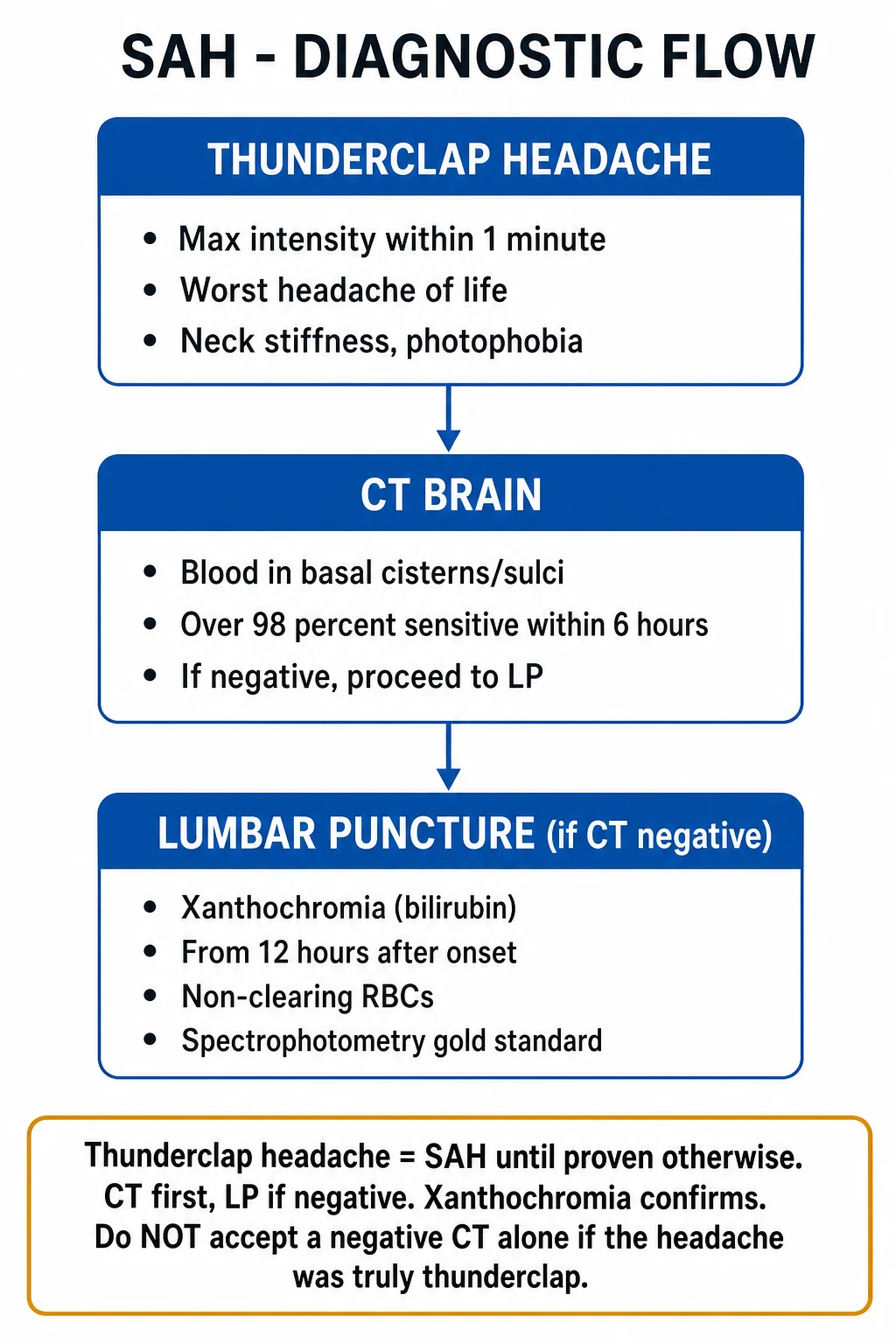
SAH diagnostic pathway
Clinical recognition
Thunderclap headache: sudden onset, maximal intensity within seconds to 1 minute, "worst headache of my life". Meningismus (neck stiffness, photophobia) develops over hours. Nausea/vomiting, altered consciousness, seizures. 10-15% die before reaching hospital.
CT brain (non-contrast)
FIRST-LINE. Sensitivity: >99% within 6h of onset. Decreases to ~86% at 24h, ~50% at 1 week. Blood in basal cisterns/sylvian fissure. Amount of blood (Fisher grade) predicts vasospasm risk. If CT positive → proceed to CT angiography to identify aneurysm.
LP if CT negative (after 6h)
Perform LP 6-12h after headache onset. Xanthochromia (yellow discolouration of CSF supernatant) = breakdown products of RBCs = SAH. Check for bilirubin (specific — persists for 2 weeks). RBC count: elevated, may decrease from tube 1 to 4 (traumatic tap) or stay elevated (true SAH). Spectrophotometry for xanthochromia.
CT angiography (CTA)
Identify the aneurysm: location, size, neck morphology. Guides treatment decision (coiling vs clipping). If CTA negative but SAH confirmed: consider digital subtraction angiography (DSA) — gold standard for small aneurysms.
Epidemiology and pathophysiology
Annual incidence
~6-16 per 100 000 population (age-adjusted); higher in Finland and Japan (~20-25/100 000). 30-day case fatality 25-35%; ~10-15% of patients die before reaching hospital. Of survivors, only ~30-40% return to full pre-morbid function.
Aneurysm prevalence
~2-3% of adults harbour an unruptured intracranial aneurysm at autopsy/imaging. Annual rupture risk ~0.5-2% for small (<7 mm) aneurysms, higher for posterior circulation, larger, or symptomatic lesions (ISUIA study).
Most common aneurysm sites
Anterior communicating artery (AcomA) ~30%; posterior communicating artery (PcomA) ~25%; middle cerebral artery bifurcation ~20%; internal carotid artery terminus ~8%; basilar tip ~5-10%.
Modifiable risk factors
Hypertension (strongest — doubles risk); current smoking (dose-dependent, ~3× risk); heavy alcohol use; cocaine / amphetamine use; oral contraceptives (small effect); anticoagulants worsen outcome but not rupture risk.
Non-modifiable risk factors
Female (1.6× after age 55); age >50; family history (≥2 first-degree relatives → ~4× risk); autosomal dominant polycystic kidney disease; Ehlers-Danlos type IV (vascular); Marfan syndrome; fibromuscular dysplasia; alpha-1 antitrypsin deficiency.
SAH pathophysiology begins with rupture of a saccular (berry) aneurysm at a vessel branch point where haemodynamic stress is greatest. The sudden release of arterial blood into the subarachnoid space produces three immediate consequences: (1) a sharp rise in intracranial pressure (often to diastolic BP) that can transiently halt bleeding but also causes reduced cerebral perfusion and the characteristic sudden, worst-ever headache; (2) meningeal irritation from blood breakdown products producing neck stiffness and photophobia over hours; and (3) acutely reduced consciousness from raised ICP and global ischaemia. The major downstream killers — rebleeding, hydrocephalus, vasospasm/DCI, and neurocardiac injury — unfold over the following 2 weeks and dominate ICU management. [1]
Pathophysiological cascade of SAH complications
Moment of rupture
Sudden arterial jet into subarachnoid space → ICP spike (may approach MAP) → transient cessation of bleeding, brief loss of consciousness in ~50% ("thunderclap" headache). Volume of blood on CT (Fisher grade) predicts downstream vasospasm.
Hours 0-6
Acute hydrocephalus if intraventricular extension; neurogenic pulmonary oedema (sympathetic surge); apnoea; cardiac stunning (catecholamine-mediated). Rebleeding risk maximal in first 24h (~4%).
Days 1-3
Blood breakdown → oxyhaemoglobin release → smooth-muscle contraction in vessel walls begins. Sodium dysregulation (CSW) appears. Cardiac stunning peaks (troponin, QTc).
Days 4-14 (peak 7-8)
Vasospasm (angiographic 30-70%; symptomatic DCI 20-30%). Mechanisms: endothelin-1 ↑, nitric oxide ↓, microthrombosis, cortical spreading depolarisation, inflammation. DCI — not vasospasm per se — drives poor outcome.
Days 14-28 and beyond
Chronic communicating hydrocephalus (impaired CSF reabsorption → VP shunt in ~10-20%). Cognitive impairment, fatigue, depression, late epilepsy.
Hunt and Hess grading
Hunt and Hess grade (1968, pre-CT era — still widely used in North America)
Drowsy or confused, mild focal neurological deficit
Moderate-severe. Altered mentation is the defining feature.
Fisher and modified Fisher grading (CT blood burden → vasospasm risk)
Original Fisher grade (1980 — amount/location of SAH on initial CT)
Localised clot and/or vertical layer of SAH ≥1 mm thick
Thick clot — HIGHEST symptomatic vasospasm risk in original Fisher.
Modified Fisher grade (Frontera 2006 — incorporates IVH; superior vasospasm/DCI predictor)
Thin SAH WITH IVH
IVH adds DCI risk.
WFNS
Clinical (GCS + focal deficit)
- Most widely used worldwide; objective and reproducible
- Grade I GCS 15 no deficit; II GCS 13-14 no deficit; III GCS 13-14 WITH deficit; IV GCS 7-12; V GCS 3-6
- Predicts functional OUTCOME (mortality at 1 month doubles per grade)
- Ignores CT blood burden — cannot predict vasospasm alone
Hunt-Hess
Clinical (1968, pre-CT)
- Historical; entrenched in North American charting
- Subjective terms (drowsy, stupor, moribund) → poor inter-rater reliability
- Mortality figures over-estimate current outcomes (pre-modern-treatment era)
- Useful for trend documentation alongside WFNS
Fisher / mFisher
Radiological (CT blood)
- Predicts VASOSPASM / DCI risk rather than mortality
- Modified Fisher (incorporates IVH) is the modern standard — better calibrated
- Thick cisternal blood + IVH = highest DCI risk → informs intensity of TCD surveillance
- Does NOT predict outcome independent of vasospasm
PAASH / VLASSO
Newer clinical scales
- PAASH (Prognosis with Aneurysmal SAH) uses GCS with different cut-points (GCS 13-15 mild, 9-13 moderate, ≤8 severe)
- Better calibrated for outcome prediction than WFNS in some validations
- Not yet routine in clinical practice but increasingly cited in trials
WFNS grading
WFNS (World Federation of Neurosurgical Societies) grade (click each)
GCS 14-13 WITH deficit
Mild impairment with focal neurological deficit.
Management
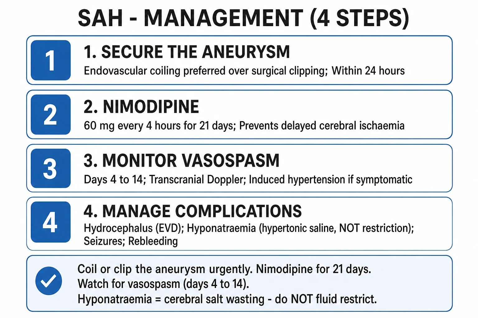
SAH ICU management protocol
Secure the aneurysm EARLY (within 24h)
Coiling (endovascular — preferred if anatomy suitable, ISAT trial: better outcomes). OR Clipping (surgical craniotomy — for wide-neck, complex aneurysms). Securing the aneurysm prevents REBLEEDING (#1 early cause of death). Once secured, BP can be liberalised.
Nimodipine 60 mg PO/NG every 4h for 21 days
Calcium channel blocker. Reduces incidence of delayed cerebral ischaemia (DCI) from vasospasm by ~33%. Improves outcome (NNT ~10). Does NOT reverse established vasospasm — prophylactic use. Give for full 21 days regardless of clinical course.<Cite id="1" />}
BP management
BEFORE aneurysm secured: SBP <160 (prevent rebleeding). Use labetalol/nicardipine. AFTER securing: permissive hypertension (maintain SBP 160-200) to improve cerebral perfusion through vasospasm. Do NOT routinely lower BP after securing unless evidence of haemorrhage expansion.
Maintain euvolaemia
Avoid hypovolaemia (worsens vasospasm). Target euvolaemia to mild hypervolaemia. Crystalloid maintenance infusion. Monitor fluid balance, CVP. Do NOT fluid restrict (even if hyponatraemia from CSW — give salt + water).
Monitor for vasospasm (days 4-14)
Daily transcranial Doppler (TCD): rising velocities (>120 cm/s mild, >200 severe) suggest vasospasm. Clinical monitoring: new neurological deficit (confusion, weakness, aphasia) = DCI. If symptomatic: induced hypertension (noradrenaline to raise MAP), maintain euvolaemia. Consider angioplasty or intra-arterial vasodilator (nimodipine/verapamil).<Cite id="3" />}
Manage complications
Hydrocephalus (from blood blocking CSF absorption): EVD (external ventricular drain). Seizures: prophylactic levetiracetam (controversial — some centres only if seizure occurs). Cerebral salt wasting: give salt + water (NOT fluid restriction). Cardiac: stress cardiomyopathy (takotsubo), arrhythmias — monitor ECG, troponin. DVT prophylaxis.
Vasospasm and delayed cerebral ischaemia
Vasospasm
Days 4-14 (peak day 7)
- Narrowing of cerebral arteries from blood breakdown products in subarachnoid space
- Clinical: new neurological deficit (confusion, weakness, aphasia, decreased GCS)
- TCD: rising mean velocities (>120 mild, >200 severe)
- CT perfusion: reduced cerebral blood flow in affected territory
- Treatment: induced hypertension (noradrenaline → raise MAP), euvolaemia ± angioplasty
- Angiographic vasospasm in 30-70% of SAH patients; symptomatic DCI in 20-30%
Triple-H therapy (historical)
Now modified
- OLD: Hypervolaemia + Haemodilution + Hypertension
- CURRENT: Maintain euvolaemia (NOT hypervolaemia — risk of pulmonary oedema)
- Induced hypertension if symptomatic DCI (raise MAP with noradrenaline)
- Haemodilution not specifically targeted (may worsen oxygen delivery)
Cerebral salt wasting vs SIADH
[1]Landmark trials — what the evidence shows
Molyneux 2002/2005 — ISAT: International Subarachnoid Aneurysm Trial (Lancet) (PMID 15708160)
Source
Lancet 2002 (initial) and 2005 (1-year follow-up) — multinational RCT, 2143 patients with ruptured aneurysms suitable for EITHER coiling OR clipping, randomised to endovascular coiling vs neurosurgical clipping. Molyneux A, Kerr R, Stratton I, et al.
What it found
At 1 year, **dependency or death (modified Rankin 3-6) was significantly lower with coiling (23.7%) vs clipping (30.6%)** — absolute risk reduction 6.9%, relative risk reduction 22.6%, NNT ~14. The benefit was concentrated in anterior circulation aneurysms. Long-term 18-year follow-up (Molyneux 2015, PMID 25706011) confirmed the outcome advantage persists, although re-bleeding risk after coiling is slightly higher (late re-rupture ~3%/10 yr vs ~1%/10 yr after clipping).
Key contribution
Established endovascular coiling as the DEFAULT treatment for ruptured aneurysms with favourable anatomy (favourable dome:neck ratio, narrow neck <4 mm). Drove a global shift — now >70% of ruptured aneurysms are coiled in most developed countries. Clipping reserved for wide-neck, complex, branch-incorporating, or MCA bifurcation aneurysms, or where coiling fails.
Limitations & caveats
Selection bias — only patients suitable for BOTH techniques were enrolled (~22% of screened). Posterior circulation under-represented. Stent- and balloon-assisted techniques have since widened coiling applicability. BRAT (Barrow Ruptured Aneurysm Trial, Lawton 2020) showed equipoise for MCA aneurysms when both techniques are feasible — surgical clipping outcomes were non-inferior.
Clinical bottom line
Coil if anatomy permits; clip for complex/wide-neck/MCA bifurcation aneurysms. The decision is multidisciplinary (neurovascular MDT): neurosurgeon + interventional neuroradiologist + intensivist.
Perry 2017 — Ottawa SAH Rule for acute headache triage (JAMA) (PMID 28403209)
Source
JAMA 2017;317(9):940-948 — prospective multicentre cohort of 1861 neurologically intact adults with acute non-traumatic headache peaking within 1 hour, across 10 Canadian EDs. Perry JJ, Sivilotti MLA, Sutherland J, et al.
What it found
The **Ottawa SAH Rule** (6 criteria: age ≥40; neck pain or stiffness; witnessed loss of consciousness; onset during exertion; thunderclap/instant-onset headache; limited neck flexion on examination) identified ALL cases of SAH (sensitivity 100%, specificity 13-15%) — i.e., if NO criterion is present, SAH is effectively excluded without CT or LP.
Key contribution
Standardised which headache patients warrant an SAH workup. Companion CT study (Perry 2011, PMID 21257119) showed CT within 6h of headache onset has ~100% sensitivity — incorporated into AHA 2021 and NICE guidelines: a negative CT within 6h in a low-risk patient may rule out SAH without LP.
Caveats
Sensitivity of CT falls steeply after 6h (~86% at 24h, ~50% at 1 week) — LP for xanthochromia remains mandatory for delayed presentations or CT-negative cases >6h. Does NOT apply to patients with altered mental status, focal deficit, prior SAH, or new headache in known aneurysm.
Clinical bottom line
Use the Ottawa Rule to risk-stratify who needs workup; perform CT <6h if possible; LP for xanthochromia if presentation delayed or CT-negative after 6h.
Vergouwen 2010 — Consensus Definition of Delayed Cerebral Ischaemia (Stroke) (PMID 20557354)
Source
Stroke 2010;41(10):2391-2395 — multidisciplinary ad-hoc consensus group (Vergouwen, Ilodigwe, Macdonald, Muizelaar, et al.) on behalf of the SAH Outcomes Working Group.
What it established
Defined DCI as: (a) clinical deterioration (focal neurological deficit OR ≥2-point GCS drop) lasting ≥1 hour, OR (b) a new cerebral infarct on imaging, NOT attributable to other causes (rebleeding, hydrocephalus, infection, electrolyte disturbance, seizure, sedation). Explicitly distinguished 'DCI' (clinical/imaging endpoint) from 'cerebral vasospasm' (angiographic/TCD finding) — two related but NON-identical concepts.
Key contribution
Created a uniform endpoint that all subsequent SAH trials use — without it, comparing trials was impossible. Enabled recognition that ~half of DCI episodes occur WITHOUT angiographic vasospasm, redirecting research to microcirculatory, microthrombotic, and cortical spreading depolarisation mechanisms.
Clinical bottom line
When you say 'DCI', mean it: a global or focal neurological decline persisting >1h, after excluding mimics (hyponatraemia, sepsis, rebleed, hydrocephalus, sedation, non-convulsive seizures). Always re-image to document infarction.
Kirkpatrick 2014 — STASH: Simvastatin in Aneurysmal SAH (Lancet Neurology) (PMID 24903427)
Source
Lancet Neurology 2014;13(7):666-673 — multicentre RCT, 803 SAH patients randomised to simvastatin 40 mg daily vs placebo for 21 days. Kirkpatrick PJ, Turner CL, Smith C, Hutchinson PJ, Murray GD; STASH Collaborators.
What it found
**No benefit** — no reduction in delayed cerebral ischaemia, no improvement in functional outcome (modified Rankin 0-2 at 6 months), no mortality difference. Earlier single-centre phase-2 studies (Tseng 2005, 2007) had suggested benefit, but STASH definitively refuted routine statin use. Infection rates were similar between groups.
Key contribution
A high-quality NEGATIVE trial that changed practice — simvastatin is NOT recommended for SAH. A paradigm of how positive phase-2 trials can fail to replicate in definitive multicentre RCTs, and of why prophylactic neuroprotectants must be tested rigorously.
Clinical bottom line
Do NOT give statins for SAH unless the patient has an independent cardiovascular indication.
Macdonald 2008/2011 — CONSCIOUS-1/2/3: Clazosentan for vasospasm (PMID 18725526)
Source
Lancet Neurology 2008 (CONSCIOUS-1) — 413 patients, dose-finding RCT of the endothelin receptor antagonist clazosentan to prevent angiographic vasospasm. CONSCIOUS-2 (2011) — 1147 patients with secured aneurysms, clazosentan 5 mg/hr vs placebo on functional outcome. CONSCIOUS-3 added a 2.5 mg/hr dose.
What it found
Clazosentan **reduced angiographic vasospasm** by ~30-65% (dose-dependent) and reduced vasospasm-related morbidity. BUT it did NOT improve good functional outcome (mRS 0-2 at 3 months) in CONSCIOUS-2 — and trended toward harm in surgical patients. It also caused anaemia, hypotension, and pulmonary complications.
Key contribution
Demonstrated that reversing angiographic vasospasm does NOT translate into better outcomes — a pivotal lesson that 'vasospasm' and 'DCI' are not synonymous. DCI has important non-vasospasm mechanisms (microthrombosis, cortical spreading depolarisation, neuroinflammation, autoregulatory failure). Clazosentan is not licensed in most countries for SAH.
Clinical bottom line
Improving the angiographic appearance of vessels is not the goal — improving outcome is. Nimodipine (which does improve outcome) remains the only drug proven to do so after SAH.
Diringer 2011 — Neurocritical Care Society Guidelines for SAH (PMID 21131147)
Source
Neurocritical Care 2011;9(1):1-25 — comprehensive multidisciplinary NCS guideline (Diringer MN, Bleck TP, Claude Hemphill J, et al.) covering diagnosis, aneurysm securing, haemodynamic management, vasospasm/DCI, and complications.
What it established
Formalised: (1) early aneurysm securing within 24h; (2) nimodipine 60 mg q4h × 21 days as standard; (3) euvolaemia (NOT hypervolaemia) with induced hypertension for DCI; (4) routine TCD surveillance; (5) avoidance of prophylactic hypervolaemia/haemodilution; (6) cardiac and sodium monitoring. Pre-dated and aligned with the AHA 2012 and 2021 updates.
Key contribution
The operational playbook that ICUs still follow. Drove the abandonment of triple-H therapy and its replacement with 'euvolaemia + induced hypertension' for symptomatic DCI.
Clinical bottom line
The standard-of-care reference for ICU management; complement with the AHA 2021 update for the latest evidence.
Complications — temporal profile
SAH complications by day — when to worry about what
0-24h (hyperacute)
REBLEEDING (~4% risk in first 24h; case fatality >50%). Acute hydrocephalus from intraventricular extension. Apnoea, neurogenic pulmonary oedema. Seizures at onset (~10%). Sudden ICP spike may mimic brain death.
Days 1-3
Cardiac stunning (neurogenic stunned myocardium / takotsubo), QT prolongation, malignant arrhythmia. Acute hydrocephalus — insert EVD for declining GCS. Hyponatraemia begins (CSW).
Days 4-14 (peak 7-8)
Vasospasm (angiographic 30-70%) and DCI (clinical 20-30%) — leading cause of morbidity. CSW ~30% (Na loss, volume depletion). Pneumonia, VTE, sepsis, pressure injury.
Days 14-28
Persistent communicating hydrocephalus → VP shunt in ~10-20%. Cognitive impairment, fatigue, depression. Late seizures. Critical illness myopathy from prolonged immobility.
Long-term
Cognitive deficits in ~50% (executive function, processing speed, memory). Mood disorders in ~30%. Epilepsy ~7%. Return to independent living in only ~30-40% of all SAH survivors.
Rebleeding
Greatest early mortality
- Highest in first 24h (~4%); cumulative risk ~20-30% over first 2 weeks if untreated
- Risk factors: poor-grade SAH, larger aneurysm (>10 mm), elevated SBP pre-securing, posterior circulation location, intracerebral extension
- Presents as sudden new headache, Cushing response, new fixed dilated pupil, GCS drop
- Eliminated by securing the aneurysm — coiling or clipping within 24h
- Case fatality of re-rupture >50%; survivors often devastatingly disabled
Hydrocephalus
Acute obstructive & communicating
- Acute (days 0-3): intraventricular/third-ventricular blood obstructs CSF flow → insert EVD
- Subacute communicating (days 7-21): impaired arachnoid granulation reabsorption from blood products
- Chronic (~10-20%): persistent communicating hydrocephalus → VP shunt
- Signs: progressive GCS decline, gait apraxia, urinary incontinence; confirm on CT (ventriculomegaly)
- Drain slowly — target 10-15 mL/h, ICP <20 mmHg. Over-drainage risks aneurysm re-rupture (lowers intracranial pressure below transmural gradient)
Seizures
Onset & late epilepsy
- Onset seizures in ~10% (cortical blood, intracerebral haematoma, MCA aneurysm)
- Late epilepsy in ~7%; risk factors: MCA aneurysm, ICH, infarct, poor grade, craniotomy
- Prophylactic anticonvulsant controversial — NCS suggests short course (3-7d) only for poor-grade / ICH / ICP monitors; NOT routine for all
- Continuous EEG if unexplained coma — occult non-convulsive seizures in ~10-20% of poor-grade SAH
- Levetiracetam is preferred first-line (no hepatic metabolism, no interaction with nimodipine)
Cardiac (neurocardiac)
Days 1-3
- Troponin elevated in ~30%; neurogenic stunned myocardium / takotsubo (apical ballooning)
- QTc prolongation, polymorphic VT/VF (torsades), atrial fibrillation
- Mechanism: catecholamine surge from hypothalamic ischaemia → myofibrillar degeneration
- Echo: reduced EF (usually recovers in 5-10 days); ECG changes often persist longer
- Manage haemodynamically — usually self-limited; correct K/Mg; avoid beta-blockade in acute QTc >500
Sodium (CSW & SIADH)
Days 3-14
- Hyponatraemia in 30-50% of SAH — usually CSW (volume-depleted)
- BNP/catecholamine-mediated natriuresis → renal sodium loss + volume depletion
- Treat CSW with hypertonic saline (3% NaCl) ± fludrocortisone; NEVER fluid-restrict (worsens vasospasm)
- SIADH (euvolaemic) is the less common mimic — fluid restriction safe in SIADH but dangerous in CSW
- Differentiate by volume status: CSW = hypovolaemic (↓CVP, ↑haematocrit); SIADH = euvolaemic
Other systemic
Throughout admission
- Fever (central + infectious) — every 1°C fever doubles poor-outcome odds; treat aggressively (paracetamol, cooling)
- Hyperglycaemia — associated with poor outcome; target glucose 6-10 mmol/L
- Anaemia — transfuse to maintain Hb >80-90 g/L (debated; avoid both extremes)
- DVT/PE — start prophylactic LMWH 24h after securing the aneurysm (NOT before); mechanical prophylaxis from admission
- Pneumonia ~20%, VAP risk with prolonged ventilation — head-of-bed 30°, oral chlorhexidine, daily sedation interruption
Delayed cerebral ischaemia — stepwise management
Suspected DCI (new neurological deficit, days 4-14) — ICU protocol
1. Confirm DCI and exclude mimics
Check Na (hyponatraemia mimics), glucose, temperature (infection/central fever), ABG (hypoxia/hypercapnia), sedation off if possible. CT to exclude rebleed, hydrocephalus, new infarct. Continuous EEG to exclude non-convulsive status. TCD: rising velocities (>200 cm/s, or >50 cm/s rise in 24h). CT perfusion: ↑MTT, ↓CBF.
2. Maintain euvolaemia
Target CVP 8-12 mmHg (or PiCCO GEDI 680-800, ITBI 850-950). Bolus 250-500 mL crystalloid or 4-5% albumin. Insert central line + arterial line. Daily weight, strict I/O, serum Na Q6-12h. Avoid hypovolaemia.
3. Induced hypertension
Raise MAP with noradrenaline (drug of choice — alpha-mediated vasoconstriction without coronary steal). Start at baseline MAP + 20%, titrate to clinical improvement. Typical SBP target 180-220 (post-securing). Continue until deficit resolves, then wean slowly over 24-48h. Use cardiac output monitoring if EF reduced.
4. Refractory DCI → endovascular therapy
If no improvement at maximal medical therapy → urgent DSA. Options: (a) intra-arterial vasodilator (nimodipine, verapamil, milrinone — short acting, may need repeat); (b) transluminal balloon angioplasty (durable for proximal spasm, ~1% rupture/stroke risk). Repeat daily if recurrent spasm.
5. Avoid harmful interventions
Do NOT fluid-restrict. Do NOT lower BP. Do NOT stop nimodipine (continue full 21 days even with DCI — IV nimodipine if PO not tolerated, but watch BP). Do NOT use steroids. Do NOT use prophylactic hypervolaemia (no outcome benefit; risk of pulmonary oedema). Do NOT transfuse above Hb 100 g/L unless ischaemia proven (TRANSAH suggested harm from liberal transfusion).
Prognosis
Overall case fatality
25-35% at 30 days. ~10-15% die pre-hospital. Of survivors, ~30-50% have significant cognitive impairment and only ~30-40% return to full pre-morbid functional level (mRS 0-1).
Predictors of poor outcome
Poor WFNS grade (IV-V) at admission, thick SAH on CT (mFisher 3-4), posterior circulation aneurysm, large aneurysm (>10 mm), rebleeding, symptomatic DCI with cerebral infarct, hyperglycaemia, sustained fever, anaemia, older age.
Predictors of good outcome
WFNS I-II, young age, anterior circulation aneurysm, secured within 24h, no DCI, normoglycaemia, afebrile, euvolaemia maintained throughout.
Long-term
Even 'good outcome' (mRS 0-2) patients often have measurable cognitive slowing, fatigue, sleep disturbance, and mood disorder at 1 year. Counsel patients and families on realistic recovery trajectory — recovery continues for 12-18 months.
Exam practice — SAQs
SAQ — Symptomatic vasospasm and delayed cerebral ischaemia on day 7
10 minutes · 10 marks
A 54-year-old woman was admitted 6 days ago with a WFNS grade II aneurysmal subarachnoid haemorrhage (modified Fisher 4) from a ruptured anterior communicating artery aneurysm, secured by endovascular coiling on day 1. She has been receiving nimodipine 60 mg via nasogastric tube every 4 hours and has remained euvolaemic and neurologically intact. On the morning of day 7 the nursing staff report that she is drowsy (GCS 13, E3V4M6, from a baseline of 15) with a new right-sided pronator drift and mild expressive dysphasia. Transcranial Doppler shows the left middle cerebral artery mean velocity has risen from 120 to 210 cm/s over 24 hours. Serum sodium 131 mmol/L. CT brain shows no rebleed, no hydrocephalus, no new infarct. BP 138/78.
SAQ — SAH hyponatraemia: cerebral salt wasting versus SIADH
10 minutes · 10 marks
A 62-year-old man is on day 5 of a WFNS grade III aneurysmal subarachnoid haemorrhage from a ruptured posterior communicating artery aneurysm, secured by coiling on day 0. He is receiving nimodipine 60 mg via nasogastric tube every 4 hours. Over the last 48 hours his serum sodium has fallen from 138 to 124 mmol/L. He has a negative fluid balance of 1.5 L, a central venous pressure of 2 mmHg, and a haematocrit that has risen from 0.36 to 0.45. Urine output is 3.5 mL/kg/h with urine sodium 78 mmol/L; serum osmolality 268 mOsm/kg and urine osmolality 420 mOsm/kg. The registrar has commenced 1 L of 5 percent dextrose and written for fluid restriction to 1 L per day in order to correct the sodium.
Clinical pearls
Red flags
References
- [1]Connolly ES, Rabinstein AA, Carhuapoma JR, et al. Court decisions in criminal proceedings for dental malpractice in Taiwan J Formos Med Assoc, 2022.PMID 34663527
- [2]Macdonald RL, Schweizer TA. Fast isolation and ex vivo culture of circulating tumor cells from the peripheral blood of lung cancer patients Yi Chuan, 2017.PMID 28115307
- [3]Dodd WS, Laurent D, Dumont AS, et al. Older adult Alexander Technique practitioners walk differently than healthy age-matched controls J Bodyw Mov Ther, 2016.PMID 27814855
- [4]Diringer MN, Bleck TP, Claude Hemphill J, et al. Cognitive deficit in preschoolers born late-preterm Early Hum Dev, 2011.PMID 21131147
- [5]Molyneux AJ, Kerr RS, Yu LM, et al.; ISAT Collaborative Group. Internet security and privacy protection for the health care professional Curr Surg, 2005.PMID 15708160
- [6]Molyneux AJ, Birks J, Clarke A, et al. Curriculum revitalization initiative at Tulane Am J Public Health, 2015.PMID 25706011
- [7]Perry JJ, Sivilotti MLA, Sutherland J, et al. Correction: How Much Can the USA Reduce Health Care Costs by Reducing Smoking? PLoS Med, 2017.PMID 28403209
- [8]Vergouwen MD, Vermeulen M, van Gijn J, et al. Dental surgery with minimal factor support in the inherited bleeding disorder population at the Alfred Hospital Haemophilia, 2011.PMID 20557354
- [9]Macdonald RL, Kassell NF, Mayer S, et al.; CONSCIOUS-1 Investigators. Dicer-dependent microRNA pathway safeguards regulatory T cell function J Exp Med, 2008.PMID 18725526
- [10]Frontera JA, Fernandez A, Schmidt JM, et al. Inhibition of autophagy causes tau proteolysis by activating calpain in rat brain J Alzheimers Dis, 2009.PMID 19158420
- [11]Perry JJ, Stiell IG, Sivilotti ML, et al. Obese adolescents are less active than their normal-weight peers, but wherein lies the difference? J Adolesc Health, 2011.PMID 21257119
- [12]Connolly ES, Rabinstein AA, Carhuapoma JR, et al. Guidelines for the management of aneurysmal subarachnoid hemorrhage: a guideline for healthcare professionals from the American Heart Association/american Stroke Association Stroke, 2012.PMID 22556195
- [13]Frontera JA, Claassen J, Schmidt JM, et al. Syndecan-4 clustering induces cell migration in a PDZ-dependent manner Circ Res, 2006.PMID 16675718