ICU · Neurocritical
ICU-acquired weakness: critical illness polyneuropathy and myopathy
Also known as ICU-acquired weakness · ICUAW · Critical illness polyneuropathy · CIP · Critical illness myopathy · CIM · Critical illness neuromyopathy
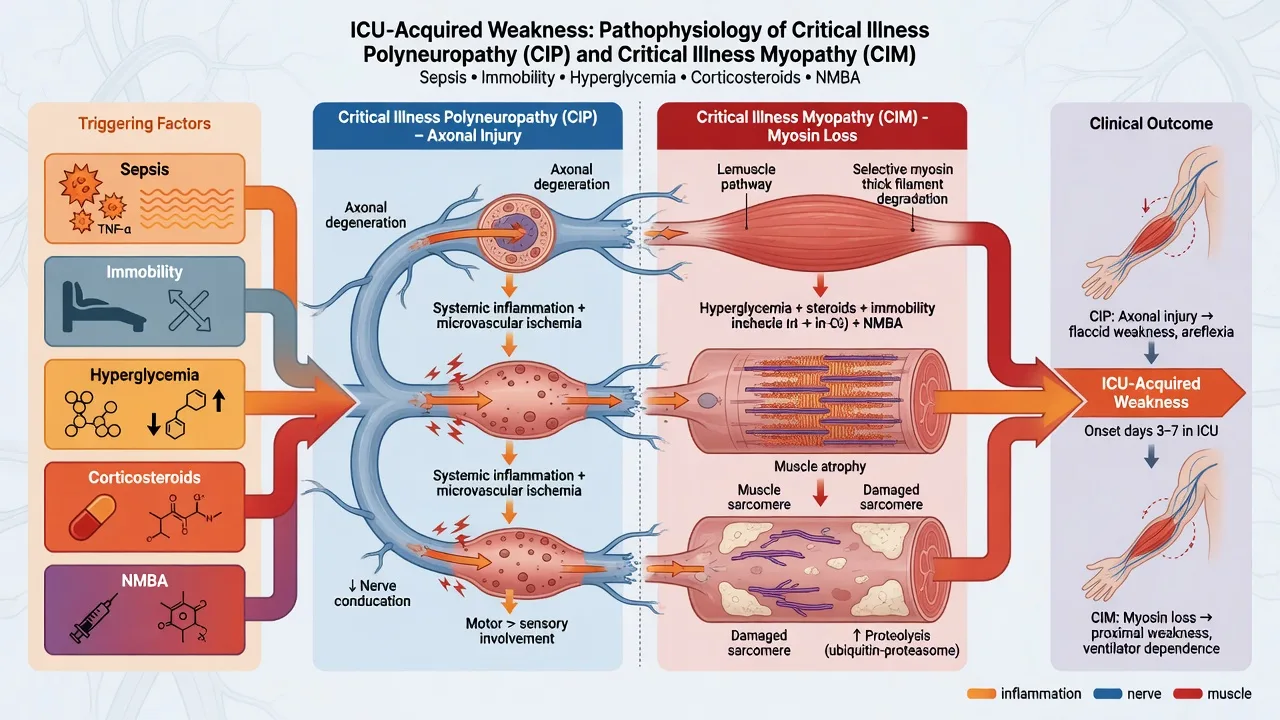
ICU-acquired weakness (ICUAW): muscle weakness developing during ICU stay, not attributable to other causes. Affects 25-50% of long-stay ICU patients. Types: (1) CIP (critical illness polyneuropathy) — AXONAL sensorimotor polyneuropathy (distal). (2) CIM (critical illness myopathy) — MYOPATHY (muscle fibre atrophy/necrosis). (3) Combined (CINM — most common). Risk factors: sepsis, multi-organ failure, prolonged immobilisation, corticosteroids, neuromuscular blockade, hyperglycaemia. Diagnosis: clinical (MRC sum score <48), electrophysiology (NCS/EMG), muscle biopsy. Prevention: minimise sedation, early mobilisation, glycaemic control, avoid steroids/NMBA if possible, nutrition. Treatment: supportive (rehabilitation, physiotherapy). Recovery: months-years, may be incomplete.
On this page & tools
Your progress
Saved locally on this device.
Target exams
Red flags
CIP vs CIM vs CINM
| Feature | CIP (Polyneuropathy) | CIM (Myopathy) | CINM (Combined) |
|---|---|---|---|
| Site | Peripheral NERVE (axonal) | MUSCLE (myopathy) | Both nerve + muscle |
| Pathology | Distal axonal degeneration | Myofibre atrophy, myosin loss | Both |
| Weakness | Distal > proximal (sensorimotor) | Proximal > distal (or generalised) | Generalised |
| Sensation | Reduced (sensory neuropathy) | Normal | May be reduced |
| NCS | Reduced CMAP + SNAP (axonal) | Reduced CMAP, normal SNAP | Reduced CMAP, ± SNAP |
| EMG | Denervation (fibrillation, positive sharp waves) | Myopathic (small motor units, early recruitment) | Mixed |
| Biopsy | Nerve: axonal loss | Muscle: atrophy (myosin loss), necrosis | Both |
| Most common | ~30% | ~20% | ~50% (most common) |
| Recovery | Slow (months-years) | Faster (weeks-months) | Variable |
Diagnosis and management of ICU-acquired weakness
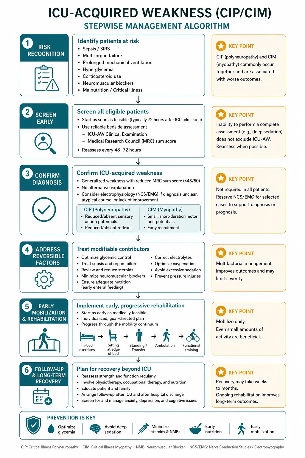
- SUSPECT ICUAW — difficulty weaning from ventilator, flaccid limbs, unable to lift arms/legs off bed, in patient with prolonged ICU stay (>1 week), sepsis, steroids, NMBA exposure
- EXCLUDE other causes — (a) Residual neuromuscular blockade (train-of-four, reversal). (b) Electrolyte disturbance (K+, Mg2+, Ca2+, phosphate). (c) Spinal cord injury. (d) Guillain-Barré. (e) Myasthenia gravis. (f) Steroid myopathy. (g) Cachexia/malnutrition
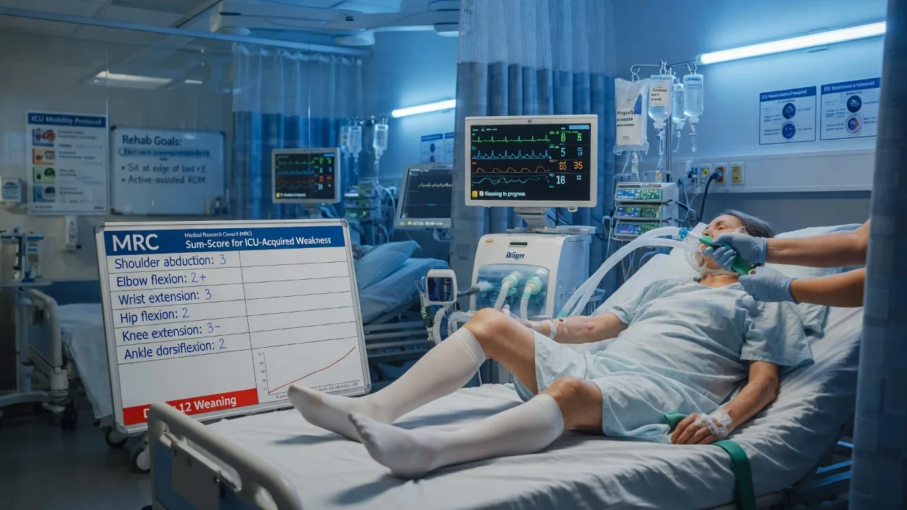
- CLINICAL DIAGNOSIS — MRC sum score: assess 6 muscle groups (arm flexion, arm abduction, wrist extension, leg flexion, leg extension, dorsiflexion), bilateral, 0-5 each. Score <48/60 = ICUAW. Requires: awake, cooperative, able to follow commands
- ELECTROPHYSIOLOGY (if available) — NCS (nerve conduction study): reduced CMAP + SNAP (axonal). EMG: denervation (CIP) or myopathic (CIM). Helps distinguish CIP vs CIM
- PREVENTION (most effective strategy): (a) Minimise sedation (dexmedetomidine, SAT). (b) Early mobilisation (day 1-2 — ABCDE trial). (c) Glycaemic control (6-10 mmol/L — NICE-SUGAR). (d) Avoid/minimise corticosteroids + NMBA. (e) Adequate nutrition (protein 1.2-1.5 g/kg/day). (f) Avoid prolonged bed rest
- TREATMENT — supportive: (a) Rehabilitation (physiotherapy, occupational therapy). (b) Early mobilisation (progressive — passive → active → walking). (c) Nutritional support (adequate protein). (d) Manage expectations (recovery is SLOW — months-years)
- RECOVERY — monitor over weeks-months. Most improve, but may take 1-5 years. Some never fully recover (permanent disability). Predictors of poor recovery: severe weakness, prolonged ICU stay, older age
SAQ — Difficulty weaning after prolonged septic ICU stay: diagnosing CIP
10 minutes · 10 marks
A 64-year-old man is day 16 of a prolonged ICU admission for pneumococcal pneumonia complicated by septic shock and ARDS. He has received 5 days of cisatracurium infusion and a 7-day course of hydrocortisone 200 mg/day for refractory vasopressor-dependent shock. Sedation has now been lightened and he is awake and cooperative, but he has failed three spontaneous breathing trials and is unable to lift his arms or legs off the bed. He has flaccid, areflexic limbs with reduced sensation distally. He asks why he cannot move.
SAQ — Preventing critical illness myopathy in a high-risk ARDS patient
10 minutes · 10 marks
A 52-year-old woman with severe ARDS from influenza pneumonitis (PaO₂/FiO₂ 70) has been ventilated for 48 hours on volume-control ventilation with a PEEP of 14, FiO₂ 0.8. She is on propofol and fentanyl infusions, has received 48 hours of cisatracurium for profound hypoxaemia per the ACURASYS protocol, and was started on dexamethasone 20 mg daily. Blood glucose is 14 mmol/L. The consultant asks you to outline a strategy to prevent ICU-acquired weakness, particularly critical illness myopathy (CIM).
Clinical pearls
Red flags
Prognosis
ABCDE trial (Schweickert 2009, Lancet) — early mobilisation for ICUAW prevention
RCT: 104 ICU patients on mechanical ventilation. Early mobilisation + sedation interruption vs usual care.
- Primary outcome (days alive without delirium or coma): early 2.9 vs usual 0 (more days without delirium)
- Independent at discharge (able to perform ADLs): early 59% vs usual 35% (p=0.02 — SIGNIFICANT)
- ICUAW at discharge (MRC <48): early 30% vs usual 60% (trend — not significant, but clinically important)
- Ventilation days: similar (no difference)
- CONCLUSION: Early mobilisation (with sedation interruption) IMPROVES functional outcomes and reduces ICUAW. Get patients OUT OF BED from day 1-2 (even if ventilated). [1]
Puthucheary (JAMA 2013): quadriceps mass decreased ~12.5% in first week (2%/day). Worse with sepsis. Van den Berghe (2001): intensive insulin reduced CIP from 52% to 29% (but NICE-SUGAR — moderate control now recommended). Recovery: 1-year: 30-50% still weak. Some never fully recover (permanent disability).
Electrophysiological discrimination — CIP vs CIM vs CINM
Electrophysiological discrimination: nerve conduction, direct muscle stimulation, and needle EMG
| Test | CIP (polyneuropathy) | CIM (myopathy) | CINM (combined) |
|---|---|---|---|
| Pathology site | Distal sensory + motor AXON degeneration | MUSCLE fibre (myosin loss, atrophy) | Both nerve + muscle |
| Nerve CMAP (compound muscle action potential — nerve stimulation) | REDUCED (axonal loss — fewer axons reaching muscle) | REDUCED (muscle can't generate force) | REDUCED |
| SNAP (sensory nerve action potential) | REDUCED (sensory axonal loss) | NORMAL (sensory nerve intact) | REDUCED (±normal) |
| Direct muscle stimulation CMAP (stimulate muscle directly — bypasses nerve) | NORMAL (muscle intact — nerve is the problem) | REDUCED (muscle itself is diseased) | REDUCED |
| KEY: Nerve CMAP vs direct muscle CMAP | Nerve CMAP < direct muscle CMAP (nerve is problem) | Nerve CMAP ≈ direct muscle CMAP (both low — muscle is problem) | Both reduced |
| Conduction velocity | NORMAL or mildly reduced (axonal — NOT demyelinating) | NORMAL | NORMAL or mildly reduced |
| Distal latency | Normal (no demyelination) | Normal | Normal |
| Needle EMG | Denervation potentials (fibrillation, positive sharp waves) — neurogenic | Myopathic units (small, short-duration polyphasic, EARLY recruitment) | MIXED (denervation + myopathic units) |
| Repetitive stimulation (to exclude MG) | Normal decrement | Normal decrement | Normal decrement |
| Phrenic nerve / diaphragm EMG | Abnormal if respiratory involvement | May be abnormal | May be abnormal |
| Interpretation tip | Reduced CMAP + REDUCED SNAP = AXONAL NEUROPATHY (CIP) | Reduced CMAP + NORMAL SNAP + reduced direct muscle CMAP = MYOPATHY (CIM) | Reduced CMAP + reduced SNAP + reduced direct muscle CMAP = COMBINED (CINM) |
Risk factors for ICU-acquired weakness — modifiable vs non-modifiable
| Risk factor | Mechanism | Relative risk | Modifiable? |
|---|---|---|---|
| Sepsis / systemic inflammation (STRONGEST) | Microvascular dysfunction, cytokine-mediated catabolism, mitochondrial dysfunction in nerve + muscle | RR 2-4 | Partly (early source control, antibiotics) |
| Multi-organ failure | Cumulative catabolism, hypoxia, oxidative stress | RR 3-5 | Partly |
| Corticosteroids (high-dose, prolonged) | Steroid myopathy — preferential type IIb fibre atrophy, myosin loss; synergistic with NMBA | RR 2-3 | YES — minimise dose/duration |
| Neuromuscular blocking agents (NMBA) | Prolonged blockade, upregulation of acetylcholine receptors, synergistic myopathy with steroids | RR 1.5-2 | YES — minimise duration, monitor TOF |
| Hyperglycaemia | Cellular damage via glycation, oxidative stress, impaired axonal transport | RR 2 | YES — glycaemic control (6-10 mmol/L) |
| Immobilisation / bed rest | Disuse atrophy (type I fibre), anabolic resistance, insulin resistance | RR 1.5-2 | YES — early mobilisation |
| Mechanical ventilation (prolonged) | Diaphragm dysfunction (VIDD — ventilator-induced diaphragm dysfunction), bed rest | RR 2 | Partly — minimise ventilation days |
| ARDS / hypoxaemia | Hypoxic nerve/muscle damage, high ventilator pressures, inflammation | RR 3-4 | Partly — lung-protective ventilation |
| Age > 60 | Reduced baseline muscle mass (sarcopenia), impaired regeneration | RR 1.5-2 | No |
| Female sex | Lower baseline muscle mass | RR 1.3-1.5 | No |
| Alcohol misuse / pre-existing neuropathy | Additive nerve damage | RR 2 | Partly |
| Renal failure (AKI/dialysis) | Uraemic neuropathy, electrolyte disturbance, volume shifts | RR 2 | Partly |
| Severe illness (APACHE II high) | Surrogate for severity/inflammation | RR 2-3 | No |
| Vasopressors (catecholamines) | Peripheral vasoconstriction → nerve/muscle ischaemia; surrogate for severity | RR 1.5-2 | Partly |
Prevention — the ABCDEF bundle
ABCDEF bundle for ICUAW prevention (SCCM/PADIS)
| Element | Meaning | ICUAW relevance |
|---|---|---|
| A — Assess, prevent, and manage pain | Validated pain tools (CPOT, BPS); multimodal analgesia (opioid-sparing) | Pain → immobility → atrophy; opioids contribute to weakness |
| B — Both spontaneous awakening trials (SAT) AND spontaneous breathing trials (SBT) | Daily sedation interruption (SAT); daily SBT to reduce ventilation days | Fewer sedation/ventilation days = less ICUAW risk (Kress SAT trial) |
| C — Choice of analgesia and sedation | Prefer dexmedetomidine/propofol over benzodiazepines (PADIS) | Benzos → more delirium, more immobility, more ICUAW |
| D — Delirium assess, prevent, manage | Screen (CAM-ICU, ICDSC); treat cause (not antipsychotics first-line) | Delirium → immobility, prolonged stay → more ICUAW |
| E — Early mobility and exercise | Progressive mobility (passive ROM → active → sitting → standing → walking) from day 1-2 | MOST DIRECTLY reduces ICUAW (ABCDE trial — Schweickert 2009) |
| F — Family engagement and empowerment | Family participation in care, mobility, communication | Family assists mobility, reduces delirium |
Prevention of ICU-acquired weakness — practical bundle implementation
- MINIMISE SEDATION (B + C of bundle) — Daily spontaneous awakening trial (SAT). Prefer dexmedetomidine or propofol over benzodiazepines (PADIS 2018). Target light sedation (RASS 0 to -1) unless indication for deep sedation. Avoid over-sedation → fewer ICU days → less ICUAW.[7] }
- DAILY SPONTANEOUS BREATHING TRIAL (SBT) (B of bundle) — Reduces ventilation days → less VIDD, less immobilisation, less ICUAW. SAT + SBT together = 'wake up and breathe' protocol.
- EARLY MOBILISATION (E of bundle) — Start day 1-2. PROGRESSIVE ladder: (a) passive range of motion (if unconscious/paralysed), (b) active-assisted, (c) active ROM, (d) sitting over edge of bed, (e) standing/transfer to chair, (f) walking. Even ventilated patients can mobilise (with adequate staffing — physio, nurse, respiratory therapist). ABCDE trial: early mobility improved functional outcomes.[4] }
- GLYCAEMIC CONTROL — Moderate targets: 6-10 mmol/L (NICE-SUGAR). Avoid hyperglycaemia (worsens ICUAW) and hypoglycaemia (harmful). Do NOT use intensive insulin therapy (4.4-6.1) — excess hypoglycaemia.[8] }
- MINIMISE CORTICOSTEROIDS — Use when indicated (ARDS — DEXA-ARDS; septic shock with CIRCI). Use LOWEST dose, SHORTEST duration. Avoid simultaneous high-dose steroids + prolonged NMBA (highest myopathy risk).
- MINIMISE NMBAs — Use only when indicated (severe ARDS — ACURASYS 48h cisatracurium; severe hypoxaemia). Monitor with train-of-four (target 1-2/4 twitches). Minimise duration. AVOID simultaneous high-dose steroids.
- ADEQUATE NUTRITION — Start enteral nutrition early (within 24-48 h). Protein 1.2-1.5 g/kg/day (supports muscle synthesis — though anabolic resistance limits efficacy). Avoid overfeeding (REEDS — refeeding syndrome). Consider protein supplementation, HMB.[3] }
- DELIRIUM PREVENTION (D of bundle) — Screen (CAM-ICU). Reduce modifiable factors (sedation, sleep disruption, immobility). Delirium → prolonged stay → more ICUAW. Early mobility reduces delirium AND ICUAW.
- FAMILY ENGAGEMENT (F of bundle) — Family present, assists with mobility, communication. Reduces delirium, supports rehabilitation.
Recovery timeline and prognosis
Recovery trajectory of ICU-acquired weakness
- ACUTE PHASE (ICU stay, days-weeks) — Weakness maximal at time of diagnosis. Muscle wasting ongoing (2%/day without intervention). Diaphragm dysfunction → weaning difficulty. Management: prevention (ABCDEF), rehabilitation (passive → active). Do NOT expect recovery during acute phase.
- EARLY RECOVERY (hospital discharge, weeks-months) — CIP recovery begins: axonal regrowth at ~1 mm/day (SLOW — months). CIM recovery: faster (myofibre regeneration — weeks). Most patients improve but remain weak at discharge. Many require rehabilitation facility. MRC score improves slowly.
- MEDIUM-TERM (3-6 months) — Continued improvement. CIP (axonal regrowth) still recovering. Many patients regain ability to walk independently. CIM often recovered. Residual: distal weakness, sensory symptoms (numbness, paraesthesia) if CIP. Fatigue common.
- LONG-TERM (6-12 months) — Plateau for most. ~30-50% still have some weakness at 1 year (especially elderly, severe ICUAW). CIM usually fully recovered. CIP: distal weakness/sensory may persist (axonal regrowth is slow). Functional recovery (walking, ADLs): better than electrophysiological recovery.
- LONG-TERM (>1 year) — Some never fully recover. Severe ICUAW (MRC <36): worse prognosis. Permanent disability in 10-20% (wheelchair, walking aid). Quality of life reduced (physical domain). Cognitive and psychological PICS may compound disability.
Prognostic factors — good vs poor recovery
| Factor | Good prognosis | Poor prognosis |
|---|---|---|
| ICUAW subtype | CIM (myopathy — regenerates faster) | CIP (neuropathy — axonal regrowth 1 mm/day, SLOW) |
| Severity | MRC 36-47 (mild-moderate) | MRC <36 (severe) |
| Duration of critical illness | Short (<1 week) | Prolonged (>2 weeks) |
| Age | Young (<60) | Elderly (>60, sarcopenia, impaired regeneration) |
| Baseline frailty | Robust (pre-morbid fit) | Frail (pre-existing low muscle mass) |
| Comorbidities | Few | Multiple (diabetes, alcohol, pre-existing neuropathy) |
| Rehabilitation | Early, intensive | Delayed, limited |
| Persistent electrophysiology | Recovery of CMAP/SNAP | Persistent severe axonal loss |
Differential diagnosis — mimics of ICUAW
Differential diagnosis of weakness in the ICU patient
| Condition | Key distinguishing features |
|---|---|
| Residual NMBA | Onset: hours. Check TOF (4/4 = recovered). Reversal: sugammadex (rocuronium), neostigmine. Resolves in hours. |
| Electrolyte disturbance | Hypo/hyper-kalaemia, Mg2+, Ca2+, phosphate. Check labs. Corrects rapidly with replacement. |
| Guillain-Barré syndrome (GBS) | PRE-ICU onset (ascending weakness). Demyelinating (slowed conduction velocity, conduction block — UNLIKE CIP). Albuminocytologic dissociation in CSF. May be the REASON for ICU admission (not acquired IN ICU). |
| Myasthenia gravis | Fluctuating, fatigable weakness. Decrement on repetitive stimulation. Ptosis, diplopia. May be reason for admission. |
| Spinal cord injury | Sensory level, sphincter disturbance, MRI abnormality. Upper motor neuron signs (if past shock phase). |
| Steroid myopathy | Proximal weakness, slowly progressive, related to chronic high-dose steroids. CK normal. EMG myopathic. Overlaps with CIM. |
| Mitochondrial/toxic myopathy | Drug exposure (statins, colchicine, antiretrovirals). CK elevated. Biopsy: mitochondrial abnormalities or toxic changes. |
| Cachexia/malnutrition | Generalised wasting, low albumin. Insidious onset. No electrophysiological abnormalities (or mild). |
| Hydrocephalus / CNS lesion | Focal signs, altered mental status. Imaging abnormal. |
| Hypothyroid myopathy | Proximal weakness, delayed reflexes, elevated TSH. |
Diagnostic approach summary
Step-by-step diagnostic approach to the weak ICU patient
- RECOGNISE weakness — Difficulty weaning from ventilator, flaccid limbs, unable to lift arms off bed or dorsiflex feet. Patient with prolonged ICU stay, sepsis, steroid/NMBA exposure.
- EXCLUDE REVERSIBLE CAUSES FIRST — Check: (a) Train-of-four (residual NMBA), (b) Electrolytes — K+, Mg2+, Ca2+, phosphate, (c) Glucose, (d) CK (rhabdomyolysis/toxic), (e) Thyroid function, (f) Drug history (statins, colchicine).
- CLINICAL DIAGNOSIS — MRC sum score (if awake, cooperative, follows commands). 6 muscle groups × 2 sides × 0-5 = maximum 60. MRC <48 = ICUAW. Document WHICH groups weak (distal vs proximal — helps distinguish CIP [distal] vs CIM [proximal]).
- ELECTROPHYSIOLOGY (if diagnosis uncertain, or to distinguish CIP vs CIM). NCS: CMAP + SNAP. DMS (direct muscle stimulation) if available. Needle EMG. Key: SNAP reduced = CIP; SNAP normal + reduced DMS-CMAP = CIM; both = CINM.
- IMAGING (adjunct) — Muscle ultrasound: increased echogenicity, reduced muscle thickness (correlates with ICUAW). Diaphragm ultrasound: reduced thickening fraction (diaphragm weakness). Not diagnostic but supportive.
- BIOPSY (rarely needed) — Reserved for atypical cases (suspect mitochondrial myopathy, inflammatory myopathy, toxic myopathy). Muscle: type II atrophy, myosin loss, necrosis. Nerve: axonal degeneration. Not routine in ICUAW.
- CLASSIFY — CIP (electrophysiology: axonal neuropathy), CIM (myopathy), CINM (both), or ICUAW-unspecified (clinical diagnosis, no electrophysiology). Classification affects prognosis (CIM recovers faster).
- PLAN REHABILITATION — Severity-based: passive ROM → active → standing → walking. Respiratory muscle training. Nutritional support. Set realistic expectations (months-years recovery).
Additional red flags
Additional landmark trials
EPIC study (Hermans 2014, Intensive Care Medicine) — prevalence and risk factors for ICUAW
Multicentre European observational study. Patients ventilated >7 days.
- Prevalence of ICUAW (MRC <48) at ICU discharge: ~30-40% in long-stay patients
- Highest-risk subgroup: sepsis, ARDS, multi-organ failure → up to 60-80%
- Risk factors (independent): sepsis, female sex, duration of organ failure, corticosteroids, hyperglycaemia, immobilisation
- CONCLUSION: ICUAW is COMMON in long-stay ICU patients. Risk factor profile confirms importance of prevention (glycaemic control, minimise steroids, early mobility).
PADIS guidelines (Devlin 2018, Critical Care Medicine) — pain, agitation, delirium, immobility, sleep
SCCM clinical practice guideline update (PAD → PADIS, adding immobility + sleep).
- Key recommendations for ICUAW: (1) prefer dexmedetomidine/propofol over benzodiazepines for sedation, (2) light sedation target (RASS 0 to -1), (3) daily SAT, (4) early mobilisation, (5) glycaemic control 6-10 mmol/L, (6) minimise corticosteroids/NMBA, (7) treat pain (multimodal, opioid-sparing).
- Mobility recommendation: 'Rehabilitation should be provided to critically ill adults' — graded to functional capacity.
- CONCLUSION: PADIS formalises the ABCDEF bundle. The 'I' (immobility) element directly addresses ICUAW prevention — early mobilisation is now a guideline-level recommendation.
TEAM trial (Denehy 2019, AJRCCM) — early active mobilisation during mechanical ventilation
RCT: 750 mechanically ventilated adults. Early active mobilisation (progressive, goal-directed) vs usual care.
- Primary outcome (days alive and out of hospital at 180 days): NO significant difference (early 143 vs usual 145 days, p=0.62)
- Adverse events: MORE in early group (desaturation 0.8%, arrhythmia 0.5%) — but low overall
- Secondary outcomes: mortality, function, quality of life — no significant difference
- CONCLUSION: AGGRESSIVE early active mobilisation in ALL ventilated patients did NOT improve 180-day outcomes vs usual care (which now includes some mobilisation). IMPORTANT NUANCE: Does NOT negate ABCDE trial (different population, different intervention intensity). TAKEAWAY: mobilise early but TAILOR intensity to the individual — 'one size fits all' aggressive mobilisation may not help all.
MVP trial (Schaller 2016, Lancet Respiratory Medicine) — early goal-directed mobilisation
RCT: structured, goal-directed early mobilisation protocol in ICU vs usual care.
- Feasibility and safety: mobilisation protocol was safe and feasible; achieved higher mobility levels in intervention group
- Functional outcomes: trend to better functional outcomes
- CONCLUSION: Structured, GOAL-DIRECTED mobility protocols (not ad hoc) are safe and feasible. Supports protocolised mobilisation as part of ICUAW prevention.
Puthucheary (JAMA 2013, Thorax 2013) — acute muscle wasting in critical illness
Prospective cohort: ultrasound measurement of quadriceps muscle mass in ICU patients.
- Muscle wasting: quadriceps mass decreased ~12.5% in first week (rate ~2% per day)
- Worse with: sepsis (septic patients lost ~18% in first week vs ~6% non-septic)
- Mechanism: ubiquitin-proteasome proteolysis + autophagy; impaired protein synthesis (anabolic resistance). Genomic/proteomic: upregulation of catabolic pathways.
- CONCLUSION: Muscle wasting in ICU is RAPID (2%/day), proportional to illness severity, and occurs DESPITE nutrition (anabolic resistance). Prevention must start EARLY (day 1) — cannot 'catch up' once wasting established.
NICE-SUGAR (Finfer 2009, NEJM) vs Van den Berghe (2001, NEJM) — glycaemic control and ICUAW
- Van den Berghe (Leuven 2001): intensive insulin (4.4-6.1 mmol/L) vs conventional (10-11.1) in surgical ICU. CIP electrophysiological abnormalities: intensive 29% vs conventional 52% (REDUCED). Also reduced mortality.
- NICE-SUGAR (2009): intensive glucose control (4.5-6.0) vs moderate (≤10) in 6100 ICU patients. Intensive → MORE hypoglycaemia (6.8% vs 0.5%), NO mortality benefit (slightly higher mortality).
- CONCLUSION: Intensive insulin reduced ICUAW BUT caused harmful hypoglycaemia. Current consensus: MODERATE glycaemic control (target 6-10 mmol/L) — avoid both hyperglycaemia (worsens ICUAW) and hypoglycaemia (harmful). The 'Leuven protocol' is no longer recommended.
References
- [1]Stevens RD, et al. Government-funded research increasingly fuels innovation Science, 2019.PMID 31221848
- [2]Hermans G, et al. Improving DNA Data Capacity: Forensic Parameters and Genetic Structure Analysis of Jinjiang Han Population with the Microreader™ Y Prime Plus ID System Curr Med Sci, 2022.PMID 35403953
- [3]Puthucheary ZA, et al. Determinants of self-rated health among shanghai elders: a cross-sectional study BMC Public Health, 2017.PMID 29029627
- [4]Schweickert WD, et al. Can sand nourishment material affect dune vegetation through nutrient addition? Sci Total Environ, 2020.PMID 32278174
- [5]Fan E, et al. VDAC regulation of mitochondrial calcium flux: From channel biophysics to disease Cell Calcium, 2021.PMID 33529977
- [6]Needham DM, et al. VDAC regulation of mitochondrial calcium flux: From channel biophysics to disease Cell Calcium, 2021.PMID 33529977
- [7]Kress JP, et al. Insurance type and minority status associated with large disparities in prelisting dialysis among candidates for kidney transplantation Clin J Am Soc Nephrol, 2008.PMID 18199847
- [8]Devlin JW, et al. Progress Toward Ruling Out Sepsis Crit Care Med, 2018.PMID 30113374
- [9]Pun BT, et al. Metal-organic aerogel as a coating for solid-phase microextraction Anal Chim Acta, 2017.PMID 28502427
- [10]Hermans G, et al. Conjugation of vitamin E analog α-TOS to Pt(IV) complexes for dual-targeting anticancer therapy Chem Commun (Camb), 2014.PMID 24452361
- [11]Latronico N, et al. Two Cases of Urinary Tract Endometriosis - Two Reconstruction Method After Segmental Resection J Minim Invasive Gynecol, 2015.PMID 27678689
- [12]Zifko UA, et al. Antivascular endothelial growth factor for retinopathy of prematurity Curr Opin Pediatr, 2009.PMID 19300261
- [13]Witt NJ, et al. VaDE: a manually curated database of reproducible associations between various traits and human genomic polymorphisms Nucleic Acids Res, 2015.PMID 25361969
- [14]Tipping CJ, et al. Helicobacter pylori Infection and the Development of Advanced Colorectal Neoplasia J Clin Gastroenterol, 2020.PMID 31651570
- [15]Schaller SJ, et al. Influence of reduction quality on functional outcome and quality of life in treatment of tibial plafond fractures: a retrospective cohort study BMC Musculoskelet Disord, 2019.PMID 31722696
- [16]Nordholt H, et al. Association of interleukin-17A rs2275913 polymorphism with rheumatoid arthritis susceptibility in Sudanese population SAGE Open Med, 2021.PMID 34104441