ICU · Neurocritical
Sepsis-associated encephalopathy and ICU delirium update
Also known as Sepsis-associated encephalopathy · SAE · Septic encephalopathy · ICU delirium · CAM-ICU · Delirium in ICU
Sepsis-associated encephalopathy (SAE): brain dysfunction from systemic sepsis (NOT direct CNS infection). Presents as DELIRIUM (acute, fluctuating disturbance of attention/cognition). Mechanisms: blood-brain barrier disruption, neuroinflammation (cytokines cross BBB), microvascular dysfunction, neurotransmitter imbalance, mitochondrial dysfunction. Affects 50-80% of septic ICU patients. WORSE outcomes: longer ICU stay, higher mortality, long-term cognitive impairment. Management: treat sepsis (source control, antibiotics), minimise sedation (dexmedetomidine, avoid benzodiazepines), promote sleep-wake cycle, early mobilisation, family presence, treat pain. Monitor with CAM-ICU. Pharmacological treatment controversial (haloperidol does NOT prevent — MIND-USA trial).
On this page & tools
Your progress
Saved locally on this device.
Target exams
Red flags
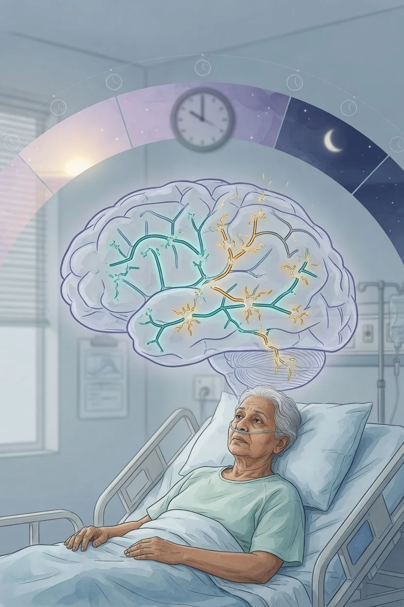
Management of delirium in the septic ICU patient
- Diagnose delirium — CAM-ICU (Confusion Assessment Method for ICU): acute onset + fluctuating + inattention + altered consciousness OR disorganised thinking. Positive CAM-ICU = delirium
- Treat SEPSIS first — source control, antibiotics within 1h, fluids, vasopressors. SAE resolves as sepsis improves
- Identify and treat CONTRIBUTING factors — (a) Medications (benzodiazepines — STOP, opioids — reduce). (b) Metabolic (hypoxia, hypoglycaemia, hyponatraemia, uraemia, hepatic). (c) Infection (UTI, pneumonia — sepsis source). (d) Pain (untreated — assess with CPOT). (e) Sleep deprivation (minimise night disruption). (f) Sensory deprivation (glasses, hearing aids)
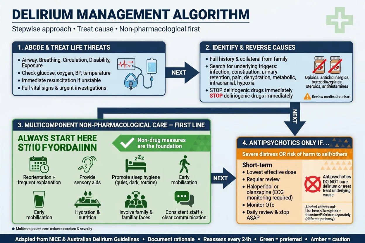
- Non-pharmacological management (FIRST-LINE) — (a) Promote sleep-wake cycle (lights on day, off night). (b) Reorient daily (time, place, person). (c) Family presence (bedside). (d) Early mobilisation (day 1-2). (e) Minimise restraints, catheters, lines
- Pharmacological (if distressed/dangerous) — dexmedetomidine (preferred — reduces delirium vs benzos). Haloperidol/quetiapine (if agitated — but MIND-USA: no preventive benefit). AVOID benzodiazepines (strongest delirium risk factor)
- Monitor and follow up — CAM-ICU daily. After discharge: cognitive assessment (long-term impairment in 40%)
Exam practice
SAQ — Sepsis-associated encephalopathy: diagnostic approach
10 minutes · 10 marks
A 72-year-old woman is admitted to ICU with septic shock from a urinary tract source. She is intubated and ventilated on noradrenaline 0.3 mcg/kg/min, sedated with propofol. On day 4 sedation is held for a spontaneous awakening trial; her RASS is -1 and CAM-ICU is positive. She has no focal neurology and no neck stiffness; her sepsis is improving (lactate 1.4 mmol/L). CT brain two days ago was normal.
SAQ — Management and prognosis of sepsis-associated encephalopathy
10 minutes · 10 marks
A 68-year-old man is in ICU on day 5 of ventilation for pneumococcal pneumonia with septic shock. He is sedated with a midazolam infusion at 8 mg/h and fentanyl 100 mcg/h; his RASS is -3. When sedation is lightened he is agitated and CAM-ICU positive. He has not yet been mobilised. His wife asks what his long-term recovery will look like.
Clinical pearls
Red flags
Prognosis
MIND-USA trial (Girard 2018, NEJM) — antipsychotics for delirium
RCT: 566 ICU patients with delirium. Haloperidol vs ziprasidone vs placebo.
- Primary outcome (days alive without delirium): NO difference between groups
- Secondary outcomes (time to extubation, ICU discharge, mortality): NO difference
- CONCLUSION: Antipsychotics (haloperidol, ziprasidone) do NOT improve delirium outcomes in ICU. Focus on: non-pharmacological (ABCDEF bundle), treat underlying cause, reduce sedation. [1]
Pandharipande (NEJM 2013): 3 months post-ICU: 40% had cognitive impairment (moderate TBI equivalent), 26% (mild Alzheimer's). 12 months: 25% still impaired. Strongest predictor: delirium duration. Ely (JAMA 2004): delirium = 2x higher 6-month mortality, independent of severity.
SAE — definition and clinical spectrum
Definition
Sepsis-associated encephalopathy (SAE) is defined as diffuse cerebral dysfunction caused by the systemic inflammatory response to infection, in the absence of direct CNS infection and without another identifiable cause of encephalopathy. It is a diagnosis of exclusion.[5] }
The term covers a clinical continuum: [1]
- Early/mild SAE — inattention, disorientation, subtle thought disorganisation. Easily missed unless formally screened (CAM-ICU, ICDSC).
- Established SAE — overt delirium (hyperactive, hypoactive or mixed), agitation or lethargy, sleep–wake disruption.
- Severe SAE — obtundation or coma; occasionally the presenting feature of occult sepsis/septic shock in the elderly ("quiet, confused, dropped GCS"). [1]
Terminology: SAE vs delirium vs metabolic encephalopathy
| Term | What it is | Key distinction |
|---|---|---|
| SAE | Brain dysfunction from SYSTEMIC sepsis, no CNS infection | Aetiological label (exclusion diagnosis) |
| Delirium (DSM-5 / ICD-11) | Acute, fluctuating disturbance of attention/awareness + cognition, due to a medical condition | The clinical SYNDROME by which SAE is detected |
| Septic encephalopathy | Older synonym | Now largely replaced by SAE |
| Metabolic/toxic encephalopathy | Brain dysfunction from any metabolic or toxin cause | SAE is one subtype |
| ICU encephalopathy | SAE + contributions from sedatives, organ failure, sleep deprivation, electrolytes | Often multifactorial in ICU |
| Sub-syndromal delirium | ICDSC 1–3, CAM-ICU negative with subjective concerns | Already associated with worse outcomes — act early |
SAE and delirium overlap but are NOT identical. SAE is the aetiological label; delirium is the phenotypic syndrome observed clinically. Early SAE may be present before formal CAM-ICU positivity (sub-syndromal — elevated ICDSC, not yet delirium).[5] }
Epidemiology
- Affects 50–80% of septic ICU patients; near-universal in septic shock.
- Independent of infection source (pneumonia, UTI, abdominal, bacteraemia, line infection).
- More common with: age > 65, prior cognitive impairment, severity of illness (APACHE/SOFA), alcohol excess, smoking, pre-existing cerebrovascular disease, diabetes.
- Hypoactive motoric subtype predominates in SAE (≥ 60%) — easily missed.
- SAE independently predicts mortality, length of stay, mechanical ventilation duration and long-term cognitive impairment.[5] }
Pathophysiology of SAE
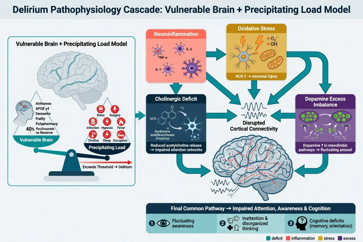
SAE is a multi-mechanism, diffuse brain disorder — no single pathway explains every case. The four dominant, interacting mechanisms below converge on diffuse synaptic and network dysfunction.[5] }
1. Blood–brain barrier (BBB) disruption
- Circulating cytokines (TNF-α, IL-1β, IL-6) and activated leukocytes upregulate endothelial adhesion molecules (ICAM-1, VCAM-1) on brain microvascular endothelium.
- Endothelial tight junctions (claudin-5, occludin) are loosened → increased BBB permeability.
- Result: cytokines, complement, leukocytes and albumin leak into brain parenchyma → vasogenic oedema and microglial activation.
- Demonstrated in animal sepsis models and human post-mortem studies (albumin extravasation).
- CSF albumin index (CSF albumin / serum albumin × 1000) can demonstrate this clinically.
2. Neuroinflammation and microglial activation
- Microglia (resident brain macrophages) switch to a pro-inflammatory (M1) phenotype.
- They release IL-1β, TNF-α, reactive oxygen species, glutamate and nitric oxide.
- Sustained activation → synaptic dysfunction, neuronal injury, impaired neurotransmission.
- Astrocyte dysfunction (loss of glutamate uptake, water homeostasis via AQP4) compounds injury.
- Neuroinflammation persists AFTER the systemic infection resolves — this is the proposed mechanism linking SAE to long-term cognitive impairment.
3. Cerebral microcirculatory dysfunction
- Endothelial activation + leukocyte adhesion → capillary plugging / no-reflow.
- Microthrombi (DIC, platelet–leukocyte aggregates).
- Loss of cerebral autoregulation — both CO2 and pressure reactivity impaired → cerebral blood flow becomes pressure-passive.
- Microvascular shunting → regional hypoxia despite adequate global perfusion.
- Clinical implication: SAE can worsen despite MAP > 65. Autoregulation failure means some patients need a higher MAP; there is no universal "brain MAP target," but a trial of higher MAP (75–85 mmHg) is reasonable in a patient with worsening SAE.
4. Mitochondrial dysfunction and cellular energetic failure
- Sepsis-induced mitochondrial damage (oxidative stress; NO inhibition of cytochrome c oxidase) → ATP depletion.
- "Cytopathic hypoxia": cells cannot utilise oxygen even when delivery is adequate.
- Brain (high metabolic demand) is exquisitely sensitive → neuronal dysfunction WITHOUT overt cell death early on.
- Explains why SAE can occur with normal brain imaging, normal lactate and normal perfusion indices.
5. Neurotransmitter imbalance
- Aromatic amino acids (phenylalanine, tryptophan) accumulate (impaired hepatic clearance + BBB leak) → ↑ dopamine, false neurotransmitters, ↓ serotonin.
- Tryptophan–kynurenine shunt activated → quinolinic acid (NMDA agonist, neurotoxic) accumulates.
- Acetylcholine deficiency (reduced choline, increased breakdown by microglial cholinesterase).
- GABAergic and glutamatergic dysregulation → altered arousal and excitotoxicity.
- These disturbances explain why SAE resembles other metabolic encephalopathies clinically and on EEG.
Biomarker correlation (research / supportive — not routine)
- S100B (astrocytic protein) — elevated in SAE; correlates with severity and mortality.
- Neuron-specific enolase (NSE) — neuronal injury marker; elevated with worse outcome.
- GFAP and UCH-L1 — research markers of glial and neuronal injury.
- These biomarkers are NOT yet part of routine clinical diagnosis but are commonly examined as supportive evidence. [1]
Net effect
Diffuse, largely reversible (early) synaptic and network dysfunction, with risk of permanent injury if severe or prolonged. The two strongest modifiable predictors of long-term cognitive outcome are delirium duration and depth of sedation.[1] }
Diagnosis of SAE
SAE is a diagnosis of exclusion. Confirm the delirium syndrome, then exclude other causes.[5] }
Step 1 — Recognise the syndrome (delirium screen)
- Use a validated tool at least once per shift: CAM-ICU, ICDSC, 4AT or local protocol.
- Document RASS (Richmond Agitation-Sedation Scale) before delirium assessment.[12] }
- If RASS −4 or −5 (deep sedation/coma) → cannot reliably assess delirium that day; reassess when sedation is lightened.
Step 2 — Exclude other causes (the differential)
| Category | Examples | Key tests |
|---|---|---|
| Direct CNS infection | Meningitis, encephalitis, brain abscess | LP (cells, protein, glucose, Gram stain, HSV PCR), CT/MRI |
| Structural brain lesion | Ischaemic/haemorrhagic stroke, SOL, SDH | CT/MRI brain |
| Metabolic | Hypoglycaemia, Na/Ca/Mg derangement, uraemia, hepatic failure, hypoxia, hypercapnia | VBG, U&E, LFT, Ca/Mg/PO4, glucose, lactate |
| Endocrine | Thyroid storm/myxoedema coma, adrenal crisis, DKA/HHS | TFT, cortisol, glucose, ketones |
| Drug / withdrawal | Benzodiazepines, opioids, anticholinergics; alcohol withdrawal | Medication chart, alcohol history, tox screen |
| Seizure-related | Non-convulsive status epilepticus (NCSE), post-ictal state | EEG (preferably continuous — cEEG) |
| Hypoperfusion | Shock (any type), severe anaemia | Lactate, ScvO2, Hb, MAP |
Routine lumbar puncture is NOT required in typical SAE (no meningeal signs, septic source identified, delirium consistent with severity of illness). Perform LP if: meningitic signs, severe unexplained headache, atypical course, immunocompromise, suspected HSV, or deterioration despite adequate sepsis treatment. [1]
Step 3 — Investigations that support SAE
Electroencephalography (EEG)
- Typical SAE pattern: diffuse background slowing — loss of posterior dominant alpha (8–13 Hz), emergence of theta (4–7 Hz) and delta (< 4 Hz) activity.
- Triphasic waves: blunt, intermittent waves with phase 1 (small negative) → phase 2 (large positive) → phase 3 (slow negative), maximal anteriorly. Classic for hepatic encephalopathy but also seen in SAE, uraemia, hypoxia, hyponatraemia and drug toxicity — NOT specific.[5] }
- Severity grading (Young's classification) correlates with outcome.
- Main role of EEG: exclude non-convulsive status epilepticus (NCSE) — essential if delirium fluctuates, is prolonged, or is associated with subtle facial/limb twitching. cEEG ≥ 24–48 h increases yield.
- EEG does NOT distinguish SAE from other metabolic encephalopathies.
Biomarkers (CSF and serum)
- CSF: usually normal; mild protein elevation in ~50%. Cell count usually normal (a pleocytosis suggests an alternative diagnosis).
- Serum S100B and NSE: elevated in SAE; correlate with severity, BBB disruption and mortality. Not yet routine — research/adjunctive.[5] }
- CSF albumin index (CSF albumin / serum albumin × 1000) — demonstrates BBB dysfunction (elevated in SAE).
Neuroimaging (CT then MRI as indicated)
- CT: typically normal in SAE; performed to exclude haemorrhage, mass effect, hydrocephalus, large stroke.
- MRI (when obtained): may show
- DWI/FLAIR white-matter hyperintensities — diffuse leukoencephalopathy (delayed; often reversible).
- Watershed-zone restriction — microvascular hypoperfusion.
- Posterior reversible encephalopathy syndrome (PRES) — parieto-occipital vasogenic oedema.
- Acute necrotising encephalopathy — rare (thalamic, brainstem involvement; very poor prognosis).
- Microbleeds (T2*/SWI) — microvascular/inflammatory.
- MRI is not required for diagnosis, but valuable when the picture is atypical, the course does not track with sepsis, or focal signs develop.
Practical diagnostic summary
- Septic patient with acute brain dysfunction (delirium, altered arousal or coma).
- No direct CNS infection (clinically and, if indicated, LP/imaging).
- No alternative explanation (metabolic, structural, drug, seizure).
- Course tracks with sepsis (improves as sepsis resolves). [1]
Sedative choice and pharmacology
| Agent | Receptor | Delirium risk | Notes |
|---|---|---|---|
| Dexmedetomidine | α2A agonist | Lowest | Analgo-sedative; preserves respiratory drive; reduces delirium vs benzodiazepines (MENDS, SEDCOM). Bradycardia, hypotension. Costly. |
| Propofol | GABA-A | Low–moderate | Rapid offset; hypotension; propofol infusion syndrome (PRIS) at high/long doses (> 4 mg/kg/h for > 48 h). |
| Midazolam | GABA-A (benzo) | High | AVOID if possible; strong, independent delirium risk factor. |
| Lorazepam | GABA-A (benzo) | High | AVOID except alcohol withdrawal / status epilepticus / catatonia. |
| Ketamine | NMDA antagonist | Low (analgesic) | Useful adjunct for analgesia and induction; preserves haemodynamics. |
| Opioids (fentanyl, morphine) | μ-opioid | Dose-dependent | Essential analgesia; over-use → sedation, constipation, delirium. Use multimodal opioid-sparing. |
Dexmedetomidine — practical points
- Dose: infusion 0.2–0.7 (up to 1.5) mcg/kg/h, titrated to RASS −1 to 0. No loading bolus in ICU sedation (bolus reserved for procedural/anaesthesia use — causes transient hypertension then bradycardia/hypotension).
- No significant respiratory depression → useful for weaning/extubation and "no sedation" protocols.
- Adverse effects: bradycardia (including severe), sinus arrest, hypotension — caution in low-output states, AV block, severe valvular disease.
- Evidence: MENDS[7] } and SEDCOM[8] } — reduced delirium vs lorazepam/midazolam. HOPE-ICU[9] } — early dexmedetomidine in agitated patients did NOT improve outcome (so NOT for prevention in all). Reade 2009 (NEJM) review summarises sedative/delirium pharmacology.[16] }
- Cost remains a barrier in many systems.
ABCDEF bundle — components in detail
The ABCDEF bundle (SCCM "ICU Liberation") is the single best-evidenced non-pharmacological approach to reducing SAE/delirium:[6] }[15] }
| Letter | Component | Action |
|---|---|---|
| A | Assess, prevent and manage pain | CPOT (ventilated) / NRS (able); multimodal analgesia; opioid-sparing |
| B | Both SAT and SBT | Spontaneous awakening trial + spontaneous breathing trial daily (when safe); reduces ventilation days and delirium |
| C | Choice of analgesia and sedation | Analgesia-first; dexmedetomidine/propofol over benzodiazepines; light target (RASS −1 to 0) |
| D | Delirium: assess, prevent, manage | CAM-ICU or ICDSC each shift; treat cause; non-pharmacological first |
| E | Early mobility and exercise | Out of bed from day 1–2 when safe; PT/OT; passive → active progression |
| F | Family engagement and empowerment | Bedside presence; orientation; communication; shared decision-making |
Implementation data: complete bundle performance is associated with lower delirium, more coma-free days, more ventilator-free days, lower mortality, and shorter ICU and hospital length of stay (Pun 2019 — large multicentre cohort).[15] }
Daily SAT/SBT safety screen
Before performing a spontaneous awakening trial (SAT) and spontaneous breathing trial (SBT):
- No active sedative up-titration in prior 12 h
- No neuromuscular blocker; TOF ≥ 4 if recently used
- No agitation with the previous SAT failure
- Acceptable SpO2/FiO2 (typically FiO2 ≤ 50%, PEEP ≤ 8)
- No significant vasopressor escalation, no new myocardial ischaemia, no intracranial hypertension concern [1]
Kress (NEJM 2000) established that daily interruption of sedation reduced ventilation days and ICU stay without adverse events.[14] }
Comparison: delirium screening tools
Delirium screening and severity tools
| Tool | What it measures | Time | Best use |
|---|---|---|---|
| CAM-ICU[11] } | Acute/fluctuating + inattention + (altered LOC OR disorganised thinking) — binary | 2–4 min | Ventilated or unable to converse; high specificity (~ 95%) |
| ICDSC[13] } | 8-item checklist, score 0–8; ≥ 4 = delirium; 1–3 = sub-syndromal | 1–2 min | Nursing routine; tracks severity; detects sub-syndromal |
| 4AT | Alertness + AMT (age, DOB, place, year/month) + attention (months backwards) | 1–2 min | Non-ICU wards; quick cognitive screen |
| DRS-R-98 | Delirium Rating Scale — Revised-98 (severity) | 20–30 min | Research / specialist |
| RASS[12] } | Sedation/agitation depth (−5 to +4) | < 1 min | Mandatory BEFORE any delirium assessment |
| CPOT / NRS | Pain intensity | < 1 min | CPOT for ventilated; NRS for those able to report |
CAM-ICU — feature-by-feature
- Acute onset or fluctuating course — change from baseline ± fluctuation over 24 h.
- Inattention — squeeze hand on hearing the letter 'A' in a 10-letter sequence (SAVEHAART). Errors (squeeze on wrong letter or fail to squeeze 'A') indicate inattention.
- Altered level of consciousness — RASS anything other than 0.
- Disorganised thinking — 4 simple yes/no questions + 1 command (e.g. "Are there stones in water? Will a stone float on water?"). [1]
Positive CAM-ICU: features 1 + 2 + (3 OR 4). Sensitivity ~ 80%, specificity ~ 95%. Takes 2–3 min. Perform at least once per shift.[11] }
Comparison: SAE vs other encephalopathies
SAE vs hepatic vs uraemic vs meningitis (mimics)
| Feature | SAE | Hepatic | Uraemic | Meningitis (mimic) |
|---|---|---|---|---|
| Onset | Acute, tracks sepsis | Subacute | Gradual | Acute |
| Fever/sepsis source | Yes | No (unless infected) | No | Yes |
| Meningeal signs | No | No | No | Often |
| CSF | Normal / mild ↑ protein | Normal | Normal | Pleocytosis, ↑ protein, ↓ glucose |
| EEG | Diffuse slowing, ± triphasic | Triphasic waves (classic) | Diffuse slowing | May be normal or focal |
| Imaging | Normal / DWI white-matter changes | Often normal | Often normal | Meningeal enhancement, hydrocephalus, infarct |
| Treatment | Treat sepsis + ABCDEF bundle | Lactulose, rifaximin, treat liver | Dialysis, renal support | Antibiotics ± LP before imaging |
Comparison: motoric subtypes of delirium
Hyperactive vs hypoactive vs mixed delirium
| Subtype | % of ICU delirium | Features | Prognosis |
|---|---|---|---|
| Hypoactive | 40–65% | Quiet, withdrawn, lethargic, slow | WORST (often missed; lowest recognition; highest mortality) |
| Hyperactive | 5–15% | Agitated, restless, pulling tubes | More readily recognised; somewhat better than hypoactive |
| Mixed | 20–50% | Fluctuates between hypo- and hyperactive | Intermediate |
Hypoactive is the dominant subtype in SAE — the septic patient who is "just sleepy" must be assessed formally with CAM-ICU.[4] }
Pharmacological treatment — what the trials say
MENDS (Pandharipande 2007, Crit Care Med) — dexmedetomidine vs lorazepam in sepsis
- RCT, 106 mechanically ventilated medical/neuro ICU patients
- Dexmedetomidine vs lorazepam for up to 5 days, targeted to RASS
- Result: dexmedetomidine group had more days alive without delirium or coma (median 7 vs 3)
- Strongest effect in septic patients — biological plausibility for SAE benefit
SEDCOM (Riker 2009, JAMA) — dexmedetomidine vs midazolam
- RCT, 375 mechanically ventilated patients sedated ≥ 4 days
- Dexmedetomidine vs midazolam to RASS −2 to +1
- Result: dexmedetomidine non-inferior for time at target sedation; lower prevalence of delirium (54% vs 76.6%); more bradycardia and hypotension
HOPE-ICU (Page 2017, Lancet Respir Med) — early dexmedetomidine for agitated delirium
- RCT, 194 patients with agitation-associated delirium
- Low-dose dexmedetomidine vs placebo
- Result: NO improvement in days alive without delirium or coma
- Take-home: routine dexmedetomidine for established agitation/delirium is NOT supported; the benefit is from avoidance of benzodiazepines and lighter sedation
DICONS (van den Boogaard 2015, Crit Care) — rivastigmine for ICU delirium
- RCT, addition of rivastigmine (cholinesterase inhibitor) to haloperidol for ICU delirium
- Stopped early for harm: longer delirium duration in the treatment group
- Take-home: cholinesterase inhibitors are NOT recommended for ICU delirium
Hughes 2024 (Lancet) — dexmedetomidine individual-patient-data meta-analysis
- ~ 4000 patients across multiple RCTs (SPICE, MENDS, HOPE-ICU, others)
- Dexmedetomidine associated with small reduction in delirium duration and a modest mortality signal
- Benefit greatest in agitated/hyperactive delirium; caution in bradycardia/hypotension
FlowSteps: systematic workup of new-onset delirium in a septic ICU patient
New-onset delirium in a septic ICU patient — systematic workup
- Confirm delirium — CAM-ICU after recording RASS. If RASS ≤ −3, defer; reassess when lighter.[11] }
- Re-check the basics — VBG (glucose, Na, Ca, lactate), SpO2/PaO2, PaCO2 (hypercapnia is a commonly missed cause), temperature, MAP, urine output. Correct any metabolic derangement.
- Review medications — chart for benzodiazepines, opioids, anticholinergics, corticosteroids, sleeping tablets. Stop or reduce deliriogenic agents; convert to dexmedetomidine-based sedation if needed.[4] }
- Identify and treat infection — review sepsis source; new fever, raised lactate or rising WCC suggests progression or a new source; send cultures, escalate antibiotics as needed.
- Exclude structural brain lesion — focal neurology, head trauma, anticoagulation, unequal pupils or rapid coma → urgent CT brain.
- Exclude non-convulsive status epilepticus — fluctuating/atypical delirium, subtle facial or limb twitching, history of seizures → arrange cEEG ≥ 24–48 h.[5] }
- Consider lumbar puncture — only if meningitic signs, severe unexplained headache, immunocompromise, atypical course, or deterioration despite sepsis treatment.
- Implement the ABCDEF bundle — analgesia, light sedation, mobilisation, family, sleep hygiene.[6] }
- Reassess daily — CAM-ICU each shift; track duration (the key prognostic variable).[2] }
Implementing the ABCDEF bundle on the morning ward round
- A — Pain score recorded (CPOT if ventilated, NRS if able)? Treating to target? Multimodal plan in place?
- B — SAT/SBT safety screen passed today? If yes, performed? If failed, why (FiO2/PEEP, vasopressor excess, sedation excess, agitation)?[14] }
- C — Sedation target RASS −1 to 0? On a benzodiazepine — can it be stopped? Switch to dexmedetomidine/propofol?
- D — CAM-ICU done this shift? If positive, contributing factors addressed? If sub-syndromal, prevention in place?
- E — Mobilised today? What level (passive, in-chair, standing, walking)? Barriers addressed?
- F — Family updated? Bedside visit facilitated? Goals of care current?
More high-yield clinical pearls
Red flags — when SAE is something else
Prognosis and follow-up
Short-term outcomes
SAE/delirium independently predicts:[2] }
- 2–3× higher 6-month mortality (Ely 2004, JAMA)
- Each additional day of delirium: ~ 10% higher mortality risk
- Longer mechanical ventilation
- Longer ICU and hospital length of stay
- Higher rates of self-extubation, line removal and falls
- Higher ICU-acquired weakness (shared risk factors and bidirectional causation) [1]
Long-term outcomes — post-intensive care syndrome (PICS)
- Cognitive: 25–40% of ICU survivors have measurable cognitive impairment at 6–12 months (memory, executive function, processing speed). Severity resembles moderate TBI (40%) or mild Alzheimer's (26%) at 3 months. Strongest predictor: delirium duration.[1] }
- Psychiatric: depression, anxiety, PTSD — up to 30–50% have at least one.
- Physical: ICU-acquired weakness (CIP/CIM), impaired functional capacity.
- Highest risk of permanent impairment: older age, prior cognitive impairment, prolonged delirium, severe sepsis, deep/ prolonged sedation, benzodiazepine exposure.
Prevention is the most effective treatment
There is no pharmacological "cure" for delirium. The most evidence-based interventions to improve long-term cognitive outcomes are:
- Minimise sedation — light target (RASS −1 to 0), daily SAT/SBT, dexmedetomidine or propofol over benzodiazepines.[7] }[8] }[14] }
- Early mobilisation from day 1–2 (passive → active progression).[6] }
- The ABCDEF bundle as a coordinated daily package.[6] }[15] }
- Sleep hygiene and family engagement.
- Identify at-risk patients early and act BEFORE delirium is established.
Follow-up after discharge
- ICU follow-up clinic at 2–3 months (mandated by some guidelines — e.g. NICE CG83 in the UK).
- Formal neuropsychological assessment if cognitive concerns.
- Cognitive rehabilitation programmes (ABCDEF principles extended to ward and outpatient).
- Screen for depression, anxiety and PTSD (PHQ-9, GAD-7, IES-R).
- Treat sleep disturbance, persistent pain and substance withdrawal actively.
Quick-revision summary (exam cram)
| Question | One-line answer |
|---|---|
| What is SAE? | Diffuse brain dysfunction from SYSTEMIC sepsis, no direct CNS infection — a diagnosis of exclusion |
| Frequency in septic ICU patients? | 50–80% (near-universal in septic shock) |
| Dominant motoric subtype? | Hypoactive (≥ 60%) — easily missed |
| Pathophysiology — name four mechanisms | BBB disruption, neuroinflammation/microglial activation, microcirculatory dysfunction, mitochondrial/energetic failure |
| EEG finding | Diffuse background slowing; ± triphasic waves (NON-specific) — use to exclude NCSE |
| Serum biomarkers (research) | S100B, NSE — elevated, correlate with mortality |
| MRI may show | DWI/FLAIR white-matter change, watershed infarct, PRES, rarely acute necrotising encephalopathy |
| Best delirium screening tool | CAM-ICU (binary); ICDSC (severity + sub-syndromal) |
| Sedative of choice | Dexmedetomidine or propofol — AVOID benzodiazepines |
| Does haloperidol prevent delirium? | NO (MIND-USA) |
| Does dexmedetomidine treat established delirium? | Modest effect only (HOPE-ICU negative; Hughes 2024 small signal) |
| Best non-pharmacological intervention | ABCDEF bundle |
| Long-term cognitive outcome | 25–40% impaired at 1 year; duration of delirium = strongest modifiable predictor |
| Mortality impact | 2–3× 6-month mortality, independent of severity; +10% per delirium day |
References
- [1]Pandharipande PP, et al. Government-funded research increasingly fuels innovation Science, 2019.PMID 31221848
- [2]Ely EW, et al. Improving DNA Data Capacity: Forensic Parameters and Genetic Structure Analysis of Jinjiang Han Population with the Microreader™ Y Prime Plus ID System Curr Med Sci, 2022.PMID 35403953
- [3]Girard TD, et al. Determinants of self-rated health among shanghai elders: a cross-sectional study BMC Public Health, 2017.PMID 29029627
- [4]Devlin JW, et al. Can sand nourishment material affect dune vegetation through nutrient addition? Sci Total Environ, 2020.PMID 32278174
- [5]Gofton TE, et al. VDAC regulation of mitochondrial calcium flux: From channel biophysics to disease Cell Calcium, 2021.PMID 33529977
- [6]Ely EW, et al. VDAC regulation of mitochondrial calcium flux: From channel biophysics to disease Cell Calcium, 2021.PMID 33529977
- [7]Pandharipande PP, et al. Oxygen depletion in relation to water residence times J Environ Monit, 2007.PMID 17968445
- [8]Riker RR, et al. High-throughput multiplex sequencing to discover copy number variants in Drosophila Genetics, 2009.PMID 19528327
- [9]Page VJ, et al. Evidence-based review on temporomandibular disorders among musicians Occup Med (Lond), 2017.PMID 28472414
- [10]van den Boogaard M, et al. Target (In)Validation: A Critical, Sometimes Unheralded, Role of Modern Medicinal Chemistry ACS Med Chem Lett, 2015.PMID 26101559
- [11]Ely EW, et al. Broken kidney: traumatic fracture of a renal allograft Am J Kidney Dis, 2001.PMID 11273903
- [12]Sessler CN, et al. Dialysis therapies for end-stage renal disease Semin Dial, 2002.PMID 12191021
- [13]Bergeron N, et al. Stem cell differentiation, genetic reprogramming and programmed cell death. Abstracts from an international conference of molecular and tumor biology. September 2-7, 2001, Santorini, Greece Oncol Res, 2001.PMID 11534549
- [14]Kress JP, et al. Daily interruption of sedative infusions in critically ill patients undergoing mechanical ventilation N Engl J Med, 2000.PMID 10816184
- [15]Pun BT, et al. Successful Treatment of Pemphigus Vulgaris With Ofatumumab J Drugs Dermatol, 2018.PMID 30586270
- [16]Reade MC, Finfer S Automatic image segmentation by dynamic region growth and multiresolution merging IEEE Trans Image Process, 2009.PMID 19535323