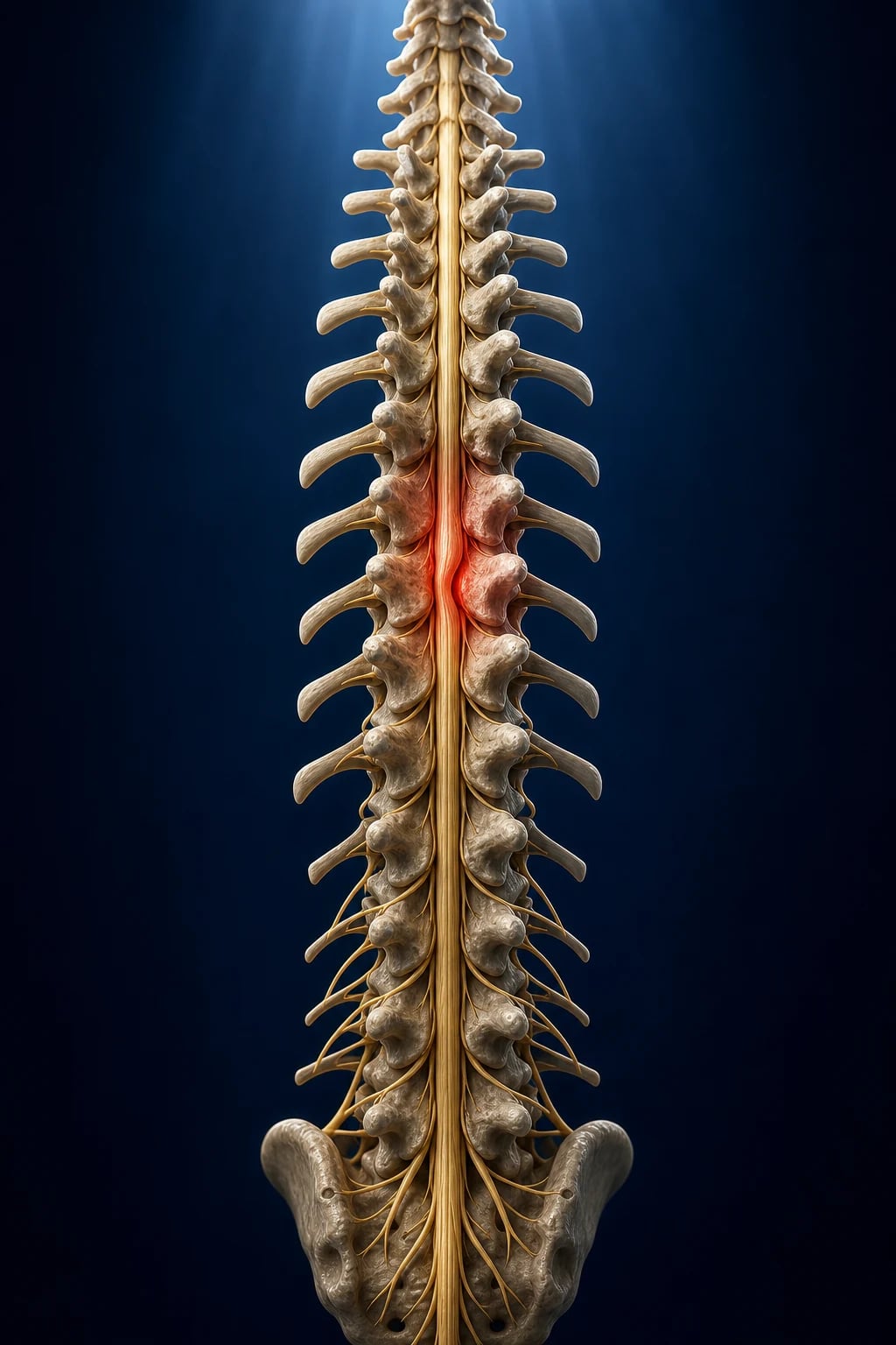
Figure Acute spinal cord injury — neurogenic shock is the hypotension with the bradycardia, the lost sympathetic tone below a lesion above T6 (not the tachycardia of hypovolaemia). Hold the MAP at 85–90 mmHg for 7 days for the cord perfusion (noradrenaline), immobilise and decompress early (<24h), and watch the respiratory failure of the cervical lesion (the diaphragm is C3–5). Methylprednisolone is not standard. Prevent the DVT and the pressure areas. Figure Neurogenic shock — distributive hypotension with bradycardia from lost sympathetic outflow; always exclude bleeding because mixed shock is common. Spinal cord injury (SCI) : neurogenic shock = hypotension + BRADYcardia (loss of sympathetic tone below T6 lesion). ICU: ABC + C-spine immobilisation, MAP target 85-90 mmHg for 7 days (noradrenaline), early surgical decompression (<24h), methylprednisolone controversial (NOT standard), prevent DVT/pressure areas. Respiratory failure in cervical SCI (diaphragm C3-5).
[1]
Neurogenic shock vs spinal shock vs hypovolaemic shock
[1]
Acute management of suspected spinal cord injury
ABC + C-spine immobilisation — airway (jaw thrust, NOT head tilt — may worsen cord injury), hard collar, head blocks, spinal board. Maintain in-line stabilisation during all movementsResuscitate — IV access, fluids (beware: neurogenic shock needs vasopressors, not just fluids — excessive fluids worsen cord oedema). Noradrenaline if hypotensiveNeurological assessment — ASIA score (motor + sensory levels, complete vs incomplete). Document level of lesion (cervical, thoracic, lumbar)Imaging — CT spine (first-line — fast, available). MRI spine (cord parenchyma — oedema, haematoma, compression). Clear C-spine per NEXUS/Canadian rulesMAP target 85-90 mmHg for 7 days — noradrenaline first-line. Avoid hypotension (worsens secondary cord injury). Avoid hypertension (risk of haemorrhage)Early surgical decompression (<24h) — STASCIS trial showed better outcomes with early surgery (<24h). Remove compression, stabilise spineMethylprednisolone — CONTROVERSIAL, NOT standard — NASCIS trials (high-dose within 8h). May modestly improve motor function but increases infection, GI bleed. Many guidelines do NOT recommend. Individual decisionICU management — ventilation (cervical SCI), DVT prophylaxis (HIGH risk — LMWH after 24-72h), pressure area care (turn q2h), bowel/bladder management, thermoregulation (poikilothermia below lesion)Rehabilitation — early physiotherapy, occupational therapy, spinal injuries unit referral [1]
[1]
Exam practice
SAQ — Cervical spinal cord injury with neurogenic shock and respiratory failure 10 minutes · 10 marks
Reveal all A 24-year-old man is brought in by ambulance after a diving accident. He is awake but cannot move his arms or legs, has a sensory level at C5, and is hypotensive (BP 82/48) with a heart rate of 48. Respiratory rate is shallow at 16, SpO2 90% on high-flow oxygen. He is on a spinal board in a hard collar.
a Define the haemodynamic syndrome, explain the mechanism, and outline your ICU resuscitation with a specific perfusion-pressure target and its duration.
b Discuss the respiratory management of a cervical spinal cord injury, including the indication and timing of intubation.
c What is the role of surgical decompression and of high-dose methylprednisolone in acute spinal cord injury?
[1]
SAQ — Autonomic dysreflexia in a chronic SCI patient 10 minutes · 10 marks
Reveal all A 30-year-old man with a T4 complete spinal cord injury from 2 years ago presents to the ED with a sudden severe throbbing headache, sweating above the nipples, nasal congestion and a recorded blood pressure of 220/130. He was admitted with urinary retention and a blocked urethral catheter.
a What is the diagnosis and mechanism? Outline your immediate step-wise management.
b Which patients are at risk of autonomic dysreflexia, and what are the common triggers to search for?
c How does the bradycardia and the distribution of symptoms (flushing/sweating above the lesion) help confirm the diagnosis at the bedside?
Clinical pearls
High-yield spinal cord injury points for CICM/FFICM exam
Neurogenic shock = HYPOTENSION + BRADYCARDIA. This is the KEY distinguishing feature from hypovolaemic shock (hypotension + tachycardia). If a trauma patient is hypotensive AND bradycardic → think neurogenic shock (SCI above T6). Do NOT give excessive fluids (will worsen cord oedema) — use vasopressors.[3] }Neurogenic shock occurs with lesions ABOVE T6. Sympathetic outflow T1-L2. Lesion above T6 → most sympathetic outflow lost → unopposed parasympathetic (vagus) → bradycardia + vasodilation. Lesion below T6 → some sympathetic preserved → less severe shock. Cervical lesions → most severe (also lose cardiac sympathetic T1-4).[5] }MAP target 85-90 mmHg for 7 days. Rationale: maintain cord perfusion, prevent secondary ischaemic injury. Noradrenaline first-line (alpha vasoconstriction supports BP, modest beta supports heart). Avoid hypotension (SBP <90 → cord ischaemia → worse outcome). Use arterial line for close monitoring.[4] }Spinal shock ≠ neurogenic shock. SPINAL SHOCK: temporary areflexia, flaccidity, loss of autonomic function below lesion (neurological phenomenon). Resolves over days-weeks (reflexes return — bulbocavernosus first). NEUROGENIC SHOCK: haemodynamic (hypotension, bradycardia). Different concepts — don't confuse.[3] }Respiratory failure in cervical SCI. Diaphragm innervated by phrenic nerve (C3-5, mainly C4). Cervical SCI above C5 → diaphragmatic paralysis → ventilatory failure. C5-C8 → intercostal paralysis (diaphragm intact but accessory muscles lost → weak cough, atelectasis). Need: early intubation if respiratory distress, ventilatory support, secretion management.[1] }Early surgical decompression (<24h) improves outcomes. STASCIS trial (Fehlings 2012): early decompression (<24h) vs late. Result: 2.8x more likely to improve ≥2 ASIA grades with early surgery. Current recommendation: surgical decompression within 24h for cervical SCI with cord compression.[2] }Methylprednisolone — CONTROVERSIAL, NOT standard. NASCIS trials (1990s): high-dose methylprednisolone (30 mg/kg bolus, then 5.4 mg/kg/h for 23-48h) within 8h of injury → modest motor improvement. BUT: increased infection, GI bleed, hyperglycaemia, no mortality benefit. AANS/CNS guidelines: treatment OPTION (not recommendation). Many centres do NOT use. Individual decision.[6] }DVT risk is VERY HIGH in SCI (highest of any patient group). Without prophylaxis: DVT 60-100%, PE 5-10%. Prophylaxis: LMWH (enoxaparin 40mg SC daily) starting 24-72h after injury (balance with risk of epidural haematoma). Mechanical (IPC, TEDS) initially. Duration: 3 months (or until mobilisation). IVC filter if anticoagulation contraindicated.[1] }Pressure area care is critical. SCI patients have lost sensation and mobility below lesion → pressure ulcers develop within HOURS. Turn every 2 hours, use pressure-relieving mattress, check pressure areas (sacrum, heels, occiput) each turn. Pressure ulcers → infection, osteomyelitis, prolonged hospitalisation.[1] }Autonomic dysreflexia (chronic SCI above T6). Triggered by noxious stimulus below lesion (bladder distension, constipation, pressure area, ingrown toenail). Massive sympathetic discharge below lesion → HYPERTENSIVE CRISIS (SBP >200). Above lesion: compensatory parasympathetic → bradycardia, flushing, headache. EMERGENCY: sit upright, remove trigger (catheterise bladder), antihypertensives (nifedipine bite-and-swallow, nitrates).[3] }Poikilothermia — temperature dysregulation. Below lesion: loss of sympathetic vasomotor control → body temperature matches environment (poikilothermia). Patient may become hypothermic (cold environment) or hyperthermic (hot environment, infection — but cannot mount fever normally). Monitor temperature, use warming/cooling blankets.[5] }Bowel and bladder management. Neurogenic bladder (flaccid → retention; spastic → urgency, incontinence). Early urinary catheter (monitor output, prevent retention). Bowel: neurogenic bowel (constipation — flaccid; incontinence — spastic). Stool softeners, regular bowel regime, suppositories.[1] }Gastric stasis and ileus. SCI causes gastroparesis and paralytic ileus (loss of autonomic input). NGT for decompression. Consider prokinetics (metoclopramide, erythromycin). Enteral nutrition when bowel function returns. Stress ulcer prophylaxis (PPI) — SCI patients at risk.[1] }C-spine clearance. NEXUS criteria (no clearance needed if ALL: no midline tenderness, no focal neurological deficit, normal alertness, no intoxication, no painful distracting injury). Canadian C-spine rule (age <65, no dangerous mechanism, no paresthesia → assess range of motion). CT first-line. MRI if ligamentous injury suspected. DO NOT remove collar until cleared.[1] } Red flags
Critical spinal cord injury red flags
Hypotension + bradycardia in trauma → neurogenic shock (SCI above T6), use noradrenaline.[3] }MAP <85 mmHg → cord ischaemia, secondary injury. Maintain MAP 85-90 for 7 days.[4] }Cervical SCI above C5 → diaphragmatic paralysis, respiratory failure, early intubation.[1] }Autonomic dysreflexia (chronic SCI) → hypertensive crisis, sit upright, remove trigger, nifedipine.[3] }DVT/PE → highest risk of any patient group, LMWH prophylaxis essential.[1] }Cord compression (tumour, abscess, haematoma) → urgent decompression, steroids for tumour.[2] }C-spine not cleared → maintain immobilisation, do not remove collar until excluded.[1] } Prognosis
STASCIS trial (Fehlings 2012, PLoS ONE) Multicentre cohort study. 313 patients with cervical SCI and cord compression.
Early surgery (<24h) vs late surgery (>24h) Primary outcome (improvement ≥2 ASIA motor grades at 6 months):
Early: 19.8% vs Late: 8.8% (OR 2.8, p<0.001)
CONCLUSION : Early decompression (<24h) associated with BETTER neurological recovery. Guidelines now recommend early surgery for cervical SCI with cord compression. [1] NASCIS-3 (methylprednisolone) : modest motor benefit if given within 8h, BUT increased infection/GI bleed. Controversial — not standard. Many guidelines: option, not recommendation.
Prognosis : Complete (ASIA A) → 10-20% convert to incomplete. Incomplete → 50-70% improve. Mortality: 5-10% acute, 20-30% 1-year.
[1]
Spinal cord syndromes
Spinal cord syndromes in SCI
[1]
Differentiating the cord syndromes (exam technique)
First, determine AIS grade (A-E) — is the injury complete or incomplete?Test dorsal columns (vibration/proprioception) — if preserved but motor + pain/temp lost → ANTERIOR cordTest each side separately — motor loss ipsilateral + pain/temp loss contralateral → BROWN-SÉQUARD (hemisection)Compare upper vs lower limbs — upper > lower weakness after hyperextension → CENTRAL cordCheck sacral sensation (S4-5) and deep anal pressure — preserved = SACRAL SPARING = incomplete injuryCheck saddle anaesthesia, sphincter tone, bladder retention — present = CONUS/CAUDADocument level precisely — neurological level (most caudal with intact motor + sensory), motor level, sensory levelRepeat examination at 72h — early exam may underestimate recovery; serial exams refine prognosis [1]
Cord syndromes — exam high-yield
Anterior cord syndrome — anterior spinal artery territory. Loss of corticospinal + spinothalamic. DORSAL COLUMNS SPARED (vibration/proprioception intact). Mechanisms: aortic surgery/dissection, hyperflexion, vertebral body fracture. Prognosis poor — ~10-20% functional motor recovery.Brown-Séquard — hemisection. IPSILATERAL motor + dorsal column loss; CONTRAlateral spinothalamic (pain/temp) loss 2-3 segments BELOW lesion (fibres cross in anterior white commissure). Best prognosis — up to 90% regain bowel/bladder, ambulation often possible.Central cord syndrome — MOST COMMON incomplete SCI. Hyperextension in older patient with cervical spondylosis (e.g. fall down stairs, RTA). UPPER > lower limb weakness (hands more affected than legs — medial corticospinal tract fibres to hand are most central). Bladder usually spared. Often improves; surgery if compression/instability.Sacral sparing — preserved perianal sensation or voluntary anal contraction = incomplete injury. CRITICAL for prognosis and surgical decision-making. ALWAYS document S4-5 pinprick/light touch and deep anal pressure on every exam.Posterior cord syndrome — rare; loss of dorsal columns only (vibration, proprioception, ROMberg positive). Motor and pain/temp preserved. Causes: B12 deficiency, syphilis (tabes dorsalis), tumour.Conus medullaris vs cauda equina — conus (T12-L1 vertebral, mixed UMN/LMN, early/severe bladder-bowel); cauda (below L2, pure LMN, asymmetric, radicular pain). Both need urgent imaging and decompression. [1]
Respiratory management in cervical SCI
Figure ICU priorities — protect the cord (airway, oxygenation, perfusion), distinguish shock types, plan decompression, and prevent secondary complications. Respiratory impact by level of SCI
[1]
Tracheostomy timing in cervical SCI
Anticipate prolonged ventilation in high cervical SCI (C1-C4) — early tracheostomy within 7-10 daysC5-C8 — assess weaning capacity (vital capacity, NIF, peak cough flow); trial extubation may be attempted firstSurgical vs percutaneous — surgical tracheostomy if anterior cervical fixation (avoid track through surgical field); percutaneous dilatational feasible after posterior fixationSpeaking valve — early to facilitate communication, swallow assessment, weaningWeaning — progressive ventilator-free breathing (SVB); assist control → PSV → trachy mask; cuff down trialDecannulation criteria — ventilator-free, effective cough (peak expiratory flow >160 L/min), manageable secretions, intact bulbar function [1]
Respiratory pearls in SCI
Vital capacity < 1 L (or <15 mL/kg) predicts need for ventilation. Serial bedside spirometry guides weaning decisions — improvement precedes successful extubation by 24-48h.Peak cough flow <160 L/min predicts secretion retention → consider mechanical insufflation-exsufflation (MIE/CoughAssist) and macrolide prophylaxis.Paradoxical breathing — intercostal paralysis → chest retracts on inspiration, abdomen protrudes. Sign of high cervical injury and impending fatigue.Aspiration risk is HIGH — vocal cord paralysis (recurrent laryngeal, especially after anterior cervical surgery) + weak cough + gastroparesis. Bedside swallow assessment before any oral intake.Secretion management is the key to weaning — chest physio, mucolytics, MIE, inhaled salbutamol (also helps prevent autonomic dysreflexia).Ventilator-associated pneumonia — common in prolonged ventilation. Head of bed 30°, oral chlorhexidine, subglottic suction, early enteral nutrition.Sleep-disordered breathing common post-decannulation — overnight oximetry, CPAP/BiPAP. Respiratory reserve worst in REM. [1]
Vasopressor strategy in neurogenic shock
Vasopressor / chronotrope choice in neurogenic shock
[1]
Vasopressor pearls in neurogenic shock
Noradrenaline is FIRST-LINE. Provides α1 vasoconstriction (raises SVR/BP) + modest β1 (supports cardiac output without excessive tachycardia). Start 0.05-0.1 mcg/kg/min, titrate to MAP 85-90 mmHg.Avoid phenylephrine as SOLE agent if bradycardic — pure α1 raises BP but reflex bradycardia may worsen unopposed vagal tone. Useful adjunct if HR controlled.Atropine for SYMPTOMATIC bradycardia (HR <50 with hypotension, arrhythmia, low CO). Often transient — escalate to dopamine/isoprenaline infusion if persistent.Volume loading first — give 500-1000 mL crystalloid bolus to restore preload, but STOP early. Excessive crystalloid → cord oedema, ARDS. Most patients need vasopressor within 24-48h of injury.Duration of vasopressor support typically 1-3 weeks — wean as neurogenic shock resolves (reflexes return, HR rises, SVR normalises).Arterial line is MANDATORY — beat-to-beat BP for vasopressor titration; cuff pressures unreliable in vasodilated patients.CVP/ScvO2 targets — CVP 8-12 mmHg, ScvO2 >65%. Avoid over-resuscitation. Some centres use PiCCO/LiDCO for goal-directed therapy.Beware occult haemorrhage — bradycardia may coexist with hypovolaemia from associated injuries (chest, abdomen, pelvis, long bones). Always EXCLUDE bleeding before attributing hypotension solely to neurogenic shock. [1]
Autonomic dysreflexia
Managing acute autonomic dysreflexia
RECOGNISE — SBP >20-40 mmHg above baseline in SCI above T6 (often SBP 200-300). Pounding headache, flushing/blurred vision above lesion, sweating/piloerection above lesion, bradycardia belowSIT UPRIGHT (head up) — use gravity to lower BP; loosen tight clothing, abdominal binder, stockings, strapsCHECK BP every 2-5 min until resolved; baseline in tetraplegics may be 90/60 — 'normal' BP may already be ADIDENTIFY AND REMOVE TRIGGER (most common: bladder ~75-85%). Check catheter for kinks/blockage; irrigate if needed; change catheter; do bladder scan; treat UTIIf no bladder trigger — check bowel (digital rectal exam with lignocaine gel, remove stool slowly), skin (pressure area, ingrown toenail, burn), other (fracture, acute abdomen, pregnancy)ANTIHYPERTENSIVE if SBP still elevated after trigger removed — short-acting, rapid-onset: nifedipine (bite-and-swallow 10 mg), GTN paste/sublingual, captopril 25 mg SL, hydralazine. AVOID sustained-release or long-acting agentsRECHECK BP for 2 hours after resolution — recurrence common; admit to monitored areaDOCUMENT and educate — write the episode in chart; review bowel/bladder regimen; medic-alert bracelet; patient/family education [1]
Autonomic dysreflexia pearls
Occurs in SCI above T6 (splanchnic sympathetic outflow lost). Develops after spinal shock resolves — usually 4-6 weeks post-injury. Lifetime risk in 50-90% of tetraplegics.Massive unopposed sympathetic discharge below lesion → vasoconstriction → hypertension. Above lesion: compensatory vagal (bradycardia) + vasodilation (flushing). Body cannot modulate sympathetic below lesion.Most common trigger = BLADDER (~75-85%): blocked catheter, urinary retention, UTI, instrumentation. Next: BOWEL (~15-20%): constipation, faecal impaction, bowel programme.Hypertension can be LIFE-THREATENING — intracranial haemorrhage, MI, seizures. SBP may exceed 300 mmHg. Bradycardia may be the only clue if BP cuff not cycled.Patient education is the cornerstone — patients carry wallet cards, alert bracelets. Teach trigger recognition, self-management. Bowel/bladder regimen must be REGULAR and predictable.Lignocaine gel during DRE — reduces further noxious stimulation. Use generous gel + wait 5 min before manipulation.Pregnancy and labour — epidural anaesthesia is PREFERRED to prevent autonomic dysreflexia during childbirth. Anaesthetic input mandatory; epidural placed early in labour. [1]
Autonomic dysreflexia red flags
SBP >200 mmHg in chronic SCI → autonomic dysreflexia emergency, sit upright, find trigger, nifedipineHeadache + sweating above lesion + piloerection below → autonomic dysreflexia even if BP 'normal' (baseline may be 90/60)Trigger not found within 5 min → escalate to senior, consider life-threatening cause (occult fracture, acute abdomen, pregnancy)Recurrent episodes → review bowel/bladder regimen, consider prophylaxis (nifedipine, gabapentin, clonidine) [1]
Pharmacology
Pharmacology in acute SCI
Methylprednisolone (NASCIS protocol) — 30 mg/kg IV bolus over 15 min, then 5.4 mg/kg/h for 23h (started within 3h of injury) or 48h (started 3-8h). After 8h: NOT indicated. Side effects: hyperglycaemia, infection (wound, pneumonia, sepsis), GI bleed, pancreatitis, myopathy. Many centres no longer use.[6] }GM-1 ganglioside (Sygen trial) — no benefit; not recommended.Nimodipine, thyrotropin-releasing hormone, naloxone, tirilazad — investigated in SCI, NO proven benefit; not used.Riluzole — Phase III trial ongoing (RISCIS); glutamate modulator. Not yet standard.Minocycline — Phase II showed trend; Phase III (MASC) completed; not yet standard.DVT prophylaxis — enoxaparin 40 mg SC daily from 24-72h. Beware epidural haematoma if neuraxial. Mechanical (IPC) from admission. Fondaparinux alternative. Clexane in renal impairment (1 mg/kg SC bd).Stress ulcer prophylaxis — PPI for first 4 weeks (mechanical ventilation, coagulopathy, shock). STOP after — C. difficile, pneumonia risk.Bowel regimen — stool softener (docusate) + stimulant (senna/bisacodyl) + glycerine/bisacodyl suppository every other day. Aim formed stool, no impaction.Spasticity — baclofen (oral or intrathecal pump), tizanidine, botulinum toxin. Avoid sudden withdrawal (severe rebound spasm).Neuropathic pain — gabapentin/pregabalin first-line; add TCA (amitriptyline) or SNRI (duloxetine). Opioids only for breakthrough nociceptive pain. Investigations and monitoring
[1]
Whole-spine CT in polytrauma — 10-15% have non-contiguous spinal injuries. Clear the WHOLE spine, not just the symptomatic level.MRI within 24-48h if neurological deficit — demonstrates cord oedema/haematoma, disc herniation, ligamentous injury. SCOPEC (spinal cord oedema, haemorrhage >10 mm, two-level oedema) predicts poor outcome.[9] }Vertebral artery injury in 20-40% of cervical subluxation/foramen transversarium fractures — CT angiogram mandatory. Posterior circulation stroke risk.Tracheostomy: percutaneous vs surgical — surgical if anterior cervical fixation (avoid track through surgical field). PDT feasible after posterior fixation.Continuous EEG if unconscious — 10-30% of severe TBI/SCI co-occur; non-convulsive seizures possible.CT head if decreased GCS — exclude concurrent TBI (10-50% have associated head injury).Serial lactate, venous/arterial blood gas — guide resuscitation; high lactate suggests occult hypoperfusion/haemorrhage rather than neurogenic shock alone. Surgical management
STASCIS (Fehlings 2012, PLoS ONE)
Design : Multicentre cohort, 313 cervical SCI patients with cord compressionComparison : Early (<24h) vs late (>24h) decompressionOutcome : ≥2 ASIA motor grade improvement at 6 monthsResult : 19.8% (early) vs 8.8% (late), OR 2.8 (95% CI 1.6-5.1), p<0.001CONCLUSION : Early surgical decompression (<24h) associated with better neurological recovery. Now standard of care. [1]
NASCIS-3 (Bracken 1997, JAMA)
Design : RCT, 499 acute SCI patientsComparison : 24h vs 48h methylprednisolone, and tirilazadResult : Modest motor improvement if started within 3-8h (48h course)Complications : Increased infection, GI bleed, sepsis, hyperglycaemia, pancreatitisCONCLUSION : Methylprednisolone is an OPTION (not recommendation). Many guidelines discourage. Benefit marginal, harms significant.[7] }
Surgical decompression within 24h : RECOMMENDED for cervical SCI with cord compression (Level B)MAP augmentation 85-90 mmHg for 7 days : SUGGESTED (Level C)Methylprednisolone : OPTION within 8h (Level C); discuss risks/benefits with patientCONCLUSION : Multidisciplinary, internationally endorsed — basis of current practice.[8] }
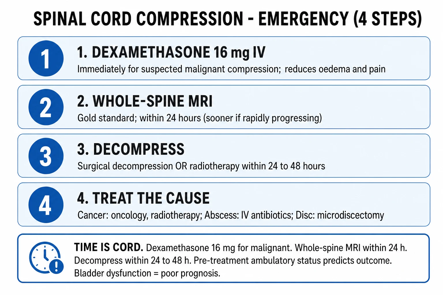
Early decompression <24h is standard for cervical SCI with cord compression (STASCIS). Reduction of fracture-dislocation is URGENT if incomplete injury deteriorating.Closed reduction (Gardner-Wells tongs, halo) for cervical facet dislocation — often performed in ED/ICU before surgery. May avoid surgery if alignment restored.Anterior vs posterior approach depends on compression site and instability. Combined approaches for severe injuries.Timing of surgery in complete SCI — still debated; decompression may allow root recovery (improve ambulation even if cord complete).Non-traumatic SCI (cord compression from tumour, abscess, haematoma) — URGENT decompression (within 6-24h). High-dose dexamethasone for tumour (10 mg IV then 4 mg q6h).Surgery does not prevent neurogenic shock — even after fixation, sympathetic outflow lost below lesion; vasopressors still needed for 1-3 weeks. [1]
Neurological level — functional implications
Functional outcomes by level of SCI
[1]
C3-C5 = diaphragm — phrenic nerve roots. Above C5 → ventilator-dependent. C3 alone → permanent ventilator (some units offer diaphragm pacing).C6 = wrist extension — KEY for functional tenodesis grip (passive finger flexion with wrist extension). Allows self-feeding, writing, typing.T1 = hand intrinsics — preserved = full hand function. T1 lesion → claw hand, no fine motor.T6 = intercostals — vital for cough. Below T6 = adequate respiratory reserve for independent living.L2-L4 = quadriceps/hip flexors — knee extension, hip flexion. With orthotics (KAFO), ambulation possible.S2-S4 = sacral (bowel/bladder/sexual) — preserved = volitional voiding, erection, ejaculation.S2-S4 sparing also indicates intact sacral reflex arc — bulbocavernosus reflex returns first as spinal shock resolves. [1]
Complications (long-term)
Complications of SCI — high-yield
DVT/PE — HIGHEST risk of any patient group. Without prophylaxis 60-100% DVT, 5-10% PE fatal. LMWH 24-72h post-injury. Duration 3 months (extended if not mobile). IVC filter if anticoag contraindicated. Doppler ultrasound surveillance in some units.Pressure injuries — NPUAP staging I-IV. Sacrum, heels, occiput, scapulae common. Turn q2h, pressure-relieving mattress, nutrition (zinc, vitamin C, protein 1.5-2 g/kg/day), shear-minimising transfers. Stage IV → osteomyelitis, sepsis.Neurogenic bladder — flaccid (areflexic) early → retention → overflow. Spastic (reflexic) later → detrusor-sphincter dyssynergia → high-pressure bladder → hydronephrosis, reflux, renal failure. Intermittent catheterisation (qid, <500 mL each); anticholinergics (oxybutynin, solifenacin); botox; ileal conduit if refractory.Neurogenic bowel — flaccid → constipation/impaction; reflexic → frequent small stools. Regimen: stool softener, stimulant, glycerine suppository/digital stimulation every other day. Colostomy if refractory.Heterotopic ossification — 20-30% (especially hip). Pain, swelling, reduced ROM. Bisphosphonates, NSAIDs, surgery when mature.Osteoporosis — disuse below lesion; calcium/vitamin D, weight-bearing (standing frame). Pathological fractures common.Spasticity — develops after spinal shock. Baclofen (PO or intrathecal pump), tizanidine, botulinum toxin, stretching.Neuropathic pain — at-level (segmental) and below-level burning/shooting. Gabapentin/pregabalin, amitriptyline, duloxetine. Often refractory.Depression, suicide — 25-30% depression; suicide rate 4-6x general population. Psychological support from admission. Peer support (other SCI patients).Cardiovascular deconditioning — resting bradycardia (40s common), orthostatic hypotension (especially during rehab). Compression stockings, abdominal binder, midodrine, fludrocortisone.Sexual/reproductive — men: erectile dysfunction (PDE5 inhibitors, penile injection, vacuum device, prosthesis); ejaculation (vibrator, electroejaculation for fertility). Women: usually resume menses within 6 months; pregnancy possible, autonomic dysreflexia during labour (epidural mandatory).Sleep-disordered breathing — OSA common (50%+). Sleep study, CPAP/BiPAP especially if tracheostomy decannulated.Renal calculi — immobilisation hypercalciuria, urinary stasis, infection. Hydration, treat infection, lithotripsy.Latex allergy — ~5-20% of chronic SCI patients (repeated catheter exposure). Use latex-free equipment.Post-traumatic syringomyelia — 3-30% develop over years; expanding cavity, ascending sensory level, new weakness, neuropathic pain → shunt surgery. [1]
Late complications red flags
Fever in chronic SCI — may not have localising signs; consider UTI, pressure area, osteomyelitis, DVT, autonomic dysreflexiaNew headache or hypertension in tetraplegic — autonomic dysreflexia until proven otherwiseSudden leg swelling — DVT (high baseline risk)Confusion + bradycardia + hypoxia — consider occult sepsis from pressure area or UTINew weakness or sensory change months-years post-injury — syrinx (post-traumatic syringomyelia) [1]
Prognosis and outcomes
EMSCI (European Multicenter Spinal Cord Injury) — prognosis
MRI-based prediction : brainstem and spinal cord oedema, haemorrhage extent predict AIS conversionAIS A at admission → 20% convert to B, 10% to C, 5% to D at 1 yearAIS B-D at admission → 60-80% improve ≥1 gradeMortality at 1 year: 5-15% (cervical higher than thoracic)Median life expectancy (cervical complete): 20-30 years less than uninjured peers [1]
AIS grade at admission is strongest predictor. AIS A → poor motor recovery; AIS D → near-complete recovery.MRI cord haemorrhage = poor prognosis. Oedema alone = better. Haemorrhage length >10 mm predicts poor outcome.Level matters — cervical worse functional outcome than thoracic (respiratory, hand function).Age — older patients recover less. >65 worse outcome at every level.Time to surgery <24h improves AIS conversion (STASCIS).MAP <85 in first week worsens outcome (secondary ischaemic injury).Somatosensory evoked potentials — bilateral absent cortical response = poor motor outcome.Post-traumatic syringomyelia develops in 3-30% over years — expanding cavity, ascending sensory level, new weakness → shunt surgery. [1]
C-spine clearance in ICU
C-spine clearance in obtunded ICU patient
NEXUS / Canadian C-spine Rule — only valid in ALERT, non-intoxicated patients. Most ICU patients cannot be cleared clinically.CT cervical spine — high-quality CT negative excludes unstable bony injury in obtunded patient. Clearance possible if no clinical signs on awakening.MRI if concern for ligamentous injury (hyperflexion/extension, distracting injury, persistent pain). Best within 48-72h; MRI negative excludes ligamentous injury.If MRI cannot be obtained (pacemaker, instability) — leave collar in place, repeat CT at 48h, clearance deferred. Some units use dynamic fluoroscopy (controversial).Once cleared — remove collar promptly (pressure injury risk within 5 days: occiput, chin, shoulders).Document clearance clearly — date, time, modality, responsible clinician. [1]
Prolonged collar (≥5 days) → pressure injury (occiput 20%) . Goal: clearance within 48h. Use Miami-J/Aspen with padding; remove collar briefly each turn for skin check.Obtunded patient + CT negative + MRI negative = SAFE to clear. MRI within 72h for ligamentous integrity.SCIWORA (Spinal Cord Injury Without Radiographic Abnormality) — more common in children; MRI may show cord oedema without bony injury. Treat as SCI.Polytrauma patient: 10-15% have non-contiguous spine fractures — clear the WHOLE spine.In-line stabilisation during intubation — manual in-line stabilisation (MILS), video laryngoscopy (GlideScope, C-MAC) reduces C-spine movement. Avoid nasal intubation in basal skull fracture. AFOI (awake fibreoptic) for known difficult airway/unstable C-spine.Log-rolling for transfers/turns until thoracolumbar spine cleared. 4-person lift preferred for transfers. [1]
C3-C5 lesion → diaphragmatic paralysis, prepare for early intubationFalling vital capacity, rising PaCO2, weak cough → impending respiratory failureDirect laryngoscopy in unstable C-spine without MILS → worsening cord injuryAnterior cervical fixation + early percutaneous trachy → risk of surgical field contaminationVocal cord dysfunction (recurrent laryngeal, post-anterior approach) → aspiration, stridor [1]
Non-traumatic SCI
Non-traumatic causes of SCI
Spinal epidural abscess — Staph aureus, diabetes, IV drug use, immunosuppression. Triad: back pain, fever, neurological deficit. Urgent surgical decompression + IV antibiotics (4-6 weeks). Dexamethasone controversial.Spinal epidural haematoma — anticoagulation, coagulopathy, post-procedure (epidural). Sudden severe back pain + rapidly progressive deficit. EMERGENCY surgery within 6-24h. Reverse anticoagulation.Malignancy — breast, lung, prostate, myeloma most common. Vertebral metastasis with cord compression. High-dose dexamethasone (10 mg IV then 4 mg q6h) + radiotherapy + surgical decompression (separation surgery).Disc herniation — acute large central disc → cord compression. Surgical decompression.Vascular — anterior spinal artery syndrome (aortic surgery, dissection, AVM, vasculitis). Often sudden, painless, no trauma. Pattern: motor + pain/temp loss, dorsal columns spared.Inflammatory — transverse myelitis, NMOSD, MOGAD, MS. MRI shows T2 hyperintensity, often >3 segments. Steroids, plasma exchange, immunotherapy.Infection — Pott's disease (TB), herpes zoster, schistosomiasis. Endemic regions. [1]
Non-traumatic SCI red flags
Back pain + fever + deficit → spinal epidural abscess, urgent MRIAnticoagulated patient + sudden back pain + weakness → epidural haematoma, urgent MRI + reversalKnown cancer + new weakness → metastatic cord compression, steroids + urgent imagingPainless acute paraplegia → anterior spinal artery infarct (recent aortic surgery?)Bilateral sensory level + bowel/bladder → transverse myelitis vs compression; MRI mandatory [1]
Special considerations
Paediatric SCI — SCIWORA common (ligamentous laxity). Higher cervical injuries. Cervical collar may exacerbate injury (relative oversized head) — use flat immobilisation with padding under shoulders. Lower threshold for MRI.Elderly — central cord syndrome (cervical spondylosis + hyperextension, e.g. fall down stairs). Higher mortality, less recovery. Aggressive respiratory care.Pregnancy — autonomic dysreflexia during labour (T6 above). Epidural mandatory. Multidisciplinary planning. Higher risk DVT — LMWH throughout.Anticoagulated / coagulopathic — high suspicion for epidural haematoma; reverse anticoagulation if suspicion. Avoid neuraxial.Pre-existing myelopathy (cervical stenosis, MS) — minor trauma may cause major deficit. Lower threshold for imaging.Penetrating SCI (stab, GSW) — often incomplete (Brown-Séquard pattern). Steroids NOT indicated. Surgical only for retained object, vascular injury, CSF leak. [1]
Sedation and analgesia
Sedation/analgesia in SCI
Neurological assessment must be possible — avoid prolonged deep sedation; daily sedation holds (once stable) for serial examinations.Propofol favoured for short-acting, titratable sedation. Avoid high-dose/long propofol infusion (PRIS).Fentanyl for analgesia; avoids histamine release (morphine — avoid in hypotensive neurogenic shock).Ketamine — historically avoided (raised ICP); now evidence supports safety, useful for refractory pain, bronchodilation in asthma.Avoid benzodiazepines for routine sedation — prolonged effect, delirium risk. Use for specific indications (seizure, alcohol withdrawal).Pain assessment — polytrauma + sensory loss below lesion makes pain assessment difficult. Use behavioural pain scales (BPS, CPOT) if unable to report.Neuropathic pain — gabapentin/pregabalin first-line. Add TCA (amitriptyline) or SNRI (duloxetine). Opioids only for breakthrough nociceptive pain.Titrate sedation to lightest level permitting ventilation/assessment — RASS -1 to 0 in stable patients; SATs daily. [1]
ICU bundle of care
Daily SCI bundle in ICU
Neurological check — motor, sensory, AIS grade (or appropriate serial assessment if sedated/ventilated)MAP 85-90 mmHg — adjust vasopressor; daily plan to wean as shock resolvesRespiratory — vital capacity, peak cough flow, secretion management, ventilator weaning trialC-spine clearance status — has it been cleared? Is collar off? Skin check under collarDVT prophylaxis — LMWH given? Mechanical on? Surveillance ultrasound per protocolSkin — turn q2h, pressure areas, mattress adequacyBowel/bladder — IDC in situ/output, bowel regimen, abdominal examNutrition — enteral established? Gastric residual volumes? Calorie/protein targets metGI prophylaxis — PPI (if indicated), glucose control (avoid hyperglycaemia, especially if methylprednisolone)Rehabilitation — passive ROM, splints, early mobilisation as permitted, MDT meeting weeklyFamily communication — prognosis discussions, goal-of-care, spinal injuries unit referral [1]
Summary
SCI with neurogenic shock : lesion above T6 → unopposed parasympathetic → HYPOTENSION + BRADYCARDIA + WARM DRY SKIN (≠ hypovolaemic shock). ICU priorities: (1) ABC + C-spine immobilisation; (2) MAP 85-90 mmHg for 7 days with noradrenaline (avoid fluid overload → cord oedema); (3) early surgical decompression <24h (STASCIS); (4) methylprednisolone controversial, NOT standard (NASCIS); (5) DVT prophylaxis (highest risk group); (6) pressure area care (q2h turns); (7) respiratory support for cervical SCI (C3-5 diaphragm); (8) bowel/bladder management; (9) autonomic dysreflexia prophylaxis (chronic). AIS scale grades severity (A=complete → E=normal). Cord syndromes: complete, anterior (motor + ST loss, dorsal spared), Brown-Séquard (hemisection), central (UE>LE).
[1]
Final high-yield exam pearls (CICM/FFICM/EDIC)
ALWAYS suspect neurogenic shock in hypotensive + bradycardic trauma patient — examine for SCI. Exclude occult haemorrhage first.Noradrenaline FIRST-LINE for neurogenic shock; atropine for symptomatic bradycardia; phenylephrine may worsen bradycardia.MAP 85-90 × 7 days — cord perfusion. Arterial line mandatory.Early decompression <24h (STASCIS) for cervical SCI with compression — improves AIS conversion.Methylprednisolone = OPTION not standard (NASCIS) — increased infection/GI bleed; many units do NOT use.Respiratory failure in cervical SCI is leading cause of early death — vital capacity <15 mL/kg predicts ventilation.DVT risk highest of any patient — LMWH from 24-72h, 3 months duration.Autonomic dysreflexia — chronic SCI above T6, SBP >200, find trigger (bladder/bowel), nifedipine bite-and-swallow.C-spine clearance within 48h — prolonged collar → occipital pressure injury (20%).Sacral sparing (S4-5) preserved = incomplete injury → better prognosis, surgical candidate.Spinal shock ≠ neurogenic shock — different concepts; reflexes return after spinal shock (bulbocavernosus first).Poikilothermia — body temperature tracks environment; control room temperature, warming/cooling blankets.Whole-spine imaging — 10-15% non-contiguous injuries in trauma.Tracheostomy within 7-10 days for high cervical (C1-C4) — early PDT after fixation.MDT approach — intensivist, neurosurgeon, spinal surgeon, rehab physician, physio, OT, social work, psychology — from admission. [1] References [1] Hadley MN, et al. The influence of crossbite in early development of mandibular bone asymmetries in paediatric patients J Clin Exp Dent , 2017.PMID 29075414 [2] Fehlings MG, et al. Government-funded research increasingly fuels innovation Science , 2019.PMID 31221848 [3] Saadeh YS, et al. Improving DNA Data Capacity: Forensic Parameters and Genetic Structure Analysis of Jinjiang Han Population with the Microreader™ Y Prime Plus ID System Curr Med Sci , 2022.PMID 35403953 [4] Evans L, et al. Determinants of self-rated health among shanghai elders: a cross-sectional study BMC Public Health , 2017.PMID 29029627 [5] Furlan JC, Fehlings MG. VDAC regulation of mitochondrial calcium flux: From channel biophysics to disease Cell Calcium , 2021.PMID 33529977 [6] Guly JA, et al. Can sand nourishment material affect dune vegetation through nutrient addition? Sci Total Environ , 2020.PMID 32278174 [7] Bracken MB. Differences in hemodynamic parameters and exercise capacity between patients with pulmonary arterial hypertension and chronic heart failure J Cardiopulm Rehabil Prev , 2012.PMID 23143688 [8] Fehlings MG, et al. A screen for inhibitory peptides of hepatitis C virus identifies a novel entry inhibitor targeting E1 and E2 Sci Rep , 2017.PMID 28638089 [9] Aarabi B, et al. Is vulvovaginal atrophy due to a lack of both estrogens and androgens? Menopause , 2017.PMID 27875388 [10] Casha S, et al. [New trends and novel possibilities in the clinical use of SPECT/CT imaging] Magy Onkol , 2015.PMID 25763909 [11] Krassioukov A, et al. Lipids and lipidomics in brain injury and diseases AAPS J , 2006.PMID 16796382 [12] Consortium for Spinal Cord Medicine. OmpT: molecular dynamics simulations of an outer membrane enzyme Biophys J , 2004.PMID 15315948