ICU · Obs/Gynae
Acute fatty liver of pregnancy (AFLP)
Also known as Acute fatty liver of pregnancy (AFLP) · Acute yellow atrophy of pregnancy · Swansea criteria · Microvesicular fatty liver of pregnancy · Acute yellow atrophy · Mitochondrial fatty-acid-oxidation disorder of pregnancy · LCHAD deficiency in pregnancy · Third-trimester acute liver failure · HELLP mimic
AFLP is a rare (1 in 7000-15,000 pregnancies), life-threatening liver disorder occurring in the third trimester. Fatty infiltration of hepatocytes → microvesicular steatosis → acute liver failure. Presents with: nausea, vomiting, abdominal pain, jaundice, polydipsia/polyuria (DI from pancreas involvement), encephalopathy (progressive). Diagnosis: Swansea criteria (6+ of 11 clinical/laboratory features). Management: URGENT DELIVERY (the only definitive treatment — liver function recovers after delivery), supportive ICU care (coagulopathy, hypoglycaemia, AKI, encephalopathy), NAC may help. Distinguishes from HELLP: AFLP has more severe coagulopathy, encephalopathy, and hypoglycaemia. Mortality: 1-12% (maternal), 7-58% (fetal) — has improved with early recognition and delivery.
On this page & tools
Your progress
Saved locally on this device.
Target exams
Red flags

Overview
Acute fatty liver of pregnancy (AFLP) is a rare, potentially fatal third-trimester hepatocellular crisis characterised histologically by microvesicular steatosis — small cytoplasmic lipid droplets crowding hepatocytes with strikingly little necrosis and minimal inflammation. It is pathologically and mechanistically allied to Reye's syndrome, sodium valproate hepatotoxicity, tetracycline/high-dose methotrexate injury, Jamaican vomiting sickness (hypoglycin A), and the inborn errors of mitochondrial β-oxidation. The unifying pathogenic thread is mitochondrial fatty-acid β-oxidation failure.[1][2]
The fellowship-critical insight — and the single most examinable concept in AFLP — is that the fetus drives the maternal disease. A fetus homozygous (or compound heterozygous) for long-chain 3-hydroxyacyl-CoA dehydrogenase (LCHAD) deficiency cannot complete mitochondrial β-oxidation of long-chain fatty acids. The accumulated toxic 3-hydroxy-fatty-acid metabolites (and their acylcarnitines) cross the placenta into the maternal circulation, overwhelm the residual β-oxidation capacity of the heterozygous-carrier mother, and produce hepatic mitochondrial dysfunction and microvesicular steatosis.[4] This fetal–maternal metabolic crosstalk is why the definitive treatment is delivery of the fetus — removing the source of toxic metabolites — and why AFLP typically resolves postpartum. It is also why the mother (and infant) must be tested for a fatty-acid oxidation disorder after the event, because recurrence in future pregnancies (and the child's long-term prognosis) depends on it.[1][4]
AFLP sits in the spectrum of third-trimester liver disease alongside pre-eclampsia/HELLP and intrahepatic cholestasis of pregnancy, and the intensivist's central tasks are fourfold: (1) recognise the syndrome early (the prodrome is easily dismissed as 'gastric flu'); (2) apply the Swansea criteria to make the diagnosis without subjecting a coagulopathic patient to liver biopsy; (3) distinguish AFLP from HELLP, because the two share the third-trimester setting yet differ in their dominant laboratory signature, their complications, and their management emphasis; and (4) deliver the baby without delay while supporting the failing liver, the coagulation system, the glucose, and the kidney — in most cases the liver regenerates fully within 1–4 weeks and the mother returns to complete health without chronic liver disease.[1][2]
Pathophysiology — the fetal–maternal β-oxidation defect
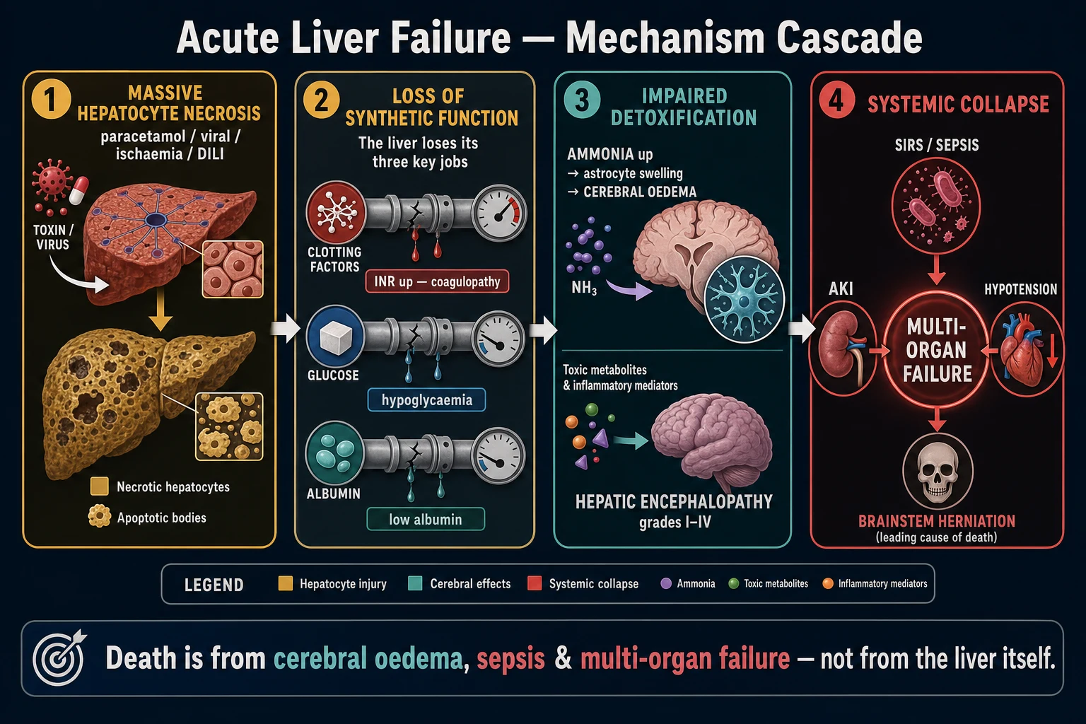
Understanding the pathophysiology is the key to remembering the clinical syndrome and to articulating an exam answer that distinguishes AFLP from HELLP. The mechanism is a disorder of mitochondrial fatty-acid β-oxidation.[2][4]
Mitochondrial β-oxidation and the pathogenesis of AFLP — the mechanistic cascade
- LONG-CHAIN FATTY ACIDS ARE THE FASTED-LIVER FUEL — in late pregnancy the mother is relatively energy-deficient (fasting, increased fetoplacental demand, physiologic insulin resistance driving adipose lipolysis), so the liver, heart, and skeletal muscle depend heavily on β-oxidation of long-chain fatty acids (LCFA, C12–C18) for ATP.
- CARNITINE SHUTTLE + MTP COMPLEX — LCFAs enter the mitochondrion via the carnitine shuttle (CPT-I → carnitine–acylcarnitine translocase → CPT-II) and are progressively shortened by the mitochondrial trifunctional protein (MTP): a hetero-octamer on chromosome 2p23 with two α-subunits (gene HADHA, carrying long-chain enoyl-CoA hydratase and LCHAD activities) and four β-subunits (gene HADHB, carrying long-chain 3-ketoacyl-CoA thiolase activity).
- LCHAD IS THE THIRD STEP OF THE β-OXIDATION SPIRAL — long-chain 3-hydroxyacyl-CoA dehydrogenase catalyses the NAD⁺-dependent oxidation of 3-hydroxyacyl-CoA → 3-ketoacyl-CoA. Loss of this step blocks β-oxidation of C12–C16 fatty acids at the 3-hydroxy intermediate.
- FETAL LCHAD DEFICIENCY — a fetus homozygous or compound heterozygous for loss-of-function HADHA mutations (the common pathogenic allele is the G1528C / E474Q missense mutation, accounting for ~60–70% of LCHAD-deficient alleles) cannot complete β-oxidation. The mother is typically a heterozygous carrier (autosomal recessive).
- PLACENTAL TRANSFER OF TOXIC METABOLITES — long-chain 3-hydroxy-fatty acids and their acylcarnitines accumulate in the affected fetus and cross the placenta into the maternal circulation.
- MATERNAL OVERLOAD — the carrier mother's ~50% residual LCHAD activity is normally sufficient, but in the metabolically stressed third trimester it is overwhelmed by the fetal load; her own β-oxidation fails.
- HEPATIC MITOCHONDRIAL DYSFUNCTION → MICROVESICULAR STEATOSIS — the accumulating 3-hydroxy-fatty acids are directly toxic to hepatocyte mitochondria (uncoupling of oxidative phosphorylation, impaired ATP synthesis, oxidative stress). Unable to oxidise incoming fatty acids, the liver esterifies them into triglyceride, which accumulates as small (microvesicular) lipid droplets in zone-3 (centrilobular) hepatocytes. Histology shows fatty change with minimal necrosis and little inflammation — the key distinction from viral or autoimmune hepatitis.[2]
- HEPATIC FAILURE — SYNTHETIC + DETOXIFYING — mitochondrial failure produces loss of synthetic function (coagulation factors II, V, VII, IX, X and fibrinogen → coagulopathy; albumin; and crucially gluconeogenesis → hypoglycaemia) and detoxifying function (ammonia and bilirubin clearance → encephalopathy and jaundice).
- MULTI-ORGAN STEATOSIS — the same microvesicular lipid infiltration affects renal tubular cells (AKI), pancreatic acinar cells (pancreatitis), and the posterior pituitary / hypothalamus (transient central diabetes insipidus → the polydipsia/polyuria of Swansea criterion 3).[1][2]
Two essential caveats the candidate must articulate. First, not all AFLP is LCHAD-mediated. Approximately 15–25% of AFLP cases (and a proportion of HELLP cases) are associated with a fetal fatty-acid oxidation disorder; conversely, only a minority (~15–20%) of pregnancies carrying an LCHAD-deficient fetus actually develop AFLP. The association is nevertheless strong enough that both mother and infant must be tested (plasma acylcarnitine profile, HADHA sequencing, fibroblast enzyme assay) after any AFLP (and arguably any atypical HELLP) pregnancy.[4] Second, the microvesicular pattern (small droplets, central nucleus) — as opposed to the macrovesicular steatosis of non-alcoholic fatty liver disease, alcohol misuse, or chronic starvation — is the histological hallmark of mitochondrial/toxic/metabolic injury, shared by AFLP, Reye's, valproate, and the inborn errors. Liver biopsy is the gold standard but is rarely required (bleeding risk in a coagulopathic patient; the Swansea criteria are diagnostic).[1][3]
Epidemiology and risk factors
AFLP complicates approximately 1 in 7,000 to 1 in 15,000–20,000 pregnancies (the higher UKOSS-reported incidence reflects tertiary-centerral referral bias). It occurs almost exclusively in the third trimester (median ~36 weeks, range 28–40 weeks), occasionally in the late second trimester, and rarely presents for the first time postpartum.[1][2]
Risk factors for AFLP and outcomes — incidence and associations
| Factor | Detail / magnitude | Exam relevance |
|---|---|---|
| Gestational age | Third trimester (28–40 wk); median ~36 wk; rare postpartum | Essentially never before 20 wk — distinguishes from hyperemesis gravidarum (1st trimester) and cholestasis (late 2nd/3rd) |
| Parity | Primigravidae over-represented | A common exam stem: '34-year-old primigravida at 35 weeks…' |
| Multiple gestation | Twin pregnancy increases risk ~2–3× | Increased fetoplacental load of toxic metabolites |
| Fetal sex | Male fetus slight excess | Reflects X-linked skew / reporting bias |
| Maternal LCHAD carrier status | Heterozygous carrier + LCHAD-deficient fetus | The mechanistic core; ~15–25% of AFLP associated with fetal FAOD |
| Low BMI / undernutrition | Reduced metabolic reserve | Exacerbates the fasted-state β-oxidation dependence |
| Pre-existing metabolic disease | Family history of sudden infant death, Reye-like illness, FAOD | Screen the family; recurrence risk up to 25% |
| Maternal mortality | 1–12.5% (modern series ~1–3%; historically higher) | Markedly improved by early recognition + delivery |
| Perinatal / fetal mortality | 7–58% (modern ~10–20%; older series up to 58%) | Driven by prematurity, placental insufficiency, IUD |
| Recurrence in next pregnancy | ~25% overall; higher (up to 70%) if confirmed FAOD | Counsel; intensive surveillance; consider delivered early |
Clinical presentation
The presentation is subacute then accelerating over days. The early symptoms are non-specific and frequently dismissed as ordinary late-pregnancy complaints — a delay that the intensivist must actively overcome. A high index of suspicion in any third-trimester woman with persistent nausea, vomiting, or abdominal pain is the single most important diagnostic step.[1][2]
AFLP presentation — the chronological cascade
- PRODROME (days to 1 week) — malaise, fatigue, anorexia, and nausea and vomiting that is persistently worse than ordinary pregnancy. Often misattributed to 'gastric flu', reflux, or hyperemesis.
- ABDOMINAL PAIN — typically epigastric or right-upper-quadrant, dull to moderately severe, reflecting Glisson's capsule distension by the fatty liver. May radiate through to the back if pancreatitis coexists (which it commonly does).
- JAUNDICE — appears within 1–2 days of the prodrome as bilirubin rises; dark urine and pale stools may be reported, but pruritus is NOT prominent (distinguishes from intrahepatic cholestasis of pregnancy).
- PROGRESSIVE HEPATIC FAILURE — (a) Encephalopathy: irritability and confusion progressing to somnolence and coma (West Haven grade I→IV); (b) Coagulopathy: easy bruising, mucosal bleeding, epistaxis, and — critically — postpartum haemorrhage risk; (c) Hypoglycaemia: sweating, tremor, seizures, masked by the encephalopathy (always check glucose).
- TRANSIENT CENTRAL DIABETES INSIPIDUS — polydipsia and polyuria (Swansea criterion 3) from microvesicular infiltration of the posterior pituitary / hypothalamus; responds to DDAVP and resolves postpartum.
- AKI — oliguria and rising creatinine (hepatorenal physiology + acute tubular injury from fatty infiltration ± nephrotoxins).
- PANCREATITIS — coexists in a significant minority; epigastric pain radiating to the back, raised lipase. Easy to miss; check lipase routinely.
- ASCITES — may develop from a combination of hypoalbuminaemia, portal hypertension, and capillary leak.
- FETAL FEATURES — intrauterine growth restriction, fetal distress (abnormal CTG), preterm labour, and intrauterine death — often the presenting sign and a major contributor to perinatal mortality.
The archetypal vignette the examiner will give: a primigravida at 34–36 weeks with 5 days of 'gastric flu' (nausea, vomiting, malaise), now jaundiced and mildly confused, with glucose 2.1 mmol/L, INR 2.1, creatinine 190 μmol/L, platelets 120 × 10⁹/L, and AST 300 U/L. That constellation — coagulopathy + hypoglycaemia + encephalopathy with only mild thrombocytopenia — is AFLP until proven otherwise.[1]
Swansea criteria
[1] [2]The Swansea criteria were derived by Ch'ng et al (Gut, 2002) from a prospective regional study of pregnancy-related liver dysfunction and remain the most widely used and most examined bedside diagnostic tool for AFLP. They are attractive because they are clinical and laboratory, do not require a liver biopsy in a coagulopathic patient, and perform well when applied early. The canonical list is given below; some sources group items to 6 of 11 (as above, combining ammonia/renal impairment) while the full derivation is 6 of 14 including coagulopathy and biopsy as standalone items.[3]
Swansea criteria — the full canonical list (>=6 of 14 required; Ch'ng 2002)
| # | Criterion | Threshold |
|---|---|---|
| 1 | Vomiting | — |
| 2 | Abdominal pain | — |
| 3 | Polydipsia / polyuria | transient central DI |
| 4 | Encephalopathy | any West Haven grade |
| 5 | Elevated bilirubin | >14 μmol/L |
| 6 | Hypoglycaemia | <4 mmol/L |
| 7 | Elevated urea | >5.7 mmol/L (or creatinine >150 μmol/L) |
| 8 | Leucocytosis | >11 × 10⁹/L |
| 9 | Ascites OR bright liver on ultrasound | radiological |
| 10 | Elevated transaminases | AST or ALT >42 IU/L |
| 11 | Elevated ammonia | >47 μmol/L |
| 12 | Renal impairment | creatinine >150 μmol/L |
| 13 | Coagulopathy | PT >14 s OR aPTT >34 s |
| 14 | Microvesicular steatosis on liver biopsy | histological (gold standard, rarely needed) |
Diagnosis: >=6 of 14 (with no alternative explanation). Sensitivity/specificity high when applied in the correct population; biopsy reserved for atypical or equivocal cases.[3]
Typical laboratory pattern in AFLP
The laboratory derangement follows directly from the pathophysiology: impaired synthetic + detoxifying + metabolic hepatic function plus multi-organ involvement. The pattern — and crucially how it differs from HELLP — is the core of the diagnostic exam answer.[1][2]
The typical laboratory pattern in AFLP (and how it differs from HELLP)
| Test | AFLP pattern | Rationale / note |
|---|---|---|
| AST / ALT | Elevated, typically 300–500 IU/L (sometimes up to 1000); rarely >1000 | Hepatocellular (not cholestatic) injury; lower than viral hepatitis |
| Bilirubin | Elevated (>14 μmol/L; may reach 100–300); mixed direct/indirect | Impaired conjugation/excretion ± mild haemolysis |
| Glucose | HYPOGLYCAEMIA (<4 mmol/L) — frequently severe | Impaired gluconeogenesis + hyperinsulinaemia (impaired insulin clearance) — the hallmark distinguishing AFLP from HELLP |
| INR / PT | PROLONGED EARLY (INR often >1.5–2.0) | Synthetic failure of vitamin-K-dependent factors — appears early in AFLP |
| Fibrinogen | Low (consumption + reduced synthesis → overt DIC in ~70%) | AFLP is a consumptive coagulopathy, not just synthetic |
| D-dimer | Markedly elevated | Confirms DIC |
| Platelets | Mild–moderately low (often 100–150 × 10⁹/L) | Consumed (DIC) but thrombocytopenia is NOT the dominant feature (cf HELLP) |
| Creatinine / urea | Elevated (>150 μmol/L; AKI) | Hepatorenal + acute tubular injury from fatty infiltration |
| Ammonia | Elevated (>47 μmol/L) — drives encephalopathy | Impaired urea-cycle clearance; high ammonia predicts severity |
| White cell count | Leucocytosis (>11 × 10⁹/L) | Stress / inflammation / SIRS |
| Lipase / amylase | May be elevated (coexisting pancreatitis) | Check routinely — easily missed |
| Albumin | Low | Reduced synthesis |
| Lactate | May be elevated | Impaired clearance + mitochondrial dysfunction |
| Blood gas | Metabolic acidosis (lactic/renal); low bicarbonate | Reflects severity; arterial pH is a King's-college-type marker |
| Urate | Often elevated | Non-specific; reflects pre-eclampsia overlap |
Imaging and biopsy
Ultrasound typically shows a bright (echogenic, fatty) liver with or without ascites, but has low sensitivity and specificity — a normal scan does NOT exclude AFLP. Its main value is to exclude biliary obstruction (cholestasis, gallstones) and hepatic vein thrombosis (Budd–Chiari). CT/MRI demonstrate hepatic steatosis (reduced liver attenuation on CT; signal drop-out on opposed-phase MRI) and may reveal hepatic infarction or — in HELLP — haematoma/rupture.[1]
Liver biopsy is the gold standard (oil-red-O staining shows microvesicular fat; electron microscopy shows mitochondrial abnormalities), but it is rarely required because (a) the Swansea criteria are diagnostic, and (b) the patient is usually coagulopathic, making percutaneous biopsy hazardous (consider transjugular route if biopsy is genuinely essential, e.g. atypical/overlap cases). It is reserved for equivocal presentations or when the diagnosis is in doubt after delivery.[2]
Differential diagnosis — AFLP versus HELLP (the key distinction)
The single most examined comparison in obstetric liver disease is AFLP versus HELLP, because both occur in the third trimester, both cause deranged liver function, and both are treated by delivery — yet they differ fundamentally in their dominant laboratory signature, complications, and management emphasis. A candidate who cannot make this distinction fluently will not pass the viva.[1][2]
AFLP versus HELLP — the single most examined distinction in obstetric liver disease
| Feature | AFLP | HELLP (Haemolysis, Elevated Liver enzymes, Low Platelets) |
|---|---|---|
| Pathology | Microvesicular steatosis (centrilobular); mitochondrial β-oxidation failure | Periportal / focal haemorrhage and hepatocyte necrosis; platelet-fibrin deposition |
| Spectrum | Distinct entity (fetal FAOD link) | Severe variant of pre-eclampsia |
| Timing | Third trimester (28–40 wk); rare postpartum | Third trimester OR postpartum (~30% present postpartum) |
| Hallmark signature | Coagulopathy + hypoglycaemia + encephalopathy (synthetic/metabolic failure) | Haemolysis + thrombocytopenia (haematological/microangiopathic) |
| Platelets | Mild–moderate ↓ (consumption, DIC) — not the dominant feature | Markedly low (<100 × 10⁹/L) — defining criterion |
| AST / ALT | 300–500 IU/L (sometimes higher) | Elevated (typically 70–600 IU/L; 'elevated' = Mississippi/ Tennessee class threshold) |
| Bilirubin | ↑ (mixed) | ↑ — predominantly indirect (haemolysis) |
| INR / PT | ↑↑ EARLY (synthetic failure) | Usually normal (until advanced/ DIC) |
| aPTT | Prolonged | Usually normal |
| Fibrinogen | Low (DIC) | Usually normal until late |
| Glucose | ↓↓ Hypoglycaemia (prominent, dangerous) | Normal |
| Haemolysis | Mild / absent | Yes — schistocytes, ↑LDH, ↓haptoglobin, anaemia |
| LDH | Mildly ↑ | Markedly ↑ (haemolysis + liver injury) |
| Encephalopathy | Common and prominent | Rare (unless progresses to eclampsia/ infarction) |
| Coagulopathy / DIC | Common (~70%) and early | Less common (~20%) |
| Hypertension / proteinuria | Present in ~50% (may overlap with pre-eclampsia) | Almost always (part of pre-eclampsia spectrum) |
| Polyuria / polydipsia (DI) | Yes (transient central DI) | No |
| Pancreatitis | May coexist | May coexist (less typical) |
| Hepatic complication | Fulminant liver failure, cerebral oedema | Subcapsular haematoma, hepatic infarction or RUPTURE (surgical emergency) |
| Diagnostic tool | Swansea criteria (≥6 of 14) | Mississippi/Tennessee criteria (haemolysis + AST + platelets) |
| Definitive treatment | DELIVER (+ supportive liver-failure care) | DELIVER (+ magnesium sulphate for eclampsia prophylaxis; ± steroids for platelets — controversial) |
| Recovery | INR/platelets improve within days; full recovery 1–4 weeks | Platelets recover within ~3–5 days postpartum |
The mnemonic for the exam distinction: AFLP = "C-H-E" (Coagulopathy, Hypoglycaemia, Encephalopathy) versus HELLP = "H-T" (Haemolysis, Thrombocytopenia). If the dominant derangement is a prolonged INR and a low glucose, think AFLP; if the dominant derangement is a low platelet count and haemolysis, think HELLP. Note that overlap exists — up to half of AFLP patients have hypertension/proteinuria, and HELLP can evolve DIC — so the two are best thought of as a spectrum of third-trimester liver injury with a shared definitive treatment (delivery).[1]
AFLP versus other causes of third-trimester liver disease — the broader differential
| Condition | Key distinguishing feature |
|---|---|
| HELLP syndrome | Microangiopathic haemolysis + thrombocytopenia dominant; part of pre-eclampsia; ↑LDH, schistocytes |
| Pre-eclampsia with hepatic involvement | Hypertension + proteinuria + RUQ pain; AST/ALT up to ~10× ULN; usually normal glucose/INR |
| Intrahepatic cholestasis of pregnancy (ICP) | Severe pruritus (palms/soles, worse at night); ↑bile acids; normal glucose/INR/platelets; minimal AST/ALT; recurs; fetal risk (stillbirth) |
| Hyperemesis gravidarum | First trimester; intractable vomiting, ketosis, ↑transaminases (sometimes high), deranged TFTs; no jaundice/encephalopathy/coagulopathy |
| Viral hepatitis (HAV, HBV, HEV, HSV) | Exposure/travel history; viral serology; HEV in pregnancy can be fulminant (high mortality in developing countries); HSV in immunosuppressed/pregnant |
| Biliary disease (cholelithiasis, cholangitis) | Pain ± fever/rigors; obstructive LFTs (↑ALP/GGT); dilated ducts on USS/MRCP |
| Budd–Chiari syndrome | Hepatic vein thrombosis; hypercoagulable states; painful hepatomegaly, ascites; outflow obstruction on imaging |
| Drug-induced liver injury | Antibiotics, anti-epileptics, anti-TB, paracetamol, herbal supplements; temporal relationship |
| Haemolysis elevated liver low platelets from other MHA | TTP/HUS, antiphospholipid syndrome — different ADAMTS13, complement, histology |
| Sepsis-related cholestasis / shock liver | Identifiable source; haemodynamic instability; rapid recovery with resuscitation |
Management — deliver, then support the failing liver
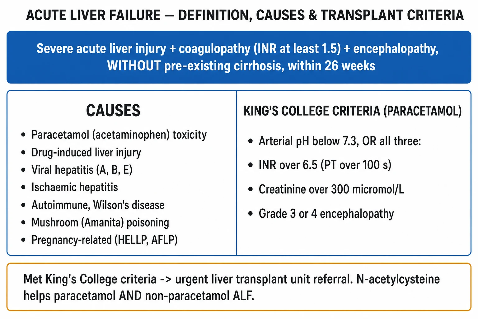
AFLP management rests on two non-negotiable pillars: (1) urgent delivery of the fetus — the definitive, causal treatment that removes the source of toxic metabolites — and (2) intensive supportive care of the failing liver, the coagulation system, the glucose, the kidney, and the brain, as a bridge to the postpartum hepatic regeneration that follows in the overwhelming majority of cases.[1][2]
Pillar 1 — deliver (the definitive treatment)
AFLP management — the two-pillar framework
- MAKE THE DIAGNOSIS (Swansea) AND CALL THE MULTIDISCIPLINARY TEAM — apply the Swansea criteria; immediately involve obstetrics, intensive care, anaesthesia, haematology, hepatology/transplant, and neonatology. AFLP is a maternal critical-care emergency. Do NOT delay delivery to complete exhaustive investigations — the Swansea criteria + basic bloods + bedside ultrasound are sufficient to commit to delivery.[1]
- STABILISE AND CORRECT COAGULOPATHY / GLUCOSE BEFORE DELIVERY — in AFLP you ARE about to perform a procedure (delivery), so UNLIKE idiopathic acute liver failure (where the INR is prognostic and left uncorrected), you correct the coagulopathy first. Targets pre-delivery: fibrinogen >1.5–2.0 g/L, platelets >50 × 10⁹/L, INR <1.5–1.7. Give vitamin K 10 mg IV, FFP, cryoprecipitate (for fibrinogen), platelets, tranexamic acid, and activate the major obstetric haemorrhage / PPH protocol — guided by TEG/ROTEM where available. Correct hypoglycaemia with 10% dextrose (glucose check hourly).[1]
- DELIVER — WITHOUT DELAY — mode: caesarean section if maternal or fetal distress, rapid deterioration, or unfavourable cervix; vaginal delivery may be appropriate if the mother is stable and labour established. Anaesthesia: regional (epidural/spinal) is preferred for cardiovascular stability IF coagulopathy is corrected; general anaesthesia with rapid-sequence induction if emergent, coagulopathy uncorrected, or the patient is encephalopathic (airway protection). Avoid aortocaval compression — left lateral tilt / manual uterine displacement throughout. If pre-eclampsia/eclampsia overlap, give magnesium sulphate for seizure prophylaxis.[1][2]
- SUPPORTIVE ICU CARE FOR LIVER FAILURE (post-delivery) — admit to ICU for the immediate postpartum period (often 1–2 weeks). The liver typically regenerates; the job is to bridge safely (see organ-support table below).[2]
- POSTPARTUM SURVEILLANCE — monitor INR/platelets/glucose/creatince/bilirubin/ammonia trend (should improve over days); screen for infection; watch for PPH; counsel regarding recurrence. If liver failure persists or worsens beyond ~7 days postpartum and King's College criteria are met, involve transplant for the rare refractory case.
- TEST MOTHER AND INFANT FOR FATTY-ACID OXIDATION DISORDER — plasma acylcarnitine profile + HADHA sequencing; arrange neonatal follow-up (early dietary therapy — low fat, high carbohydrate, medium-chain triglycerides — prevents lethal metabolic crises in an affected child).[4]
Pillar 2 — supportive organ support
System-by-system organ support in AFLP (post-delivery)
| System | Problem | ICU management |
|---|---|---|
| Neurological | Encephalopathy → cerebral oedema (ammonia-driven) | Head of bed 30° neutral; treat hypoglycaemia; lactulose (debatable benefit, low harm); if grade III–IV, intubate for airway/CO₂ control, hypertonic saline to Na 145–155, mannitol/ICP monitoring as for any acute liver failure; avoid hypotension, hypoxia, hypercapnia |
| Hepatic | Synthetic + detoxifying failure | N-acetylcysteine (NAC) — extrapolated from acute liver failure; many units give to all AFLP (antioxidant, improves microcirculation); supportive only (no specific 'liver drug'); liver regenerates postpartum |
| Haematological / coagulation | Coagulopathy + DIC | Correct before delivery/procedures (fibrinogen >1.5, platelets >50, INR <1.5): vitamin K, FFP, cryoprecipitate, platelets, tranexamic acid; TEG/ROTEM-guided; activate PPH protocol; DVT prophylaxis once bleeding controlled (AFLP is prothrombotic) |
| Metabolic — glucose | Hypoglycaemia (impaired gluconeogenesis) | 10% dextrose infusion; check glucose hourly; keep glucose 4–8 mmol/L. Always exclude hypoglycaemia before attributing deterioration to 'worsening encephalopathy' |
| Renal | AKI (hepatorenal + ATN) | Optimise perfusion (noradrenaline for MAP ≥65); continuous RRT (CRRT) if needed (preferred over intermittent — avoids solute-shift/ICP rise; removes ammonia); avoid nephrotoxins |
| Cardiovascular | Vasoplegia (SIRS-like) ± haemorrhage | Noradrenaline first-line (target MAP ≥65, uteroplacental then cerebral perfusion); cautious fluid (avoid overload); treat PPH aggressively; left lateral tilt pre-delivery |
| Respiratory | Aspiration (encephalopathy), pulmonary oedema, ARDS | Intubate at grade III encephalopathy; lung-protective ventilation; pre-oxygenase before RSI (reduced FRC in pregnancy → fast desaturation) |
| Gastrointestinal / metabolic | Pancreatitis coexists; stress ulceration; nutrition | Check lipase; PPI for stress-ulcer prophylaxis; early enteral nutrition once stable (medium-chain triglyceride feeds may be favoured if FAOD suspected) |
| Endocrine | Transient central DI | If polyuria/hypernatraemia: DDAVP (desmopressin); monitor UO + serum Na; resolves postpartum |
| Infectious | High infection risk (liver failure + instrumentation) | Surveillance cultures; low threshold for broad-spectrum antibiotics + antifungal cover in severe disease; rigorous line/ventilator bundles |
Coagulopathy correction — the AFLP-specific principle
A common and dangerous error is to treat AFLP's coagulopathy exactly like idiopathic acute liver failure. They are different situations. In non-pregnancy acute liver failure the INR is prognostic (the backbone of the King's College criteria) and is deliberately not corrected unless the patient is bleeding, because of 'rebalanced haemostasis'. In AFLP, however, the patient is about to undergo delivery — a procedure with a high bleeding risk (caesarean section, uterine atony, PPH) — and postpartum haemorrhage is a leading cause of maternal death. Therefore correct the coagulopathy before delivery.[1][2]
Coagulopathy targets and products in AFLP (pre-delivery / bleeding)
| Parameter | Target before delivery / procedure | Product |
|---|---|---|
| Fibrinogen | >1.5–2.0 g/L (pregnancy-specific; fibrinogen is the FIRST factor to fall in obstetric haemorrhage) | Cryoprecipitate (or fibrinogen concentrate) — 2 pools typically raise fibrinogen by ~1 g/L |
| Platelets | >50 × 10⁹/L (active bleeding / caesarean: aim >75) | Platelets — 1 adult dose raises count by ~20–40 × 10⁹/L |
| INR / PT | <1.5–1.7 | FFP 10–15 mL/kg; vitamin K 10 mg IV (corrects any deficiency) |
| Active fibrinolysis | Reduce (TEG LY30 >3% or clinical bleeding) | Tranexamic acid 1 g IV (within 3 h of bleeding) — WOMAN trial |
| Guiding test | TEG / ROTEM preferred over static INR/platelets | Rational, rapid, point-of-care product selection |
| PPH readiness | Activate major obstetric haemorrhage protocol | Cross-match blood; cell salvage if available; obstetric + haematology on site |
Anaesthesia considerations
Pregnancy + liver failure + coagulopathy + full stomach create a high-risk anaesthetic. Regional anaesthesia (spinal/epidural) is preferred for cardiovascular stability and to avoid airway manipulation, but only after coagulopathy has been corrected (risk of spinal haematoma). General anaesthesia with rapid-sequence induction is required for emergencies, uncorrected coagulopathy, or encephalopathy (aspiration + airway protection). Note that pseudocholinesterase (plasma cholinesterase) is reduced in liver failure, prolonging the action of suxamethonium — consider rocuronium + sugammadex reversal. Pre-oxygenate fully (3–5 min of 100% O₂) because the reduced functional residual capacity of late pregnancy causes rapid desaturation. Avoid hepatotoxic agents; volatile agents (sevoflurane/isoflurane) are generally acceptable at standard doses.[1][2]
N-acetylcysteine (NAC)
There are no RCTs of NAC specifically in AFLP, but it is widely used (and biologically plausible) by extrapolation from acute liver failure, where NAC improves transplant-free survival in early non-paracetamol ALF (Lee 2009). NAC is a glutathione precursor, antioxidant, and microcirculatory agent — all potentially relevant to AFLP's mitochondrial injury. Many units therefore give NAC to all AFLP patients. Standard IV regimen: 150 mg/kg over 1 h, then 50 mg/kg over 4 h, then 100 mg/kg over 16 h (total 300 mg/kg). It is cheap and safe.[2]
Postpartum course and prognosis
The defining feature of AFLP — and the source of hope after a terrifying presentation — is that the liver regenerates after delivery. The synthetic functions recover fastest: INR and platelets typically improve within 2–4 days, signalling the turning point; bilirubin and transaminases fall more slowly over 1–2 weeks; encephalopathy clears as ammonia falls. ICU stay is typically 1–2 weeks. Complete biochemical and histological recovery occurs within 1–4 weeks in survivors, with no residual chronic liver disease — unlike cirrhosis.[1][2]
Persistent or worsening liver failure beyond ~7 days postpartum is atypical and should prompt (a) reconsideration of the diagnosis (Wilson's, viral, autoimmune, drug-induced), and (b) liver-transplant assessment if King's College criteria are met — though transplant is required in fewer than 1% of AFLP cases. Breastfeeding is generally safe once the mother has recovered, unless an inborn error of metabolism in the infant dictates otherwise.[1]
Outcomes, prognostic factors and recurrence in AFLP
| Domain | Finding |
|---|---|
| Maternal mortality | 1–12.5% (modern tertiary series ~1–3%; historically up to 75% before modern ICU/delivery) |
| Perinatal / fetal mortality | 7–58% (modern ~10–20%); driven by prematurity, placental insufficiency, intrauterine death |
| Cause of maternal death | Sepsis, haemorrhage (PPH + DIC), cerebral oedema/herniation, multi-organ failure, pancreatitis |
| Favourable signs postpartum | Falling INR/creatinine/rising platelets within 48–72 h; clearing encephalopathy |
| Unfavourable signs | Persistent/worsening encephalopathy, rising ammonia/lactate, refractory coagulopathy, sustained AKI, uncontrolled sepsis |
| Recurrence | ~25% overall in subsequent pregnancies; up to 70% if confirmed maternal/fetal fatty-acid oxidation disorder |
| Predictors of poor outcome | Delayed diagnosis, delayed delivery, multi-organ failure, encephalopathy grade III–IV, need for transplant |
| Recovery | Full biochemical + histological recovery within 1–4 weeks; no chronic liver disease |
| Transplant | Required in <1%; consider if failure persists >7 days postpartum + King's criteria met |
SAQ — AFLP with acute liver failure
SAQ — AFLP with acute liver failure
10 minutes · 10 marks
A 31-year-old primigravida at 36 weeks gestation presents with 5 days of nausea, vomiting and epigastric pain. She is now jaundiced and drowsy (GCS 13). HR 110, BP 102/64, RR 22, SpO2 96% on room air, temp 37.1°C. Bloods: glucose 1.9 mmol/L, INR 2.3, fibrinogen 1.2 g/L, platelets 105 × 10⁹/L, AST 380 IU/L, bilirubin 95 μmol/L, creatinine 178 μmol/L, ammonia 110 μmol/L, WCC 16 × 10⁹/L, lipase normal. CTG shows reduced baseline variability.
SAQ — Swansea criteria and diagnosis of AFLP
SAQ — Swansea criteria for AFLP diagnosis
10 minutes · 10 marks
A 28-year-old woman at 34 weeks gestation with a twin pregnancy presents with 4 days of vomiting, abdominal pain and new polydipsia/polyuria. She is mildly confused (GCS 14). Examination: BP 148/96, HR 104, jaundiced, mild RUQ tenderness, no asterixis. Bloods: bilirubin 36 μmol/L, glucose 3.0 mmol/L, urea 7.2 mmol/L, creatinine 162 μmol/L, WCC 13.5 × 10⁹/L, AST 95 IU/L, INR 1.4, fibrinogen 2.1 g/L, platelets 142 × 10⁹/L, ammonia 60 μmol/L, LDH 320, haptoglobin normal. Bedside ultrasound shows a bright echogenic liver.
Clinical pearls
Red flags
Prognosis
Modern intensive care and early delivery have transformed AFLP from a near-uniformly fatal disease (historical maternal mortality up to 75%) into a survivable one with maternal mortality of 1–12.5% (modern tertiary series ~1–3%) and perinatal mortality of 7–58% (modern ~10–20%). The dominant causes of maternal death are sepsis, haemorrhage (PPH + DIC), cerebral oedema/herniation, and multi-organ failure; fetal loss reflects prematurity, placental insufficiency, and intrauterine death. The favourable turning point postpartum is a falling INR and rising platelet count within 48–72 hours; conversely, persistent encephalopathy, rising ammonia/lactate, refractory coagulopathy, sustained AKI, or uncontrolled sepsis predict a poor outcome. In survivors the liver regenerates completely within 1–4 weeks with no residual chronic liver disease, and emergency transplantation is required in fewer than 1%. Recurrence in subsequent pregnancies is approximately 25% overall and up to 70% when a fatty-acid oxidation disorder is confirmed — mandating counselling and intensive surveillance in future pregnancies.[1][2][4]
Key trials and evidence
Ch'ng 2002 — Swansea criteria for AFLP (Gut; PMID 12427793)
Source
Prospective 15-month regional study of liver dysfunction in pregnancy, South West Wales (Ch'ng CL, Morgan M, Hainsworth I, Kingham JGC)
Objective
Define the incidence and spectrum of pregnancy-related liver disease and derive practical diagnostic criteria for AFLP
Key finding
Liver dysfunction was seen in ~3% of deliveries and was usually directly related to pregnancy with spontaneous recovery in the puerperium; the incidence of AFLP and HELLP was far higher than previously reported
Derivation
Swansea criteria: AFLP diagnosed if >=6 of 14 clinical/laboratory/imaging features are present in the absence of another explanation — biopsy not required
Clinical bottom line
The Swansea criteria remain the most widely used bedside diagnostic tool for AFLP, allowing diagnosis without a risky biopsy in a coagulopathic patient
Ibdah 1999 — fetal fatty-acid oxidation disorder as a cause of AFLP/HELLP (NEJM; PMID 10352164)
Source
Landmark case–control molecular study, Vanderbilt (Ibdah JA, Bennett MJ, Rinaldo P, Zhao Y, Gibson B, Sims HF, Strauss AW)
Objective
Test whether a fetal disorder of mitochondrial fatty-acid oxidation (LCHAD deficiency) causes maternal liver disease in pregnancy
Intervention/observation
Mothers of children with confirmed LCHAD deficiency were studied for a history of AFLP/HELLP; the common G1528C (E474Q) HADHA mutation was sought in fetuses and heterozygous carrier mothers
Key finding
A striking proportion of mothers who carried an LCHAD-deficient fetus had developed AFLP or HELLP; affected fetuses were homozygous/compound heterozygous while mothers were heterozygous carriers — establishing the fetal–maternal pathogenic link
Mechanism
The fetus cannot beta-oxidise long-chain fatty acids; toxic 3-hydroxy-fatty-acid metabolites cross the placenta and overwhelm the carrier mother's residual beta-oxidation -> hepatic microvesicular steatosis
Clinical bottom line
Test infants born to mothers with AFLP/HELLP for LCHAD deficiency; early dietary therapy (low fat, high carbohydrate, medium-chain triglycerides) prevents lethal metabolic crises in the child
References
- [1]Nelson DB, et al. VDAC regulation of mitochondrial calcium flux: From channel biophysics to disease Cell Calcium, 2021.PMID 33529977
- [2]Goel A, et al. Notum palmitoleoyl-protein carboxylesterase regulates Fas cell surface death receptor-mediated apoptosis via the Wnt signaling pathway in colon adenocarcinoma Bioengineered, 2021.PMID 34402722
- [3]Ch'ng CL, Morgan M, Hainsworth I, Kingham JGC Prospective study of liver dysfunction in pregnancy in Southwest Wales Gut, 2002.PMID 12427793
- [4]Ibdah JA, Bennett MJ, Rinaldo P, Zhao Y, Gibson B, Sims HF, Strauss AW A fetal fatty-acid oxidation disorder as a cause of liver disease in pregnant women N Engl J Med, 1999.PMID 10352164