ICU · Obstetric critical care
Amniotic Fluid Embolism
Also known as Amniotic fluid embolism · AFE · Anaphylactoid syndrome of pregnancy · AFE biphasic
The amniotic the fluid the embolism (the AFE) — the catastrophic the peripartum the anaphylactoid the reaction to the amniotic the fluid the entering the maternal the circulation. The the biphasic: the phase the 1 (the sudden the hypoxia + the hypotension / the cardiovascular the collapse + the altered the mental the state / the seizure / the cardiac the arrest) → the phase the 2 (the coagulopathy / the DIC + the massive the bleeding + the multi-organ). The clinical the diagnosis (the exclusion). The resuscitation, the haemodynamic the support, the correct the coagulopathy, the delivery, the ECMO. The mortality the 20 to the 40 per cent.
On this page & tools
Your progress
Saved locally on this device.
Target exams
Overview & definition
The amniotic the fluid the embolism (the AFE) — the catastrophic the peripartum the anaphylactoid the reaction. The amniotic the fluid + the fetal the debris the enter the maternal the circulation → the complement / the leukotriene / the thromboxane the cascade → the pulmonary the vasoconstriction + the LV the depression + the DIC. The sudden, the unpredictable, the mortality the 20 to the 40 the per cent.[1][1]

The biphasic the clinical
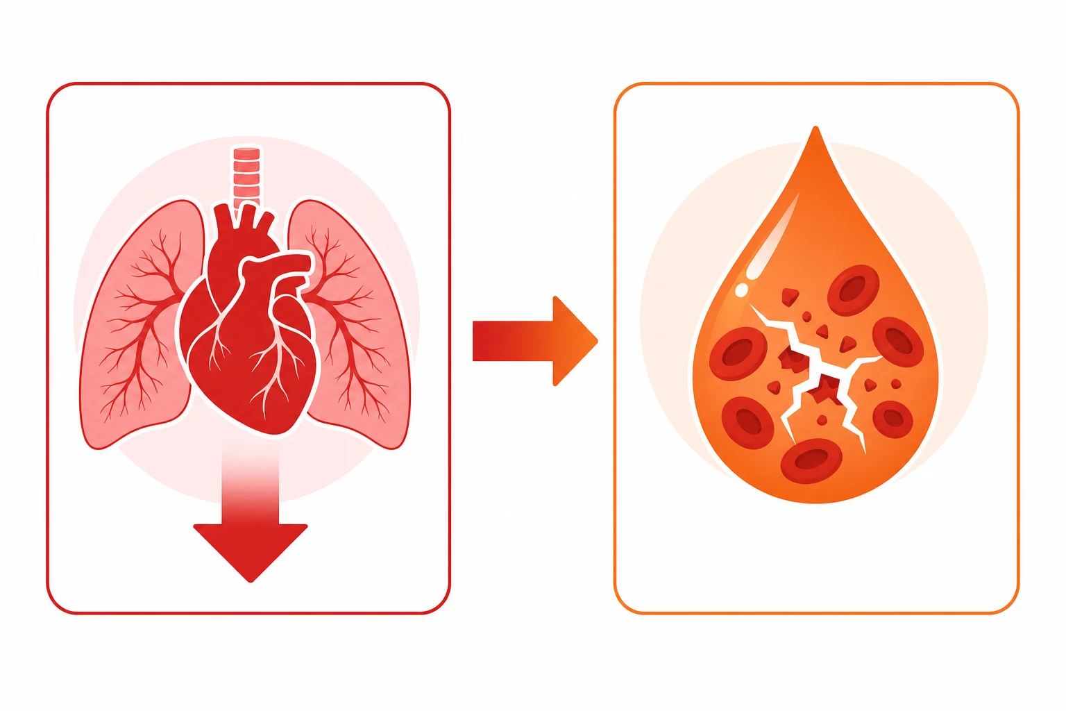
The phase the 1 — the cardiopulmonary the collapse.[1][2][1]
- The the sudden the hypoxia (the dyspnoea, the cyanosis — the pulmonary the vasoconstriction + the V/Q the mismatch).
- The the hypotension / the cardiovascular the collapse (the LV the depression + the pulmonary the hypertension → the right-the-heart the failure → the low the output).
- The the altered the mental the state / the seizure / the cardiac the arrest. The often the within the minutes of the labour / the delivery / the Caesarean / the postpartum.[1][2]
The phase the 2 — the coagulopathy / the DIC.[1][1]
- The massive the bleeding (the vaginal, the surgical-the-site, the IV-the-site the oozing — the DIC).
- The multi-organ the failure (the renal, the hepatic). The if the phase the 1 the survived.[1][1]
The diagnosis
The the clinical the diagnosis (the triad: the sudden the hypoxia + the hypotension / the collapse + the coagulopathy in the peripartum the period); the the exclusion (the exclude the pulmonary the embolism, the high the spinal, the local-the-anaesthetic the toxicity, the eclampsia, the sepsis, the MI, the haemorrhage the primary). The NOT the "the squamous the cells the in the pulmonary the vessels" the (the historical, the non-the-specific). The suspect the AFE the in the ANY the unexplained the peripartum the collapse.[1][2][1]
The treatment
1. The resuscitation.[1][1][1]
- The ABCDE; the oxygen; the intubation (the hypoxia, the altered the mental); the CPR the if the arrest.
- The left the lateral the tilt (the 20 the weeks).[1]
2. The haemodynamic the support.[1][1][1]
- The vasopressors (the noradrenaline — the LV the failure + the vasodilation); the inotrope (the dobutamine, the milrinone — the LV the depression).
- The cautious the fluid (the pulmonary the oedema, the right-the-heart the failure). The echocardiography (the RV the failure, the LV the depression).[1][1]
3. The correct the coagulopathy.[1][1]
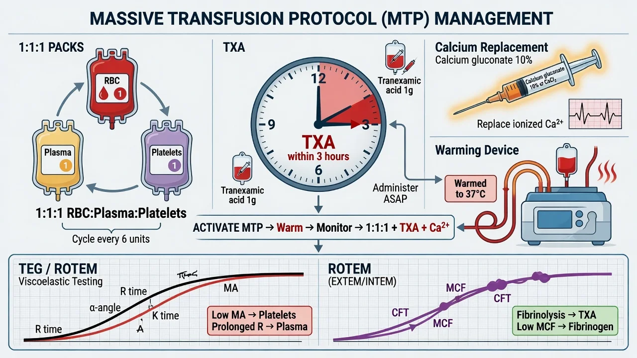
- The massive the transfusion the protocol (the 1:1:1; the cryoprecipitate for the fibrinogen; the TXA the 1 g the IV).
- The factors, the platelets, the ROTEM the guided.[1]
4. The delivery (the if the antepartum — the resuscitative, the once the maternal the stabilised).[1][1]
5. The ECMO (the VA-the-ECMO) for the refractory the cardiopulmonary the collapse (the bridge the to the recovery). The increasingly the used the for the AFE.[1][1]
6. The multidisciplinary (the obstetrics, the anaesthesia, the haematology, the ICU, the neonatology).[1][1]
Prognosis
The AFE the mortality the 20 to the 40 the per cent (the historical the higher — the 60 to the 80; the improved the with the early the recognition + the aggressive the resuscitation + the MTP). The survivors the often the complete the recovery (the no the chronic). The neurologically the intact the if the CPR the prompt.[1][2][1]
Red flags
Detailed pathophysiology — the biphasic response
AFE biphasic response — the two phases
| Phase | Timing | Mechanism | Clinical features | Management |
|---|---|---|---|---|
| Phase 1 — Anaphylactoid + pulmonary vasospasm | 0-30 min | Amniotic fluid + fetal debris enter maternal circulation (uterine venous sinuses → IVC → right heart → pulmonary vasculature) → massive complement activation + cytokine storm (NOT a mechanical obstruction — it's an IMMUNE response to fetal antigens) → pulmonary vasospasm → acute pulmonary hypertension → RV failure → hypoxaemia + hypotension | Sudden cardiovascular collapse: hypoxaemia (SpO2 drops to 50-70%), hypotension (SBP <90), cardiac arrest in 50%. Seizures. Disseminated intravascular coagulation begins. | 100% O2 + intubate + noradrenaline for MAP + vasopressin + inotropes for RV failure (milrinone) ± VA-ECMO |
| Phase 2 — Coagulopathy + left ventricular failure | 30 min - 12h | Complement + cytokines → massive tissue factor release → explosive DIC (fibrinogen <1.0 g/L within 60-90 min). Simultaneously: LV stunning (cytokine-mediated cardiomyopathy). | Massive haemorrhage from ALL sites (uterine, surgical wounds, IV sites, mucosa). Fibrinogen <1.0. Platelets <50. PT/APTT prolonged. LV dysfunction on echo (EF 20-30%). | MASSIVE TRANSFUSION PROTOCOL (RBC:FFP:platelets 1:1:1). CRYOPRECIPITATE EARLY (fibrinogen target >4 g/L — much higher than standard bleeding). TXA 1g IV. Hydrocortisone (controversial — may suppress complement cascade). Factor VIIa for refractory bleeding. |
Diagnostic criteria — the SMG 2016 criteria
AFE is a CLINICAL diagnosis — there is NO specific confirmatory test. The Society for Maternal-Fetal Medicine (SMG) 2016 criteria require ALL of the following: [1]
- Sudden cardiovascular collapse (cardiac arrest OR SBP <90 for ≥15 min) AND/OR
- Acute hypoxaemia (SpO2 <90% OR requiring intubation for hypoxia) AND
- Coagulopathy (fibrinogen <1.0 g/L within 60 min of onset OR abnormal PT/APTT + low platelets) AND
- Temporal relationship to labour/delivery/cesarean/postpartum (onset during or within 30 min of these) AND
- No alternative explanation for the clinical picture [1]
EXCLUSION criteria (these RULE OUT AFE):
- Infection/sepsis with a clear source
- Amniotic fluid contamination during Caesarean (common, NOT AFE)
- Eclampsia (seizures + hypertension — NOT the sudden collapse pattern of AFE)
- Pulmonary embolism (can be distinguished by echo — PE shows RV strain, AFE shows biventricular failure)
- Anaesthetic reaction (anaphylaxis — tryptase elevated, complement not) [1]
Management — the multidisciplinary approach
AFE management — the ICU protocol
- CALL FOR HELP: Anaesthetist + obstetrician + haematologist + neonatal team (if baby not delivered)
- AIRWAY + OXYGENATION: 100% O2 via non-rebreather → INTUBATE (may need RSI — AFE patients desaturate rapidly → pre-oxygenate with 100% O2 for 3 min). If cardiac arrest → CPR + perimortem Caesarean if uterus >20 weeks (within 5 min of arrest)
- CIRCULATION: Noradrenaline first-line for MAP >65 (vasoplegia from complement + cytokines). Add vasopressin 0.04 U/min if refractory. Inotropes (milrinone 0.125-0.5 mcg/kg/min) for LV/RV dysfunction. Hydrocortisone 200mg IV (may suppress complement cascade — controversial but commonly given)
- COAGULOPATHY: ACTIVATE MASSIVE TRANSFUSION PROTOCOL. Target ratios: RBC:FFP:platelets 1:1:1. GIVE CRYOPRECIPITATE EARLY (10-15 units) — fibrinogen target >4 g/L in AFE (HIGHER than standard bleeding — pregnancy has high baseline fibrinogen 4-6 g/L; <2.0 in pregnancy = SEVERE deficiency). TXA 1g IV within 3h (WOMAN trial — reduces mortality). Recombinant factor VIIa for refractory bleeding (last resort — risk of thrombosis)
- HAEMORRHAGE CONTROL: Uterotonics (oxytocin + ergometrine + carboprost + misoprostol). Bakri balloon. B-Lynch suture. Hysterectomy (life-saving — do NOT delay if bleeding uncontrolled). Interal iliac artery ligation/embolisation
- CARDIAC SUPPORT: Echocardiography (assess LV + RV function — both may be impaired). Mechanical support: VA-ECMO for refractory cardiogenic shock (AFE is a good indication for ECMO — reversible cardiomyopathy)
- FETAL DELIVERY: If baby not delivered → emergency Caesarean (perimortem if cardiac arrest). Neonatal team present. Baby may also be affected (hypoxia from maternal collapse)
- POST-EVENT: ICU admission. Monitor: coagulation (DIC may persist 12-24h), cardiac function (LV dysfunction usually recovers in 48-72h — cytokine-mediated stunning), renal function (AKI common from hypoperfusion + DIC microthrombi), neurological (hypoxic brain injury from initial collapse)
Additional clinical pearls
[1]Differential diagnosis — the peripartum collapse mimics
AFE is a diagnosis of exclusion. The first minutes are spent running through the differential — the wrong diagnosis delays the right therapy (e.g. giving thrombolysis for "PE" in a patient who is bleeding from AFE-DIC = catastrophic). The clinical setting (labour, Caesarean, immediate postpartum, recent uterine manipulation, cervical cerclage, termination) is the trigger to put AFE at the TOP of the differential alongside the mimics.[1][2][8]
AFE vs the peripartum collapse mimics — the discriminator table
| Feature | AFE | Pulmonary embolism | Eclampsia | Local-anaesthetic toxicity | Anaphylaxis | Sepsis | Myocardial infarction |
|---|---|---|---|---|---|---|---|
| Timing | During labour / delivery / within 30 min postpartum | Any time (postpartum risk high for weeks) | ≥20 weeks to 6 weeks postpartum | Within min of LA injection / infusion | Within min of drug / antigen exposure | Gradual onset over hours | Any time; high in older / pre-eclamptic |
| Hallmark | Triad: hypoxia + hypotension + coagulopathy | Isolated hypoxia + RV strain; NO coagulopathy early | Hypertension → seizure | CNS excitation → seizures → cardiac arrest | Bronchospasm + urticaria + hypotension | Fever, tachycardia, vasoplegia, leukocytosis | Chest pain, ECG changes, troponin rise |
| Blood pressure | Hypotension (sudden) | Hypotension (if massive) | Hypertension (then may fall post-ictal) | Variable, then collapse | Hypotension | Hypotension (warm or cold shock) | Variable |
| Coagulopathy | EXPLOSIVE (fibrinogen <1.0 g/L within 60 min) | Usually absent early; may develop with prolonged shock | Low platelets, haemolysis (HELLP overlap) | Absent | Absent | Variable (DIC in late sepsis) | Absent |
| Echo | Biventricular dysfunction (LV stunning + RV strain from pulmonary HTN) | Isolated RV strain / McConnell sign | Usually normal; LV diastolic dysfunction if pre-eclampsia | Acute LV failure late | Empty hyperdynamic LV | LV hyperdynamic early; globally impaired late | Regional wall motion abnormality (coronary territory) |
| Fibrinogen | <1.0 g/L (within 60 min) — pathognomonic | Normal | Normal or low (HELLP) | Normal | Normal | Variable | Normal |
| Tryptase | Often elevated (mast-cell activation) | Normal | Normal | Normal | Markedly elevated | Normal | Normal |
| Complement C3/C4 | Low (consumed in the complement cascade) | Normal | Low (complement activation in pre-eclampsia) | Normal | Variable | Low (in septic shock) | Normal |
| Discriminating clue | Coagulopathy is the first and most severe feature; chest pain/chest tightness/panic ("I'm going to die") precedes collapse | Isolated respiratory failure with clear lung fields; DVT signs; ECG S1Q3T3 | Preceding hypertension, proteinuria, headache, visual disturbance, hyper-reflexia | Preceding LA infusion / bolus; tinnitus, perioral tingling, metallic taste | Exposure to antibiotic / latex / blood product; rash, wheeze, angioedema | Fever, source (chorioamnionitis, pyelonephritis), warm peripheries early | Chest pain, ECG ST changes, regional wall motion; coronary risk factors |
The classic clinical triad — the exam-key pattern
The AFE triad in the peripartum period is (1) sudden hypoxaemia + (2) hypotension/cardiovascular collapse + (3) coagulopathy — onset during labour, delivery, Caesarean, or within 30 min postpartum. Some patients present with a typical prodrome of chills, restlessness, anxiety, chest tightness, dyspnoea, and a sensation of impending doom ("I feel like I am going to die") in the 5-10 min before collapse. This prodrome is highly characteristic but easy to dismiss as anxiety or reaction to uterotonics.[2][11]
A biphasic presentation is seen in ~80%: phase 1 (cardiopulmonary collapse) is followed within 30 min to several hours by phase 2 (coagulopathy / haemorrhage). In ~15-20% the bleeding/DIC is the dominant or sole initial feature — these patients may not have a dramatic cardiovascular collapse first. A third phenotype — isolated cardiac arrest at presentation — carries the highest mortality. The phase dominance and severity varies with the load and type of fetal antigenic material that enters the maternal circulation.[1][3]
Incidence, mortality, and risk factors — the epidemiology
The reported incidence is approximately 1 in 40,000 deliveries in modern population-based studies (range 1:15,000 to 1:80,000 depending on case-finding methodology and registry vs administrative coding). The maternal mortality is 20-40% in contemporary series — a substantial improvement on the 60-86% reported in older series, attributable to early recognition, massive transfusion protocols, and VA-ECMO. The case-fatality has been halved over two decades.[3][4][8]
Perinatal mortality is also significant (5-15%), driven by maternal hypoxia-induced fetal distress during the collapse. Of note, AFE mortality figures vary widely because (a) the diagnosis is clinical and registry-based, (b) milder cases are missed, and (c) surviving patients often make a complete neurological recovery if CPR is prompt — distinguishing AFE favourably from many other causes of maternal cardiac arrest.[1][3]
AFE risk factors — those consistently associated (vs historical associations since discarded)
| Well-supported risk factors | Weaker / inconsistent associations |
|---|---|
| Advanced maternal age (≥35 yr) | Oxytocin use (causality questioned — confounded by labour itself) |
| Caesarean delivery (especially emergency) | Placenta previa / accreta |
| Operative vaginal delivery (forceps / vacuum) | Cervical cerclage |
| Polyhydramnios | Female fetal sex |
| Multi-parity (≥4) | Eclampsia (overlapping diagnosis) |
| Uterine tachysystole / hyperstimulation | Premature labour |
| Abdominal trauma in pregnancy | Induction of labour (any agent) |
| Cervical cerclage removal / intra-amniotic instrumentation | Previous AFE (recurrence is rare but documented) |
| Medical induction / termination (second trimester) | Drug / substance use |
Critically, the majority of cases occur in women with NO identifiable risk factor — AFE remains fundamentally unpredictable and unpreventable. Screening or prophylaxis is not possible. Risk factors are useful for clinical vigilance, not for risk stratification.[2][4]
Laboratory and imaging features — what to send, what to expect
There is no specific confirmatory test for AFE. The laboratory picture is that of (a) sudden cardiovascular collapse and (b) explosive DIC. Send immediately on suspicion: fibrinogen (Clauss), PT, APTT, platelet count, D-dimer, blood gas (lactate), full blood count, group-and-save / crossmatch, TEG/ROTEM if available, tryptase (within 1-2 h of the event for anaphylaxis overlap), and complement C3/C4 (if available — research/confirmatory).[1][1]
AFE laboratory pattern — the expected abnormalities
| Test | AFE typical value | Why / mechanism | Differential clue |
|---|---|---|---|
| Fibrinogen (Clauss) | <1.0 g/L (often <0.5); normal pregnancy 4-6 g/L | Massive consumption by tissue-factor–triggered DIC | A low fibrinogen in pregnancy is the single most sensitive marker for AFE |
| PT / INR | Prolonged (INR >1.5) | Consumption of factors V, VII, X | Less rapid to change than fibrinogen |
| APTT | Prolonged (>1.5× control) | Consumption of intrinsic factors | Also prolonged by heparin — check timing |
| Platelets | Falling rapidly (<100, often <50) | Consumption | A falling trend over 30-60 min is more telling than absolute value |
| D-dimer | Massively elevated | Active fibrinolysis | Non-specific; supports DIC vs simple dilution |
| Arterial gas | Severe metabolic + lactic acidosis; hypoxaemia; often low PaCO2 from hyperventilation then rising | Tissue hypoperfusion + V/Q mismatch + mitochondrial dysfunction | Lactate >5 mmol/L = severe shock |
| Tryptase | Often mildly elevated (mast-cell activation — NOT IgE-mediated) | Complement-mediated mast-cell degranulation | Marked elevation (>2× baseline + >20 μg/L) favours anaphylaxis |
| Complement C5a / C3 / C4 | Low (consumed); C5a elevated | The complement cascade is central to AFE pathophysiology | Confirms complement activation; not yet routine |
| Insulin-like growth factor binding protein-1 (IGFBP-1) | Elevated in maternal serum | Amniotic fluid marker leaking into maternal circulation | Research only — not used clinically |
| Cervical/uterine zinc coproporphyrin-I | Elevated in older studies | Amniotic fluid component | Non-specific; abandoned |
The historical "fetal squames in pulmonary vessels" finding (on aspiration of pulmonary artery catheter blood or at autopsy) is non-specific — squames are found in many pregnant and postpartum women without AFE. They are not required for the diagnosis and should not delay empiric treatment.[2][9]
Echocardiography in AFE — the bedside discriminator
Bedside echo is one of the most useful early investigations in suspected AFE. The typical pattern reflects the biphasic injury: (a) acute right ventricular dilatation / hypokinesis from pulmonary vasospasm and acute pulmonary hypertension (phase 1), followed within minutes to hours by (b) global or apical LV stunning — the takotsubo-reversible cardiomyopathy pattern (phase 2). The combination of biventricular failure, in the peripartum setting, with coagulopathy, is highly suggestive of AFE.[6]
AFE echo findings vs the mimics
| Pathology | Right ventricle | Left ventricle | Other |
|---|---|---|---|
| AFE | Acute dilatation + hypokinesis (pulmonary HTN phase); may recover | Global / apical ballooning (takotsubo-like); EF 10-30%; usually recovers in 48-72 h | Often pericardial effusion; no valvular pathology |
| Massive PE | RV dilatation + hypokinesis (McConnell sign: free-wall hypokinesis with apical sparing); septal bowing into LV | Small, hyperdynamic, D-shaped LV (underfilled) | IVC plethoric; tricuspid regurgitant jet >2.6 m/s |
| Peripartum cardiomyopathy | Usually normal; may be dilated late | Global dilation; EF <45%; thrombus common | Onset typically last month of pregnancy to 5 months postpartum |
| Pre-eclampsia / HELLP | Normal; may show LV diastolic dysfunction | Diastolic dysfunction; hypertensive heart | Pulmonary oedema; preserved EF |
| Anaphylaxis | Small, hyperdynamic (vasodilation) | Hyperdynamic | IVC <1 cm collapsible |
| Septic cardiomyopathy | Variable | Biventricular global hypokinesis; recovers if survives | Often hyperdynamic early; low SVR |
A normal biventricular echo in a haemodynamically stable patient with coagulopathy makes AFE less likely — but does NOT exclude it, especially if the patient has already been resuscitated. Conversely, biventricular dysfunction in the peripartum setting with coagulopathy is practically diagnostic.[6][5]
Massive transfusion and coagulopathy — AFE-specific targets
AFE causes the most explosive coagulopathy in obstetrics. The standard postpartum-haemorrhage fibrinogen threshold of 2.0 g/L is INSUFFICIENT in AFE — pregnancy baseline is 4-6 g/L, and the goal in AFE is fibrinogen >4 g/L. ROTEM/TEG-guided therapy is preferred where available; the FIBTEM MCF target is ≥10-12 mm. Cryoprecipitate (10-15 U) reaches the fibrinogen target faster than FFP.[1][1]
AFE coagulopathy — the transfusion algorithm
- ACTIVATE THE MASSIVE TRANSFUSION PROTOCOL IMMEDIATELY on clinical suspicion of AFE — do NOT wait for laboratory confirmation. Alert the blood bank to the high fibrinogen / cryoprecipitate requirement.
- RBC : FFP : platelets in a 1:1:1 ratio (PROPPR-derived principle for massive haemorrhage). Aim Hb ≥80 g/L, platelets ≥75 ×10⁹/L (≥100 if active bleeding).
- GIVE CRYOPRECIPITATE EARLY — 10-15 units (or fibrinogen concentrate 4-6 g) — to push fibrinogen >4 g/L. Recheck every 30 min during active bleeding.
- TRANEXAMIC ACID 1 g IV within 3 h of onset (WOMAN trial — early TXA reduces maternal mortality from haemorrhage). Repeat 1 g after 30 min if bleeding continues (maximum 3 g in 24 h).[7]
- CALCIUM CHLORIDE 10 mmol (1 g) IV — citrate toxicity from massive transfusion ionises calcium; maintain ionised Ca >1.0 mmol/L.
- WARM THE PATIENT — hypothermia worsens coagulopathy (the trauma triad of death: hypothermia, acidosis, coagulopathy applies equally to AFE). Forced-air warmer, fluid warmer, ambient theatre temperature >24°C.
- TREAT ACIDOSIS — pH <7.2 disables clotting factors; correct hypoperfusion with vasopressors / inotropes / blood rather than bicarbonate.
- RECOMBINANT FACTOR VIIA (90 mcg/kg) — last resort for refractory bleeding when fibrinogen >1.5 g/L, platelets >50, pH >7.2, and normothermia have been achieved. Risk of thrombosis — discuss with haematology.
- AVOID FIBRINOGEN DEPLETING RECOVERY ERRORS — recheck fibrinogen every 30 min for the first 2 h; DIC may persist or recur for 12-24 h.
- SOURCE CONTROL — uterotonics, Bakri balloon, B-Lynch / brace suture, internal iliac ligation/embolisation, hysterectomy. Bleeding source control MUST happen in parallel with transfusion, not sequentially.
WOMAN trial — tranexamic acid for postpartum haemorrhage (PMID 28980957)
Design
International, multicentre, randomised, double-blind, placebo-controlled trial of 20,060 women with postpartum haemorrhage across 193 hospitals in 21 countries
Population
Women with clinically diagnosed PPH after vaginal or caesarean delivery; included regardless of cause (including AFE-related haemorrhage)
Intervention
Tranexamic acid 1 g IV over 10 min vs placebo, IN ADDITION to standard care, as soon as possible after bleeding onset (within 3 h)
Primary outcome
All-cause mortality within 42 days of giving birth
Key result
TXA reduced death from bleeding (1.5% vs 1.9%; RR 0.81, 95% CI 0.65-1.00; p=0.045). NNT ≈ 250 to prevent one bleeding death. No increase in thromboembolic events or complications.
Time-dependence
Mortality benefit confined to women treated within 3 h of bleeding onset; benefit greatest when given within 1 h. Late (>3 h) TXA showed no benefit and a trend to harm.
Clinical bottom line
TXA 1 g IV is SAFE and reduces mortality from PPH — give EARLY (within 3 h), alongside the first uterotonic, to every woman with major PPH, INCLUDING AFE-associated haemorrhage.
Extracorporeal membrane oxygenation for AFE — systematic review of extracorporeal therapies (PMID 31599831)
Design
Systematic review of the Extracorporeal Life Support Organization (ELSO) registry + literature — case series of AFE managed with ECMO
Population
Women with AFE and refractory cardiopulmonary collapse despite conventional resuscitation
Intervention
VA-ECMO (the dominant modality; provides both cardiac and respiratory support) — initiated a median of 1-9 h after AFE onset in reported cases
Key result
Survival to discharge 70-80% in ECMO-treated AFE cases — substantially higher than the 20-40% historical AFE mortality. The cardiac dysfunction is reversible (cytokine-mediated stunning), making AFE an excellent indication for VA-ECMO.
Timing of cannulation
Best outcomes when VA-ECMO is initiated within 60-120 min of refractory shock onset. Prolonged CPR (>60 min) before ECMO is associated with poorer neurological outcome.
Mode
VA-ECMO preferred over VV-ECMO — AFE causes biventricular failure, not isolated respiratory failure. Peripheral (femoro-femoral) cannulation feasible during CPR (eCPR).
Clinical bottom line
Refractory cardiogenic shock or cardiac arrest in AFE should prompt IMMEDIATE referral to an ECMO centre / mobile eCPR team — not a salvage therapy after all else fails.
VA-ECMO for AFE — the rescue pathway
The cardiac injury in AFE is reversible (cytokine-mediated stunning, like takotsubo cardiomyopathy; EF usually recovers to normal in 48-72 h). This makes AFE one of the best indications for VA-ECMO in obstetrics — a time-limited bridge to myocardial recovery. Indications, contraindications, and the practical steps:[5][1]
VA-ECMO in AFE — selection
| Indication (any one of) | Relative contraindication (weigh against) | Absolute contraindication |
|---|---|---|
| Refractory cardiogenic shock (cardiac index <2.0 L/min/m² despite 2 inotropes/vasopressors) | Late presentation (>6 h of refractory shock) — neurologically uncertain | Brain death / devastating hypoxic brain injury |
| Cardiac arrest not responding to conventional ALS within 10-15 min (consider eCPR) | High bleeding risk from unreversed DIC (relative — heparin-bonded circuits exist) | Patient/family goals of care inconsistent with escalation |
| Refractory hypoxaemia (P/F <80) with biventricular failure | Limited ECMO centre access (transfer risks) | Pre-existing terminal condition |
| Inability to achieve MAP ≥65 with escalating vasopressors | Maternal weight >140 kg (cannulation technical challenge) | Unwitnessed arrest with >30 min downtime before CPR |
Practical points: (a) femoro-femoral peripheral cannulation is fastest and feasible during CPR; (b) the pregnant/postpartum uterus is no barrier to femoral cannulation — left lateral tilt not needed once cannulated; (c) anticoagulation should be minimised in the active bleeding phase — heparin-free or low-dose heparin circuits with close circuit monitoring; (d) a maternal perimortem Caesarean (within 5 min of cardiac arrest) improves both maternal and fetal outcomes and is part of the ECMO pathway if the fetus is in utero; (e) the fetus can be delivered on ECMO if needed; (f) decannulation is typically possible within 48-72 h as cardiac function recovers.[1][5]
Anaesthetic considerations in AFE
The anaesthetist is usually present at the moment of AFE collapse (labour epidural in situ, theatre for Caesarean). Key principles:[1][11]
- Airway: rapid sequence induction (RSI) with the most cardiovascular-stable agent the operator is familiar with — ketamine 1-2 mg/kg or etomidate 0.2-0.3 mg/kg are reasonable; propofiol should be avoided or dose-reduced (severe vasodilation in a shocked patient). Pre-oxygenate with 100% O₂ for 3 min (desaturation is rapid in pregnancy — FRC reduced, O₂ consumption increased).
- Circulation: large-bore IV access (≥2 × 14-16G peripheral + a multi-lumen central line once resuscitated). Intraosseous access EARLY if peripheral access lost. Arterial line as soon as practical. Activate the massive transfusion protocol on suspicion.
- Vasopressors: noradrenaline first-line (vasoplegia + maintains coronary perfusion); vasopressin 0.04 U/min as second agent; inotropes (milrinone 0.125-0.5 mcg/kg/min, dobutamine) for LV/RV dysfunction. Adrenaline may be needed for profound shock but worsens lactate and arrhythmia risk.
- Avoid fluid overload — patients develop pulmonary oedema from LV failure + capillary leak. 500 mL crystalloid boluses, reassessed; consider 250 mL aliquots in the post-collapse phase. Use dynamic indices (PPV, SVV, PLR) rather than CVP.
- Regional vs general: an in-situ functioning epidural may provide surgical anaesthesia for Caesarean / hysterectomy; conversion to GA may be necessary for haemodynamic control. Top-up with the obstetric-anaesthesia-equivalent of a Caesarean dose; avoid if coagulopathy is established (epidural haematoma risk).
- Oxytocin is given AFTER delivery — but a bolus of syntometrine or carboprost may be required for uterine atony secondary to DIC.
- Steroids: hydrocortisone 200 mg IV q6h is commonly given (may suppress the complement cascade — controversial, weak evidence, low risk). Avoid high-dose methylprednisolone (no evidence, side-effect burden).
- Prophylaxis against venous thromboembolism once bleeding controlled — these patients are at very high VTE risk postpartum + post-surgery + post-massive transfusion. [1]
Perimortem Caesarean and neonatal considerations
Perimortem Caesarean (PMCD) is part of the maternal resuscitation, NOT a separate fetal salvage procedure. Performed at the bedside by the obstetric team, within 5 minutes of maternal cardiac arrest, at ≥20 weeks gestation / fundus above umbilicus. Mechanism: relieves aortocaval compression, restores venous return, increases cardiac output by 25-60%, improves CPR effectiveness.[1]
Perimortem Caesarean during AFE cardiac arrest
- Start CPR immediately with manual left uterine displacement (or left lateral tilt if displacement not possible). Do NOT interrupt CPR for positioning.
- Call for the obstetric + neonatal team as soon as the arrest is recognised — state "maternal cardiac arrest, gravid uterus, planning perimortem section."
- Start a timer at the onset of arrest. Target: delivery by 5 minutes (4 min from arrest = knife to skin).
- No anaesthesia needed in the true perimortem setting — patient is in arrest. Antiseptic. Vertical midline incision (rapid access). Classical uterine incision if lower segment not accessible.
- Neonatal team present to receive baby. Resuscitate baby with the standard NLS algorithm — baby may be profoundly acidotic / hypoxic.
- Continue maternal CPR throughout — CPR on the empty uterus is dramatically more effective. The most common cause of maternal ROSC after PMCD is the relief of aortocaval compression.
- Address the cause (AFE management continues): oxygenation, vasopressors, MTP, VA-ECMO referral.
- Post-delivery: uterus and abdominal wall closed once ROSC achieved; OR continued resuscitative surgery if hysterectomy / bleeding control needed.
Long-term prognosis and recurrence counselling
Survivors of AFE, if neurologically intact at discharge, typically make a complete recovery. The cardiac stunning resolves in 48-72 h; DIC resolves in 24-48 h with appropriate factor replacement; AKI is usually reversible. Long-term follow-up should include: echocardiography at 6 weeks (confirm LV recovery), psychological support (post-traumatic stress and post-ICU syndrome are common in this abrupt catastrophic illness), and future-pregnancy counselling.[1][3]
The recurrence risk of AFE in subsequent pregnancies is unknown — fewer than 100 cases of pregnancy-after-AFE are reported in the literature, with very few recurrences. Most experts counsel that the absolute recurrence risk is low (likely <5%), but subsequent pregnancies should be managed in a high-risk obstetric unit with ICU access, an individualised AFE-alert plan, and an elective mode/delivery timing discussion. Avoiding unnecessary uterine instrumentation and using intra-partum vigilance are the only practical preventive measures.[4]
Additional clinical pearls (advanced)
[1]Additional red flags
Summary — the exam one-minute answer
SAQ — Amniotic fluid embolism with cardiopulmonary collapse
10 minutes · 10 marks
A 33-year-old woman (Gravida 4 Para 3) collapses 8 minutes after an uneventful forceps delivery at 39 weeks. She had just said she felt like she was going to die. She is cyanosed, gasping, GCS 7, HR 148, BP 58/32, SpO2 72% on 15 L mask, then has a tonic-clonic seizure. Bedside echocardiography shows a dilated hypokinetic right ventricle and a globally stunned LV with EF 25%. Blood is oozing from her IV cannula sites and the perineal repair site. Fibrinogen returns at 0.7 g/L, platelets 42, INR 2.4, APTT 1.8x control.
SAQ — Explosive disseminated intravascular coagulation in AFE
10 minutes · 10 marks
A 36-year-old woman underwent emergency Caesarean section for placental abruption at 38 weeks. Twenty minutes after delivery she develops torrential bleeding from the wound, IV sites, gums and vagina — without any preceding cardiovascular collapse. BP 84/50, HR 138, lactate 6.2. Labs: fibrinogen 0.5 g/L, platelets 38, INR 2.8, APTT 65 sec, D-dimer massively elevated, PT prolonged. ROTEM FIBTEM MCF 4 mm. She has received 4 units of RBC and 2 of FFP.
References
- [1]Pacheco LD, et al. Amniotic Fluid Embolism: a comprehensive review of diagnosis and management J Perinat Med, 2025.PMID 40842297
- [2]Clark SL, et al. Amniotic fluid embolism: diagnosis and management Am J Obstet Gynecol, 2016.PMID 26987420
- [3]Fitzpatrick KE, et al. Association of Pregnancy Characteristics and Maternal Mortality With Amniotic Fluid Embolism JAMA Netw Open, 2022.PMID 36399343
- [4]Clark SL, et al. Amniotic fluid embolism Obstet Gynecol, 2014.PMID 24402585
- [5]Luo X, et al. Extracorporeal Therapies for Amniotic Fluid Embolism Obstet Gynecol, 2019.PMID 31599831
- [6]O'Carroll J, et al. Echocardiography findings in amniotic fluid embolism: a systematic review of the literature Can J Anaesth, 2023.PMID 36307749
- [7]Miller S, et al. Tranexamic acid for post-partum haemorrhage in the WOMAN trial Lancet, 2017.PMID 28980957
- [8]Coggins AS, et al. Pulmonary Embolism and Amniotic Fluid Embolism Obstet Gynecol Clin North Am, 2022.PMID 36122978
- [9]Plantzas I, et al. The Anaphylactoid Syndrome of Pregnancy: Two Autopsy Cases Cureus, 2023.PMID 37842488
- [10]Nichols L Amniotic fluid embolism: lessons for rapid recognition and intervention Autops Case Rep, 2021.PMID 34458179
- [11]Moore J, et al. Amniotic fluid embolism Crit Care Med, 2005.PMID 16215348