ICU · Obstetric critical care
Obstetric Haemorrhage & DIC
Also known as Postpartum haemorrhage · PPH · Major obstetric haemorrhage · Uterine atony · Tranexamic acid · Massive transfusion protocol · Pregnancy DIC
The major obstetric the haemorrhage (the leading the cause of the maternal the mortality the globally). The causes — the the 4 Ts: the Tone (the uterine the atony — the commonest), the Trauma (the lacerations, the rupture), the Tissue (the retained, the accreta), the Thrombin (the coagulopathy / the DIC). The massive the transfusion the protocol (the 1:1:1; the cryoprecipitate for the fibrinogen), the tranexamic acid (the WOMAN the trial — the early the within the 3 h), the uterotonics (the oxytocin, the carboprost, the misoprostol), the surgical the escalation (the Bakri the balloon, the B-Lynch, the embolisation, the hysterectomy).
On this page & tools
Your progress
Saved locally on this device.
Target exams
Overview & definition
The major the obstetric the haemorrhage — the leading the cause of the maternal the mortality the globally. The leading: the uterine the atony (the 70 per cent); the trauma (the lacerations, the rupture); the tissue (the retained, the accreta); the thrombin (the coagulopathy / the DIC). The rapid the recognition + the massive the transfusion the protocol + the tranexamic acid (the WOMAN the trial) + the cause-the-specific the treatment the essential.[1][1]

The causes — the 4 Ts
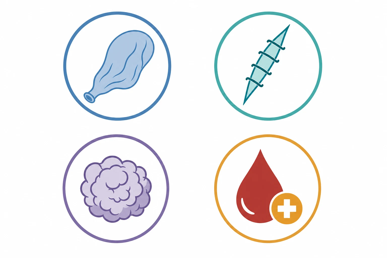
- The Tone (the uterine atony — the 70 per cent) — the uterus the fails to the contract. The risk: the over-the-distension (the multiple, the macrosomia, the polyhydramnios), the prolonged the labour, the high the parity, the oxytocin the prolonged, the chorioamnionitis, the GA the volatile.[1][1]
- The Trauma — the cervical / the vaginal / the perineal the lacerations, the uterine the rupture (the VBAC, the previous the scar), the inversion.[1][1]
- The Tissue — the retained the placenta / the membranes, the placenta the accreta / the increta / the percreta (the abnormal the invasion — the previous the Caesarean, the placenta the praevia).[1][1]
- The Thrombin — the coagulopathy (the DIC — the abruption, the pre-eclampsia, the sepsis, the amniotic the fluid the embolism, the retained the dead the fetus; the inherited — the von the Willebrand, the haemophilia; the anticoagulant — the LMWH, the warfarin).[1][1][1]
The management
1. The ABCDE + the massive the transfusion the protocol.[1][3][1]
- The oxygen; the IV access (the 2 the large the bore); the rapid the infuser + the warmer.
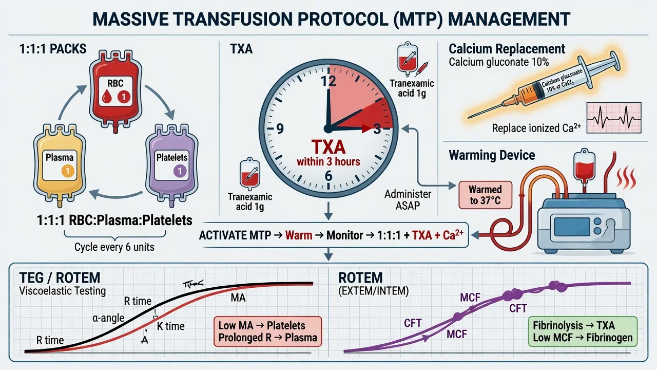
- The the massive the transfusion the protocol (the MTP) — the RBC : the FFP : the platelets the 1 : the 1 : the 1. The cryoprecipitate (the 10 the units) for the fibrinogen (the pregnancy the baseline the 4 to the 6 g/L; the keep the above the 2). The TXA the 1 g the IV the early (the WOMAN — the within the 3 h the reduces the mortality).[2][3]
- The ROTEM / the TEG the guided (the rapid the viscoelastic; the fibrinogen, the clot).[1]
2. The tranexamic acid — the 1 g the IV the within the 3 h (the WOMAN the trial — the reduced the mortality the from the bleeding the by the 19 per cent; the no the thromboembolic). The EARLY; the NOT the late (the above the 3 h the no the benefit / the possible the harm).[2]
3. The Tone — the uterotonics.[1][1]
- The oxytocin (the 5 to the 10 the IU the IV / the IM; the infusion) — the first-the-line.
- The ergometrine (the 0.25 mg the IM) — the AVOID the hypertension / the pre-eclampsia.
- The carboprost (the PGF2-alpha; the 250 mcg the IM) — the AVOID the asthma.
- The misoprostol (the PGE1; the 800 mcg the PR / the sublingual).[1]
4. The Tone — the mechanical / the surgical.[1][1]
- The bimanual the compression (the temporising).
- The the uterine the balloon the tamponade (the Bakri).
- The the B-Lynch the compression the suture (the laparotomy).
- The the uterine the artery / the internal the iliac the ligation OR the the embolisation (the interventional the radiology).
- The the hysterectomy (the life-the-saving the last the resort — the NOT the delay).[1][1]
5. The Trauma — the surgical the repair (the inspection, the suturing; the uterine the rupture the laparotomy).[1]
6. The Tissue — the manual the removal (the retained), the curettage, the accreta the hysterectomy (the often the planned the Caesarean-the-hysterectomy).[1][1]
7. The Thrombin / the DIC — the correct (the FFP, the cryoprecipitate [the fibrinogen the above the 2], the platelets the above the 50, the TXA). The exclude / the treat the trigger (the abruption, the sepsis, the AFE).[1][1]
Prognosis
The early the recognition + the MTP + the TXA the reduce the mortality. The delay → the coagulopathy (the massive the transfusion → the dilutional), the multi-organ, the death. The hysterectomy the life-the-saving (the counselling).[1][2][1]
Red flags
Pathophysiology — pregnancy physiology and the haemorrhage–coagulopathy spiral
Understanding major obstetric haemorrhage requires understanding how pregnancy physiology differs from the non-pregnant baseline — these differences explain both why pregnant women bleed and why they tolerate bleeding differently, and they drive every resuscitation target.[1][1]
The pregnant cardiovascular system — volume and reserve
- Plasma volume rises by ~40–50% (to ~5 L by the third trimester), with a smaller rise in red-cell mass (~20–30%) producing the physiologic haemodilutional anaemia of pregnancy (haematocrit ~32–34%). The expanded volume is a preloaded resuscitation reservoir: a pregnant woman can lose 1000–1500 mL (15–25% of blood volume) before exhibiting classical signs of shock. Tachycardia and a narrowed pulse pressure are early; hypotension is a LATE and pre-terminal sign. Underestimating blood loss because the vitals look fine is the classic trap.[1]
- Cardiac output rises ~30–50% (from ~5 to ~7 L/min), driven by a stroke-volume increase and a ~15–20 bpm rise in heart rate. This high-flow, low-resistance circulation means shock state evolves rapidly once compensation fails.
- Aorto-caval compression. From ~20 weeks gestation, the gravid uterus compresses the inferior vena cava and aorta in the supine position → reduced venous return and uteroplacental perfusion. Always nurse the critically ill pregnant/postpartum patient with LEFT LATERAL tilt (≥15°) or manual uterine displacement during resuscitation. This is a First-Part viva favourite and a reversible cause of unexplained hypotension.[1][1]
The hypercoagulable state — a double-edged sword
Pregnancy is a pro-thrombotic state designed to protect the mother from exsanguination at placental separation: [1]
- Fibrinogen rises to 4–6 g/L by term (non-pregnant 2–4 g/L) — the single most important clotting factor for placental-bed haemostasis.
- Factors VII, VIII, IX, X, XII and von Willebrand factor all rise; free Protein S falls; acquired activated Protein C resistance is common.
- Fibrinolysis is suppressed (PAI-1 and PAI-2 — produced by the placenta — rise markedly), so clots form readily and resist breakdown. [1]
This protects against normal delivery blood loss (~500 mL vaginal, ~1000 mL caesarean) but means that DIC, when triggered, is explosive and sustained because the clotting cascade is already primed and fibrinolysis is pathologically deranged. It also means the recovery phase is thrombogenic — VTE prophylaxis is mandatory.[7]
The vicious spiral — bleeding begets coagulopathy
Obstetric haemorrhage differs from controlled surgical bleeding: it enters a self-perpetuating spiral unless interrupted. Massive bleeding → consumption of clotting factors and platelets + dilution by crystalloid/blood products → dilutional and consumptive coagulopathy. This is compounded by the lethal triad: hypothermia (cool resus room, exposed abdomen, cold fluids) → acidosis (hypoperfusion, lactic acidosis) → coagulopathy. Each element worsens the others: hypothermia impairs clotting enzyme kinetics (the cascade is temperature-dependent), acidosis reduces thrombin generation and fibrinogen synthesis. Damage-control resuscitation exists to break this spiral: warm the patient, give balanced blood products early, avoid over-resuscitation with clear fluid, correct acidosis and calcium.[1][7]
Why estimated blood loss is unreliable in obstetrics
Visual estimation of blood loss underestimates true loss by 30–50%, more so at higher volumes. Blood is absorbed into drapes, swabs, sheets, under the patient, and mixed with amniotic fluid. The blood-soaked floor pad holds ~500 mL; a saturated perineal pad ~50–100 mL. Weigh swabs and drapes (1 g = 1 mL) for accuracy. Resuscitate on clinical and haemodynamic status, not the estimated number. Shock index (heart rate / systolic BP) >0.9 in pregnancy suggests significant occult loss; a rising shock index predicts transfusion need.[1]
DIC in pregnancy — diagnosis and triggers
Disseminated intravascular coagulation in pregnancy is always secondary — there is no idiopathic obstetric DIC. It is systemic activation of coagulation generating intravascular thrombin and fibrin, with consumption of platelets and clotting factors, leading simultaneously to thrombosis (microvascular) and bleeding (consumptive). In the obstetric context, bleeding dominates the clinical picture.[7]
The ISTH overt-DIC score (apply in the bleeding patient)
The International Society on Thrombosis and Haemostasis (ISTH) overt-DIC score is calculated on a single set of labs and trended. A score of ≥5 = compatible with overt DIC (repeat daily): [1]
| Parameter | 0 points | 1 point | 2 points | 3 points |
|---|---|---|---|---|
| Platelet count (×10ˆ9/L) | >100 | <100 | <50 | — |
| Fibrinogen (g/L) | >1.0 | <1.0 | — | — |
| D-dimer / FDP | no increase | — | moderate increase | strong increase |
| Prolongation of PT (s above upper limit) | <3 s | >3 s but <6 s | — | >6 s |
Pregnancy caveat: the fibrinogen arm is insensitive in pregnancy because baseline is 4–6 g/L — a non-zero fibrinogen of 2.0 scores 0 on the ISTH tool yet is profoundly abnormal for a pregnant patient and predicts severe haemorrhage. Do not be reassured by a normal-range fibrinogen in PPH: interpret it against the pregnancy baseline and treat to a target >4 g/L (some units target the ROTEM FIBTEM A10 >10 mm).[7]
The five classic pregnancy-specific DIC triggers
DIC in pregnancy is almost always one of these — identify and remove the trigger alongside correction: [1]
- Placental abruption — the commonest cause of pregnancy-related DIC. Retroplacental clot releases thromboplastin (tissue factor) into the maternal circulation → explosive cascade activation. Clinical: abdominal pain (constant, severe), tense/tender woody-hard uterus, vaginal bleeding (concealed or revealed), fetal distress. Diagnosis is clinical; ultrasound is insensitive for abruption. Deliver the fetus (the trigger is the retroplacental clot — delivery removes it).[7]
- Pre-eclampsia / HELLP — endothelial activation + platelet consumption produces a low-grade coagulopathy that can decompensate into overt DIC, especially with placental abruption or eclampsia. Platelets fall, AST/ALT rise, haemolysis (schistocytes, raised LDH) — HELLP. Magnesium sulphate for seizure prophylaxis, urgent delivery for severe disease.[7]
- Amniotic fluid embolism (AFE) — the highest-mortality trigger. Amniotic fluid enters the maternal circulation at delivery → biphasic reaction: (i) acute pulmonary vasospasm + right heart failure + hypoxia (the collapse phase), then (ii) left ventricular dysfunction + catastrophic DIC (the bleeding phase). The PPH from AFE is coagulopathy-driven (Thrombin) — uterotonics alone fail. Resuscitate with blood products, TXA, vasopressors, ventilation. Mortality 20–40%.[7]
- Sepsis — Gram-negative / Gram-positive endotoxin activates the extrinsic and intrinsic pathways and consumes antithrombin and Protein C. Genital-tract sepsis (chorioamnionitis, postpartum endometritis), pyelonephritis, or community-acquired sepsis all qualify. Treat the source + give antibiotics within the hour + supportive care.[1][1]
- Intrauterine fetal death / retained dead fetus — prolonged retention (>4 weeks) of a dead fetus releases tissue factor from degenerating placental/fetal tissue → subacute DIC. Modern obstetric practice (prompt delivery after diagnosis) has made this rare, but it remains a classic exam cause.[1]
Non-pregnancy triggers coexisting: severe pre-eclampsia with hepatic infarction, acute fatty liver of pregnancy (AFLP) with consumptive coagulopathy, massive-transfusion dilutional coagulopathy itself. [1]
Management of the coagulopathy (Thrombin / DIC)
The principle: treat the trigger AND replace what is consumed, guided by viscoelastic testing (ROTEM/TEG) where available.[7]
- Fibrinogen — the linchpin. Target >4 g/L in PPH (pregnancy baseline 4–6). Give cryoprecipitate 10 units (≈6–8 g fibrinogen) or fibrinogen concentrate 4–6 g. ROTEM FIBTEM <10 mm → give cryo/concentrate.
- Platelets — target >50 ×10ˆ9/L (>75 if actively bleeding). Give 1 adult therapeutic dose (raises by ~20–40).
- Fresh frozen plasma (FFP) — for clotting factor replacement (PT/APTT >1.5× normal). Dose 15 mL/kg (≈4 units).
- Tranexamic acid — 1 g IV within 3 h (anti-fibrinolytic; addresses the pathologically deranged fibrinolysis).
- Prothrombin complex concentrate (PCC) is NOT first-line in obstetric DIC (risk of microvascular thrombosis); reserve for warfarin reversal or specific factor deficits.
- Recombinant activated Factor VII (rFVIIa) — last-resort salvage only, after correction of fibrinogen, platelets, pH and temperature; high thrombosis risk. Off-label in most units.[7]
The 4 Ts — exam-exhaustive comparison
The 4 Ts (Tone, Trauma, Tissue, Thrombin) is the diagnostic mnemonic that covers virtually every cause of PPH. The frequencies below are the standard Fellowship-exam figures. The first move at the bedside is to palpate the fundus: a soft, boggy fundus = atony (Tone) — treat while you inspect for the others.[1][5]
The 4 Ts — frequency, clue, and first action
| Cause | Frequency | Pathophysiology | Bedside clue | First action |
|---|---|---|---|---|
| Tone (uterine atony) | 70% (#1) | Myometrial failure to contract around the spiral arteries after placental separation | Fundus is soft, boggy, not contracted; bleeding is dark red, steady | Bimanual compression + uterotonic ladder (oxytocin → ergometrine → carboprost → misoprostol); if fails → Bakri balloon → B-Lynch → hysterectomy |
| Trauma (laceration / rupture) | ~20% | Cervical, vaginal, perineal lacerations; uterine rupture (VBAC, previous scar); uterine inversion | Fundus is firm/contracted but bleeding persists → examine cervix/vagina; rupture = severe abdominal pain + fetal bradycardia + loss of station + collapse | Surgical: inspection + repair under anaesthesia; rupture → laparotomy (repair vs hysterectomy) |
| Tissue (retained / accreta) | ~9% | Retained placenta/membranes prevent contraction; accreta = abnormal trophoblastic invasion of myometrium | Placenta not delivered or incomplete; accreta = placenta adherent, cannot be separated | Manual removal / curettage; accreta: do NOT avulse — leave in situ or planned caesarean-hysterectomy |
| Thrombin (coagulopathy / DIC) | ~1% (but amplifies the other three) | Consumption / dilution; triggers: abruption, AFE, sepsis, pre-eclampsia/HELLP, AFLP, inherited (vWD), anticoagulants (LMWH) | Oozing from IV sites/gums + abnormal coagulation labs (low fibrinogen, low platelets) | Treat trigger + replace: cryo (fibrinogen >4 g/L) + platelets >50 + FFP + TXA |
The percentages are not coincidence: atony dominates because the mechanical contraction of the uterus is the primary haemostatic mechanism at the placental bed (there are no ligated vessels — the myometrial fibres act as living ligatures). Any factor that impairs contraction (overdistension, prolonged labour, chorioamnionitis, volatile anaesthesia) predisposes to atony.[1]
The management cascade — the first 30 minutes
Obstetric haemorrhage — the escalating cascade (uterotonics → Bakri → B-Lynch → hysterectomy)
- CALL FOR HELP — the single most important first step. Mobilise the full team: senior obstetrician + obstetric anaesthetist + haematologist (to release blood) + midwives + theatre/IR alert. Activate the major obstetric haemorrhage / massive transfusion protocol (MTP). This is never a one-person resuscitation.
- RESUSCITATE (ABCDE) — in parallel, not in series:
- Left lateral tilt / uterine displacement (relieve aorto-caval compression).
- High-flow oxygen; secure the airway if obtunded.
- Two large-bore IV cannulae (14–16 G); send cross-match, FBC, coagulation, fibrinogen, U&E, lactate, blood gas.
- Rapid infuser + fluid warmer; warm crystalloid (Hartmanns) as a bridge — but do NOT over-resuscitate with crystalloid (dilutional coagulopathy). Use permissive hypotension cautiously in the undelivered second stage.
- Emergency-release blood: O-negative RBC if no cross-match available; switch to group-specific then fully cross-matched as soon as possible.
- Tranexamic acid 1 g IV over 10 min — EARLY, within 3 h (WOMAN trial: reduces death from bleeding by ~19%). Do NOT wait for refractory — give alongside the first uterotonic.
- Keep the patient warm (Bair Hugger, warmed fluids, raise room temp → target core >36°C).
- IDENTIFY & TREAT THE CAUSE — palpate the fundus first:
- Soft/boggy fundus → ATONY (70%) → go to step 4.
- Fundus firm but bleeding persists → TRAUMA → speculum inspection of cervix/vagina under anaesthesia; repair lacerations; suspect rupture (→ laparotomy).
- Placenta not delivered / incomplete → TISSUE → manual removal / curettage; if accreta, do NOT avulse.
- Oozing + abnormal labs → THROMBIN/DIC → correct (step 7) + find the trigger (abruption, AFE, sepsis).
- UTEROTONIC LADDER for atony (escalate at each failure — different receptors, so the ladder is mechanistically additive):
- Oxytocin 5 IU slow IV (over ≥1 min) + infusion 40 IU in 500 mL Hartmanns at 125 mL/hr (first-line).
- Ergometrine 0.25 mg IM (AVOID pre-eclampsia/hypertension; AVOID cardiac disease). Repeat after 5 min; max 0.5 mg.
- Carboprost 250 mcg deep IM q15min, max 8 doses / 2 mg (AVOID asthma — bronchoconstrictor).
- Misoprostol 800 mcg PR (or 600 mcg SL) — safe in asthma; the workhorse in resource-limited settings.
- MECHANICAL / BALLOON TAMPONADE (if uterotonics fail within ~10–15 min):
- Bimanual compression (temporary bridge).
- External aortic compression (compress aorta at umbilical level against the vertebra — buys time).
- Bakri balloon — silicone balloon inserted into uterine cavity, filled with 300–500 mL warm saline → tamponades the bleeding sinuses; left 12–24 h. Success ~60–80% for atony. Avoid if trauma/infection suspected at insertion site.
- SURGICAL ESCALATION (proceed to theatre once balloon fails — time-critical):
- B-Lynch compression suture — brace-like suture around the uterus physically compresses it; uterine-sparing.
- Cho / Hayman square sutures — alternative compression sutures for lower-segment atony.
- Uterine artery ligation (ascending branch) — reduces uterine inflow; uterine-sparing.
- Internal iliac (hypogastric) artery ligation — reduces pelvic pulse pressure by ~50%; technically demanding; uterine-sparing.
- Uterine artery embolisation (interventional radiology) — gelatin sponge particles; uterine-sparing; needs IR availability (often not emergently available).
- Hysterectomy — LIFE-SAVING LAST RESORT; do NOT delay. Decide early (typically once >4–6 units transfused and bleeding uncontrolled despite uterotonics + balloon + compression sutures). The coagulopathy worsens with every minute of delay.
- CORRECT COAGULOPATHY (the Thrombin arm) — guided by ROTEM/TEG:
- Fibrinogen → target >4 g/L (cryoprecipitate 10 units, or fibrinogen concentrate 4–6 g).
- Platelets → target >50 ×10ˆ9/L (1 adult dose).
- PT/APTT → INR <1.5 (FFP 15 mL/kg ≈ 4 units).
- Calcium — citrate toxicity from massive transfusion → hypocalcaemia → give calcium gluconate 10 mmol IV per 4 units RBC (calcium is a cofactor for the clotting cascade and myocardial function).
- Correct acidosis and hypothermia (the lethal triad — they inactivate clotting enzymes).
- POST-HAEMORRHAGE ICU CARE:
- ICU admission for ongoing bleeding surveillance, coagulopathy, AKI (hypoperfusion), DIC evolution, and transfusion reactions (TRALI/TACO).
- Correct anaemia (IV iron; RBC if Hb <70 g/L).
- VTE prophylaxis — mandatory (pregnancy + tissue factor + stasis = pro-thrombotic); LMWH 6–12 h after bleeding controlled, continue 6 weeks postpartum.
- Debrief + psychological support (traumatic birth → PTSD risk).
- Sheehan syndrome surveillance (pituitary apoplexy — failure to lactate, amenorrhoea, hypothyroidism, adrenal insufficiency weeks–months later).
Massive transfusion protocol (MTP) in pregnancy
The obstetric MTP borrows its evidence base from trauma (PROPPR) but applies pregnancy-specific targets — chiefly a higher fibrinogen threshold. Activation triggers major-haemorrhage protocols in blood bank: pre-made packs delivered rapidly, with a haematology scientist assigned.[4][7]
When to activate
Activate the MTP early, on predicted need, not on units already given: [1]
- Blood loss >1500 mL ongoing, OR
- Haemodynamic instability (shock index >0.9, sustained tachycardia, hypotension), OR
- Abnormal coagulation/low fibrinogen at presentation, OR
- Clinical judgement of uncontrolled ongoing loss. [1]
The balanced ratio — 1:1:1
From the PROPPR trial (Holcomb 2015, trauma): RBC : FFP : platelets 1:1:1 achieved earlier haemostasis and reduced death from exsanguination at 24 h versus 1:1:2, with no increase in complications. This is extrapolated (not directly tested) to obstetrics but is the universal standard. Pre-made MTP packs deliver 1 unit RBC + 1 unit FFP + 1 adult-dose platelets (≈1 pool); aim for at least this ratio. Add cryoprecipitate separately for fibrinogen.[4]
Pregnancy-specific transfusion targets
| Product | Pregnancy / PPH target | Trigger to give | Dose |
|---|---|---|---|
| RBC | Hb >70 g/L (>80 if ongoing bleed / symptomatic) | Hb <70, or anticipated ongoing major loss | 1 unit raises Hb ~10 g/L |
| FFP | PT/APTT <1.5× normal | INR >1.5 or predicted factor depletion | 15 mL/kg (≈4 units) |
| Cryoprecipitate | Fibrinogen >4 g/L (NOT 1.5) | Fibrinogen <4 in PPH; FIBTEM A10 <10 mm | 10 units (≈6–8 g fibrinogen) |
| Platelets | >50 ×10ˆ9/L (>75 if bleeding) | <50, or predicted ongoing loss | 1 adult dose (raises ~20–40) |
| Calcium | Ionised Ca in normal range | Per 4 units RBC (citrate chelation) | Calcium gluconate 10 mmol IV |
| TXA | Anti-fibrinolysis | All major PPH, within 3 h | 1 g IV over 10 min |
MTP-specific pitfalls in the obstetric patient
- Fibrinogen is the keystone. A normal fibrinogen of 2.0 g/L is low in pregnancy — give cryoprecipitate early. Hypofibrinogenaemia (<2 g/L) is the strongest single laboratory predictor of progression to severe PPH.[7]
- Citrate toxicity → hypocalcaemia. Stored RBC and FFP contain citrate anticoagulant; rapid transfusion overwhelms hepatic metabolism → ionised calcium falls → both coagulopathy (calcium is cofactor for factors II, VII, IX, X) and myocardial depression/hypotension. Give calcium gluconate 10 mmol per 4 units RBC, guided by ionised calcium monitoring.[1]
- Hypothermia → coagulopathy. Warm everything (fluids, patient, room). Target core >36°C.
- Potassium load. Stored RBC supernatant K rises; massive transfusion can cause hyperkalaemia → arrhythmia. Use fresh (<7 day) RBC where possible in truly massive transfusion; monitor ECG and potassium.
- TRALI / TACO. Transfusion-related acute lung injury (donor antibodies) and transfusion-associated circulatory overload are the leading transfusion-related causes of maternal morbidity — minimise unnecessary units, monitor for hypoxaemia and overload, and stop + report if suspected.
- Rh immunisation. Rh-negative mother receiving Rh-positive blood → anti-D. Ensure anti-D prophylaxis.[1]
Uterotonic pharmacology — in depth
Uterotonics are the cornerstone of atonic PPH management (70% of cases). The four agents act at different receptors — this is why the ladder works (a second-line agent uses a distinct mechanism when oxytocin is tachyphylactic or contraindicated). The receptor pharmacology makes the contraindications logical rather than rote.[5]
Uterotonic agents — receptor, dose, contraindication
| Agent | Receptor / class | Dose & route | Onset | Key contraindications | Hallmark adverse effects |
|---|---|---|---|---|---|
| Oxytocin (Syntocinon) | OXTR (Gq) on myometrium → IP3 → Caˆ2+ release | 5 IU slow IV (≥1 min) or 10 IU IM; then infusion 40 IU / 500 mL at 125 mL/hr | 1 min (IV); 3–5 min (IM) | Rapid IV = hypotension (give slowly); caution in cardiac disease | Hypotension/tachycardia (fast push); water intoxication/hyponatraemia at high rates (weak ADH activity); flushing |
| Ergometrine / methylergometrine | Ergot; alpha-agonist + 5-HT2 + dopamine → tetanic contraction | 0.25 mg IM; repeat after 5 min; max 0.5 mg (0.125–0.25 mg IV if urgent) | 5–7 min (IM); 1 min (IV) | Pre-eclampsia/hypertension (alpha-agonist → crisis); cardiac disease (afterload/coronary spasm); sepsis; before placenta delivery | Hypertension; nausea/vomiting (CTZ); reflex bradycardia; coronary spasm → MI |
| Carboprost (15-methyl PGF2α; Hemabate) | PGF2α analogue → FP receptor (Gq); resists degradation | 250 mcg deep IM q15min; max 8 doses (2 mg); NOT IV | ~5 min | ASTHMA (absolute) — bronchoconstrictor; cardiac disease | Diarrhoea (~40%); vomiting; fever (hypothalamic set-point); bronchospasm |
| Misoprostol (PGE1; Cytotec) | PGE1 → EP receptors (Gq/Gi); vasodilates (opposite of PGF2α) | 800 mcg PR (single); alt 600 mcg SL, 800 mcg oral | SL ~10 min; PR ~20 min | (Generally safe; off-label) | Shivering (30–50%); fever; diarrhoea; nausea |
| Carbetocin (long-acting oxytocin analogue) | OXTR; t½ ~40 min (vs ~5 min) | 100 mcg IV single (prophylaxis at LSCS) | ~2 min | As for oxytocin | Mild flushing, nausea |
Oxytocin — first line
Synthetic nonapeptide; binds the oxytocin receptor (OXTR, Gq-coupled) on myometrial smooth muscle → phospholipase C → IP3 + DAG → sarcoplasmic Caˆ2+ release → myosin-light-chain phosphorylation → contraction. OXTR expression rises 100–200-fold across gestation, peaking at term — hence the late-pregnant uterus is exquisitely sensitive.[5]
Tachyphylaxis (burnout). Prolonged/saturating oxytocin exposure downregulates OXTR → the uterus becomes oxytocin-resistant and escalating doses yield diminishing contraction. This is the pharmacological rationale for escalating to a second-line agent (different receptor) after ~1–2 h of high-dose infusion rather than endlessly pushing oxytocin. It also explains failed inductions after prolonged augmentation.[5]
Bolus danger. Rapid IV oxytocin triggers endothelial nitric-oxide release + direct vascular smooth-muscle relaxation → sudden venodilation + tachycardia → in a hypovolaemic patient, profound hypotension and arrest. Always give the 5 IU IV bolus over ≥1 min. If the patient is shocked, prefer IM or a slow infusion.[5]
Ergometrine — the tetanic constrictor
Ergot alkaloid; agonist at alpha-adrenergic, 5-HT2 and dopamine receptors → sustained tetanic (cramp-like) contraction that mechanically strangulates the spiral arteries and venous sinuses. The contraindications follow directly from its alpha-agonist/vasoconstrictor action: pre-eclampsia/hypertension (absolute — precipitates hypertensive crisis → stroke/ICH/pulmonary oedema), cardiac disease (afterload + coronary spasm), sepsis (worsens perfusion), and before placental delivery (the tetanic contraction traps the placenta). Light-sensitive — store in the dark.[5]
Carboprost — the prostaglandin F2α analogue
15-methyl modification resists degradation by 15-hydroxyprostaglandin dehydrogenase → prolonged half-life. Binds the FP prostanoid receptor (Gq) → strong rhythmic contractions + uterine vasoconstriction. ASTHMA is an absolute contraindication — PGF2α is a bronchoconstrictor and can trigger fatal bronchospasm; always check asthma status first. Diarrhoea (~40%), vomiting, and non-infective fever (prostaglandins reset the hypothalamic set-point) are characteristic and must be distinguished from sepsis.[5]
Misoprostol — the off-label workhorse
PGE1 analogue; binds EP receptors → contractions. Crucially, PGE1 vasodilates (opposite of PGF2α) and does not bronchoconstrict → safe in asthma. Cheap, heat-stable (no cold chain) → the agent of choice in resource-limited settings. Shivering (30–50%) within 10–20 min is a benign prostaglandin hypothalamic effect (not sepsis/transfusion reaction) — reassure; meperidine 25–50 mg IV aborts it.[5]
Carbetocin — long-acting prophylaxis, not treatment
t½ ~40 min (vs oxytocin ~5 min) → sustained tone from a single 100 mcg IV dose → used for third-stage prophylaxis at caesarean, not established atonic PPH (no bolus effect). Heat-stable carbetocin (WHO CHAMPION 2018) tolerates 30°C/75% humidity for years → solves the cold-chain problem in tropical settings.[1]
Tranexamic acid and the WOMAN trial
Mechanism. Tranexamic acid is a synthetic lysine analogue that reversibly blocks the lysine-binding sites on plasminogen → prevents plasminogen converting to plasmin → inhibits fibrinolysis → stabilises the clot. In PPH, fibrinolysis is pathologically upregulated (clots form then break down); TXA preserves the clot. The effect is time-dependent — clots that have already lysed cannot be salvaged, hence the benefit only early.[2][1]
Dosing. 1 g IV over 10 min within 3 h of bleeding onset; a second 1 g dose can be given after 30 min if bleeding continues. Give it alongside the first uterotonic, not as a salvage drug. [1]
The WOMAN trial (Shakur 2017) — the practice-defining evidence
WOMAN trial — tranexamic acid for postpartum haemorrhage (PMID 28456565)
Design
International, randomised, double-blind, placebo-controlled — 20,060 women across 193 hospitals in 21 countries
Population
Women with postpartum haemorrhage after vaginal or caesarean delivery (clinical diagnosis)
Intervention
Tranexamic acid 1 g IV over 10 min vs placebo, IN ADDITION to standard care, within 3 h of bleeding onset
Primary outcome
Death from bleeding: 1.5% (TXA) vs 1.9% (placebo) — relative reduction ~19% (RR 0.81, p=0.045)
Key finding
Benefit ONLY within 3 h. No benefit / possible harm after 3 h. NO increase in thromboembolic events (DVT/PE/stroke/MI)
Secondary
No significant reduction in hysterectomy or laparotomy rates
Clinical bottom line
TXA 1 g IV is SAFE and reduces mortality from PPH — give EARLY (within 3 h), alongside the first uterotonic, to every woman with major PPH
The time-dependence is the exam-critical point: a subgroup analysis showed the mortality benefit was confined to women treated within 3 h, and an excess of bleeding deaths was seen with late (>3 h) administration. The mechanism is thought to be that late TXA cannot salvage already-lysed clots and may, in the pro-thrombotic recovery phase, tilt the balance toward thrombosis. Give it first-line, not last-resort.[2][1]
The PROPPR trial — the evidence for 1:1:1
PROPPR trial — 1:1:1 vs 1:1:2 transfusion ratio in severe trauma (PMID 25339133)
Design
Phase III, multicentre, randomised — 680 patients with severe trauma at 12 North American Level I centres
Population
Adults with major haemorrhage (trauma)
Intervention
Plasma:platelets:RBC ratio of 1:1:1 vs 1:1:2
Primary outcome
No difference in 24-h or 30-day all-cause mortality
Key secondary
1:1:1 achieved earlier haemostasis and significantly fewer deaths from exsanguination at 24 h; no increase in complications
Clinical bottom line
1:1:1 (balanced) resuscitation is safe and improves early haemostasis — adopted into obstetric MTPs
Placenta accreta spectrum — the surgical catastrophe
Rising epidemic driven by the caesarean-section rate. Previous caesarean scar + placenta praevia in the next pregnancy → placenta implants on the scar and invades the myometrium to varying depths: accreta (adherent to myometrium, ~75%), increta (invades myometrium, ~15%), percreta (penetrates serosa/bladder, ~10%). Incidence ~1:533 and rising. Risk factors: previous caesarean (dose-dependent — each one increases risk), placenta praevia, advanced maternal age, multiparity, previous uterine surgery.[6]
Diagnosis. Antenatal ultrasound (loss of the normal hypoechoic retroplacental clear space, irregular lacunae, bladder wall interruption); MRI for mapping percreta. The goal is antenatal diagnosis → planned multidisciplinary delivery. [1]
Management — planned caesarean-hysterectomy. The adherent placenta is the haemostatic plug. Attempting manual removal opens the uncontractile lower-segment sinuses → catastrophic, often fatal bleeding. Standard of care: elective caesarean-hysterectomy at 34–36 weeks, with the placenta left in situ, performed by a multidisciplinary team (obstetrics + gynae-oncology surgery, anaesthetics, haematology, urology for bladder involvement, interventional radiology for prophylactic internal iliac balloon catheters). Conservative management (leaving placenta, methotrexate) is selective and high-risk.[6]
The exam error to avoid: a clinician pulls on a cord that will not deliver → the placenta is accreta → STOP, do not avulse, leave in situ, escalate to planned hysterectomy.[6]
Amniotic fluid embolism — collapse + DIC
The highest-mortality obstetric emergency (~20–40%). AFE presents in two phases during/shortly after labour and delivery:[7]
- Phase 1 — collapse. Acute pulmonary vasospasm → right heart failure + severe hypoxaemia + hypotension. The patient arrests or collapses suddenly, often after a contraction or delivery. Often fatal within minutes.
- Phase 2 — left ventricular dysfunction + DIC. Survivors of phase 1 develop LV failure and catastrophic, coagulopathy-driven PPH (oozing from every site). The bleeding is Thrombin-driven, not atonic → uterotonics alone fail; you must correct the coagulopathy with massive blood-product support. [1]
Diagnosis is clinical (there is no reliable test in real time; histology shows fetal squames in pulmonary vasculature post-mortem). Management is supportive + treat the DIC: high-quality CPR/advanced life support, ventilation, vasopressors (noradrenaline), MTP with balanced ratios + cryoprecipitate + TXA, delivery of the fetus if undelivered (perimortem caesarean within 4 min of arrest if ≥20 weeks). Recognise that the consumption is massive — anticipate enormous blood-product needs.[7]
Damage-control resuscitation — the obstetric patient is a trauma patient with a bleeding pelvis
The trauma-derived principles transfer in full to major obstetric haemorrhage:[4]
- Permissive hypotension during uncontrolled bleeding — target SBP ~80–90 mmHg until bleeding is controlled. Caveat: less validated during the second stage for uteroplacental perfusion; apply with judgement once delivered.
- Haemostatic (balanced) 1:1:1 ratio — minimises dilutional coagulopathy.
- Avoid the lethal triad — keep warm (target core >36°C), correct acidosis, give calcium, avoid hypoxia.
- Tranexamic acid EARLY (within 3 h).
- Damage-control surgery — the obstetric equivalent is definitive haemorrhage control (hysterectomy) over uterine preservation when the patient is exsanguinating and coagulopathic. [1]
Clinical pearls
Additional red flags
Prognosis and complications
Outcomes and complications of major obstetric haemorrhage
| Scenario / complication | Outcome | Notes |
|---|---|---|
| Overall PPH mortality | 2–5% (developed); 10–30% (developing) | The #1 cause of maternal death globally |
| Massive transfusion (>10 units) | 10–20% mortality | DIC + multi-organ failure are the killers |
| Hysterectomy for PPH | 5–15% mortality | Depends on delay — early decision = better outcome |
| Placenta accreta (planned surgery) | 5–10% mortality | Antenatal diagnosis + multidisciplinary team dramatically improves outcome |
| Placenta accreta (emergency) | 30%+ mortality | Undiagnosed accreta at delivery is catastrophic |
| Amniotic fluid embolism | 20–40% mortality | Highest-mortality obstetric emergency |
| Sheehan syndrome | Variable (late) | Weeks–months later: failure to lactate, amenorrhoea, hypothyroidism, adrenal insufficiency |
| Acute kidney injury | Common | Hypoperfusion; usually recovers with volume — may need RRT if severe |
| TRALI / TACO | Variable | Leading transfusion-related causes of maternal morbidity |
| VTE (post-PPH rebound) | Significant | Pro-thrombotic rebound — mandatory LMWH, 6 weeks postpartum |
| Psychological (PTSD) | Common | Traumatic birth — debrief + counselling |
The dominant determinants of survival are (1) early recognition, (2) rapid activation of the MTP, (3) early TXA, and (4) timely definitive haemorrhage control (notably, not delaying hysterectomy). Systems-level factors — a rehearsed major-haemorrhage protocol, multidisciplinary team training (OB + anaesthesia + haematology + IR), and massive-haemorrhage drills — reduce mortality more than any individual drug.[1][5]
Exam practice — SAQs
SAQ — Massive obstetric haemorrhage from uterine atony after prolonged second stage
10 minutes · 10 marks
A 34-year-old woman (G3P2, two previous caesarean sections) delivers vaginally at 39 weeks gestation after a prolonged second stage. Immediately postpartum she bleeds an estimated 2000 mL of dark red blood. The fundus is soft and boggy. HR 128, BP 84/48, SpO2 96 percent on room air. She is pale and drowsy. IV access is a single 18-gauge cannula. The midwife has given 5 IU oxytocin IM. You are the ICU registrar called to assist.
SAQ — Obstetric disseminated intravascular coagulation complicating severe placental abruption
10 minutes · 10 marks
A 36-year-old woman at 38 weeks gestation presents with severe constant abdominal pain and vaginal bleeding. She is pale, diaphoretic and restless. HR 138, BP 76/44, SpO2 92 percent on 15 L oxygen. The uterus is tense, woody-hard and tender. The fetal heart is absent. Bloods: Hb 71 g/L, platelets 65 x 10^9/L, INR 2.4, APTT 58 s, fibrinogen 1.3 g/L, D-dimer markedly elevated. The obstetric team diagnoses a severe placental abruption. You are asked to manage the coagulopathy.
References
- [1]Lakhi NA, et al. Postpartum Hemorrhage: A Comprehensive Review of Guidelines Obstet Gynecol Surv, 2022.PMID 36345105
- [2]WOMAN Trial Collaborators. Effect of early tranexamic acid administration on mortality, hysterectomy, and other morbidities in women with post-partum haemorrhage (WOMAN): an international, randomised, double-blind, placebo-controlled trial Lancet, 2017.PMID 28456509
- [3]Yamasaki A, et al. Transfusion of blood and blood products for the management of postpartum haemorrhage Cochrane Database Syst Rev, 2025.PMID 39911088
- [4]Holcomb JB, et al. Management of cryptoglandular supralevator abscesses in the magnetic resonance imaging era: a case series Int J Colorectal Dis, 2014.PMID 25339133
- [5]Sentilhes L, et al. Interaction of insulin-like growth factor-I and insulin resistance-related genetic variants with lifestyle factors on postmenopausal breast cancer risk Breast Cancer Res Treat, 2017.PMID 28478612
- [6]Silver RM, et al. A Novel Recombinant Anti-CD22 Immunokinase Delivers Proapoptotic Activity of Death-Associated Protein Kinase (DAPK) and Mediates Cytotoxicity in Neoplastic B Cells Mol Cancer Ther, 2016.PMID 26826117
- [7]Collins P, et al. Colorectal cancer screening in Australia Lancet Public Health, 2017.PMID 29253456