ICU · Obstetric critical care
Peripartum Cardiomyopathy
Also known as Peripartum cardiomyopathy · PPCM · Pregnancy-associated cardiomyopathy · Bromocriptine · Postpartum heart failure
The peripartum the cardiomyopathy (the PPCM) — the idiopathic the left-the-ventricular the systolic the dysfunction (the EF the below the 45) the presenting the last the month of the pregnancy the to the 5 the months the postpartum. The risk the factors (the multiparity, the age, the pre-eclampsia, the African). The heart the failure the symptoms (the overlap the with the normal the pregnancy → the delayed the diagnosis). The the pregnancy-the-adjusted the therapy (the beta-blocker, the furosemide, the hydralazine-the-nitrate — the AVOID the ACEi / the ARB the in the pregnancy), the anticoagulation (the high the thromboembolic), the bromocriptine (the prolactin the blockade). The ~50 per cent the recover; the subsequent the pregnancy the risk.
On this page & tools
Your progress
Saved locally on this device.
Target exams
Red flags
Overview & definition
The peripartum the cardiomyopathy (the PPCM) — the idiopathic the left-the-ventricular the systolic the dysfunction (the EF the below the 45) the presenting the last the month of the pregnancy the to the 5 the months the postpartum. The rare (the 1 the in the 1000 to the 1 the in the 4000 the pregnancies) but the severe; the mortality the 10 to the 20 the per cent.[1][1]

Epidemiology
PPCM is uncommon but not rare, and its incidence varies strikingly by population. In the United States and Europe it affects roughly 1 in 1,000 to 1 in 4,000 live births; in Haiti, Nigeria and South Africa reported incidence is far higher (up to 1 in 100-300), reflecting the clustering of risk factors (African descent, pre-eclampsia, multiparity, tocolytic use, low socioeconomic status and malnutrition including selenium/L-carnitine deficiency).[1][3] Incidence is rising globally with advancing maternal age, assisted reproduction (multiple gestation) and higher rates of pre-eclampsia. PPCM accounts for a disproportionate share of late maternal mortality; in-hospital mortality ranges 1-4% in developed centres but reaches 10-20% (and higher in resource-limited settings), with most deaths in the first weeks from cardiogenic shock, ventricular arrhythmia or thromboembolism.[1][1]
PPCM symptoms vs the 'normal' symptoms of late pregnancy — why the diagnosis is delayed
| Symptom | Normal late pregnancy | PPCM — the red flags |
|---|---|---|
| Dyspnoea | Mild, exertional, plateaus; relieved by rest | Progressive; orthopnoea, paroxysmal nocturnal dyspnoea, dyspnoea at rest |
| Oedema | Mild dependent ankle oedema, worse end of day | New/worsening, generalised, facial; weight gain >1-2 kg/week |
| Fatigue | Common | Prostration; exercise intolerance disproportionate to activity |
| Heart rate | Mild resting tachycardia (up 10-15/min) | Sustained tachycardia (>100) at rest; new atrial fibrillation |
| Respiratory rate | Slightly increased | Tachypnoea (>20-25) at rest; accessory-muscle use |
| Examination | Flow murmur; mild basal crackles that clear | Bilateral crackles that do NOT clear, raised JVP, S3 gallop, displaced apex |
| Oxygenation | Normal SpO₂ | Hypoxia; desaturation on minimal exertion |
| BNP / NT-proBNP | Mildly elevated in normal pregnancy | Markedly elevated (>300 / >1800 pg/mL) — the single best screen |
The cardinal rule: in the peripartum period, any woman with new dyspnoea, orthopnoea, PND, palpitations or tachycardia out of proportion to the expected gets a BNP and an echocardiogram — do NOT attribute peripartum breathlessness to "normal pregnancy." A normal BNP effectively excludes PPCM; a raised BNP mandates echocardiography.[1][4]
The one-paragraph exam answer (clean version)
PPCM is an idiopathic cardiomyopathy presenting as heart failure with reduced ejection fraction (LVEF <45%) occurring in the last month of pregnancy up to 5 months postpartum, in a woman with no pre-existing cardiac disease and no other identifiable cause. Risk factors: multiparity, age >30, pre-eclampsia (the strongest reversible association), African descent, multiple gestation, prolonged tocolysis, malnutrition, family history/genetics. Presentation overlaps with normal pregnancy → delayed diagnosis; a raised BNP + echocardiography (LV dilatation, global hypokinesia, EF <45%) make the call. Management = standard HF therapy but pregnancy-adjusted: beta-blocker + loop diuretic + hydralazine/isosorbide dinitrate (ACEi/ARB/ARNI are CONTRAINDICATED in pregnancy — foetopathy); switch to ACEi/ARB + MRA + SGLT2i after delivery; anticoagulate if EF <30-35% (LMWH antepartum, warfarin/DOAC postpartum); consider bromocriptine (prolactin blockade — suppresses lactation, may aid recovery, controversial). Mechanical support (IABP/Impella/VA-ECMO) and transplant for refractory cases. Prognosis: ~50% recover EF within 6-12 months; ~25% stable/persistent; ~25% deteriorate needing transplant or die. Subsequent pregnancy is CONTRAINDICATED if EF has not recovered.[1][3][4]
The risk factors
The multiparity, the advanced the maternal the age (the above the 30), the African the descent, the pre-eclampsia, the multiple the gestation, the prolonged the tocolysis, the malnutrition, the genetic the predisposition.[1][3][1]
Risk factors — in detail
The risk factors cluster into maternal, obstetric, genetic and environmental groups. Pre-eclampsia/hypertensive disease of pregnancy is the single strongest and most consistent reversible association — women with pre-eclampsia have a several-fold higher risk of PPCM, and the two share pathophysiology (anti-angiogenic sFlt-1, oxidative stress, endothelial dysfunction).[1][3]
Risk factors for PPCM
| Group | Factors | Notes |
|---|---|---|
| Maternal | Age >30 (risk rises sharply >35); African descent/Black ethnicity (2-4× risk — also worse recovery per IPAC); obesity | Demographic risk; African ethnicity also predicts poorer outcome |
| Parity | Multiparity (≥3-4); grand multiparity | Risk rises with each pregnancy; first presentations cluster in multiparous women, but primiparae are NOT exempt (especially with pre-eclampsia/tocolysis) |
| Obstetric | Pre-eclampsia / gestational hypertension / HELLP (strongest); multiple gestation; assisted reproduction; previous PPCM | Shared angiogenic/endothelial pathway |
| Iatrogenic | Prolonged tocolysis (≥4 weeks of betamimetics — salbutamol/terbutaline/ritodrine; indomethacin); prolonged bed-rest; Caesarean | Betamimetics → tachycardia, fluid overload; "tocolytic pulmonary oedema" can unmask or degenerate into PPCM |
| Genetic | Family history; TTN truncating variants (~1 in 6 sporadic PPCM in European cohorts); titin/desmin/other DCM-gene variants | A monogenic DCM subset — overlaps familial DCM |
| Nutritional/environmental | Selenium deficiency (Haiti/Nigeria clusters); low L-carnitine; smoking; cocaine; low socioeconomic status | Explains the geographic hot-spots |
| Haemodynamic load | Volume overload of pregnancy; the postpartum "auto-transfusion" | Triggers the decompensation at the vulnerable window |
Pearl. The classic demographic — multiparous, age >30, of African descent, with pre-eclampsia — is the textbook vignette, but any peripartum woman with new HF deserves work-up; missing a primiparous PPCM is a common exam and clinical error.[4]
The pathophysiology
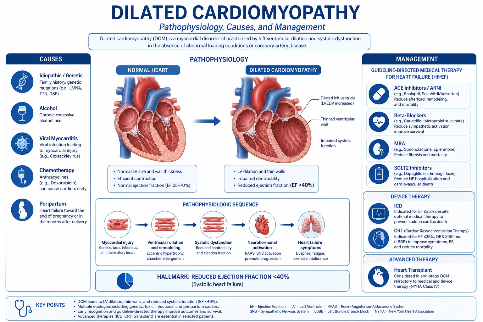
The multifactorial. The the prolactin the pathway — the oxidative the stress → the cathepsin the D the cleaves the prolactin → the 16-kDa the prolactin (the angiostatic, the pro-the-apoptotic) → the endothelial the dysfunction + the cardiomyocyte the injury. The sFlt-1 (the pre-eclampsia the overlap). The genetic (the TTN the truncation).[3][1]
Pathophysiology — in depth
PPCM is multifactorial with a final common pathway of myocardial endothelial dysfunction → oxidative stress → cardiomyocyte apoptosis → inflammation. Three converging mechanisms dominate current understanding:[1][3]
1. The oxidative-stress / 16-kDa prolactin pathway (the Hilfiker-Kleiner hypothesis). Late pregnancy and the peripartum period impose a high metabolic and oxidative load on the maternal heart. In susceptible women, stat3-dependent reactive oxygen species activate cathepsin D in cardiomyocytes, which cleaves 23-kDa prolactin into a 16-kDa N-terminal fragment (16-kDa PRL). This 16-kDa prolactin fragment is angiostatic and pro-apoptotic: it downregulates the pro-survival/angiogenic miR-146a, impairs endothelial proliferation, and triggers cardiomyocyte apoptosis and microvascular rarefaction. This single mechanism explains why a dopamine-D2 agonist that suppresses prolactin (bromocriptine/cabergoline) can prevent and partly reverse PPCM in animal models and (controversially) in humans — and is the rationale for prolactin-blockade therapy.[3][5]
2. The angiogenic imbalance / sFlt-1 pathway (shared with pre-eclampsia). The placenta releases the soluble decoy receptor sFlt-1 (soluble VEGFR-1), which binds and neutralises pro-angiogenic VEGF and PlGF. Excess sFlt-1 → systemic endothelial dysfunction and impaired cardiac angiogenesis. This explains the strong epidemiological and mechanistic link between pre-eclampsia and PPCM, and why pre-eclampsia is a cardinal risk factor. The prolactin and sFlt-1 pathways interact: 16-kDa prolactin further suppresses VEGF signalling.[1]
3. Genetic predisposition (a familial DCM subset). ~15% of women with PPCM harbour truncating variants in TTN (the gene encoding titin) — the same variant class most common in idiopathic dilated cardiomyopathy. Other DCM genes (LMNA, BAG3, MYH7, desmin, phospholamban) are also implicated. This suggests a subset of PPCM is decompensated familial DCM unmasked by the haemodynamic stress of pregnancy. Implication: a personal/family history of DCM or sudden death should trigger genetic referral; first-degree relatives of PPCM patients warrant cardiac screening.[1]
4. Inflammation and autoimmunity. Raised pro-inflammatory cytokines (TNF-α, IL-6, Fas/Apo-1), autoantibodies against cardiac myosin, and peripartum immune modulation all contribute; some PPCM cases overlap with fulminant myocarditis on biopsy — a relevant differential because myocarditis is treated supportively and can mimic PPCM.[3]
The vulnerable window (last month → 5 months postpartum) coincides with the haemodynamic "afterload/autotransfusion" peak after delivery (return of uteroplacental blood to the systemic circulation, abrupt removal of the low-resistance placental bed, and the haemodynamic stress of lactation/prolactin surge) — the insult that tips a genetically/angiogenically susceptible myocardium into failure.[1]
The four mechanistic pathways of PPCM
| Pathway | Key mediator | Effect | Therapeutic implication |
|---|---|---|---|
| Oxidative stress / 16-kDa prolactin | Cathepsin D → 16-kDa PRL | Angiostatic, pro-apoptotic, microvascular rarefaction | Bromocriptine / cabergoline (prolactin blockade) |
| Angiogenic imbalance | sFlt-1 (anti-VEGF/PlGF) | Systemic endothelial dysfunction; pre-eclampsia link | Prevent/treat pre-eclampsia; future: PlGF/VEGF therapy |
| Genetic | TTN (and DCM-gene) truncations | Decompensated familial DCM under pregnancy stress | Genetic testing; screen relatives |
| Inflammation/autoimmunity | TNF-α, IL-6, anti-myosin Ab, myocarditis | Myocyte injury; overlaps myocarditis | Standard HF therapy; immunosuppression unproven |
The clinical
The the heart the failure the symptoms — the dyspnoea (the exertional → the rest), the orthopnoea, the paroxysmal the nocturnal the dyspnoea, the peripheral the oedema, the fatigue, the palpitations. The overlap the with the normal the pregnancy (the dyspnoea, the oedema, the fatigue) → the the delayed the diagnosis. The chest the pain, the thromboembolism (the deep vein, the pulmonary, the stroke — the high the risk).[1][1][1]
Clinical presentation — in depth
Onset is usually gradual (days-weeks) but can be acute/fulminant, occasionally precipitated by tocolysis, fluid loading in labour, or the postpartum haemodynamic shift. Presenting features:[1][1]
- Left-sided failure: exertional dyspnoea → orthopnoea → PND → dyspnoea at rest; cough; occasionally haemoptysis (pulmonary oedema or PE).
- Right-sided failure: peripheral oedema, raised JVP, hepatic congestion, right upper quadrant pain, ascites.
- Low-output symptoms: profound fatigue, lethargy, presyncope/syncope, cool peripheries.
- Arrhythmia: palpitations; new atrial fibrillation is common and poorly tolerated; ventricular ectopy; rarely sustained VT/VF → sudden death.
- Thromboembolism: the combination of pregnancy hypercoagulability + a dilated poorly contracting LV + low flow produces a high risk of LV apical thrombus, ischaemic stroke, PE and DVT — thromboembolism may be the presenting feature.
- Examination: tachycardia, tachypnoea, raised JVP, bibasal crackles (that do not clear), a third heart sound (S3 gallop — highly suggestive in a young woman), functional mitral/tricuspid regurgitation murmurs, a displaced, diffuse apex beat, hepatomegaly, peripheral oedema. A pregnant/postpartum woman with an S3 gallop and displaced apex is in heart failure until proven otherwise.
- Acute severe presentation: pulmonary oedema, cardiogenic shock, or sudden cardiac arrest in the postpartum period — an obstetric/cardiac emergency requiring immediate echocardiography and a heart-failure/ICU pathway. [1]
Examiner's discriminating signs. An S3 gallop, a persistently raised JVP, displaced apex, crackles that persist, and orthopnoea/PND are NOT features of normal pregnancy and should trigger echocardiography immediately.[4]
The diagnosis
- The echo (the LV the dilated, the EF the below the 45; the global the hypokinesia). The key.[1]
- The BNP / the NT-proBNP (the elevated — the sensitive).[1]
- The exclude the other the causes (the valvular, the congenital, the ischaemic, the thyrotoxic, the viral the myocarditis).[1][1]
- The ECG (the non-the-specific; the arrhythmia — the AF), the troponin, the CXR (the pulmonary the oedema).[1]
Diagnostic criteria (the PPCM definition — know all four)
PPCM is a diagnosis of inclusion and exclusion. The full definition requires ALL of:[1][3]
- Heart failure (symptoms/signs) developing in the last month of pregnancy OR within 5 months of delivery.
- LV systolic dysfunction: LVEF <45% (classic), or <45-50% with reduced global longitudinal strain; LV often dilated (LVEDD above normal for gestation).
- No pre-existing structural or functional heart disease (no prior cardiomyopathy).
- No other identifiable cause of the cardiomyopathy (exclude ischaemic, valvular, congenital, thyrotoxic, viral myocarditis, sepsis, anaemia, drug toxicity).
Note the temporal window: HF before the last month of pregnancy is NOT PPCM (consider pre-existing/unmasked DCM, valvular disease, pre-eclampsia with pulmonary oedema); HF >5 months postpartum is also not classical PPCM. [1]
Diagnostic workup of suspected PPCM
- Recognise and escalate. Any peripartum woman with new dyspnoea/orthopnoea/PND/palpitations/tachycardia out of keeping with pregnancy → cardiology + ICU/maternal-medicine referral. Do not attribute to "normal pregnancy."
- Bedside bloods: BNP/NT-proBNP (screen — normal effectively excludes PPCM; markedly raised supports it), FBC, U&E, LFT, troponin (often mildly raised — myocardial injury, not necessarily infarction), CRP, TSH (exclude thyrotoxicosis), iron studies, and a septic/infective screen (exclude sepsis as the cause).
- 12-lead ECG: usually abnormal — sinus tachycardia, non-specific ST-T changes, low voltage, LV hypertrophy/strain, arrhythmia (AF, atrial/ventricular ectopy, VT). A completely normal ECG makes significant PPCM less likely (but does not exclude it).
- Chest X-ray: cardiomegaly, pulmonary venous congestion, interstitial/alveolar oedema, bilateral pleural effusions. (CXR may be deceptively normal early; pregnancy alters the cardiothoracic silhouette.)
- Transthoracic echocardiogram — THE key investigation. Confirm LVEF <45%, LV dilatation, global (sometimes regional) hypokinesia, functional MR/TR, raised estimated filling pressures; exclude valvular/congenital/pericardial disease; look for LV apical thrombus. Consider strain imaging (global longitudinal strain reduced even when EF borderline).
- Exclude other causes when atypical or severe: coronary angiography or CT coronary (ischaemia — especially in older women/risk factors; PPCM itself has normal coronaries), viral PCR/serology and (selected) cardiac MRI with tissue characterisation (late gadolinium enhancement, T1/T2 mapping) to distinguish peripartum myocarditis from PPCM, autoimmune screen, and a urine drug screen (cocaine).
- Severity stratification (guides escalation, anticoagulation, MCS): EF, haemodynamics, lactate, end-organ function, presence of thrombus, arrhythmia.
- Multidisciplinary team activation: cardiology/heart-failure, obstetrics/maternal-fetal medicine, obstetric anaesthesia, neonatology (if antepartum), cardiothoracic/transplant if severe, and haematology.
Differential diagnosis of peripartum heart failure
| Condition | Distinguishing feature(s) | Key test |
|---|---|---|
| PPCM | EF <45%, no other cause, last month pregnancy → 5 months postpartum | Echo (diagnostic); exclusion of alternatives |
| Pre-eclampsia with pulmonary oedema | Hypertension + proteinuria; diastolic dysfunction (often preserved EF); resolves postpartum | BP, proteinuria, echo (HFpEF) |
| Pre-existing/unmasked DCM or valvular disease | History/early-pregnancy symptoms; murmur; prior echo | Echo; prior records |
| Acute viral / peripartum myocarditis | Viral prodrome; chest pain; CMR mid-wall/epicardial late gadolinium enhancement (non-ischaemic); biopsy | Cardiac MRI; viral PCR; endomyocardial biopsy |
| Ischaemic cardiomyopathy (spontaneous coronary artery dissection — SCAD) | Chest pain at the time of event; regional wall motion abnormality; common in peripartum women | Coronary angiography / CTCA |
| Pulmonary embolism (massive) | Pleuritic pain, sudden dyspnoea, hypoxia; right-heart strain on echo (normal LV) | CTPA; D-dimer; echo (RV strain) |
| Sepsis-related myocardial depression | Septic source; distributive shock; global reversible dysfunction | Cultures; lactate; echo (reversible) |
| Severe anaemia / thyrotoxicosis | High-output state; tachycardia; usually preserved/hyperdynamic EF | FBC; TSH |
The treatment
1. The heart the failure the therapy — the pregnancy-adjusted.[1][1]
- The beta-blocker (the bisoprolol, the metoprolol) — the safe in the pregnancy. The titrate.
- The furosemide (the diuretic) — the safe; the cautious (the reduced the intravascular → the uteroplacental).
- The hydralazine + the nitrate (the vasodilator — the alternative to the ACEi in the pregnancy).[1]
- The digoxin (the inotrope / the rate the control) — the safe.
- AVOID the ACEi / the ARB (the teratogenic — the foetal the renal the failure, the oligohydramnios, the skull the hypoplasia) the IN the pregnancy. The START the postpartum (the enalapril — the safe in the breastfeeding).[1][1]
2. The postpartum the standard the HF the therapy.[1]
- The ACEi / the ARB (the enalapril — the breastfeeding the safe), the aldosterone the antagonist (the spironolactone), the SGLT2 the inhibitor.[1][2]
3. The anticoagulation (the high the thromboembolic — the pregnancy + the low the EF).[1][1]
- The LMWH in the pregnancy; the warfarin / the DOAC the postpartum (the switch the after the delivery). The if the EF the below the 35 (the high the risk).[1]
4. The bromocriptine (the dopamine the agonist — the prolactin the blockade; the 16-kDa the prolactin the pathway).[3]
- The emerging; the short the course (the 1 to the 8 the weeks). The individualised (the breastfeeding the contraindicated).[1][3]
5. The mechanical the support (the IABP, the Impella, the VA-the-ECMO) for the refractory; the heart the transplant.[1][1]
6. The delivery (the if the antepartum — the stabilise the maternal the first the then the deliver). The multidisciplinary (the cardiology, the obstetrics, the anaesthesia, the neonatology).[1]
Management — in depth
The principles are those of standard guideline-directed medical therapy (GDMT) for HFrEF, but temporised by pregnancy and lactation: drugs that harm the foetus or infant are withheld until delivery, then the full GDMT armamentarium is introduced. Two temporal phases have different drug menus — antenatal vs postpartum. A multidisciplinary cardio-obstetric team (cardiology/heart-failure, maternal-fetal medicine, obstetric anaesthesia, neonatology, and — if severe — cardiothoracic/transplant) is mandatory; if the diagnosis is made antepartum, stabilise the mother before delivery.[1][3]
Heart-failure drugs in PPCM — pregnancy vs breastfeeding safety at a glance
| Drug class | In PREGNANCY | While BREASTFEEDING | Note |
|---|---|---|---|
| Beta-blocker (bisoprolol, metoprolol, carvedilol) | ✅ SAFE (avoid atenolol — IUGR; watch foetal bradycardia) | ✅ Safe (metoprolol — low milk transfer) | ↓ HR, ↓ remodelling, ↑ recovery |
| Loop diuretic (furosemide) | ✅ Safe — use cautiously | ✅ Safe | ↓ preload; cautious (uteroplacental perfusion) |
| Hydralazine + isosorbide dinitrate/nitrate | ✅ SAFE (the afterload-reducer alternative to ACEi) | Can use; ACEi preferred | The "pregnancy ACEi surrogate" |
| ACE inhibitor (enalapril, captopril) / ARB | ❌ CONTRAINDICATED (foetopathy: renal dysgenesis, oligohydramnios, skull hypoplasia, foetal death) | ✅ Enalapril/captopril SAFE | START postpartum; cornerstone of recovery |
| ARNI (sacubitril/valsartan) | ❌ Contraindicated (valsartan = ARB foetopathy) | Avoid (insufficient data) | Once recovered / not breastfeeding |
| Aldosterone antagonist (spironolactone) | ⚠️ AVOID (antiandrogen → male foetus feminisation) | ⚠️ Avoid (metabolite in milk) | Start postpartum |
| SGLT2 inhibitor (dapagliflozin, empagliflozin) | ❌ AVOID (insufficient data) | ❌ Avoid | Once delivered and not breastfeeding |
| Digoxin | ✅ Safe | ✅ Safe | Inotrope / rate control of AF |
| DOAC / warfarin | ❌ Warfarin teratogenic (1st trimester) + foetal bleeding; DOACs avoided | ✅ Warfarin/DOAC postpartum safe | Anticoagulant |
| LMWH (enoxaparin) | ✅ Safe — anticoagulant of choice in pregnancy | ✅ Safe | Does not cross placenta |
Beta-blocker — the first pillar
A cardioselective beta-blocker (bisoprolol, metoprolol succinate, or carvedilol once delivered) is foundational: it lowers heart rate, reduces sympathetic drive, and is associated with improved LVEF recovery. Start once the patient is euvolaemic (never initiate a beta-blocker in acute decompensated pulmonary oedema — diurese first). Titrate gradually to the maximally tolerated dose; in pregnancy, monitor for foetal bradycardia and growth restriction (avoid atenolol — strongly linked to IUGR).[1]
Vasodilator strategy — hydralazine + nitrate in pregnancy
Because ACEi/ARB/ARNI are contraindicated in pregnancy, hydralazine (25-75 mg three times daily) plus isosorbide dinitrate (20-40 mg three times daily) is the pregnancy-safe afterload/preload-reducing combination. These are the drugs shown in older African-American HF trials (A-HeFT) to reduce mortality, and they provide the neurohormonal unloading that an ACEi otherwise would. After delivery, switch promptly to an ACEi (enalapril or captopril are breastfeeding-safe) for superior remodelling and mortality benefit.[1][1]
The postpartum switch — full GDMT quadruple therapy
Once delivered, introduce the full HFrEF quadruple as rapidly as tolerated: beta-blocker + ACEi/ARB + mineralocorticoid receptor antagonist (MRA, spironolactone/eplerenone) + SGLT2 inhibitor. This is the regimen that maximises LVEF recovery and survival; the entire rationale for the "pregnancy-adjusted" phase is that it is temporary — the sooner full GDMT starts postpartum, the better the recovery.[1][2]
Bromocriptine and the prolactin hypothesis — the controversial therapy
Mechanism. Bromocriptine is a dopamine-D2 receptor agonist that suppresses prolactin secretion from the anterior pituitary, thereby blocking production of the cardiotoxic 16-kDa prolactin fragment that drives the disease (see Pathophysiology). This makes bromocriptine the only therapy directed at a specific PPCM mechanism.[3][5]
Evidence — promising but not definitive. The multicentre German bromocriptine RCT (Hilfiker-Kleiner/Bauersachs, Eur Heart J 2017) randomised women with PPCM to short-course (2.5 mg once daily for 1-2 weeks) vs long-course (2.5 mg once daily for 1 week then 2.5 mg twice daily for 7 weeks — total 8 weeks) bromocriptine added to standard HF therapy. It showed high rates of full LVEF recovery with bromocriptine and no safety signal, but was not placebo-controlled (ethically impossible given the strong mechanistic rationale) and underpowered for hard endpoints. Subsequent meta-analyses report greater LVEF improvement with bromocriptine, but definitive proof of a mortality/recovery benefit is lacking — hence most guidelines give a Class IIb recommendation and "controversial" status.[5]
Practical dosing (expert consensus).
- Severe PPCM (LVEF <25%): long course — bromocriptine 2.5 mg once daily for 1 week, then 2.5 mg twice daily for 7 weeks.
- Milder PPCM (LVEF 25-35%): short course — bromocriptine 2.5 mg once daily for 2 weeks.
- Cabergoline (longer-acting dopamine agonist, weekly dosing) is an alternative used in some centres.
- Start ONLY after delivery (never antepartum — bromocriptine inhibits labour and is contraindicated in pregnancy).[3][5]
The trade-offs every clinician must state explicitly.
- Breastfeeding is lost — bromocriptine irreversibly (for that puerperium) suppresses lactation. This is often distressing; counsel and document consent.
- Historical safety warning — bromocriptine was withdrawn by the FDA in 1994 for routine postpartum lactation suppression after reports of hypertension, myocardial infarction, seizures and stroke in postpartum women. For PPCM the risk-benefit favours use, but avoid in uncontrolled hypertension/stroke and monitor blood pressure.
- Anticoagulation is essential when giving bromocriptine with a very low EF (additive thrombotic considerations).[5]
Anticoagulation — in depth
PPCM carries a very high thromboembolic risk (pregnancy hypercoagulability + a dilated, poorly contracting LV + low flow + possible atrial fibrillation). LV apical thrombus is common and can embolise to cause ischaemic stroke or systemic embolism; thromboembolism may be the presenting event. Anticoagulate:[1][1]
- LVEF <30% (strong recommendation) or <35% (many centres): therapeutic anticoagulation.
- Any documented LV thrombus or prior thromboembolism: therapeutic anticoagulation regardless of EF.
- New atrial fibrillation: therapeutic anticoagulation.
- In pregnancy: weight-based LMWH (enoxaparin) — does not cross the placenta; switch to warfarin or a DOAC postpartum (warfarin is breastfeeding-safe). Continue until LVEF has recovered above the threshold and the thrombus (if present) has resolved on serial echocardiography.[1]
Acute decompensation and cardiogenic shock
Acute decompensated PPCM / cardiogenic shock — the first hours
- ABCDE, sit upright, high-flow oxygen (target SpO₂ >94%); continuous cardiac monitoring and SpO₂; two large-bore IV access; urinary catheter.
- Diurese: IV furosemide bolus then infusion — relieve pulmonary congestion cautiously (the pregnant/postpartum patient is preload-sensitive; avoid over-diuresis → hypotension/uteroplacental compromise).
- Vasodilator (if not hypotensive): IV nitrate / oral hydralazine-ISDN to drop afterload (AVOID ACEi/ARB if still pregnant).
- Inotrope/vasopressor if hypotensive/hypoperfusing: dobutamine or milrinone (inotrope); noradrenaline if shock. (Dopamine avoided — arrhythmia, and it stimulates prolactin pathway.)
- Non-invasive ventilation (CPAP/BiPAP) for pulmonary oedema — reduces preload/afterload and work of breathing; intubate if failing.
- Urgent echocardiography to confirm severity, look for thrombus, and exclude alternatives (tamponade, SCAD, massive PE).
- Early mechanical circulatory support for refractory shock: IABP → Impella → VA-ECMO, as a BRIDGE TO RECOVERY (PPCM can recover dramatically over weeks) or BRIDGE TO TRANSPLANT. Do not delay MCS in a deteriorating patient.
- Deliver if antepartum (after maternal stabilisation) — multidisciplinary decision; usually vaginal if feasible and stable, Caesarean if obstetric indication or unstable.
- Arrhythmia management: rate-control AF (beta-blocker/digoxin); cardiovert haemodynamically unstable VT/VF; defibrillator (temporary) as needed.
- Postpartum immediately: start full GDMT (ACEi + MRA + SGLT2i) and consider bromocriptine; therapeutic anticoagulation.
Mechanical circulatory support and transplant
In refractory cardiogenic shock, early MCS is the key — PPCM has a real capacity for myocardial recovery over weeks to months, so MCS should be deployed as a bridge to recovery, not only to transplant. Options escalate by haemodynamic need: IABP (mild support, reduces afterload) → percutaneous microaxial flow pump (Impella) (LV unloading) → VA-ECMO (full cardiopulmonary support). Durable LVAD as destination/bridge-to-transplant for those who do not recover. Heart transplantation is reserved for irreversible end-stage failure — and importantly, women transplanted for PPCM have excellent post-transplant outcomes (among the best of any indication). Do NOT rush to transplant in the first weeks; recovery may still occur with GDMT + bromocriptine + MCS.[1][1]
Delivery planning if PPCM is diagnosed antepartum
If PPCM presents before delivery, the principle is stabilise the mother first, then deliver — expediting delivery before maternal haemodynamic stabilisation worsens outcome. Mode and timing are multidisciplinary (cardiology + obstetrics + anaesthesia + neonatology): vaginal delivery is usually preferred if the mother is stable (lower haemodynamic stress than Caesarean; consider assisted second stage with low forceps/vacuum to avoid Valsalva); Caesarean for obstetric indications or an unstable mother. Use cardiac-friendly anaesthesia (slow incremental low-dose epidural with invasive arterial/CVP monitoring; avoid sudden hypotension). Continue pregnancy-safe HF therapy through delivery; switch to full GDMT immediately postpartum.[1]
Prognosis
The ~50 per cent the recover the EF the within the 6 the months. The ~20 to the 30 per cent the persistent. The mortality the 10 to the 20 the per cent (the severe — the refractory, the transplant, the death).[1][2]
The subsequent the pregnancy the risk (the relapse): the if the EF the NOT the recovered → the high the relapse the risk + the worsening. The counsel; the avoid the subsequent the pregnancy the if the EF the not the recovered.[1][3][1]
Prognosis and recovery — in depth
PPCM has a better recovery rate than most non-ischaemic cardiomyopathies but a significant minority do poorly. The often-quoted "50/25/25" framework captures the three outcomes:[1][4]
The three outcome groups of PPCM (the '50/25/25' framework)
| Outcome | Proportion | Description |
|---|---|---|
| Full recovery | ~50% (up to 70-80% in modern GDMT + bromocriptine series, e.g. IPAC) | LVEF recovers to ≥50% within 6-12 months; symptoms resolve; excellent prognosis thereafter |
| Stable / persistent dysfunction | ~25% | LVEF improves but remains 30-50%; stable chronic HFrEF on lifelong GDMT; risk of relapse with subsequent pregnancy |
| Deterioration / severe | ~25% | Refractory HF, need for LVAD/transplant, or death; mortality 10-20% historically (lower in modern series, ~1-6%) |
Predictors of poor recovery (from the IPAC study and registries).[4]
- Baseline LVEF <30% at diagnosis — the single strongest predictor (adverse events — death/transplant/LVAD — in ~30% of those with baseline EF <30% vs ~3% with EF ≥30%).
- Greater LV dilatation (LVEDD above normal) at presentation.
- African/Black ethnicity — delayed and less complete recovery (IPAC).
- Later presentation / delay to diagnosis — longer symptom duration before treatment.
- Higher NYHA class, elevated troponin, raised filling pressures, RV dysfunction.
- Non-use of GDMT / non-use of bromocriptine in severe cases.
Monitoring recovery. Serial echocardiography (at diagnosis, then 6 weeks, 3 months, 6 months, 12 months). Full recovery is usually evident by 6-12 months; recovery beyond 12 months is uncommon but does occur. Lifelong cardiology follow-up even after apparent recovery, because subclinical dysfunction and relapse risk persist.[1][4]
Subsequent pregnancy — counselling and risk
The central clinical question after a PPCM diagnosis: is another pregnancy safe? The answer hinges almost entirely on whether LV function has recovered.[1][6]
Subsequent pregnancy risk by recovery status (Elkayam, JACC 2014)
| Pre-pregnancy LVEF | Risk of symptomatic HF in next pregnancy | Relapse/mortality risk | Counselling |
|---|---|---|---|
| NOT recovered (EF <50%) | High (~40-50%) — symptomatic HF during pregnancy | Substantial risk of permanent deterioration and death | CONTRAINDICATED — advise reliable contraception; termination if conception occurs |
| Recovered (EF ≥50%) | Moderate (~15-20%) — usually transient, recoverable | Low but NON-ZERO mortality; small chance of permanent decline | Cautioned/patient choice — pre-pregnancy cardiology review, close multidisciplinary surveillance through pregnancy and postpartum |
Practical counselling.
- EF not recovered: subsequent pregnancy is contraceptively contraindicated. Offer a reliable long-acting method (intrauterine device or sterilisation); oestrogen-containing contraception is generally avoided in HFrEF (prothrombotic).
- EF recovered: pregnancy is not contraindicated but is "high-risk cardiac" — arrange pre-pregnancy counselling, serial echocardiography through pregnancy and the postpartum window, prompt treatment of any HF symptoms, and plan delivery in a cardio-obstetric centre.
- Reassure recovered women that breastfeeding is safe (unless on bromocriptine or contraindicated drugs) and that with monitoring most do well.[6]
PPCM vs other cardiomyopathies — the exam distinctions
| Feature | PPCM | Dilated cardiomyopathy (DCM) | Myocarditis | Hypertrophic CM (HCM) |
|---|---|---|---|---|
| Onset | Last month pregnancy → 5 months postpartum | Any age; gradual | Days-weeks after viral illness | Lifelong (often asymptomatic until stress) |
| LVEF | <45% (reduced) | <45% (reduced) | Variable; can be transiently reduced | Preserved/hyperdynamic |
| Hallmark | Temporal window + risk factors | No peripartum link | CMR late gadolinium enhancement; viral prodrome | LV wall thickness ≥15 mm; dynamic LVOT obstruction |
| Coronaries | Normal | Normal | Normal | Normal |
| Recovery | Often (50%) | Variable | Often full | Never (structural) |
| Key Rx | Pregnancy-adjusted GDMT + bromocriptine | GDMT quadruple | Supportive; avoid exertion | Beta-blocker; avoid vasodilators; myectomy/ablation |
Red flags
Additional red flags
Exam practice — SAQs
SAQ — Antepartum peripartum cardiomyopathy with cardiogenic shock
10 minutes · 10 marks
A 34-year-old woman (gravida 4 para 3), of African descent, with pre-eclampsia this pregnancy, is admitted to the obstetric HDU at 36 weeks gestation with a 5-day history of progressive dyspnoea, orthopnoea and palpitations. On examination she is diaphoretic and distressed: HR 128 (sinus), RR 32, BP 96/60, SpO2 88 percent on 15 L via non-rebreather, raised JVP, bilateral crackles to mid-zones, a loud third heart sound and a displaced apex. BNP is markedly elevated (NT-proBNP 6200 pg/mL) and troponin mildly raised. Chest X-ray shows cardiomegaly and bilateral alveolar oedema. Bedside transthoracic echo shows a dilated left ventricle with global hypokinesia and an estimated LVEF of 22 percent; no pericardial effusion.
SAQ — Postpartum PPCM, recovery and counselling about a subsequent pregnancy
10 minutes · 10 marks
A 32-year-old woman was diagnosed with peripartum cardiomyopathy (PPCM) 6 weeks after an emergency Caesarean section, presenting with New York Heart Association class III heart failure and a baseline LVEF of 28 percent. She was treated with beta-blocker, ACE inhibitor, MRA, SGLT2 inhibitor, therapeutic anticoagulation and an 8-week course of bromocriptine. At 12-month follow-up her symptoms have resolved (NYHA I) and her echocardiogram shows LVEF 54 percent with a normal LV dimension. She asks whether she can have another baby.
Clinical pearls
Key trials and evidence
IPAC study — outcomes and predictors of recovery in PPCM (McNamara, JACC 2015, PMID 26293760)
Study design
Prospective, multicentre, observational cohort — 100 women with newly diagnosed PPCM across North America
Population
Women with peripartum cardiomyopathy diagnosed at a median of ~1 month postpartum; mean baseline LVEF ~30%
Intervention
Observational — standard HF therapy (beta-blocker + ACEi/ARB; most received bromocriptine)
Primary outcome
Cardiac recovery and adverse events (death, transplant, LVAD, persistent NYHA III-IV) at 12 months
Key finding
Substantial LVEF recovery in most women (~72% reached LVEF ≥50% at 12 months); **baseline LVEF <30% was the strongest predictor of poor outcome** (adverse events ~30% with EF <30% vs ~3% with EF ≥30%). Black women had less complete recovery
Clinical bottom line
Baseline EF is the key prognostic variable in PPCM — a very low EF at diagnosis warrants aggressive therapy and MCS/transplant planning; most women with EF ≥30% at diagnosis recover
German multicentre bromocriptine RCT (Hilfiker-Kleiner/Bauersachs, Eur Heart J 2017, PMID 28934837)
Study design
Prospective, multicentre, randomised open-label trial comparing short- vs long-course bromocriptine added to standard HF therapy in PPCM
Population
Women with acute PPCM and LVEF <45%
Intervention
Short course (bromocriptine 2.5 mg once daily for 1-2 weeks) vs long course (2.5 mg once daily for 1 week then 2.5 mg twice daily for 7 weeks — 8 weeks total), plus standard GDMT
Primary outcome
Change in LVEF and full recovery rate at 6 months
Key finding
Both regimens produced **high rates of LVEF recovery** with no safety signal; full recovery was frequent with bromocriptine + GDMT. NOT placebo-controlled and underpowered for hard endpoints
Clinical bottom line
Bromocriptine is **promising but not definitively proven** (Class IIb); it is the only mechanism-targeted therapy (16-kDa prolactin blockade) and is widely used in severe PPCM after delivery, with explicit counselling about loss of lactation and blood-pressure monitoring
Subsequent pregnancy in PPCM — risk by recovery status (Elkayam, JACC 2014, PMID 25301468)
Study design
Comprehensive review and pooled analysis of women with a history of PPCM undertaking a subsequent pregnancy
Population
Women with prior PPCM, stratified by whether LV function had recovered (LVEF ≥50%) or remained impaired (LVEF <50%) before the next pregnancy
Intervention
Observational — comparison of subsequent-pregnancy outcomes by recovery status
Primary outcome
Symptomatic heart failure, LVEF decline, and mortality in the subsequent pregnancy
Key finding
Risk is **determined by pre-pregnancy EF**: women with **persistent dysfunction** had a high rate of symptomatic HF (~40-50%) and substantial mortality; women with **recovered function** had a moderate rate (~15-20%) of usually-reversible HF with low but non-zero mortality
Clinical bottom line
**Subsequent pregnancy is CONTRAINDICATED if EF has not recovered** (offer reliable contraception); if EF has recovered, pregnancy is high-risk-cardiac but permissible with multidisciplinary surveillance
Worked exam scenario — acute peripartum heart failure
Presentation. A 34-year-old woman (gravida 4 para 3), of African descent, with pre-eclampsia this pregnancy, presents 3 weeks after an emergency Caesarean section with progressive dyspnoea over 10 days, now orthopnoeic, palpitations and a productive cough. On examination: HR 118 (irregular), RR 28, BP 104/68, SpO₂ 90% on room air, raised JVP, bilateral crackles to mid-zones, an S3 gallop, and a displaced apex. BNP is markedly elevated; troponin mildly raised. [1]
Step-by-step reasoning. (1) Diagnosis = PPCM until proven otherwise — fits all four criteria (HF within 5 months postpartum; risk factors multiparity/age/African descent/pre-eclampsia; no prior heart disease). (2) Confirm with urgent echocardiography (expect LVEF <45%, LV dilatation, global hypokinesia, possible apical thrombus; check for AF — she is irregularly irregular). (3) Acute management: sit upright, high-flow oxygen, IV furosemide, CPAP, beta-blocker deferred until euvolaemic, hydralazine + isosorbide dinitrate for afterload (she is postpartum — so an ACEi is acceptable, but if recently delivered or any chance of retained products/haemodynamics, hydralazine-nitrate is the safe default; start enalapril once clearly postpartum). (4) Anticoagulate (likely EF <30% + new AF — high thromboembolic risk → LMWH now, transition to warfarin/DOAC). (5) Consider bromocriptine after full counselling (lactation lost). (6) Multidisciplinary heart-failure/ICU; serial echo at 6 weeks/3/6/12 months. (7) Counsel: contraception; subsequent pregnancy contraindicated unless EF recovers. [1]
Exam teaching points. (a) The vignette embodies the classic risk-factor cluster (multiparity, age >30, African descent, pre-eclampsia). (b) The S3 gallop and orthopnoea are the signs that lift this out of "normal postpartum" into heart failure. (c) ACEi is safe now because she is postpartum — the contraindication is pregnancy, not the puerperium; the postpartum switch to full GDMT is exactly what maximises recovery. (d) New AF + low EF = anticoagulate. (e) Prognosis depends on baseline EF — counsel accordingly and plan serial follow-up. [1]
References
- [1]Arany Z, Elkayam U. Peripartum Cardiomyopathy: JACC State-of-the-Art Review J Am Coll Cardiol, 2020.PMID 31948651
- [2]Bento L, et al. Peripartum cardiomyopathy: A review Rev Port Cardiol, 2023.PMID 37414337
- [3]Sliwa K, et al. Pathophysiology, diagnosis and management of peripartum cardiomyopathy: a position statement from the Heart Failure Association of the European Society of Cardiology Study Group on peripartum cardiomyopathy Eur J Heart Fail, 2019.PMID 31243866
- [4]McNamara DM, Elkayam U, et al. (IPAC Investigators) Clinical Outcomes for Peripartum Cardiomyopathy in North America: Results of the IPAC Study (Investigations of Pregnancy-Associated Cardiomyopathy) J Am Coll Cardiol, 2015.PMID 26293760
- [5]Hilfiker-Kleiner D, Bauersachs J, et al. Bromocriptine for the treatment of peripartum cardiomyopathy: a multicentre randomized study Eur Heart J, 2017.PMID 28934837
- [6]Elkayam U. Risk of subsequent pregnancy in women with a history of peripartum cardiomyopathy J Am Coll Cardiol, 2014.PMID 25301468