ICU · Obstetric critical care
Physiology of Pregnancy Relevant to the ICU
Also known as Pregnancy physiology · Maternal physiology · Pregnancy physiological adaptations · Supine hypotensive syndrome · Pregnancy acid-base · Hypercoagulability of pregnancy · Aortocaval compression
The profound the physiological the adaptations of the pregnancy relevant to the ICU — the cardiovascular (the increased the blood the volume, the cardiac the output, the decreased the SVR; the supine the hypotensive the syndrome — the aortocaval the compression → the left the lateral the tilt), the respiratory (the hyperventilation → the respiratory the alkalosis; the decreased the FRC → the faster the desaturation; the difficult the airway + the aspiration), the renal (the increased the GFR — the lower the creatinine 'the normal'), the haematological (the dilutional the anaemia; the hypercoagulable), the GI (the delayed the gastric the emptying — the RSI). The understanding the of the 'the normal the pregnancy' the values the essential.
On this page & tools
Your progress
Saved locally on this device.
Target exams
Overview & definition
The pregnancy the triggers the profound the physiological the adaptations (the cardiovascular, the respiratory, the renal, the haematological, the GI, the endocrine) — the essential to the understanding the of the "the normal the pregnancy" the values + the safe the ICU the management. The failure to the account (the aortocaval the compression, the difficult the airway, the faster the desaturation, the hypercoagulable) → the harm.[1][4]
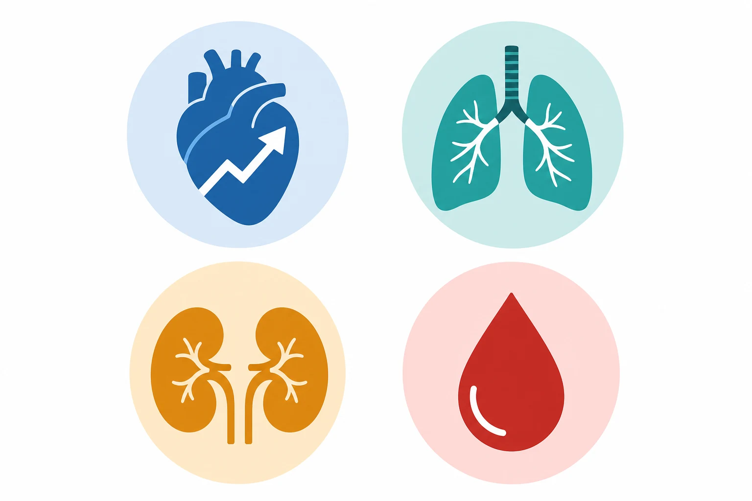
The cardiovascular
- The the increased the blood the volume (the 40 to the 50 per cent — the plasma the greater than the RBC → the dilutional the anaemia).[1][3]
- The the increased the cardiac the output (the 30 to the 40 per cent — the stroke the volume + the heart the rate). The mild the flow the murmur.[1]
- The the decreased the SVR (the progesterone the vasodilation → the BP the falls — the lowest the 2nd the trimester).[1][4]
- The the supine the hypotensive the syndrome (the aortocaval the compression — the gravid the uterus the compresses the IVC + the aorta from the 20 weeks → the reduced the venous the return, the CO, the BP → the maternal the hypotension + the uteroplacental the hypoperfusion). The the left the lateral the tilt (the 15 to the 30 the degrees) OR the manual the uterine the displacement.[1][3]
The respiratory
- The the increased the minute the ventilation (the progesterone → the hyperventilation → the respiratory the alkalosis — the PaCO2 the 28 to the 32; the metabolic the acidosis the compensation — the HCO3 the 18 to the 21). The "the normal the pregnancy the ABG."[2][4]
- The the decreased the FRC + the RV (the diaphragm the elevated → the reduced the reserve → the the faster the desaturation — the pre-oxygenation the critical).[2][5]
- The the decreased the chest the wall the compliance (the rib the cage the flared).[2]
- The the difficult the airway (the mucosal the oedema + the friability, the large the breasts, the weight the gain) + the the aspiration the risk (the delayed the gastric the emptying + the relaxed the LES) → the the RSI.[4][5]
The renal
- The the increased the GFR (the 50 to the 60 per cent — the renal the vasodilation) → the the lower the creatinine + the urea "the normal" (the creatinine the below the 60; the urea the below the 3.5). The not the interpret the "the normal" the range the as the normal in the pregnancy.[3][4]
- The glycosuria + the aminoaciduria (the tubular the load the exceeded). The ureter the dilation (the progesterone the smooth-muscle the relaxation).[3]
The haematological
- The the dilutional the anaemia (the plasma the greater than the RBC).[1]
- The leukocytosis (the mild).[1]
- The the hypercoagulable (the factors the increased, the protein the S the decreased, the fibrinolysis the decreased) → the the VTE the risk (the 4 to 5 times).[1][4]
- The gestational the thrombocytopenia (the mild — the below the 100 the benign; the not the pre-eclampsia / the HELLP).[4]
The GI + the endocrine + the acid-base
- The the delayed the gastric the emptying (the progesterone → the aspiration the risk — the RSI; the elevated the intragastric the pressure the 3rd the trimester).[4][5]
- The reflux (the LES the relaxed). The gallstones (the bile the stasis).[3]
- The endocrine: the increased the TBG (the total the T4 the high; the free the normal); the hCG (the thyroid the stimulator — the gestational the thyrotoxicosis); the insulin the resistance (the placental the hormones → the GDM).[1][4]
- The "the normal the pregnancy the ABG" — the respiratory the alkalosis (PaCO2 the 28 to the 32) + the metabolic the acidosis the compensation (HCO3 the 18 to the 21).[2][4]
The pharmacology
- The increased the volume → the decreased the concentration (the dose the adjustment).[4]
- The decreased the albumin → the increased the free the fraction.[4]
- The increased the renal the clearance (the GFR the up).[4]
- The placental the transfer (the foetal the exposure — the teratogenicity).[4][5]
Prognosis
The understanding the of the pregnancy the physiology the essential to the safe the ICU the management (the aortocaval the compression, the difficult the airway, the faster the desaturation, the hypercoagulable, the "the normal the pregnancy" the values). The failure → the maternal + the foetal the harm.[1][3][4]
Red flags
Cardiovascular adaptation — the quantitative detail
Pregnancy is the greatest cardiovascular stress test a healthy woman ever undergoes. From as early as 5 weeks' gestation, cardiac output begins to rise, driven first by a ~10–20% increase in stroke volume and a ~15–20 bpm rise in heart rate. Cardiac output peaks at +30–50% above pre-pregnancy by the late second / early third trimester (typically ~6–7 L/min, higher in multiple gestation), and climbs still further in labour (pain, and auto-transfusion of 300–500 mL with each uterine contraction). Systemic vascular resistance (SVR) falls ~20% under the combined effect of progesterone-mediated vasodilation, endothelial nitric oxide and the low-resistance placental bed — so that despite the high cardiac output, mean arterial pressure actually falls in the first and second trimester (systolic ↓5–10, diastolic ↓10–15 mmHg; nadir at ~24 weeks) before returning toward baseline in the third trimester. Blood volume expands 40–50% (plasma more than red cell mass → the dilutional / physiological anaemia).[1][3][9]
The clinical corollaries for the intensivist are unavoidable: a mild flow (ejection) murmur is normal; a third heart sound is normal; mild dependent oedema is normal; a jugular venous pressure that looks "flat" may still reflect a normal or even high-volume state. Conversely, a systolic murmur of grade 3/6 or louder, any diastolic murmur, a fourth heart sound, or a genuinely elevated JVP are NEVER physiological and demand echocardiography — pulmonary oedema in pregnancy is always pathological.[1][9]
[9]The aortocaval compression / supine hypotensive syndrome
Beyond ~20 weeks (and earlier in multiple pregnancy, obesity, or polyhydramnios), the gravid uterus mechanically compresses the inferior vena cava and, to a lesser extent, the aorta when the woman is supine. Venous return falls by 10–20%, cardiac output falls, and uteroplacental perfusion drops. In the awake patient a compensatory sympathetically-mediated tachycardia (and the maternal pelvic collateral circulation) often masks the effect — but under general anaesthesia, vasodilation, hypovolaemia, sepsis, or any loss of sympathetic tone, the drop becomes abrupt and severe. The same compression also partially occludes the aorta, so the uteroplacental bed is perfused at a pressure LOWER than the brachial cuff reads.[1][3]
The remedy is mechanical — no pharmacological manoeuvre will fix it: left lateral tilt of 15–30°, or manual left uterine displacement, for EVERY supine pregnant patient from 20 weeks — during resuscitation, intubation, transfer, surgery, cardiac arrest, and any imaging that requires the supine position. A wedge under the RIGHT hip achieves the same. Right tilt is avoided (it directs the gravid uterus ONTO the IVC and worsens compression).[3][9]
Respiratory adaptation — the "normal pregnancy ABG"
Progesterone is a direct respiratory stimulant (it increases central chemoreceptor sensitivity to CO₂). From the first trimester, tidal volume rises ~40% and minute ventilation rises ~50% while the respiratory rate is essentially unchanged — so the pregnant woman is breathing deeper, not faster. The result is a chronic respiratory alkalosis: PaCO₂ falls to 28–32 mmHg (3.7–4.3 kPa), and the kidneys compensate over days by excreting bicarbonate, so serum HCO₃⁻ falls to 18–21 mmol/L. This is the "normal pregnancy ABG".[2][4]
[2]The single most dangerous interpretation error: a PaCO₂ of 40 mmHg (5.3 kPa) in a pregnant woman is respiratory failure, not normal. It means she is no longer maintaining her progesterone-driven hyperventilation — she is tiring. Intubate, do not wait. Symmetrically, a serum bicarbonate of 24 mmol/L in pregnancy is already a metabolic acidosis, because her baseline is ~20.[2][5]
Lung volumes, FRC and the rapid desaturation
The diaphragm rises ~4 cm as the gravid uterus enlarges, and the rib cage flares (subcostal angle widens, chest circumference increases). Functional residual capacity (FRC) falls ~20% (chiefly via a fall in expiratory reserve volume) while oxygen consumption rises ~20–30% (the foetoplacental unit, increased cardiac and respiratory work, breast tissue). The combination — a smaller oxygen reservoir and a higher demand — means the pregnant patient desaturates within 60–90 seconds of apnoea (vs ~3–5 min in a healthy non-pregnant adult).[2][5]
[2]The difficult airway in pregnancy
Pregnancy is an independent risk factor for difficult intubation (~1 in 30 vs ~1 in 250 in the general population; failed intubation is roughly 8× more common). The contributors are: mucosal oedema and friability of the upper airway (progesterone + increased blood volume), engorged breast tissue obstructing the laryngoscope handle, weight gain, and a Mallampati class that worsens further in labour.[6][9]
Practical rules: have a smaller ETT ready (6.0 or 6.5 mm cuffed; oedema narrows the glottis and may make a 7.0 impossible to pass); use a short-handled laryngoscope (or videolaryngoscope) when breasts are large; always have a plan B (supraglottic airway, videolaryngoscope, an experienced second operator, the difficult-airway trolley) ready BEFORE induction; and pre-oxygenate head-up / tilted to maximise denitrogenation time. The aspiration risk (delayed gastric emptying + relaxed lower oesophageal sphincter + raised intragastric pressure) mandates rapid sequence intubation for every pregnant intubation.[4][5][6]
Renal adaptation — "normal" is a moving target
Renal plasma flow and glomerular filtration rate (GFR) rise ~50–60% by the second trimester under the influence of relaxin, progesterone, and the volume-expanded state. The practical consequence: a creatinine of 90 µmol/L is already acute kidney injury in pregnancy. A "normal" non-pregnant creatinine (60–100) is abnormal in pregnancy — the physiological range in a healthy pregnant woman is ~50–70 µmol/L, and urea is correspondingly low (<3.5 mmol/L).[3][10]
[10]Proteinuria up to 300 mg/day is physiological; ≥300 mg/day (or a protein:creatinine ratio ≥30 mg/mmol) is the threshold that activates the pre-eclampsia work-up. Physiological glycosuria is common and does NOT diagnose gestational diabetes — that diagnosis rests on plasma glucose / OGTT, not on urinary glucose. The right ureter is more dilated than the left (dextrorotation of the gravid uterus compresses the right), so right-sided hydronephrosis is common and must not be over-called as obstruction.[3][10]
Haematological adaptation — the hypercoagulable state
Pregnancy is a deliberately hypercoagulable state, an evolutionary protection against postpartum haemorrhage. The venous thromboembolism risk is 4–5× that of non-pregnant age-matched women, and up to ~20× in the postpartum period. The mechanism is multifactorial:[1][7]
- Plasma volume expands 40–50% but red cell mass only 20–30% → the physiological (dilutional) anaemia, Hb typically 105–110 g/L by term (the nadir is ~28–32 weeks). This is normal; treat only if symptomatic or Hb <100 with iron deficiency.
- Coagulation factors rise: fibrinogen (factor I) climbs to 4–6 g/L (vs 2–4 non-pregnant); factors VII, VIII, IX, X and von Willebrand factor all rise. Factor XI falls; factors II and V are essentially unchanged.
- Natural anticoagulants fall: protein S falls markedly (free and total), and an acquired resistance to activated protein C develops.
- Fibrinolysis is impaired: plasminogen activator inhibitor-1 (PAI-1) and PAI-2 (placental) rise, suppressing fibrinolysis. Thrombin-activatable fibrinolysis inhibitor (TAFI) is increased. Fibrinolysis normalises rapidly after placental delivery.
- Platelets fall slightly (gestational thrombocytopenia — by ~10%) from haemodilution and increased clearance; a count >100 × 10⁹/L in an otherwise well woman is benign and NOT pre-eclampsia / HELLP.
- White cell count rises: 8–15 × 10⁹/L is normal in the third trimester, and may reach 25–30 × 10⁹/L in labour (a left shift and even mild toxic granulation can be physiological).[1]
The DIC trap: because fibrinogen is already high, a single fibrinogen of "3.5 g/L" in a sick pregnant woman (e.g. pre-eclampsia / HELLP, placental abruption, sepsis, amniotic fluid embolism, massive PPH) is not reassuring — it may already represent consumption from a baseline of 5–6 g/L. Suspect DIC when the PT/aPTT rise, the platelets trend down, and there is clinical bleeding or microvascular thrombosis; trend the values, do not anchor on a single number. In major obstetric haemorrhage, keep the fibrinogen >2.0 g/L.[1][7]
GI, endocrine and acid-base — the supporting detail
- Gastrointestinal: progesterone relaxes smooth muscle → delayed gastric emptying and a reduced lower oesophageal sphincter tone; combined with the mechanical rise in intragastric pressure from the gravid uterus (worst in the third trimester), this is the physiological basis for the "full stomach" assumption and rapid-sequence intubation in every pregnant patient. The gallbladder also dilates and empties sluggishly → cholesterol gallstones are more common. Reflux (GORD) is near-universal by term.[3][5]
- Endocrine: oestrogen drives hepatic thyroid-binding globulin (TBG) synthesis, so total T4 and total T3 rise but free hormone stays normal — interpret TFTs on free T4 / TSH, never on totals. hCG shares its α-subunit with TSH and is a weak thyroid stimulator, producing the transient gestational thyrotoxicosis of early pregnancy (and hyperemesis gravidarum). Placental hormones (human placental lactogen, cortisol, progesterone) induce a state of insulin resistance (especially post-prandially) — the basis of gestational diabetes mellitus and the reason diabetic ketoacidosis can occur at lower glucose thresholds in pregnancy. Prolactin rises markedly to prepare lactation.[1][4]
- Acid-base: the standing summary — pregnancy is a compensated respiratory alkalosis (PaCO₂ ~30 mmHg / 4.0 kPa; HCO₃⁻ ~20 mmol/L; pH 7.42–7.48). Any deviation (a "normal" PaCO₂ of 40, a HCO₃⁻ of 24, a falling pH) signals pathology — fatigue, sepsis, ketoacidosis, or renal failure.[2][4]
Position management — left lateral tilt, everywhere
[9]Rapid sequence intubation in pregnancy — the planned approach
[6]Perimortem caesarean — resuscitative hysterotomy
In maternal cardiac arrest from 20 weeks, emptying the uterus is maternal resuscitation, not merely neonatal salvage: it relieves aortocaval compression and can restore venous return and cardiac output by up to 25%.[3][8]
[8]Pharmacology in pregnancy — the dose, the binding, the placenta
Pregnancy changes drug behaviour in three ways: the expanded volume of distribution dilutes water-soluble drugs; the lower serum albumin raises the free fraction of highly-bound drugs; and the higher renal clearance accelerates elimination of renally-cleared drugs. Dosing is therefore often higher and more frequent than in a non-pregnant adult of the same weight. The placenta is crossed by most drugs (it is a lipid membrane) — the foetus is always an unintended co-recipient, so teratogenicity (1st trimester) and foetal toxicity (3rd trimester) must be weighed; but in a critically ill pregnant woman, what is good for the mother is almost always good for the fetus, and undertreating maternal illness is the greater foetal hazard.[4][5]
[5]Trimester-by-trimester timeline
[5]SAQ — The normal pregnancy blood gas and laboratory profile: physiological or pathological?
10 minutes · 10 marks
A 28-year-old primigravida at 34 weeks gestation is admitted to ICU after a brief generalised tonic-clonic seizure at home. She is now post-ictal and drowsy (GCS 13), HR 110 (sinus), BP 96/58, RR 24, SpO2 96 percent on room air. Arterial blood gas: pH 7.44, PaCO2 32 mmHg (4.3 kPa), PaO2 98 mmHg, HCO3 20 mmol/L, base excess -4. Bloods: Hb 105 g/L, WCC 13.5 x10^9/L, platelets 220 x10^9/L, fibrinogen 4.8 g/L, creatinine 62 umol/L, urea 2.8 mmol/L, albumin 30 g/L. The obstetric registrar asks you to confirm whether everything is normal for pregnancy.
SAQ — The pregnant ICU patient with respiratory failure: airway, ventilation and thromboprophylaxis
10 minutes · 10 marks
A 32-year-old woman at 36 weeks gestation is admitted to ICU with community-acquired pneumonia and type 1 respiratory failure. She is severely hypoxic (PaO2/FiO2 90 on FiO2 0.8 via high-flow nasal cannulae, RR 34), drowsy (GCS 13), BP 102/64, HR 122 (sinus), lactate 3.2, temperature 38.6 degrees C. The team is preparing to intubate. She has a Mallampati 3 airway, marked oropharyngeal oedema, a short neck and a BMI of 33.
Clinical pearls
Key trials and evidence
OAA / DAS guideline for difficult and failed intubation in obstetrics (Mushambi et al, Anaesthesia 2015, PMID 25495759)
MBRRACE-UK: Saving Lives, Improving Mothers' Care (Knight et al; NPEU, Oxford; annual confidential enquiries)
VTE in pregnancy and the postpartum period — incidence, risk factors and mortality (James et al, Am J Obstet Gynecol 2006, PMID 16579933)
Renal hemodynamic and metabolic physiology of pregnancy (Odutayo & Hladunewich, CJASN 2012, PMID 23222983)
Additional red flags
Outcomes and prognosis
[6]The recurring lesson — codified by MBRRACE-UK and SOMANZ — is that maternal deaths in critical illness are deaths from failure to recalibrate to the pregnancy baseline: a tachycardia and a low-grade fever dismissed as "normal for pregnancy"; a creatinine of 90 accepted because it sits in the printed reference interval; a supine patient not tilted; a fibrinogen of 4 g/L called normal in a bleeding woman; a PaCO₂ of 40 mmHg watched instead of acted upon. Knowing the physiology — and acting on its consequences — is the intervention.[1][3][4][8]
References
- [1]Sanghavi M, Rutherford JD. Physiological changes in pregnancy. Cardiovascular Journal of Africa, 2016.PMID 27213856
- [2]Bettinelli S, et al. Respiratory physiological changes in pregnancy. Respiratory Medicine, 2025.PMID 40614834
- [3]Chandra S, et al. Alterations in physiology and anatomy during pregnancy. Best Practice & Research Clinical Obstetrics & Gynaecology, 2013.PMID 24012425
- [4]Karnad DR, Nor MBM, Richards GA, et al. Intensive care in severe malaria: Report from the task force on tropical diseases by the World Federation of Societies of Intensive and Critical Care Medicine. Journal of critical care, 2018.PMID 29132978
- [5]Kazma JM, van den Anker J, Allegaert K, et al. Anatomical and physiological alterations of pregnancy. Journal of pharmacokinetics and pharmacodynamics, 2020.PMID 32026239
- [6]Mushambi MC, Kinsella SM, Popat M, et al. Obstetric Anaesthetists' Association and Difficult Airway Society guidelines for the management of difficult and failed tracheal intubation in obstetrics. Anaesthesia, 2015.PMID 26449292
- [7]James AH, Jamison MG, Brancazio LR, et al. Venous thromboembolism during pregnancy and the postpartum period: incidence, risk factors, and mortality. American journal of obstetrics and gynecology, 2006.PMID 16647915
- [8]Tuffnell D, Rathod M Improving maternal safety in future: learning from the confidential enquiries. British journal of hospital medicine (London, England : 2005), 2006.PMID 16562432
- [9]Duvekot JJ, Peeters LL Maternal cardiovascular hemodynamic adaptation to pregnancy. Obstetrical & gynecological survey, 1994.PMID 7877788
- [10]Odutayo A, Hladunewich M Obstetric nephrology: renal hemodynamic and metabolic physiology in normal pregnancy. Clinical journal of the American Society of Nephrology : CJASN, 2012.PMID 22879432