ICU · Obstetric critical care
Venous Thromboembolism in Pregnancy
Also known as VTE in pregnancy · DVT in pregnancy · PE in pregnancy · Pregnancy hypercoagulable · LMWH in pregnancy · Massive PE pregnancy · Perimortem Caesarean
The venous the thromboembolism (the VTE) in the pregnancy — the leading the cause of the maternal the mortality. The hypercoagulable (the 4 to the 5 times the risk). The DVT (the left-the-sided the predominance — the left the iliac the compressed). The PE (the killer). The diagnosis (the D-dimer the unreliable; the V/Q the scan the preferred — the lower the radiation). The LMWH the preferred (the safe — the no the placental the transfer; the AVOID the warfarin / the DOAC — the teratogenic). The thrombolysis for the massive; the perimortem the Caesarean.
On this page & tools
Your progress
Saved locally on this device.
Target exams
Overview & definition
The VTE in the pregnancy — the leading the cause of the maternal the mortality. The pregnancy the hypercoagulable (the 4 to the 5 times the risk), the venous the stasis (the gravid the uterus), the reduced the mobility. The DVT (the left the predominance), the PE (the killer). The LMWH the preferred (the safe; the AVOID the warfarin / the DOAC). The thrombolysis for the massive; the perimortem the Caesarean.[1][1]

The pathophysiology
The pregnancy the hypercoagulable the state:[1][2][1]
- The the clotting the factors the increased (the I, the VII, the VIII, the IX, the X, the XII, the von the Willebrand), the protein the S the decreased, the fibrinolysis the decreased (the PAI-1, the PAI-2 the up).
- The the venous the stasis (the progesterone → the smooth-the-muscle the relaxation; the gravid the uterus → the IVC the compression; the left the iliac the compressed the by the right the iliac the artery).
- The the reduced the mobility.
The → the 4 to the 5 times the VTE the risk (the DVT the 2 in the 1000; the PE the 1 in the 1000). The DVT the left the 90 per cent (the left the iliac the compressed).[1][1]
The diagnosis
- The DVT — the compression the ultrasound (the proximal; the whole-the-leg if the negative + the high the suspicion; the MRV / the CTV if the iliac).[2][1]
- The PE — the D-dimer the unreliable in the pregnancy (the normally the elevated → the poor the specificity).[2]
- The ECG, the troponin, the BNP (the RV the strain).[1][1]
The treatment
1. The LMWH — the preferred.[1][2][1]
- The the weight-based (the enoxaparin the 1 mg/kg the BD; the tinzaparin the 175 U/kg the OD). The does NOT the cross the placenta (the safe; the no the fetal the bleeding). The preferred the over the UFH (the more the predictable, the lower the HIT, the less the monitoring).[2]
- The throughout the pregnancy; the anti-the-Xa the monitoring if the extreme the weight / the renal.[1][1]
2. The warfarin / the DOAC the CONTRAINDICATED.[2][1]
- The the warfarin (the teratogenic — the 1st the trimester the skeletal / the nasal; the 2nd / the 3rd the fetal the bleeding, the CNS).[1]
- The the DOAC (the limited the data; the NOT the recommended; the avoid).[2]
- The SWITCH the postpartum (the warfarin — the breastfeeding the safe; the DOAC — the cautious; the LMWH the transition). The 6 the weeks the minimum (the 3 to the 6 the months the total).[1][2]
3. The IV the UFH for the massive the PE / the peripartum (the short the half-the-life, the reversible, the protamine).[1]
4. The thrombolysis for the massive the PE (the life-the-threatening — the hypotension / the collapse / the cardiac the arrest).[3][1]
- The alteplase (the tPA — the crosses the placenta the minimally; the bleeding the risk; the maternal the life-the-saving).[3]
- The NOT the for the submassive (the conservative the — the LMWH).[3][1]
5. The perimortem the Caesarean (the 20+ the weeks, the cardiac the arrest → the deliver the within the 5 the minutes — the improves the maternal the CPR + the foetal).[1]
6. The prophylaxis (the risk-the-stratified: the previous the VTE, the thrombophilia, the Caesarean, the obesity, the immobility → the LMWH).[2][1]
Prognosis
The VTE the pregnancy the mortality the 1 per cent (the treated). The recurrence the (the subsequent the pregnancy / the postpartum the prophylaxis). The long-the-term the PTS (the post-the-thrombotic the syndrome).[1][2][1]
Red flags
Pregnancy as a prothrombotic state — Virchow's triad dissected
Pregnancy is a physiological hypercoagulable state engineered to protect the mother from exsanguination at delivery (placental separation leaves a 500–700 mL/min vascular bed). The price is a 4–5-fold increase in VTE risk throughout gestation, rising further in the puerperium (up to 20-fold in the first 6 weeks postpartum). VTE remains the leading direct cause of maternal mortality in the developed world.[1][2][1]
All three limbs of Virchow's triad are activated — and the intensivist must know each in detail because every element is examinable.[1][1]
1. Venous stasis (haemodynamic change)
- Gravid uterus compresses the inferior vena cava (IVC) and left common iliac vein, particularly in the supine position from the late second trimester onward. This is why the left-lateral tilt / left lateral decubitus position is mandatory in late pregnancy (and during CPR) — to relieve aortocaval compression.
- Progesterone-mediated smooth-muscle relaxation dilates venous capacitance vessels; lower-limb venous capacitance rises ~50%, and flow velocity in the leg veins falls by up to 50% by the third trimester.
- Reduced mobility — late-pregnancy discomfort, bed rest for complications (preterm labour, pre-eclampsia), and post-delivery pain all compound stasis.
- Left-sided predominance (~90% of pregnancy DVTs): the **left common iliac vein is compressed by the right common iliac artery crossing anterior to it (May–Thurner anatomy), exacerbated by the gravid uterus. An iliofemoral (left-leg) DVT in pregnancy may be missed on proximal compression ultrasound alone — request whole-leg ultrasound or MRV if clinical suspicion is high.[1][1]
2. Hypercoagulability (haematological change) — "preparation for haemostasis at delivery"
The coagulation system tilts firmly toward clot formation from early in the first trimester and peaks near term.[2][1]
The coagulation/fibrinolytic changes of pregnancy — every procoagulant lever is pulled
| Direction | Factor / pathway | Magnitude / timing | Exam significance |
|---|---|---|---|
| ↑↑ Coagulation factors | Fibrinogen (factor I), VII, VIII, IX, X, XII, von Willebrand factor | Fibrinogen doubles (to 4–6 g/L by term); factors VII/VIII/X rise 1.5–3× | High fibrinogen in pregnancy is NORMAL — do not interpret as DIC in isolation. A "normal" fibrinogen (<2 g/L) in a pregnant woman is alarming |
| ↑ Resistance to activated protein C (APC) | Acquired APC resistance (independent of factor V Leiden) | Develops progressively; maximal at term | Mimics/increases thrombophilia; contributes to clot propagation |
| ↓ Protein S (free + total) | Reduced free protein S | Up to 50–60% reduction by third trimester | Makes inherited protein S deficiency IMPOSSIBLE to diagnose reliably in pregnancy — defer testing until ≥6 weeks postpartum |
| ↓↓ Fibrinolysis | PAI-1 (placental) and PAI-2 (placenta, massive) markedly elevated; TAFI increased | PAI-2 undetectable in non-pregnant, rises ~100-fold | The placenta is the source — fibrinolysis is physiologically suppressed so clots are NOT cleared rapidly |
| ↓ Protein C | Usually unchanged or mildly reduced | Less affected than protein S | Protein C levels are relatively preserved |
| ↓ Antithrombin | Usually unchanged | Falls only if consumed (pre-eclampsia, HELLP, DIC) | Heparin requires antithrombin to work — AT deficiency causes heparin "resistance" |
| ↑ Platelet activation + marginal consumption | Increased turnover; thrombocytopenia common (gestational ~8%, pre-eclampsia, HELLP, ITP) | Platelet COUNT may fall; function enhanced | A falling platelet count in pregnancy is NOT normal physiological drift — think pre-eclampsia/HELLP/DIC/TTP/HUS |
| Net effect | Procoagulant dominance + impaired fibrinolysis | Whole pregnancy, worst at term + puerperium | 4–5× VTE risk; up to 20× postpartum |
3. Endothelial (vascular) damage — the peripartum element
- Vaginal delivery and especially Caesarean section (elective, and far more emergency) directly injure pelvic venous endothelium and the pelvic vasculature.
- Instrumental delivery (forceps, vacuum) and perineal/vaginal tears add trauma.
- Operative blood loss, venous cannulation, and pelvic haematoma all expose tissue factor and activate the coagulation cascade.
- This is why the postpartum period is the HIGHEST-risk window — stasis + peak hypercoagulability + fresh endothelial injury coincide. Caesarean section roughly doubles VTE risk vs vaginal delivery.[2][1][1]
Deep vein thrombosis (DVT) in pregnancy — diagnosis refined
Clinical features and the limits of clinical prediction rules
Leg pain, swelling (left > right), tenderness, increased circumference, and warmth are the hallmarks, BUT clinical features alone are non-diagnostic and the Wells score is NOT validated in pregnancy (every pregnant patient is "pre-test moderate-high" by definition). All suspected DVTs must be objectively imaged — do not treat empirically.[2][1]
Imaging strategy — a stepwise, radiation-conscious approach
Imaging for DVT and PE in pregnancy — dose, role, pitfalls
| Modality | Indication | Radiation (fetal) | Radiation (maternal breast) | Key pitfalls |
|---|---|---|---|---|
| Compression ultrasound (proximal) | First-line for suspected DVT | Nil | Nil | Misses isolated iliac/calf DVT; if negative but high suspicion → repeat in 5–7 days OR go to whole-leg |
| Whole-leg ultrasound | High suspicion, proximal US negative, iliac/calf symptoms suspected | Nil | Nil | Operator-dependent; needed for left-sided iliofemoral disease |
| MRV (magnetic resonance venography) | Suspected isolated iliac DVT (left leg), or to avoid radiation | Nil | Nil | Availability; claustrophobia; gadolinium (gadolinium-based contrast generally avoided unless essential) |
| CTV (CT venography) | Combined with CTPA as a single study if PE + DVT both needed | ~Low-moderate | Moderate | Higher dose than isolated CTPA; usually unnecessary |
| D-dimer | NOT diagnostic in pregnancy | Nil | Nil | Physiologically elevated in ~all pregnancies → poor specificity; only useful if NEGATIVE in a LOW-risk patient (rare). Most guidelines advise AGAINST relying on it |
| V/Q scan (ventilation-perfusion) | PREFERRED for PE when CXR normal | ~0.1–0.5 mGy (lower than CTPA) | Negligible (no breast dose) | "Intermediate" result if CXR abnormal; not available out-of-hours everywhere |
| CTPA | Alternative PE imaging when CXR abnormal, haemodynamically unstable, V/Q unavailable | ~0.1–0.3 mGy (low fetal dose) | HIGHER maternal breast dose (~10–70 mGy) — increases lifetime breast cancer risk slightly | Needs contrast (renal); may show alternative diagnosis (pneumonia) |
Exam rule of thumb: In pregnancy, for suspected PE, V/Q scan is generally preferred (lower maternal breast radiation, CXR usually normal), with CTPA reserved for an abnormal CXR, haemodynamic instability, or when an alternative diagnosis (e.g. pneumonia, dissection) is also being sought. The fetal radiation dose from EITHER test is well below the threshold for harm — never withhold diagnostic imaging for fear of fetal radiation.[2][4][6]
Pulmonary embolism in pregnancy — diagnosis and risk stratification
Pregnancy dyspnoea, tachycardia, and mild hypoxaemia overlap with NORMAL pregnancy physiology (baseline RR up, PaCO₂ ~28–32 mmHg from progesterone-driven hyperventilation). The discriminator is the degree of respiratory compromise and the change from the woman's baseline.[1][4]
PE risk stratification (ESC 2019, applied to pregnancy)
The ESC framework applies, with the caveat that haemodynamic collapse can be masked early because young, fit pregnant women compensate until late.[4][5]
PE risk strata in pregnancy — what to do at each level
| Stratum | Definition | First-line action |
|---|---|---|
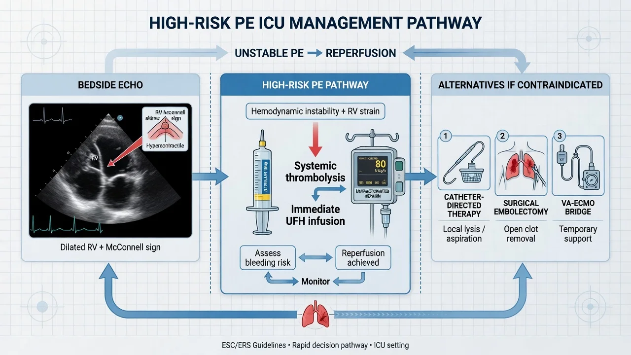
| High-risk (massive) | Haemodynamic instability: SBP <90 for >15 min, vasopressor requirement, or cardiac arrest | Immediate reperfusion — systemic thrombolysis (alteplase) as first choice; surgical/catheter embolectomy if thrombolysis contraindicated; VA-ECMO / ECPR as bridge if refractory. Do NOT delay for imaging if crashing — bedside echo for RV strain supports diagnosis |
| Intermediate-risk (submassive) | Normotensive BUT RV strain (echo RV/LV >0.9, biomarkers) | Anticoagulation (LMWH) + close monitoring (ICU/HDU). Systemic thrombolysis is NOT routine (PEITHO: bleeding/stroke excess). Rescue thrombolysis if deteriorates. Catheter-directed options if high-risk features |
| Low-risk | Normotensive, no RV strain, normal biomarkers | Anticoagulation (LMWH). Consider ward-level care |
Bedside adjuncts
- ECG: sinus tachycardia (near-universal); S1Q3T3 (uncommon); anterior T-wave inversion (V1–V4, RV strain); new RBBB. Bradycardia is a pre-arrest sign.
- Troponin / NT-proBNP: RV strain/injury markers — identify intermediate-risk PE.
- Bedside echocardiography: RV dilation, McConnell sign, septal flattening, TR jet, IVC plethora, clot-in-transit. In an unstable patient, echo evidence of RV strain is sufficient to justify thrombolysis without waiting for CTPA.[4]
DVT management — anticoagulation is the whole treatment
The cornerstone: therapeutic LMWH throughout pregnancy, switched postpartum. There is no role for warfarin or DOACs antenatally.[2][1][1]
Anticoagulants in pregnancy — the definitive comparison
| Agent | Antenatal use | Postpartum / lactation | Mechanism / monitoring | Why / why not |
|---|---|---|---|---|
| LMWH (enoxaparin, tinzaparin, dalteparin) | FIRST-LINE, entire pregnancy | FIRST-LINE | Anti-Xa; large molecule → does NOT cross placenta | Preferred — does not cross placenta (no fetal bleeding/teratogenicity), predictable pharmacokinetics, lower HIT than UFH, lower osteoporosis than UFH, outpatient-capable |
| Unfractionated heparin (UFH) IV | Reserved for massive PE, peripartum, or renal failure | Useful postpartum if rapid reversibility needed | APTT (or anti-Xa); short half-life; protamine-reversible | Short half-life + reversibility ideal around delivery and when thrombolysis may be needed. Higher HIT and osteoporosis risk than LMWH |
| Warfarin | CONTRAINDICATED (teratogenic 1st trimester; fetal bleeding/CNS 2nd–3rd) | Permitted (safe in breastfeeding) | INR 2–3; crosses placenta | Teratogenic: 1st trimester (warfarin embryopathy — nasal hypoplasia, stippled epiphyses, limb hypoplasia); 2nd–3rd trimester (fetal intracranial haemorrhage, CNS abnormalities). Crosses placenta; safe postpartum as it is NOT in breast milk in significant amounts |
| DOACs (apixaban, rivaroxaban, dabigatran, edoxaban) | CONTRAINDICATED / NOT recommended (cross placenta, limited data) | Avoid in breastfeeding (excreted in milk) | Direct anti-Xa or direct thrombin; no monitoring | Limited/contradictory reproductive data; theoretical fetal harm. Not recommended antenatally; generally avoid while breastfeeding |
| Fondaparinux | Alternative if heparin allergy/HIT | Alternative | Indirect Xa; not reversed | Used in HIT or heparin allergy; minimal placental transfer |
| Danaparoid | Alternative in HIT | Alternative | Anti-Xa | HIT-safe; availability limited (ANZ) |
| Aspirin (low-dose) | Adjuvant in antiphospholipid syndrome (with heparin) | Safe | Antiplatelet | NOT monotherapy for VTE treatment; used in APS pregnancy prophylaxis regimens |
LMWH dosing — weight-based, and the dose INCREASES in the third trimester
This is a high-yield exam point. LMWH is dosed by body weight and requirements rise as pregnancy advances because of:[2][1][1]
- Increased renal clearance of LMWH (increased GFR in pregnancy).
- Increased volume of distribution (plasma volume +50%, weight gain).
- Increased anti-Xa clearance and altered binding.
Standard therapeutic doses:
- Enoxaparin 1 mg/kg subcutaneous twice daily (BD preferred over OD in pregnancy for therapeutic dosing).
- Tinzaparin 175 IU/kg once daily.
- Dalteparin 200 IU/kg once daily (or 100 IU/kg BD). [1]
Monitoring: Routine anti-Xa monitoring is NOT required for most women. Check peak anti-Xa (4 h post-dose) in: extremes of weight (BMI <30 or >100 kg, or weight <50 / >90 kg depending on guideline), renal impairment, recurrent VTE despite therapy, and some high-risk thrombophilias. Target peak anti-Xa for BD enoxaparin ~0.5–1.0 IU/mL.[2]
Therapeutic LMWH through pregnancy — the dosing arc
- Confirm diagnosis objectively (imaging) before committing to months of therapy.
- Initiate weight-based LMWH — enoxaparin 1 mg/kg BD (or tinzaparin 175 U/kg OD). Document the woman's current weight (NOT booking weight).
- Re-weigh and re-dose each trimester — increase dose as weight and clearance rise. A woman started at 1 mg/kg in the first trimester frequently needs a higher absolute dose by the third trimester.
- Anti-Xa monitoring only if extremes of weight, renal impairment, recurrent VTE, or per local protocol (target peak 0.5–1.0 IU/mL for BD dosing).
- Continue throughout pregnancy — do NOT switch to warfarin/DOAC.
- Plan peripartum management (below) — switch to IV UFH or withhold LMWH around delivery to enable neuraxial anaesthesia.
- Postpartum — resume LMWH within 12–24 h (or switch to warfarin/DOAC); continue for minimum 6 weeks (provoked VTE: 3–6 months total from diagnosis).
Peripartum anticoagulation and neuraxial anaesthesia
The competing priorities: (a) maintain therapeutic anticoagulation (risk of PE recurrence off therapy), (b) enable neuraxial (spinal/epidural) anaesthesia safely (risk of spinal epidural haematoma). The standard strategy is a planned delivery:[2][1][1]
Elective switch to IV UFH at ~37 weeks / planned delivery
- At 37 weeks (or 36 h before planned delivery) — switch therapeutic LMWH to IV UFH infusion.
- Stop UFH ~4–6 h before planned delivery / induction / Caesarean (UFH half-life ~60–90 min → rapid offset).
- Neuraxial anaesthesia safe once APTT has normalised (no LMWH for ≥24 h, UFH stopped ≥4–6 h, APTT normal). For LMWH specifically, withhold prophylactic LMWH 12 h and therapeutic LMWH 24 h before neuraxural needle placement.
- Post-delivery — restart anticoagulation: LMWH 4–6 h after vaginal delivery / 6–12 h after Caesarean (once haemostasis secure); neuraxial catheter removal requires ≥12 h after last prophylactic LMWH (or 24 h after therapeutic) and ≥2 h before next dose.
- Never remove an epidural catheter within 12 h of an LMWH dose — spinal haematoma risk.
Massive PE in pregnancy — the resuscitation
A pregnant woman with a massive PE is the quintessential time-critical obstetric emergency: two lives, aorto-caval compression masking the circulation, and the reflex to "protect the fetus" must NOT delay maternal life-saving therapy.[3][4][7]
Massive PE in pregnancy — the first 60 minutes
- CALL FOR HELP — obstetric + anaesthetic + intensivist + haematology + neonatal MDT. Declare a maternal emergency. Move to left-lateral tilt to relieve aortocaval compression.
- ABCDE + high-flow oxygen; secure two large-bore IV cannulae; cross-match; group-and-save; send coagulation, fibrinogen, platelets, D-dimer, troponin, blood gas.
- Fluid resuscitation — CAUTIOUS (250 mL aliquots) — the failing RV tolerates volume poorly; over-resuscitation worsens RV failure.
- Vasopressors early — noradrenaline (or adrenaline) for SBP <90; vasopressin/additional agents as needed. Treat the maternal circulation first.
- Confirm diagnosis — if stable enough, bedside echo (RV strain supports PE) + CTPA; if crashing/arrest, do NOT delay for imaging — echo evidence of RV strain + compatible history justifies empirical reperfusion.
- Immediate reperfusion — systemic thrombolysis (alteplase) as first-line if no absolute contraindication. If thrombolysis contraindicated/failed → surgical embolectomy or percutaneous catheter-directed thrombectomy/thrombolysis.
- VA-ECMO / ECPR for refractory shock or arrest — as a BRIDGE to definitive reperfusion.
- Perimortem Caesarean if cardiac arrest and uterus ≥20 weeks gestation — deliver within 5 minutes of arrest (improves maternal CPR by relieving aortocaval compression AND fetal outcome).
- Therapeutic anticoagulation — IV UFH (short, reversible) once reperfusion underway; transition to LMWH postpartum.
- Senior MDT — intensivist + obstetric + anaesthetic + cardiac surgery + haematology + neonatal — for ongoing delivery-timing and reperfusion decisions.
Thrombolysis — alteplase does NOT cross the placenta significantly
This is a key reassurance that justifies maternal life-saving thrombolysis. Alteplase (recombinant tPA) is a large molecule (~65 kDa) that does NOT cross the placenta in significant amounts — fetal plasminogen activation and fetal bleeding are therefore uncommon. The predominant risks are maternal bleeding (notably peripartum/genital tract) and, rarely, placental abruption.[3][7][1]
Dose (as for non-pregnant): Alteplase 100 mg IV over 2 h for massive PE (or 50 mg bolus in cardiac arrest). Systematic reviews and case series of thrombolysis in pregnancy report maternal survival benefit with acceptable maternal bleeding and no clear fetal catastrophe attributable to tPA.[3][7]
The decision principle: In a maternal life-threatening massive PE, the mother's survival is the priority and thrombolysis should NOT be withheld for fear of fetal bleeding — the fetus dies if the mother dies. The "contraindication" of pregnancy is RELATIVE, not absolute, in this setting.[3][4][7]
Mechanical reperfusion alternatives when thrombolysis is contraindicated or fails
Reperfusion strategies for massive PE in pregnancy — beyond systemic thrombolysis
| Strategy | When | Advantages in pregnancy | Caveats |
|---|---|---|---|
| Systemic thrombolysis (alteplase) | First-line for massive PE if no absolute contraindication | Rapid; bedside; does not cross placenta significantly | Maternal bleeding; ~8% failure rate; peripartum risk |
| Surgical pulmonary embolectomy | Absolute thrombolysis contraindication; failed lysis; clot-in-transit + PFO; central clot | Avoids systemic lysis; immediate debulking | Requires on-site cardiac surgery + CPB; mobilisation time |
| Catheter-directed thrombolysis (CDT/EKOS) | Massive PE with high bleeding risk (relative thrombolysis contraindication) | Low-dose local lysis → less systemic bleeding | Needs IR; slower than systemic bolus |
| Percutaneous catheter thrombectomy (FlowTriever etc.) | Absolute thrombolysis contraindication; large central clot | Mechanical removal, NO lysis → minimal bleeding | Large-bore access; operator expertise |
| VA-ECMO / ECPR | Refractory cardiac arrest / shock; bridge to reperfusion | Immediate circulatory + oxygenation support; bridge | NOT definitive — must pair with a reperfusion strategy; bleeding, limb ischaemia, anticoagulation |
Perimortem Caesarean (PMCD) — relieve aortocaval compression to save BOTH
Perimortem Caesarean delivery — the 5-minute rule
- Trigger — maternal cardiac arrest at ≥20 weeks gestation (fundus at/above umbilicus). Start continuous high-quality CPR.
- Position — manual left-lateral uterine displacement (or left-lateral tilt) to relieve aortocaval compression during CPR.
- Decision at 4 minutes, delivery by 5 minutes — the 4-min/5-min rule: a perimortem Caesarean should be underway by 4 min, completed by 5 min from arrest.
- Why? Delivery of the gravid uterus (1) relieves aortocaval compression → restores venous return and cardiac output for maternal CPR; (2) improves maternal survival; (3) maximises fetal survival (best if delivered within 5 min of arrest).
- No consent needed — implied; the procedure is maternal life-saving. Perform at the bedside with whatever equipment is available (a scalpel; classic midline vertical incision).
- Continue CPR throughout — resuscitate the mother, then the neonate. ROSC may follow delivery as venous return is restored.
Postpartum anticoagulation — duration and switching
The postpartum period is the highest-risk window, so anticoagulation MUST continue after delivery for an adequate duration.[2][1][1]
Postpartum anticoagulation duration — match the indication
| Indication | Antepartum | Postpartum duration | Total therapy |
|---|---|---|---|
| Acute provoked VTE (antepartum DVT/PE) | Therapeutic LMWH throughout | Minimum 6 weeks (most extend to 3 months postpartum) | ≥3–6 months total from diagnosis |
| Unprovoked VTE / recurrent | Therapeutic LMWH | 6 weeks–3 months+; lifelong if persistent risk | Per non-pregnant duration |
| VTE prophylaxis (high-risk, no acute event) | Per risk (below) | 6 weeks postpartum | — |
| Antiphospholipid syndrome (on triple therapy) | Aspirin + LMWH (± hydroxychloroquine) | Continue; bridge to warfarin (safe in lactation) | Per haematology |
Postpartum drug choice
- LMWH remains first-line — convenient, no monitoring, no placental transfer issue postpartum.
- Warfarin may be used postpartum — it is safe in breastfeeding (minimal transfer). Bridge with LMWH until INR ≥2.0 for 2 consecutive days.
- DOACs — avoid in breastfeeding (excreted in milk); may be considered if not breastfeeding, after the immediate peripartum period.[2][1]
Thromboprophylaxis in high-risk pregnancy — risk stratification
Not every pregnant woman needs prophylaxis; risk stratification (RCOG Green-top 37a/37b, ASH 2018) drives the decision. The intensivist is typically involved in the highest-risk groups and postpartum.[2][1]
Risk factors for VTE in pregnancy — the search list
| Category | Specific factors |
|---|---|
| Previous VTE | Strongest single risk factor — especially unprovoked, oestrogen-related, or pregnancy-related. Mandates prophylaxis |
| Thrombophilia | Inherited (antithrombin, protein C/S deficiency, factor V Leiden, prothrombin G20210A — homozygous/double heterozygous high risk) or acquired (antiphospholipid syndrome) |
| Pregnancy-specific | Hyperemesis, ovarian hyperstimulation syndrome (OHSS), operative delivery (Caesarean — esp emergency), pre-eclampsia, postpartum haemorrhage, assisted reproduction |
| Medical/comorbidity | Obesity (BMI ≥30), age ≥35, smoking, immobility, paraplegia, sickle cell, heart/lung disease, nephrotic syndrome, IV drug use, SLE/APS, malignancy |
| Acute | Dehydration, sepsis, surgery, long-haul travel, admission/bed rest, OHSS |
Prophylaxis strategy by risk tier (RCOG/ASH-adapted)
| Risk tier | Antepartum | Postpartum |
|---|---|---|
| Previous unprovoked/oestrogen-related VTE | Prophylactic LMWH throughout | LMWH ≥6 weeks |
| Previous pregnancy-related VTE | Prophylactic LMWH throughout (or surveillance + postpartum if single, transient) | LMWH ≥6 weeks |
| High-risk thrombophilia (AT deficiency; homozygous FVL; APS) ± family history | Prophylactic LMWH throughout (± aspirin for APS) | LMWH ≥6 weeks |
| Low-risk thrombophilia + family history | Surveillance ± antenatal LMWH (haematology-led) | LMWH ≥6 weeks |
| Caesarean + additional risk factors | — | LMWH ≥10 days (extend to 6 weeks if multiple factors) |
| Multiple "current" risk factors (e.g. obesity + age + immobility) | Prophylactic LMWH if cumulative risk high | LMWH ≥6 weeks |
| Single current risk factor | Mobilisation, hydration, surveillance | LMWH only if postpartum risk factors accrue |
Non-pharmacological prophylaxis — do not forget
- Early mobilisation after delivery/surgery.
- Graduated compression stockings (anticoagulant adjunct; alone inadequate for high risk).
- Intermittent pneumatic compression intra- and post-operatively for Caesarean.
- Hydration; avoid dehydration (hyperemesis, sepsis).
Special situations the intensivist must know
Antiphospholipid syndrome (APS) in pregnancy
APS is the most thrombophilic acquired condition in pregnancy and a cause of recurrent pregnancy loss AND catastrophic thrombosis (catastrophic APS — CAPS). Standard high-risk APS prophylaxis: low-dose aspirin + prophylactic/therapeutic LMWH; ± hydroxychloroquine. An acute thrombosis (or CAPS) requires therapeutic anticoagulation ± plasma exchange, IVIG, corticosteroids, rituximab. Warfarin is the long-term agent (switch postpartum); DOACs are NOT recommended in APS (triple-positive).[2][1]
Mechanical heart valves in pregnancy — a contentious area
The highest thrombosis risk in pregnancy. Options: (a) dose-adjusted LMWH throughout (twice-daily, anti-Xa monitored — peak 0.8–1.2 IU/mL, trough >0.4); (b) continued warfarin (lowest maternal valve thrombosis but fetal risk); (c) warfarin → LMWH switch by 36 weeks. Valve thrombosis is a life-threatening emergency — managed with IV UFH ± thrombolysis (with the obstetric implications) or surgery. Multidisciplinary "Pregnancy Heart Team" decision-making is essential (ESC).[1][5]
Recurrent VTE on therapeutic LMWH
Confirm adherence, rule out missed diagnosis (e.g. iliac clot), re-check anti-Xa, screen for heparin resistance (low antithrombin), and reconsider dosing (anti-Xa-targeted or switch to IV UFH / higher-dose LMWH). Imaging for new/extension clot. MDT + haematology.[2]
Heparin-induced thrombocytopenia (HIT) in pregnancy
Rare but recognised. Falling platelet count (>50% or to <100) 5–14 days after heparin exposure → STOP all heparin (UFH and LMWH), confirm with immunoassay, switch to a non-heparin anticoagulant (danaparoid preferred in pregnancy; fondaparinux is an alternative; argatroban/bivalirudin are IV options). Do NOT use platelet transfusions unless life-threatening bleeding.[1]
Diagnosing VTE in a pregnant ICU patient — the decision tree
- Suspected DVT → compression ultrasound (whole-leg if high suspicion/left-sided/iliac); MRV if isolated iliac suspected. D-dimer NOT relied upon.
- Suspected PE, STABLE → V/Q scan preferred (normal CXR, low breast dose); CTPA if CXR abnormal/unstable/alternative dx sought.
- Suspected PE, UNSTABLE → bedside echo (RV strain); if compatible, treat empirically as massive PE — do NOT move unstable patient to CT.
- If imaging negative but suspicion high → repeat ultrasound in 5–7 days (DVT); consider MRV (iliac); withhold therapeutic anticoagulation only if low suspicion and close observation possible.
- In all cases — start therapeutic LMWH once diagnosis confirmed (or strongly suspected and imaging pending).
Key trials and evidence
ASH 2018 guidelines — VTE in the context of pregnancy (Bates et al, PMID 30482767)
Type
American Society of Hematology clinical practice guidelines
Population
Pregnant and postpartum women — diagnosis, treatment, and prevention of VTE
Key recommendations
LMWH preferred over UFH for acute VTE (strong); LMWH throughout pregnancy then ≥6 weeks postpartum (strong); compression ultrasound for suspected DVT; V/Q or CTPA for suspected PE; against routine anti-Xa monitoring (conditional)
Pregnancy relevance
The foundational reference — LMWH is the standard of care; warfarin/DOACs avoided antenatally
Bottom line
LMWH throughout pregnancy + ≥6 weeks postpartum; do not use warfarin/DOACs antenatally; image to diagnose, never treat empirically
Konstantinides et al — 2019 ESC Guidelines for acute PE (PMID 31473594)
Type
European Society of Cardiology / European Respiratory Society clinical practice guidelines
Risk stratification
High-risk (massive, unstable) → immediate reperfusion (thrombolysis first-line); intermediate-risk (submassive, RV strain) → anticoagulation + monitor; low-risk → anticoagulation
Pregnancy adaptation
Acknowledges pregnancy as a high-risk state; supports V/Q over CTPA when CXR normal; supports thrombolysis for massive PE in pregnancy (alteplase does not cross placenta significantly); recommends a PE response team
Bottom line
The PE severity framework applied to pregnancy — reperfusion only for high-risk (massive) PE; LMWH + monitoring for submassive
Regitz-Zagrosek et al — 2018 ESC Guidelines for cardiovascular diseases in pregnancy (PMID 30912108)
Type
European Society of Cardiology clinical practice guidelines
Relevance to VTE
Addresses PE in the context of maternal cardiac disease; mandates a 'Pregnancy Heart Team' for complex decisions (mechanical valves, anticoagulation strategy); supports LMWH as the anticoagulant of choice in pregnancy
Bottom line
Frames obstetric VTE within the broader maternal cardio-obstetric risk model — multidisciplinary Pregnancy Heart Team for high-risk cases
Thrombolysis for massive PE in pregnancy — pharmacology review (Hoke et al, Pharmacotherapy 2017, PMID 28891082)
Type
Pharmacology review + case series synthesis
Key message
Alteplase is a large molecule that does NOT cross the placenta in significant amounts; reported maternal outcomes favourable; bleeding risk is the main concern, particularly peripartum
Dose
Alteplase 100 mg IV over 2 h (or 50 mg bolus in arrest) — as for non-pregnant
Bottom line
Maternal life-saving thrombolysis should NOT be withheld for fear of fetal bleeding in massive PE; pregnancy is a RELATIVE, not absolute, contraindication
Shiwani et al — Thrombolysis in massive/submassive PE in pregnancy (PMID 32347509)
Type
Literature review / case series
Key finding
Thrombolysis is effective for massive PE in pregnancy; case reports/series show maternal survival benefit; for submassive PE, conservative anticoagulation preferred (consistent with PEITHO)
Bottom line
Reserve thrombolysis for high-risk (massive) PE in pregnancy; anticoagulate submassive PE
RCOG Green-top Guidelines 37a & 37b (2022) — VTE prevention and acute management in pregnancy
Type
UK Royal College of Obstetricians and Gynaecologists clinical guidelines
GT 37a
Risk assessment + thromboprophylaxis (antenatal and postpartum) — defines risk tiers and LMWH prophylaxis indications
GT 37b
Acute management of suspected/confirmed DVT and PE in pregnancy — diagnosis pathway (V/Q preferred for PE), LMWH treatment, thrombolysis for massive PE
Bottom line
The operational UK standard — every pregnant woman has a documented VTE risk assessment; prophylaxis by risk tier; postpartum LMWH ≥6 weeks for high risk
Clinical pearls
Additional red flags
One-paragraph exam synthesis
SAQ — Acute submassive pulmonary embolism in late pregnancy
10 minutes · 10 marks
A 32-year-old primigravida at 36 weeks gestation presents to the emergency department with a 4-hour history of sudden-onset dyspnoea, right-sided pleuritic chest pain and one episode of pre-syncope. She has a BMI of 33 and a 10-pack-year smoking history but no prior VTE. On examination she is distressed but peripherally warm: HR 128 (sinus), RR 32, BP 92/58, SpO2 88 per cent on room air improving to 95 per cent on 15 L via non-rebreather. The CXR is normal. ECG shows sinus tachycardia with an S1Q3T3 pattern and T-wave inversion in V1-V4. Bedside echocardiography demonstrates a dilated hypokinetic right ventricle with McConnell sign, septal flattening and a tricuspid regurgitation jet of 3.4 m/s. D-dimer is 2.8 mg/L. Troponin I is mildly raised at 0.08 ng/mL. The fetal heart rate is reassuring at 140. You are the ICU registrar.
SAQ — Anticoagulation strategy through pregnancy, delivery and the puerperium in a high-risk thrombophilia
10 minutes · 10 marks
A 34-year-old woman (gravida 2 para 1) at 12 weeks gestation is referred to the obstetric medicine clinic for ICU input on anticoagulation strategy. She has a history of an unprovoked proximal left DVT 4 years ago (treated with 6 months of warfarin), an inherited heterozygous antithrombin deficiency confirmed on genetic testing, and a BMI of 31. Her previous pregnancy ended in emergency Caesarean section at 38 weeks. She is currently on no anticoagulation. The obstetric team asks you to plan her anticoagulation through pregnancy, the peripartum period, and the puerperium.
References
- [1]Kraaijpohl N, et al. Venous thromboembolism in pregnancy: recent advances Pol Arch Intern Med, 2025.PMID 40792350
- [2]Bates SM, et al. American Society of Hematology 2018 guidelines for management of venous thromboembolism: venous thromboembolism in the context of pregnancy Blood Adv, 2018.PMID 30482767
- [3]Shiwani H, et al. Thrombolysis in massive and submassive pulmonary embolism during pregnancy and the puerperium: a systematic review J Thromb Thrombolysis, 2020.PMID 32347509
- [4]Konstantinides SV, Meyer G, Becattini C, et al. 2019 ESC Guidelines for the diagnosis and management of acute pulmonary embolism developed in collaboration with the European Respiratory Society (ERS): The Task Force for the diagnosis and management of acute pulmonary embolism of the European Society of Cardiology (ESC) Eur Respir J, 2019.PMID 31473594
- [5]Regitz-Zagrosek V, Roos-Hesselink JW, Bauersachs J, et al. 2018 ESC Guidelines for the management of cardiovascular diseases during pregnancy Kardiol Pol, 2019.PMID 30912108
- [6]D'Souza R, et al. Pulmonary embolism in pregnancy and the puerperium Best Pract Res Clin Obstet Gynaecol, 2022.PMID 35872145
- [7]Hoke ME, et al. Thrombolysis for Massive Pulmonary Embolism in Pregnancy Pharmacotherapy, 2017.PMID 28891082