ICU · Obstetrics
Critical illness in pregnancy: ICU management and obstetric emergencies
Also known as Pregnancy in ICU · Maternal ICU · Obstetric critical care · Peripartum ICU · Maternal mortality
Critical illness in pregnancy: physiological adaptations of pregnancy alter drug dosing, ventilation, circulation. Key changes: increased cardiac output (+30-40%), decreased SVR, increased blood volume, decreased albumin, dilutional anaemia, elevated diaphragm (decreased FRC), increased clotting factors (hypercoagulable). Common ICU indications: severe pre-eclampsia/eclampsia, HELLP, peripartum cardiomyopathy, amniotic fluid embolism, sepsis (pyelonephritis, chorioamnionitis), trauma, asthma, diabetic emergencies. Position: LEFT LATERAL tilt (avoid aortocaval compression from gravid uterus — especially after 20 weeks). Drugs: avoid ACEi/ARB, NSAIDs (3rd trimester), aminoglycosides, warfarin (teratogenic). Fetal monitoring.
On this page & tools
Your progress
Saved locally on this device.
Target exams
Red flags

Physiological adaptations of pregnancy
| Parameter | Non-pregnant | Pregnant | ICU implications |
|---|---|---|---|
| Cardiac output | 5 L/min | ↑30-40% (7 L/min) | Higher output needed for shock |
| Heart rate | 70 | ↑15-20 bpm | Tachycardia 'normal' |
| SVR | Normal | ↓20% | Lower BP 'normal' |
| Blood volume | 65 mL/kg | ↑40% | Tolerates more blood loss |
| Haemoglobin | 135 | ↓105-120 (dilutional) | 'Anaemia' normal |
| Albumin | 40 g/L | ↓25-30 | Affects drug binding, colloid pressure |
| FRC | Normal | ↓20% | Desaturates faster at intubation |
| Tidal volume | Normal | ↑40% | Hyperventilation 'normal' |
| PaCO2 | 40 mmHg | ↓30-32 mmHg | 'Respiratory alkalosis' normal |
| Clotting factors | Normal | ↑ (fibrinogen, factors VII-X) | Hypercoagulable (VTE risk) |
| GFR | 100 | ↑50% | Higher drug clearance |
| Urea/creatinine | Normal | ↓ | 'Low' normal for pregnancy |
General ICU management of the pregnant patient
- Position — LEFT LATERAL TILT (15-30°) if >20 weeks. Prevents aortocaval compression by gravid uterus → maintains venous return and cardiac output. Supine hypotension syndrome can be fatal
- Airway management — pregnant airway is DIFFICULT (oedema, breast engorgement). Pre-oxygenate (FRC lower → desaturates faster). Smaller ETT (6.0-6.5 mm — oedematous). Senior anaesthetist. Difficult airway equipment ready
- Ventilation — target PaCO2 30-32 mmHg (pregnancy 'normal' — lower than non-pregnant). Avoid hypocapnia (reduces uterine blood flow). Higher FiO2 if hypoxic (fetus vulnerable to hypoxia)
- Haemodynamics — MAP target >65. Vasopressors: noradrenaline (preferred — uterine blood flow), metaraminol (first trimester — phenylephrine causes bradycardia). Avoid ACEi/ARB. Fluids: balanced crystalloid
- Drug safety — AVOID: ACEi/ARB (fetal renal failure), warfarin (teratogenic), NSAIDs (premature closure ductus arteriosus, oligohydramnios), aminoglycosides (fetal ototoxicity), tetracyclines, retinoids. SAFE: penicillins, cephalosporins, paracetamol, opioids (short-term)
- VTE prophylaxis — pregnancy is HYPERCOAGULABLE. LMWH (enoxaparin 40 mg SC daily). Avoid warfarin. Mechanical (TEDS) add
- Fetal monitoring — cardiotocography (CTG) if viable (>23-24 weeks). Obstetric input. Continuous fetal heart rate monitoring
- Multidisciplinary — obstetrician, obstetric physician, anaesthetist, neonatologist (if delivery planned)
Exam practice — SAQs
SAQ — Septic shock with ARDS in a pregnant patient at 34 weeks
10 minutes · 10 marks
A 32-year-old woman, G2P1, at 34 weeks gestation, is admitted to ICU with a 3-day history of fever, productive cough and progressive dyspnoea. On examination: T 39.0 degrees C, HR 128, BP 84/50, RR 34, SpO2 88 percent on 15 L via non-rebreather, GCS 14. Lactate 3.8 mmol/L, WCC 24, platelets 95, creatinine 95 (baseline 60). Chest X-ray shows right lower and middle lobe consolidation. The cardiotocograph shows a fetal heart rate of 170 with reduced variability. The obstetric and neonatal teams have been called.
SAQ — Maternal cardiac arrest and perimortem Caesarean delivery at 38 weeks
10 minutes · 10 marks
A 34-year-old woman at 38 weeks gestation is on the obstetric ward in early labour, admitted overnight with pre-eclampsia on a magnesium sulfate infusion. She suddenly becomes unresponsive and apnoeic; the midwife finds no central pulse and starts chest compressions. The monitor shows pulseless electrical activity (PEA). You are the ICU registrar called to lead the resuscitation.
Clinical pearls
Red flags
Prognosis
Maternal critical care outcomes (Mhyre 2019)
Multicentre cohort:
- Maternal ICU admission rate: 0.5-1% of pregnancies (higher in developing countries)
- Maternal ICU mortality: 2-5% (lower than general ICU — younger, fewer comorbidities)
- Top 5 reasons: hypertensive disorders (pre-eclampsia/eclampsia — 30%), obstetric haemorrhage (20%), sepsis (10%), cardiac disease (8%), pulmonary disease (5%)
- Perimortem Caesarean: survival ~30% for mother (improved by PMCD), ~15% for fetus if delivered within 5 min
- Amniotic fluid embolism mortality: 20-60%
- Peripartum cardiomyopathy recovery: 50% full recovery, 10% transplant/VAD, 5-10% mortality [1]
Australian maternal mortality: 7 per 100,000 (top causes: cardiovascular, thromboembolism, suicide, sepsis).
Pathophysiology — physiological adaptations of pregnancy (deep dive)
Pregnancy induces a coordinated, hormonally driven re-engineering of every major organ system so that the maternal body can sustain a second, growing organism. The intensivist must know these adaptations in detail because every "normal" value on a blood test, blood gas or monitor changes — and because they are the substrate on which critical illness is superimposed.[6][9]
The hallmark is a hyperdynamic, low-resistance, hypercoagulable, hypoalbuminaemic, low-FRC circulation. The placenta is a low-resistance arteriovenous shunt, accounting for much of the fall in systemic vascular resistance (SVR) and the rise in cardiac output. Progesterone-mediated smooth-muscle relaxation produces generalised vasodilation, decreased lower oesophageal sphincter tone and reduced gut motility. Oestrogen drives water retention, mucosal oedema and a rise in clotting factors. The combined effect is that the pregnant patient has less physiological reserve at baseline — she desaturates faster, hides blood loss better, thromboses more readily, and tolerates hypotension poorly. [1]
Trimester-specific physiological landmarks
| Trimester | Cardiovascular | Respiratory | Renal/haematological | ICU relevance |
|---|---|---|---|---|
| 1st (0-13 wk) | CO ↑ begins; SVR ↓ begins | Minute ventilation ↑ (progesterone); PaCO2 ↓ to 32 | GFR ↑ starts; Hb begins to dilute | Teratogenic window — drug selection critical; vomiting may mimic surgical abdomen |
| 2nd (14-27 wk) | CO plateaus ~30-50% above baseline; BP nadir at 16-24 wk; aortocaval compression becomes relevant from ~20 wk | FRC ↓ (erect then supine); oxygen consumption ↑ 20-30% | GFR peaks (~50% above baseline); creatinine ~50-60 µmol/L; fibrinogen ↑ to 4-6 g/L | Supine hypotension begins; aortocaval compression now clinically relevant |
| 3rd (28-40 wk) | CO maximal; dependent oedema; mild LVH; functional murmur | FRC ↓20-25%; supine FRC ↓ further; rapid desaturation on apnoea | Albumin ↓ to ~25-30 g/L; platelets ~100-150 (mild fall); dilutional anaemia Hb 105-120 | Most ICU admissions; airway most difficult; term = maximal hypercoagulability |
| Peripartum | Auto-transfusion 300-500 mL on delivery; transient CO surge; high risk of pulmonary oedema in PPCM | Atelectasis from anaesthesia; increased work of breathing | Acute fall in fibrinogen/clotting factors after placental separation; massive fibrinolytic activation | Haemorrhage window; AFE window |
| Postpartum (6 wk) | CO returns to baseline by 2 weeks; SVR normalises | FRC returns to baseline by days | GFR returns to baseline by 6 weeks; VTE risk remains elevated for 6 weeks | Late PPCM (up to 5 months); late postpartum eclampsia (up to 6 weeks); VTE treatment window |
Cardiovascular adaptation — the hyperdynamic circulation
Cardiac output rises by 30-50% above baseline by the late second trimester and is sustained through term. The rise comes initially from a stroke-volume increase (due to a ~40% rise in plasma volume) and later from a 15-20 bpm rise in heart rate. Simultaneously, SVR falls ~20% under the combined effect of progesterone-mediated vasodilation, the low-resistance uteroplacental shunt, and the fall in visceral vascular tone. The net blood pressure in mid-pregnancy is therefore 10-15 mmHg lower than pre-pregnancy, with a nadir at 16-24 weeks; "normal" pregnancy blood pressure is approximately 100/60. A "normal" community blood pressure of 120/80 in a pregnant woman may already represent gestational hypertension.[6]
Two structural features drive ICU practice. First, the gravid uterus compresses the inferior vena cava and aorta in the supine position from approximately 20 weeks' gestation (uterus at umbilical level), reducing venous return by up to 30% and arterial flow to the uterus and lower limbs — the basis of the mandatory left lateral tilt. Second, an auto-transfusion of 300-500 mL of blood into the maternal circulation occurs at delivery, briefly raising preload and cardiac output — important in PPCM, where this transient load can precipitate pulmonary oedema. [1]
A benign flow murmur is common (hyperdynamic circulation); pathological murmurs (diastolic, loud systolic, radiation) warrant echocardiography. A normal ECG may show slight left-axis deviation and small Q/T-wave changes in lead III — do not over-interpret. [1]
Respiratory adaptation — the precarious airway and oxygen reserve
Respiratory adaptation is the most dangerous system for the intensivist because it produces rapid desaturation on apnoea — the single most important cause of complications at intubation in pregnancy.[7][9]
- Minute ventilation rises 30-50% (mainly via tidal volume), driven by progesterone-mediated sensitivity of the respiratory centre to CO2. PaCO2 falls to 30-32 mmHg — a "normal" PaCO2 of 40 in a pregnant woman is already hypercapnic, and a respiratory acidosis is ominous.
- Functional residual capacity (FRC) falls 20% in the erect position and up to 30% supine in the third trimester (diaphragmatic elevation by the gravid uterus + reduced chest wall compliance).
- Oxygen consumption rises 20-30% (fetoplacental unit, increased cardiac and respiratory work, breast tissue). [1]
The combined effect of low FRC and high oxygen consumption is a marked reduction in the apnoeic reserve: a healthy non-pregnant adult tolerates ~8-9 minutes of apnoea before desaturation; a third-trimester woman desaturates within 2-3 minutes, faster if obese or septic. This is the rationale for the modified RSI, including prolonged pre-oxygenation and the use of apnoeic oxygenation (nasal catheter at 5-15 L/min). [1]
Airway anatomy also changes: mucosal oedema and friability (oestrogen and increased blood volume), breast engorgement interfering with laryngoscopy, and a shortened thyromental distance. Failed intubation is 8 times more common in the obstetric population than in the general surgical population (McDonnell ANZCA study, ~1 in 274 vs 1 in 2230).[7]
Renal and gastrointestinal adaptation
Glomerular filtration rate (GFR) rises by ~50% above baseline by the early second trimester, sustained through term. The serum creatinine falls to approximately 50-60 µmol/L and urea to ~3 mmol/L. A "normal" creatinine of 90 µmol/L in a pregnant woman represents significant renal impairment (approximately 50% loss of function) and is one of the criteria for severe pre-eclampsia. Renal glycosuria and aminoaciduria are physiological, not signs of tubular injury. The upper ureters dilate (smooth-muscle relaxation + compression), predisposing to pyelonephritis — the commonest cause of maternal sepsis. [1]
Gastrointestinal adaptation centres on the progesterone-mediated reduction in lower oesophageal sphincter tone and delayed gastric emptying from mid-pregnancy, both of which combine to make the pregnant patient high-risk for aspiration (Mendelson's syndrome — chemical pneumonitis from acidic gastric contents). This is the rationale for rapid sequence induction with cricoid pressure for any obstetric intubation, despite recent debate about the efficacy of cricoid pressure itself. The stomach is also pushed cephalad by the gravid uterus, increasing intragastric pressure. [1]
Haematological adaptation — dilutional anaemia and the hypercoagulable state
Plasma volume expands ~40-50% while red cell mass expands only ~20-30%, producing a dilutional ("physiological") anaemia — Hb of 105-120 g/L is normal at term. Albumin falls proportionally to ~25-30 g/L, with consequences for drug protein binding, colloid oncotic pressure (predisposition to pulmonary oedema) and the interpretation of "calcium-corrected" tests. [1]
The hypercoagulable state is profound and is the dominant reason pregnancy is a major risk factor for venous thromboembolism (4-5x baseline, peaking postpartum). Fibrinogen rises to 4-6 g/L (a "normal" fibrinogen of 2.5 g/L in late pregnancy is abnormally low and signals DIC), factors VII, VIII, IX, X and XII rise, protein S falls, and acquired activated protein C resistance develops. Platelet count falls mildly (~10%) but generally remains above 100 × 10⁹/L; gestational thrombocytopenia is benign. The clinical correlate: a low fibrinogen in late pregnancy is always pathological and is a key marker of AFE, placental abruption and severe pre-eclampsia/HELLP with DIC. [1]
Drug dosing in pregnancy — the four pharmacokinetic changes
| Change | Magnitude | Example drugs affected | Practical effect |
|---|---|---|---|
| ↑ Renal clearance (GFR ↑50%) | β-lactam clearance ↑ 30-50% | Penicillins, cephalosporins, digoxin, vancomycin, beta-blockers (renally excreted), magnesium | Need higher and more frequent dosing; therapeutic drug monitoring (anti-Xa for LMWH, vancomycin troughs) |
| ↑ Volume of distribution | Vd ↑ from plasma volume and increased fat | β-lactams (loading doses), magnesium, succinylcholine | Higher loading doses; sux dose 1-1.5 mg/kg unchanged but onset/offset slightly prolonged |
| ↓ Plasma protein binding (albumin ↓) | Free fraction ↑ for highly bound drugs | Phenytoin, valproate, midazolam, diazepam, thyroxine | Total levels look low — interpret free (unbound) levels where possible |
| ↑ Hepatic metabolism | CYP and UGT activity changes | Paracetamol, midazolam, nifedipine, labetalol | May need higher continuous infusion rates; monitor effect |
ICU admission causes in the obstetric population
The indications for ICU admission cluster into obstetric (direct causes — pre-eclampsia/eclampsia, obstetric haemorrhage, AFE, puerperal sepsis) and non-obstetric (indirect causes — pneumonia, asthma, valvular or ischaemic heart disease, DKA, trauma, drug overdose). Globally, hypertensive disorders of pregnancy and obstetric haemorrhage together account for the majority of obstetric ICU admissions, while indirect (medical) causes increasingly dominate in high-income settings where cardiovascular disease is the leading cause of maternal death.[1][15]
Leading causes of maternal ICU admission by frequency
| Rank | Cause | Approximate % | ICU priority | Definitive treatment |
|---|---|---|---|---|
| 1 | Hypertensive disorders (severe pre-eclampsia, eclampsia, HELLP) | 25-30% | Seizure prophylaxis + BP control | Delivery |
| 2 | Obstetric haemorrhage (PPH, placenta accreta, abruption, uterine atony) | 15-25% | Massive transfusion, uterotonics, source control | Uterotonics, balloon tamponade, embolisation, hysterectomy |
| 3 | Sepsis (pyelonephritis, chorioamnionitis, puerperal sepsis, pneumonia) | 10-15% | Sepsis bundle, source control | Antibiotics + source control (uterine evacuation) |
| 4 | Cardiac disease (PPCM, valvular disease, ischaemic, congenital) | 5-10% | Haemodynamics, inotropes, MCS | Disease-specific; PPCM → bromocriptine |
| 5 | Venous thromboembolism (PE, DVT) | 3-5% | Anticoagulation, thrombolysis if massive | Anticoagulation |
| 6 | Amniotic fluid embolism | 1-2% | Resuscitation, massive transfusion | Supportive; delivery |
| 7 | Non-obstetric (pneumonia, asthma, DKA, trauma, drug overdose) | 15-20% | Disease-specific | Disease-specific |
Globally, the WHO systematic analysis (Say 2014) attributes the majority of maternal deaths to: haemorrhage (27%), sepsis (11%), hypertensive disorders (14%), unsafe abortion (8%), and indirect/medical causes (28%) — with cardiovascular disease increasingly dominant in high-income settings. The intensivist's job is not to deliver the baby (the obstetrician's) but to manage the maternal physiological derangement while the obstetric team plans delivery.[15]
Maternal sepsis — recognition and management
Sepsis is the third leading direct cause of maternal death and is rising. The Sepsis-3 framework applies — sepsis is life-threatening organ dysfunction caused by a dysregulated host response, and septic shock is sepsis with circulatory and cellular/metabolic abnormalities sufficient to substantially increase mortality — but the physiological changes of pregnancy confound the scoring.[12]
The qSOFA score performs poorly in pregnancy because tachypnoea, tachycardia and a confusional state may all be physiological in late pregnancy. A new, persistent fever, an unexplained tachycardia out of proportion to the expected pregnancy rise, a rising lactate, a new oxygen requirement, or a falling platelet count should trigger a maternal sepsis bundle rather than a wait-and-see approach. [1]
Pregnancy-specific sources of sepsis.
- Pyelonephritis — the single most common cause of maternal sepsis; physiological hydronephrosis and glycosuria predispose. Cover with a third-generation cephalosporin or piperacillin-tazobactam.
- Chorioamnionitis (intra-amniotic infection) — fever, maternal and fetal tachycardia, uterine tenderness, foul liquor; treat with ampicillin + gentamicin (or cephalosporin) and deliver.
- Puerperal sepsis (endometritis, wound infection, mastitis, episiotomy infection) — typically post-Caesarean; cover Group A and B streptococcus, anaerobes, gram-negatives; high index for Group A streptococcal (GAS) toxic shock (rapid onset, soft-tissue involvement, multi-organ failure).
- Septic abortion — historically important, still relevant where access to safe termination is limited; cover broadly and evacuate the uterus.
- Pneumonia (including influenza, COVID-19, varicella) — pregnant women are disproportionately affected by viral pneumonitides due to the immunomodulation and reduced FRC; influenza and COVID-19 vaccination are recommended.
- Appendicitis, cholecystitis, bowel obstruction — atypical presentations due to displacement of organs by the gravid uterus. [1]
Maternal sepsis six bundle (modified for pregnancy)
- Give high-flow oxygen to maintain SpO2 ≥ 94% — fetus vulnerable to maternal hypoxia. Apply nasal specs at 5 L/min plus face mask.
- Take blood cultures, serum lactate, FBC, CRP, coagulation, U&E, LFTs, and a vaginal/rectal swab — within 45 min of recognition.
- Give broad-spectrum IV antibiotics within 1 hour — piperacillin-tazobactam 4.5 g IV + gentamicin (single daily, weight-based; check levels) covers most obstetric sources. Add clindamycin if GAS toxic shock is suspected (suppresses toxin synthesis).
- Give 30 mL/kg balanced crystalloid bolus — but be cautious in pre-eclampsia (low oncotic pressure → pulmonary oedema) and in PPCM. Reassess fluid responsiveness with passive leg raise or POCUS IVC.
- Start vasopressors if MAP < 65 mmHg — noradrenaline is the first-line vasopressor in pregnancy (best evidence for fetal safety; preserves uteroplacental blood flow). Add vasopressin if escalating. Avoid pure alpha-agonists as first-line (phenylephrine reduces uterine blood flow) except for spinal-hypotension anaesthesia.
- Source control and obstetric review — uterine evacuation for retained products, drainage of abscess, debridement of infected wound, and a multidisciplinary decision about delivery (sepsis is not an automatic indication for delivery; deliver if chorioamnionitis, if the source is intrauterine, or if maternal condition is deteriorating).
Severe pre-eclampsia, eclampsia and HELLP — ICU management summary
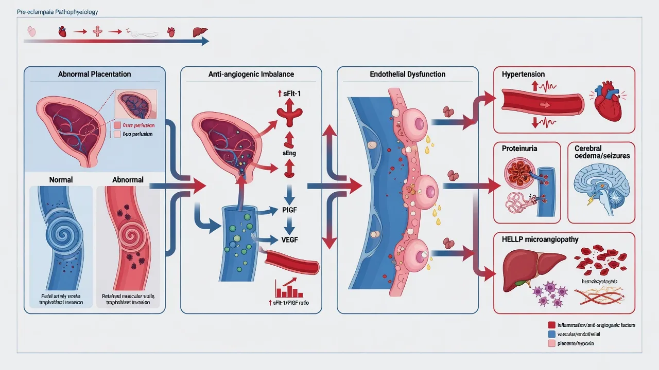
(See dedicated topic for detail.) The intensivist's role in hypertensive disorders of pregnancy is: (1) prevent seizure with magnesium sulfate for any severe pre-eclampsia or eclampsia; (2) control severe hypertension (SBP ≥ 160 or DBP ≥ 110) within 30-60 minutes to prevent intracerebral haemorrhage, using IV labetalol or hydralazine, target 140-160/90-105; (3) avoid fluid overload — these patients are oedematous but intravascularly contracted with a leaky endothelium, and pulmonary oedema is the commonest reason for ICU admission; (4) monitor for and treat the catastrophic complications — eclampsia, hepatic rupture (HELLP), DIC, AKI, PRES, pulmonary oedema; (5) plan delivery as the definitive treatment once maternal stabilisation is achieved.[2][5][16]
Obstetric haemorrhage and postpartum haemorrhage (PPH)
Postpartum haemorrhage (PPH) is defined as blood loss ≥ 500 mL after vaginal delivery or ≥ 1000 mL after Caesarean, or any blood loss causing haemodynamic instability. Major PPH is loss ≥ 1500 mL (or half the circulating volume) — the threshold for activating a massive transfusion protocol. The pregnant patient tolerates acute blood loss deceptively well (50% plasma volume expansion, tachycardia at baseline) — a systolic BP < 90 in a young pregnant woman is a pre-arrest sign, not an early warning. [1]
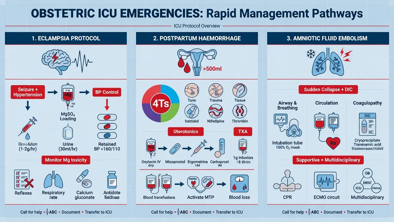
The causes are remembered by the "4 Ts": Tone (uterine atony — 70% of cases), Trauma (cervical/vaginal tears, uterine rupture), Tissue (retained placenta, placenta accreta/increta/percreta), and Thrombin (coagulopathy — pre-existing, dilutional, or DIC from abruption/sepsis/AFE). [1]
ICU management of massive obstetric haemorrhage
- Activate the massive transfusion protocol and obstetric MDT — minimum two large-bore IV cannulae, rapid-infuser warming device, group O-negative blood until type-specific available.
- Resuscitate with a 1:1:1 ratio of packed red cells : FFP : platelets to prevent the lethal triad of acidosis, hypothermia and coagulopathy. Tranexamic acid 1 g IV within 3 hours of bleeding onset — WOMAN trial showed ~19% reduction in death from bleeding (give as early as possible).[10]
- Treat uterine atony (the commonest cause) — first-line uterotonics: oxytocin 5 IU IV slow + 40 IU in 500 mL over 4 h; if refractory add ergometrine 250 µg IM (CONTRAINDICATED in hypertension), carboprost (15-methyl-PGF2α) 250 µg IM q15min max 8 doses (contraindicated in asthma), and misoprostol 800 µg rectally.
- Mechanical measures — uterine massage, bimanual compression, Bakri balloon tamponade, uterine packing.
- Correct coagulopathy aggressively — in late pregnancy the fibrinogen should be 4-6 g/L; a fibrinogen < 2 g/L in the bleeding obstetric patient is pathological and warrants cryoprecipitate (10-15 U). Aim for fibrinogen > 2 g/L, platelets > 75, INR < 1.5.
- Source control by interventional radiology or surgery — uterine/internal iliac artery embolisation, B-Lynch suture, ligation of uterine/internal iliac arteries, hysterectomy as a life-saving last resort (do not delay in refractory atony).
- Look for the underlying cause — abruption (with DIC), placenta accreta spectrum (rising with prior Caesarean), uterine rupture, retained placenta, AFE. Send coagulation and fibrinogen on admission and q30-60min during active bleeding.
WOMAN Trial (2017) — tranexamic acid for post-partum haemorrhage
International, randomised, double-blind, placebo-controlled trial in 193 hospitals across 21 countries; n = 20 021 women with post-partum haemorrhage.[10]
- Intervention: Tranexamic acid 1 g IV within 3 h of delivery vs placebo, in addition to standard care.
- Primary outcome — death from bleeding: 1.5% vs 1.9% (RR 0.81, 95% CI 0.65-1.00; p = 0.045). Number-needed-to-treat ~250.
- Laparotomy for bleeding: Reduced by about a third (RR 0.64, 99% CI 0.49-0.85).
- Timing is critical: The benefit was confined to women who received tranexamic acid within 3 hours of delivery; after 3 hours there was no benefit (and a non-significant trend to harm).
- Safety: No increase in thromboembolic events, organ failure, sepsis, or maternal complications.
- Bottom line: Give tranexamic acid 1 g IV to every woman with post-partum haemorrhage within 3 hours of delivery — cheap, safe, and life-saving. The WHO now recommends it as part of standard PPH management.
Amniotic fluid embolism (AFE) — clinical features and management
Amniotic fluid embolism is a rare, catastrophic, unpreventable peripartum syndrome classically presenting with the abrupt onset of hypoxia, hypotension and coagulopathy during labour, delivery, Caesarean section, or within 30 minutes of delivery. UKOSS data report an incidence of approximately 1.7 per 100 000 maternities with a contemporary case-fatality of ~13-20% (much better than older estimates of 60-80%, reflecting earlier recognition and better supportive care).[3]
The pathophysiology is a complement-mediated anaphylactoid response to amniotic fluid entering the maternal circulation during labour — the term "anaphylactoid syndrome of pregnancy" is more accurate. The clinical course is classically biphasic: [1]
- Phase 1 (acute, 0-30 min): Pulmonary vasoconstriction with acute right heart failure, hypoxaemia, hypotension, and often a seizure. The right heart may acutely dilate and fail (visible on POCUS as RV dilation, TR, paradoxical septal motion). Up to a third of women die in this phase.
- Phase 2 (after stabilisation): Left ventricular failure and coagulopathy dominate. Disseminated intravascular coagulation develops in up to 80%, with profuse bleeding from puncture sites, the uterus and surgical wounds. Fibrinogen falls dramatically (recall that pregnancy baseline is 4-6 g/L, so a "normal" value is already abnormal in late pregnancy). [1]
Management is supportive — there is no specific therapy. The cornerstones are: (1) immediate resuscitation with high-flow oxygen, intubation if hypoxic, and vasopressors/inotropes (noradrenaline ± dobutamine or milrinone for LV failure); (2) massive transfusion protocol for the coagulopathy, with aggressive fibrinogen replacement (cryoprecipitate aiming for > 2 g/L), FFP, platelets and red cells; (3) delivery of the fetus if AFE occurs before delivery (often dictated by maternal resuscitation needs, performed at the bedside in extremis); (4) consider VA-ECMO for refractory LV/right heart failure, which has been used successfully in case series.[3]
Differential diagnosis of acute maternal collapse in the peripartum period
| Condition | Hallmark | Distinguishing features |
|---|---|---|
| Amniotic fluid embolism | Acute hypoxia + hypotension + coagulopathy during labour/delivery | Timing (peripartum); biphasic (right then left heart); massive DIC |
| Pulmonary embolism | Acute hypoxia + hypotension ± chest pain | DVT signs; ECG (S1Q3T3, RBBB); POCUS RV strain; usually no coagulopathy |
| High spinal / total spinal | Hypotension + bradycardia + respiratory compromise after neuraxial | Onset within minutes of neuraxial; ascending sensory level; apnoea |
| Local anaesthetic toxicity (LAST) | Perioral tingling, seizure, arrhythmia after LA injection | Timing after LA bolus; circulatory arrest with asystole/VF; give Intralipid 20% 1.5 mL/kg bolus |
| Eclampsia | Generalised tonic-clonic seizure in pre-eclampsia/eclampsia | Hypertension, proteinuria, hyperreflexia before seizure; respond to magnesium |
| Anaphylaxis | Hypotension + bronchospasm + urticaria after a drug | Timing after drug (antibiotic, oxytocin, suxamethonium); give adrenaline 0.5 mg IM |
| Cardiogenic shock (PPCM, valve, ischaemic) | Pulmonary oedema + hypotension | Bilateral crackles, raised JVP; POCUS dilated/hypocontractile LV (PPCM) or valvular lesion |
| Obstetric haemorrhage (occult) | Hypovolaemic shock; uterine atony/tear | Vaginal bleeding may be concealed (intra-abdominal); high index in post-Caesarean collapse |
Peripartum cardiomyopathy (PPCM)
Peripartum cardiomyopathy is heart failure with reduced ejection fraction (LVEF ≤ 45%) presenting in the last month of pregnancy or within 5 months postpartum, in a woman with no prior heart disease and no other identifiable cause. The incidence is approximately 1 in 1000-4000 live births; risk factors include multiparity, advanced maternal age, pre-eclampsia, African descent, smoking, obesity, and long-term tocolysis.[4][11]
The pathophysiology is a peripartum oxidative stress → cleavage of prolactin into a 16-kDa anti-angiogenic fragment that drives cardiomyocyte apoptosis and endothelial dysfunction. This is the rationale for bromocriptine (a dopamine agonist that suppresses prolactin), which appears to improve LVEF recovery when given early in PPCM.[11]
Management combines standard heart-failure therapy modified for pregnancy: diuretics (furosemide) for congestion, beta-blockers (bisoprolol, metoprolol), hydralazine + nitrates in pregnancy (substitutes for ACEi/ARB, which are contraindicated in pregnancy), then ACEi/ARB + mineralocorticoid receptor antagonist added after delivery, and bromocriptine 2.5 mg BD for 8 weeks (REBICPA showed improved LVEF recovery). Anticoagulation for severe LV dysfunction (LVEF < 30%) due to high thromboembolic risk. Mechanical circulatory support (IABP, Impella, VA-ECMO) as a bridge to recovery or transplant for refractory cardiogenic shock. Breastfeeding is generally discouraged during bromocriptine therapy.[4][11]
Prognosis. Approximately 50% of women recover LVEF to normal within 6-12 months (better than non-ischaemic cardiomyopathy in older patients); 10-15% have persistent severe LV dysfunction; mortality is 5-10% in modern series; ~10% require transplant or LVAD. A subsequent pregnancy in women with persistently reduced LVEF carries a major risk of relapse and death — contraindicated if LVEF < 30% or any persistent dysfunction. [1]
Hilfiker-Kleiner 2017 — bromocriptine in peripartum cardiomyopathy
Multicentre randomised study (Germany), n = 63 women with acute PPCM (LVEF < 45%).[11]
- Intervention: Bromocriptine 1 mg BD for 8 weeks (low-dose) vs bromocriptine 2.5 mg BD for 2 weeks then 2.5 mg daily for 6 weeks (higher dose), in addition to standard heart failure therapy.
- Primary outcome — LVEF at 6 months: Marked improvement in both groups (mean LVEF improved from ~25% at baseline to ~45-50% at 6 months).
- Recovery: Full LVEF recovery (> 50%) in approximately 52% (low-dose) and 68% (high-dose) — much better than historical controls (~30%).
- Safety: Side effects of bromocriptine (hypotension, headache, dizziness) but generally well tolerated.
- Bottom line: Bromocriptine is now considered standard of care for acute PPCM, given early alongside standard heart-failure therapy; subsequent pregnancy remains contraindicated if LVEF has not fully recovered.
Venous thromboembolism in pregnancy
VTE is the leading direct cause of maternal death in high-income settings (UK and ANZ confidential enquiries); pregnancy confers a 4-5 fold increased risk, peaking in the postpartum period. Risk factors include Caesarean delivery (especially emergency), immobility, obesity, age > 35, pre-eclampsia, previous VTE, thrombophilia, multiparity, dehydration and sepsis. The 9th ACCP guideline provides the framework for risk-stratified prophylaxis and treatment.[13][14]
Diagnosis is challenging. D-dimer is physiologically elevated in pregnancy and not useful. Compression duplex ultrasound of the legs is first-line for suspected DVT; CT pulmonary angiography is the preferred imaging for suspected PE (lower radiation dose than V/Q scan and readily available; the radiation dose to the fetus is well below safety limits). The ECG may show S1Q3T3, RBBB, or T-wave inversion in V1-V4. POCUS may show RV strain. Avoid withholding anticoagulation while awaiting imaging in a haemodynamically unstable patient with a high-suspicion PE — start empirical LMWH. [1]
Treatment is with therapeutic-dose LMWH (e.g. enoxaparin 1 mg/kg BD or 1.5 mg/kg daily) — dose-adjusted with anti-Xa monitoring (target 0.5-1.0 U/mL 4 h post-dose), because the increased GFR and volume of distribution in pregnancy reduce the effective level. LMWH does not cross the placenta (unlike warfarin). Warfarin is teratogenic in the first trimester (fetal warfarin syndrome — nasal hypoplasia, epiphyseal stippling, CNS abnormalities) and causes fetal intracranial haemorrhage in the second and third trimester; it is contraindicated in pregnancy but safe in breastfeeding. Treatment is continued throughout pregnancy and for at least 6 weeks postpartum (minimum total duration 3 months). Mechanical prophylaxis (graduated compression stockings) for all hospitalised pregnant and postpartum women.[14]
Massive PE with haemodynamic compromise is treated with systemic thrombolysis (alteplase 100 mg over 2 h, or 50 mg bolus in cardiac arrest) — the fetal risk of thrombolysis is mainly placental/umbilical bleeding but maternal life takes priority. Catheter-directed thrombolysis or surgical embolectomy are alternatives if thrombolysis is contraindicated. [1]
Maternal cardiac arrest and perimortem Caesarean delivery (PMCD)
Maternal cardiac arrest is rare (1 in 12 000-30 000) but the management is modified standard ACLS with one critical obstetric intervention: perimortem Caesarean delivery (PMCD).[8][1]
The rationale for PMCD is twofold: (1) it may save the fetus if performed early (within 5 minutes of arrest); and (2) it is primarily a maternal resuscitative manoeuvre — emptying the gravid uterus relieves aortocaval compression, restores venous return, and may itself restore maternal cardiac output. The classic Katz paper re-examined the assumption that PMCD was for the fetus and made the case that the 4-minute rule (deliver by 5 minutes) is best understood as a maternal resuscitative intervention.[8]
Maternal cardiac arrest — the modified ACLS algorithm
- Call for help early — obstetrician, anaesthetist, neonatologist, ICU, blood bank. Start a timer.
- LEFT lateral tilt 15-30° (or manual left uterine displacement) if uterus at or above umbilicus (~20 weeks). Continue CPR at this angle if possible, or rotate to supine and use manual uterine displacement for the duration of compressions.
- Hand placement higher on the sternum — the diaphragm is elevated by the gravid uterus, displacing the heart cephalad and to the left.
- Standard BLS/ACLS — high-quality CPR, defibrillation at standard energies (NOT contraindicated in pregnancy — minimal current passes through the fetus), treat reversible causes (4 Hs and 4 Ts), including AFE, magnesium toxicity, eclampsia, local anaesthetic toxicity.
- Perimortem Caesarean delivery if uterus above umbilicus (≈ 20 weeks). Begin at 4 minutes after arrest, deliver by 5 minutes. Performed at the bedside, no anaesthesia needed (mother in arrest), with a classic midline vertical incision for speed. Even if the fetus is not viable, delivery may save the mother by relieving aortocaval compression.
- Continue resuscitation after delivery — uterotonics (oxytocin) to maintain uterine tone, ongoing ACLS, target TTM per local protocol. If maternal ROSC, transfer to ICU for post-arrest care including the obstetric team managing the (now possibly bleeding) uterus.
Katz 2005 — the 4-to-5-minute rule for perimortem Caesarean
A retrospective literature review of perimortem Caesarean deliveries reported since 1985 (when the original 4-min rule was proposed).[8]
- Key finding: No cases of fetal survival were reported when PMCD was performed more than 15 minutes after maternal cardiac arrest; maternal haemodynamic improvement after PMCD was documented in a substantial subset — the procedure is a maternal intervention, not solely fetal.
- Refinement of the rule: PMCD should ideally begin at 4 minutes and be completed by 5 minutes after the onset of maternal cardiac arrest (when uterus is above umbilicus), but if resuscitation is ongoing at any time and the gravid uterus is judged to be impairing maternal venous return, deliver immediately without waiting for the clock.
- Modifications since 2005: The rule is now reframed as "resuscitative hysterotomy" — emphasising that it is performed for maternal benefit at any time the gravid uterus (above the umbilicus) is contributing to cardiac arrest, regardless of the 4-5 min window.
- Bottom line: In maternal cardiac arrest, start the clock at the moment of arrest; begin PMCD at 4 minutes; this is the single highest-yield obstetric intervention in cardiac arrest and improves both maternal and fetal survival.
Timing of delivery in the critically ill mother
The decision to deliver (or to continue the pregnancy) in a critically ill mother is one of the most complex in obstetric critical care, requiring a multidisciplinary conversation (intensivist, obstetrician, obstetric physician, anaesthetist, neonatologist, the family). The guiding principles are: [1]
- The mother comes first. Maternal resuscitation always takes priority over fetal salvage. There is no benefit — and substantial harm — in attempting to prolong a pregnancy in a mother who is dying.
- Delivery is the definitive treatment for the obstetric critical illnesses (severe pre-eclampsia/HELLP, eclampsia, AFE source, chorioamnionitis, refractory obstetric haemorrhage). For these conditions the answer to "should we deliver?" is usually yes, and the question becomes "when and how".
- Delivery is NOT automatically indicated for non-obstetric critical illness (pneumonia, sepsis from a non-uterine source, DKA, trauma) — manage the maternal condition; continue the pregnancy with monitoring unless there is a separate obstetric indication.
- Gestational age matters. Before 24 weeks the fetus is generally non-viable and the decision is purely maternal. Between 24 and 34 weeks, corticosteroids for fetal lung maturity (betamethasone 12 mg IM q24h × 2) are given if delivery can be safely delayed 48 h. Beyond 34 weeks the fetus is mature and the balance favours delivery if there is any maternal or fetal indication.
- Route of delivery — Caesarean vs induction of labour — is an obstetric decision based on the maternal and fetal condition, the indication for delivery, and the gestational age. Caesarean is reserved for obstetric indications or for the patient in extremis (PMCD) — vaginal delivery is generally preferred if time allows. [1]
Pharmacology in pregnancy — vasopressors and teratogenic drugs
Vasopressors in pregnancy
The choice of vasopressor is informed by what little human data exist (mostly observational) plus animal data and an understanding of the uteroplacental circulation, which has no autoregulation — uterine blood flow is directly proportional to uterine perfusion pressure (MAP minus uterine venous pressure). Any drug that raises SVR by vasoconstricting the uterine vasculature may improve maternal BP while worsening placental perfusion.[6]
Vasopressor and inotrope safety profile in pregnancy
| Drug | Pregnancy safety | Use case | Notes |
|---|---|---|---|
| Noradrenaline | Probably safe (best human data) | First-line septic / distributive shock | Best evidence for fetal safety; preserves uteroplacental blood flow better than pure alpha-agonists |
| Phenylephrine | Safe; first-line for spinal hypotension at Caesarean | Spinal/epidural-induced hypotension | Pure alpha-agonist; high doses reduce uteroplacental flow but standard doses well studied and superior to ephedrine for maternal BP at Caesarean |
| Ephedrine | Safe but inferior to phenylephrine for spinal hypotension | Second-line at Caesarean | Mixed alpha/beta; crosses placenta, fetal acidosis at high doses |
| Metaraminol | Probably safe (limited data) | First-trimester vasopressor, peri-arrest | Mixed alpha/beta; useful when noradrenaline not yet available |
| Vasopressin | Limited data; case reports of ischaemia at high doses | Catecholamine-resistant septic shock | Use with caution; not first-line |
| Adrenaline | Safe (animal data; human data limited); may cause fetal tachycardia | Anaphylaxis (drug of choice); cardiac arrest | Always use for true anaphylaxis — the alternative is worse |
| Dobutamine / milrinone | Probably safe | Cardiogenic shock / LV failure (PPCM, AFE) | Standard inotropic use |
| Oxytocin / ergometrine / carboprost | Uterotonics (not vasopressors but haemodynamically active) | PPH (uterine atony) | Oxytocin bolus causes hypotension (give slowly); ergometrine contraindicated in hypertension; carboprost contraindicated in asthma |
Teratogenic and contraindicated drugs in pregnancy
The fetal risk of a drug is graded by FDA pregnancy categories (now replaced by the Pregnancy and Lactation Labeling Rule — PLLR) and Australian categorisation (A, B1, B2, B3, C, D, X). For the intensivist, the practical list of drugs to avoid is short and worth memorising.[6][9]
Drugs to avoid and safe alternatives in pregnancy
| Drug class | Examples | Risk | Safe alternative |
|---|---|---|---|
| ACE inhibitors / ARBs / direct renin inhibitors | Enalapril, ramipril, losartan, valsartan, aliskiren | Fetal renal agenesis, oligohydramnios, neonatal renal failure, skull hypoplasia, teratogenic in 1st trimester | Labetalol, methyldopa, nifedipine, hydralazine |
| Warfarin | Warfarin | Teratogenic (fetal warfarin syndrome — 1st trimester); fetal intracranial haemorrhage (2nd/3rd) | LMWH throughout pregnancy; switch to warfarin postpartum (breastfeeding safe) |
| NSAIDs (3rd trimester) | Ibuprofen, diclofenac, indometacin, ketorolac | Premature closure of ductus arteriosus, oligohydramnios, fetal renal impairment; avoid after 28 weeks | Paracetamol (1 g q6h PRN), opioids (short-term) |
| Aminoglycosides | Gentamicin, tobramycin, amikacin | Fetal ototoxicity (8th nerve); use only when essential and for short courses | Cephalosporins, penicillins, carbapenems, aztreonam |
| Tetracyclines | Doxycycline, tetracycline, minocycline | Discolouration of fetal teeth and bones; teratogenic | Penicillins, macrolides, cephalosporins |
| Sulfonamides (3rd trimester) | Trimethoprim, co-trimoxazole | Kernicterus (displacement of bilirubin from albumin); folate antagonism (1st trimester neural tube defects) | Penicillins, cephalosporins, nitrofurantoin (avoid at term — haemolysis) |
| Quinolones | Ciprofloxacin, levofloxacin | Cartilage toxicity in animal studies; traditionally avoided | Cephalosporins, penicillins |
| Chloramphenicol | Chloramphenicol | Grey baby syndrome (cardiovascular collapse) | Cephalosporins, penicillins |
| Retinoids | Isotretinoin, etretinate | Highly teratogenic — CNS, cardiac, craniofacial defects | Avoid entirely |
| Lithium (1st trimester) | Lithium | Ebstein's anomaly (cardiac) | Avoid in pregnancy; alternative mood stabiliser |
| Sodium nitroprusside | Nitroprusside | Theoretical cyanide / thiocyanate toxicity to fetus | Labetalol, hydralazine, nicardipine |
Additional clinical pearls
[1] [1] [1] [1] [1] [1] [1] [1] [1] [1] [1] [1] [1] [1]Additional red flags
[1] [1] [1] [1] [1] [1]Non-obstetric critical illness in pregnancy
The general principle is that pregnancy is not a contraindication to standard critical care — pregnant women should receive the same investigations, treatments and interventions as non-pregnant patients with the same condition, modified only by the physiological changes and fetal considerations described above. Examples:[9]
- Pneumonia — community-acquired, viral (influenza, COVID-19, varicella), aspiration. Pregnant women are more susceptible to severe viral pneumonitis; influenza and COVID-19 vaccination are recommended in pregnancy. Treat per standard guidelines, with attention to oxygenation (target SpO2 ≥ 94%, because the fetus is vulnerable to maternal hypoxia) and to the lower baseline PaCO2 (a PaCO2 of 40 is hypercapnic).
- Asthma exacerbation — treat per standard guidelines (β2-agonists, steroids, magnesium); the only modification is to avoid prostaglandin F2α (carboprost) in PPH (causes bronchospasm).
- Diabetic ketoacidosis — pregnant women with type 1 diabetes are at increased risk; manage per standard DKA protocol (fluids, insulin, potassium); fetal monitoring is mandatory (DKA causes fetal distress), but delivery is not the answer — fix the maternal DKA and the fetus usually recovers.
- Trauma — major trauma in pregnancy follows ATLS principles with the modifications: left lateral tilt, fetal monitoring if viable, lower threshold for imaging (CT is essential and radiation dose to fetus is acceptable), Rh-negative women with bleeding receive anti-D immunoglobulin, and domestic violence is a leading cause of maternal trauma — screen.
- Acute abdomen — appendicitis (often atypical, displaced by the gravid uterus), cholecystitis (gallstones commoner in pregnancy), pancreatitis (gallstone, hypertriglyceridaemia), bowel obstruction. Operate if indicated — pregnancy is not a contraindication to laparotomy or laparoscopy (in the second trimester if possible). [1]
Outcomes and prognosis
Maternal ICU mortality is lower than general ICU mortality (~2-5% vs 15-20%) because obstetric patients are younger, have fewer comorbidities, and most obstetric critical illnesses are reversible once delivered (pre-eclampsia, eclampsia, HELLP, haemorrhage). The exceptions are AFE (modern case fatality ~13-20%), PPCM (5-10% mortality, with 10% needing transplant), and maternal sepsis (5-10% mortality), which carry higher risk. The long-term legacy of obstetric critical illness is substantial — post-intensive care syndrome (PICS) is increasingly recognised in survivors, with persistent physical, cognitive and psychological morbidity; pregnancy-related PTSD affects up to 30% of survivors of severe obstetric emergencies; and a history of pre-eclampsia, PPCM or gestational diabetes marks a substantially elevated lifelong cardiovascular risk that should be addressed at ICU follow-up.[1][15]
The intensivist's job is to support the maternal physiology while the obstetric team delivers the placenta or treats the underlying cause, to maintain oxygenation and perfusion for both patients, to know the (short) list of contraindicated drugs, and to remember that the young pregnant patient hides blood loss and decompensates abruptly — the threshold for escalation is lower than for a non-pregnant patient with the same derangement. [1]
References
- [1]Mhyre JM, Bateman BT, Leffert LR. Maternal mortality and the role of the obstetric anesthesiologist Best Pract Res Clin Anaesthesiol, 2017.PMID 28625309
- [2]Altman D, Carroli G, Duley L, Farrell B, Moodley J, Neilson J, Smith D; Magpie Trial Collaboration Group. Do women with pre-eclampsia, and their babies, benefit from magnesium sulphate? The Magpie Trial: a randomised placebo-controlled trial Lancet, 2002.PMID 12057549
- [3]Fitzpatrick KE, Tuffnell D, Kurinczuk JJ, Knight M. Incidence, risk factors, management and outcomes of amniotic-fluid embolism: a population-based cohort and nested case-control study BJOG, 2016.PMID 25683758
- [4]Elkayam U, Akhter MW, Singh H, Khan S, Bitar F, Hameed A, Shotan A. Clinical Outcomes for Peripartum Cardiomyopathy in North America: Results of the IPAC Study (Investigations of Pregnancy-Associated Cardiomyopathy) J Am Coll Cardiol, 2015.PMID 26293760
- [5]Sibai BM. Diagnosis, controversies, and management of the syndrome of hemolysis, elevated liver enzymes, and low platelet count Obstet Gynecol, 2004.PMID 15121574
- [6]Neligan PJ, Laffey JG. Clinical review: Special populations--critical illness and pregnancy Crit Care, 2011.PMID 21888683
- [7]McDonnell NJ, Paech MJ, Clavisi OM, Scott KL; ANZCA Trials Group. Difficult and failed intubation in obstetric anaesthesia: an observational study of airway management and complications associated with general anaesthesia for caesarean section Int J Obstet Anesth, 2008.PMID 18617389
- [8]Katz V, Balderston K, DeFreest M. Perimortem cesarean delivery: were our assumptions correct? Am J Obstet Gynecol, 2005.PMID 15970850
- [9]Reitman E, Flood P. Anaesthetic considerations for non-obstetric surgery during pregnancy Br J Anaesth, 2011.PMID 22156272
- [10]WOMAN Trial Collaborators. Effect of early tranexamic acid administration on mortality, hysterectomy, and other morbidities in women with post-partum haemorrhage (WOMAN): an international, randomised, double-blind, placebo-controlled trial Lancet, 2017.PMID 28456509
- [11]Hilfiker-Kleiner D, Haghikia A, Masuko D, Nonhoff J, Held D, Libera M, Marsano-Zambrano I, et al. Bromocriptine for the treatment of peripartum cardiomyopathy: a multicentre randomized study Eur Heart J, 2017.PMID 28934837
- [12]Singer M, Deutschman CS, Seymour CW, Shankar-Hari M, Annane D, Bauer M, Bellomo R, et al. The Third International Consensus Definitions for Sepsis and Septic Shock (Sepsis-3) JAMA, 2016.PMID 26903338
- [13]Kane E, Essenmacher S, Weber C, Happe S, Baptist A. Venous thromboembolism as a cause of severe maternal morbidity and mortality in the United States Semin Perinatol, 2019.PMID 30935751
- [14]Bates SM, Greer IA, Middeldorp S, Veenstra DL, Prabulos AM, Vandvik PO; American College of Chest Physicians. VTE, thrombophilia, antithrombotic therapy, and pregnancy: Antithrombotic Therapy and Prevention of Thrombosis, 9th ed: American College of Chest Physicians Evidence-Based Clinical Practice Guidelines Chest, 2012.PMID 22315276
- [15]Say L, Chou D, Gemmill A, Tunçalp Ö, Moller AB, Daniels J, Gülmezoglu AM, Temmerman M, Alkema L. Global causes of maternal death: a WHO systematic analysis Lancet Glob Health, 2014.PMID 25103301
- [16]Duley L, Gulmezoglu AM, Henderson-Smart DJ, Chou D. Magnesium sulphate and other anticonvulsants for women with pre-eclampsia Cochrane Database Syst Rev, 2010.PMID 21069663