ICU · Oncology
Hypercalcaemia of malignancy
Also known as Hypercalcaemic crisis · Humoral hypercalcaemia of malignancy · Local osteolytic hypercalcaemia · Tumour-induced hypercalcaemia · PTHrP-mediated hypercalcaemia · Calcitriol-mediated hypercalcaemia
Hypercalcaemia of malignancy is the most common metabolic emergency in oncology and affects up to 20-30% of cancer patients at some point. Three mechanisms operate: (1) HUMORAL hypercalcaemia of malignancy (HHM, ~80%) — tumour secretion of parathyroid hormone-related peptide (PTHrP), classic in squamous cell carcinomas (lung, head/neck, oesophagus/cervix), renal cell, bladder, ovarian and some breast cancers; (2) LOCAL OSTEO LYTIC (~20%) — direct osteoclast activation by tumour cells in bone, the dominant mechanism in multiple myeloma (1 cause of osteolytic hypercalcaemia) and breast cancer bone metastases; (3) CALCITRIOL (1,25-dihydroxyvitamin D)-mediated — ectopic production by lymphoma (and a few rare tumours). Ectopic PTH is vanishingly rare. Presentation follows the mnemonic stones (nephrolithiasis, nephrocalcinosis), bones (bone pain), abdominal groans (constipation, nausea/vomiting, anorexia, abdominal pain, ileus), and psychic moans (confusion, depression, lethargy) — plus polyuria/polydipsia from nephrogenic diabetes insipidus. Severe hypercalcaemia (corrected Ca >3.5 mmol/L or ionised >1.5 mmol/L) causes altered mental status, coma, seizures, acute kidney injury, and arrhythmias with the characteristic ECG signature of SHORT QT interval (the inverse of hypocalcaemia's long QT), prolonged PR and flattened/widened T waves. Treatment is staged: (1) AGGRESSIVE IV 0.9% saline 3-6 L/day FIRST to restore volume and drive calciuresis; (2) a BISPHOSPHONATE — zoledronic acid 4 mg IV over 15 min (AFTER hydration), the gold standard, takes 2-4 days to peak and lasts weeks; (3) CALCITONIN 4 IU/kg SC q12h for a rapid but transient calcium fall (bridge while the bisphosphonate works — tachyphylaxis after 48-72 h); (4) DENOSUMAB 120 mg SC (anti-RANKL) for refractory disease or renal failure; (5) GLUCOCORTICOIDS only for calcitriol-mediated (lymphoma) hypercalcaemia; (6) HAEMODIALYSIS for severe/refractory hypercalcaemia with AKI or cardiac instability. Use IONISED calcium (corrected Ca is unreliable with hypoalbuminaemia). Treat the underlying malignancy.
On this page & tools
Your progress
Saved locally on this device.
Target exams
Red flags

Pathophysiology — the three mechanisms
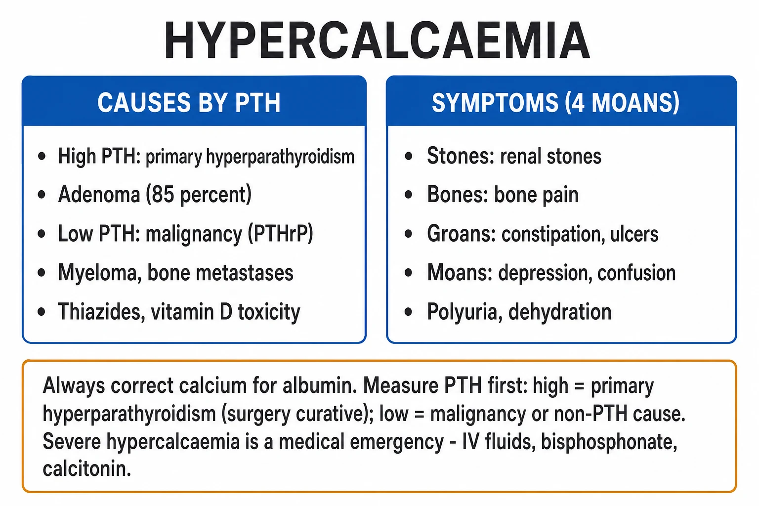
Hypercalcaemia of malignancy is not a single disease but the final common pathway of three distinct tumour-driven mechanisms that converge on one outcome: increased calcium efflux from bone into the extracellular fluid at a rate that exceeds renal calcium clearance. The 2023 Endocrine Society Clinical Practice Guideline and the classical reviews frame the disease around these mechanisms, because the mechanism determines both the tumour type and, in the case of calcitriol-mediated disease, the correct drug (glucocorticoids work only for the lymphoma/calcitriol form).[1][2][3]
1. Humoral hypercalcaemia of malignancy (HHM) — PTHrP-mediated (~80%)
The dominant mechanism. The tumour secretes parathyroid hormone-related peptide (PTHrP), which binds and activates the PTH-1 receptor in bone and kidney with the same downstream effects as parathyroid hormone itself: (a) increased osteoclastic bone resorption (calcium mobilised from bone); (b) increased distal renal tubular calcium reabsorption (calcium conserved); and (c) increased phosphate excretion with reciprocal hypophosphataemia. The net result is hypercalcaemia with low/normal phosphate — biochemically mimicking primary hyperparathyroidism, EXCEPT that PTH itself is appropriately suppressed (low) by the high ambient calcium. The discriminatory test is a PTHrP level, which is elevated.[1][4]
Classic HHM tumours are squamous cell carcinomas (lung, head and neck, oesophagus, cervix), renal cell carcinoma, bladder, ovarian, and a subset of breast cancers. These tumours frequently have little or no bone involvement — the hypercalcaemia is genuinely humoral (systemic), which is why a patient with a small squamous primary can have profound hypercalcaemia.[1]
2. Local osteolytic hypercalcaemia (~20%)
Tumour cells within the bone marrow (myeloma, lymphoma, breast cancer bone metastases) activate osteoclasts LOCALLY through cytokines (RANKL, MIP-1, interleukin-6, lymphotoxin, PTHrP produced locally) and direct cell-cell contact. The result is focal, often widespread, osteolytic bone destruction with release of calcium into the circulation. Unlike HHM, PTHrP is often normal and there is usually no renal calcium-conserving effect (in fact the kidneys try to excrete the excess calcium, producing a hypercalciuric state that drives nephrolithiasis/nephrocalcinosis).[1][3]
Multiple myeloma is the #1 cause of osteolytic hypercalcaemia — always think of (and screen for) myeloma in any hypercalcaemic adult (serum and urine electrophoresis, serum free light chains). Breast cancer bone metastases are the second classic cause.[1]
3. Calcitriol (1,25-dihydroxyvitamin D)-mediated hypercalcaemia
A minority mechanism, classically in lymphoma (and a few rare tumours such as some ovarian dysgerminomas). The tumour ectopically expresses 1-alpha-hydroxylase and converts 25-hydroxyvitamin D to active 1,25-dihydroxyvitamin D (calcitriol), which increases intestinal calcium absorption (and to a lesser degree bone resorption). PTH is appropriately suppressed. This is the ONE form of malignancy-associated hypercalcaemia that responds to glucocorticoids — steroids suppress the 1-alpha-hydroxylase and calcitriol production.[1][3]
Ectopic (true) PTH secretion by a tumour is described but is vanishingly rare.[1]
The three mechanisms of hypercalcaemia of malignancy — mechanism, tumour, biochemistry, treatment
| Feature | Humoral (HHM, PTHrP) ~80% | Local osteolytic ~20% | Calcitriol-mediated |
|---|---|---|---|
| Mediator | PTHrP (binds PTH-1 receptor) | RANKL, IL-6, MIP-1, local PTHrP — osteoclast activation | 1,25-dihydroxyvitamin D (ectopic 1-alpha-hydroxylase) |
| Main action | ↑ bone resorption + ↑ renal Ca reabsorption + ↑ phosphate excretion | Local osteoclastic bone resorption at tumour sites | ↑ intestinal Ca absorption (+ some bone resorption) |
| Typical tumours | Squamous cell (lung, H&N, oesophagus, cervix), renal cell, bladder, ovarian, some breast | Multiple myeloma (#1), breast cancer bone metastases, lymphoma | Lymphoma (and rare ovarian dysgerminoma) |
| Bone disease on imaging | Often absent (systemic/humoral) | Marked lytic lesions (myeloma "raindrop" skull, lytic bone mets) | Variable |
| PTH | LOW (suppressed by high Ca) | LOW | LOW |
| PTHrP | HIGH | Normal / low | Normal |
| Phosphate | Low (PTH-like phosphaturia) | Variable | Variable / high |
| 1,25-vit D | Low/normal | Low/normal | HIGH |
| Responds to glucocorticoids? | No | No | YES (suppresses 1-alpha-hydroxylase) |
Humoral (80%)
PTHrP-mediated
- Tumour secretes PTH-related peptide (PTHrP)
- Acts like PTH — increases bone resorption + renal Ca reabsorption + phosphate excretion
- Common tumours: squamous cell (lung, head/neck, oesophagus, cervix), renal cell, breast, bladder, ovarian
- PTH LOW (suppressed by high Ca) but PTHrP HIGH
Local osteolysis (20%)
Direct bone destruction
- Tumour cells directly activate osteoclasts → local bone resorption (RANKL, IL-6, MIP-1)
- Common: multiple myeloma (#1 cause of osteolytic hypercalcaemia), breast cancer bone metastases, lymphoma
- PTH LOW, PTHrP may be normal
Calcitriol
Lymphoma — 1,25-vit D
- Tumour expresses 1-alpha-hydroxylase → produces excess 1,25-dihydroxyvitamin D
- Increases intestinal Ca absorption; classic in lymphoma
- PTH LOW, 1,25-vit D HIGH; the ONLY form that responds to glucocorticoids
Mechanism by tumour — what to expect at the bedside
| Tumour | Dominant mechanism | Pearl |
|---|---|---|
| Squamous cell (lung, H&N, oesophagus, cervix) | HHM (PTHrP) | Profound hypercalcaemia with little/no bone disease — purely humoral |
| Renal cell carcinoma | HHM (PTHrP) | Often paraneoplastic; Stauffer syndrome (non-metastatic cholestasis) can coexist |
| Breast cancer | Mixed — local osteolysis (bone mets) + local/HHM PTHrP | Bone metastases are usually lytic/lytic-mixed |
| Multiple myeloma | Local osteolysis | #1 cause of osteolytic hypercalcaemia; always screen with SPEP/UPEP/SFLC |
| Bladder, ovarian, endometrial | HHM (PTHrP) | Less common but classic humoral |
| Lymphoma | Calcitriol-mediated (some HHM) | Steroid-responsive; check 1,25-vit D |
| Ectopic PTH (very rare) | True PTH secretion | Differentiated from HHM by an INAPPROPRIATELY HIGH PTH |
Why hypercalcaemia is dangerous — the cellular mechanism
Calcium is the master regulator of cellular excitability. The extracellular calcium concentration sets the threshold for depolarisation of nerve and muscle — a high extracellular calcium raises this threshold, making membranes HYPER-excite in subtle ways early (neuromuscular irritability is paradoxically low; the dominant CNS effect is depression of function) and then progressively DEPRESSED as the level rises (lethargy → confusion → somnolence → coma). The major end-organ threats are:[1]
- Cardiac — hypercalcaemia shortens the action potential plateau (phase 2) and shortens the ST segment, producing the hallmark short QT interval (QTc often <350 ms; a true short QT is one of the rare ECG findings in medicine). At very high levels the PR prolongs, the QRS widens, and T-wave amplitude falls (flattened/broad T waves); the risk is ventricular arrhythmia, including torsades-like polymorphic VT and sudden cardiac death.[1]
- Renal — hypercalcaemia causes (a) nephrogenic diabetes insipidus (hypercalcaemia downregulates aquaporin-2 channels → polyuria/polydipsia and free-water loss); (b) renal vasoconstriction with falling GFR; (c) nephrocalcinosis/nephrolithiasis from hypercalciuria; and (d) a pre-renal component from vomiting and poor intake. These converge to produce AKI, which then impairs calcium excretion and worsens hypercalcaemia in a vicious cycle.[1][4]
- Neurological — progressive depression of consciousness from lethargy through confusion to coma; seizures at very high levels.
- Gastrointestinal — smooth-muscle hypotonia → constipation, anorexia, nausea/vomiting, ileus; increased ulcer/pancreatitis risk.
Clinical presentation — stones, bones, abdominal groans, and psychic moans
The classical mnemonic ("stones, bones, groans, and psychic moans") maps the symptom clusters and remains the exam-friendly framework, but the modern addition — nephrogenic DI / polyuria-polydipsia — is often the earliest symptom and is frequently missed because the dehydrated, confused patient cannot report it.[1][5]
Clinical features of hypercalcaemia by system
| System | Features | Severity marker |
|---|---|---|
| CNS / "psychic moans" | Lethargy, fatigue, difficulty concentrating, depression, apathy; progressing to confusion, disorientation, somnolence, coma and seizures at high levels | Coma/seizure = severe; mental-state change is itself an indication for urgent treatment |
| GI / "abdominal groans" | Anorexia, nausea, vomiting, constipation, abdominal pain, ileus; ↑peptic ulcer and pancreatitis risk | Intractable constipation/ileus; pancreatitis is a rare but recognised trigger |
| Renal / "stones" | Polyuria/polydipsia (nephrogenic DI), dehydration, nephrolithiasis, nephrocalcinosis, acute kidney injury (volume loss + vasoconstriction + tubular injury) | AKI + oliguria = severe; AKI worsens hypercalcaemia in a vicious cycle |
| Musculoskeletal / "bones" | Bone pain (especially in myeloma/bone-metastatic disease), muscle weakness, fractures | Bone pain localises the underlying bone disease |
| Cardiac | Often asymptomatic until ECG changes — short QT, prolonged PR, flattened/broad T waves; palpitations, syncope, sudden death | Short-QT arrhythmia / cardiac arrest = peri-arrest |
Severity of hypercalcaemia (corrected total calcium, mmol/L) and clinical threshold
| Severity | Corrected Ca (mmol/L) | Typical features | Threshold to act |
|---|---|---|---|
| Mild | 2.6 – 2.85 | Often asymptomatic; mild fatigue | Investigate; outpatient workup if well |
| Moderate | 2.85 – 3.5 | Symptomatic — GI, mild cognitive change, polyuria | Admit; IV fluids + bisphosphonate |
| Severe / hypercalcaemic crisis | >3.5 (ionised >1.5) | Confusion, coma, AKI, short-QT arrhythmia; mortality high | Medical emergency — ICU, aggressive saline + bisphosphonate + calcitonin bridge ± dialysis |
The intensivist's threshold: a symptomatic patient, OR corrected Ca >3.0 mmol/L with AKI/cardiac symptoms, OR any patient >3.5 mmol/L, warrants urgent in-hospital (often ICU) treatment.[2][5]
ECG changes — the signature is a SHORT QT
The cardiac signature of hypercalcaemia is the short QT interval — the single most testable ECG fact, and the inverse of hypocalcaemia's long QT. Hypercalcaemia raises the threshold for depolarisation and shortens the cardiac action potential, particularly phase 2 (the plateau), which collapses the ST segment so the T wave appears to sit close on the QRS. As calcium rises further the PR interval prolongs, the QRS broadens, and the T wave flattens/inverts; the end-stage rhythm is polymorphic VT or asystole.[1][4]
ECG evolution with rising calcium (vs hypocalcaemia)
| ECG feature | Hypercalcaemia | Hypocalcaemia (contrast) |
|---|---|---|
| QT interval | SHORT (QTc <350-390 ms; the hallmark) | LONG (QTc >440-470 ms) |
| ST segment | Short / absent (T wave hugs QRS) | Prolonged (lengthens the QT) |
| PR interval | Prolonged at high levels | Usually normal |
| QRS | May widen at very high levels | Usually normal |
| T waves | Flattened, broadened, may invert | Normal amplitude (until very low Ca) |
| Arrhythmia risk | Polymorphic VT / torsades-like / sudden death | Torsades de pointes (long-QT) |
| Mechanism | High extracellular Ca raises depolarisation threshold + shortens phase 2 | Low Ca prolongs phase 2 → long QT |
Practical point: a short QT in an unwell cancer patient is hypercalcaemia until proven otherwise — check the ionised calcium immediately. [1]
Diagnosis and investigations — confirm, quantify, and find the mechanism
The workup confirms hypercalcaemia (on a reliable sample), quantifies severity, identifies the mechanism, and screens for end-organ damage. The first decision is whether the calcium is truly high — always measure an ionised calcium (or at minimum correct the total for albumin), because the malnourished cancer patient is typically hypoalbuminaemic and the uncorrected total underestimates the true (ionised, biologically active) calcium.[1][2]
Diagnostic workup of hypercalcaemia of malignancy
Confirm and quantify — IONISED calcium
Measure ionised calcium (the biologically active fraction). If only total calcium is available, apply the albumin correction: corrected Ca (mmol/L) = measured Ca + 0.02 × (40 − albumin g/L). In hypoalbuminaemia the corrected value still underestimates ionised Ca, so prefer the ionised measurement. Repeat to confirm and trend.
PTH (the discriminator)
Intact PTH distinguishes PTH-mediated (primary or tertiary hyperparathyroidism — PTH HIGH/normal) from non-PTH-mediated (PTH LOW). In ALL forms of malignancy-associated hypercalcaemia PTH is LOW (suppressed by the high calcium). A high/normal PTH in a cancer patient suggests coexisting primary hyperparathyroidism.
PTHrP (separates humoral from osteolytic/calcitriol)
Elevated PTHrP confirms humoral hypercalcaemia of malignancy (the ~80% PTHrP-mediated form). Normal PTHrP suggests local osteolysis or calcitriol-mediated disease. Often a research/specialised assay but increasingly available.
1,25-dihydroxyvitamin D (the lymphoma clue)
Elevated 1,25-vit D in a cancer patient (especially with lymphoma) indicates calcitriol-mediated hypercalcaemia — the form that responds to glucocorticoids. Low 1,25-vit D is typical of HHM and osteolysis (the high calcium suppresses PTH → reduces renal 1-alpha-hydroxylase).
phosphate, renal function, albumin, magnesium, ALP
Phosphate (low in HHM, variable elsewhere); U&E/creatinine for AKI; albumin for correction; magnesium (hypomagnesaemia can coexist and worsen arrhythmia); alkaline phosphatase (raised in bone turnover/liver metastases).
Screen for the tumour / mechanism
Multiple myeloma screen (serum protein electrophoresis, urine Bence-Jones protein, serum free light chains) — myeloma is the #1 osteolytic cause. Imaging: CT for primary tumour and bone lesions; bone scan / CT-PET for bone metastases; myeloma workup if lytic lesions.
ECG
Look for SHORT QT, prolonged PR, flattened/broad T waves, and arrhythmia. Continuous cardiac monitoring if severe (>3.5 mmol/L) or symptomatic.
Hypercalcaemia of malignancy vs primary hyperparathyroidism — the core differential
| Test | Hypercalcaemia of malignancy | Primary hyperparathyroidism |
|---|---|---|
| PTH | LOW (suppressed) | HIGH or inappropriately normal |
| PTHrP | High (HHM) / normal (osteolytic) | Normal |
| Calcium | High (often severe, >3.5) | Mild-moderate, chronic |
| Phosphate | Low (HHM) / variable | Low (PTH-mediated phosphaturia) |
| 1,25-vit D | Low/normal (except HIGH in lymphoma) | High/normal (PTH drives 1-alpha-hydroxylase) |
| ALP | Variable (bone metastases raise it) | Variable |
| Onset/age | Acute-subacute; known cancer | Chronic; "moans, stones, groans" over years |
Management — the staged ICU protocol
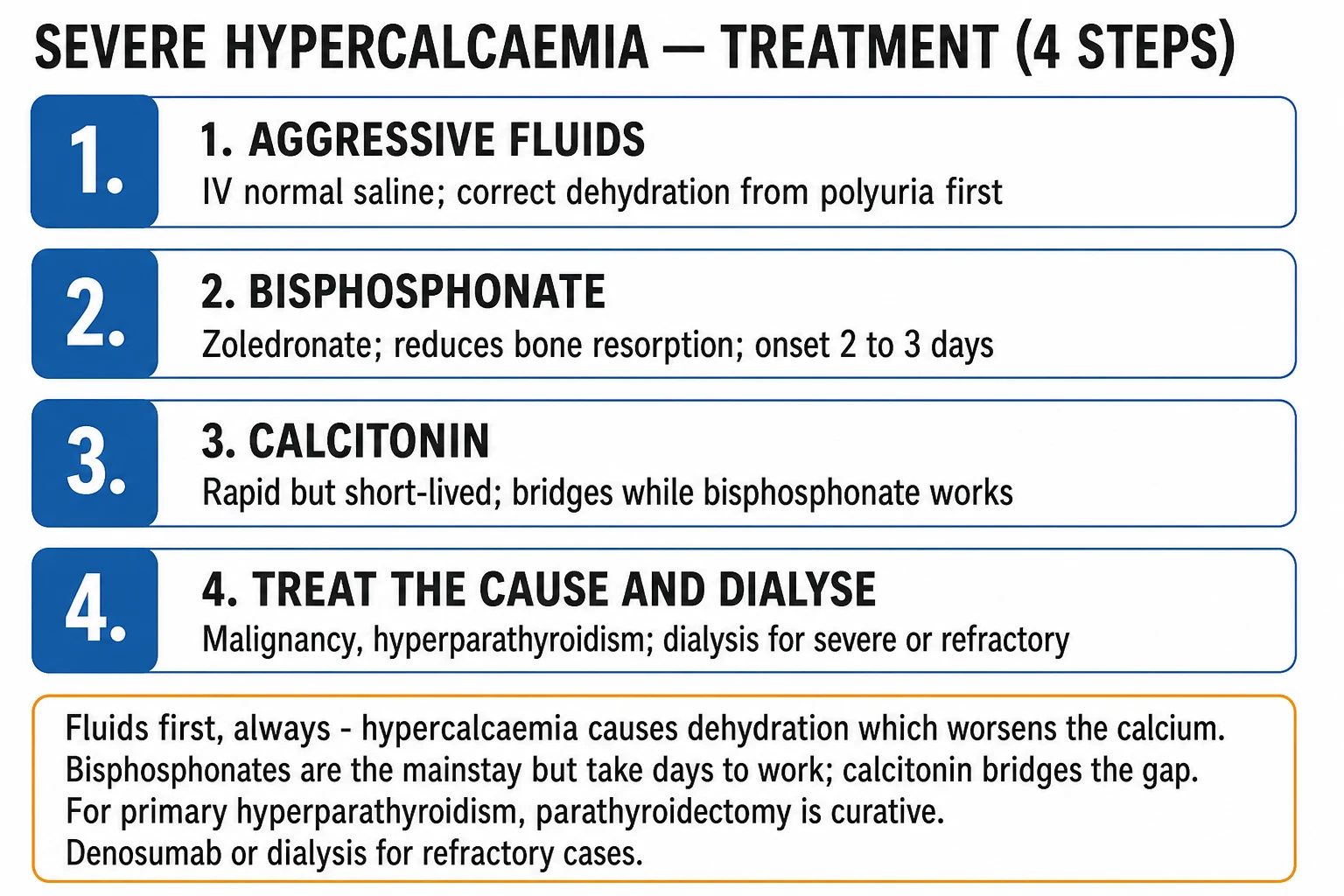
The management of hypercalcaemia of malignancy is staged and protocolised. The cornerstone is aggressive intravenous saline, given FIRST, before any drug; the bisphosphonate (zoledronic acid) is added once the patient is volume-replete; calcitonin provides a rapid bridge while the bisphosphonate reaches peak effect (2-4 days); denosumab is reserved for refractory disease or renal failure; glucocorticoids only for the calcitriol-mediated (lymphoma) form; and haemodialysis for severe/refractory hypercalcaemia with AKI or cardiac instability. Treat the underlying malignancy.[1][2][5]
Hypercalcaemia management protocol
Aggressive IV saline FIRST
0.9% saline 200-300 mL/h for the first 24h (3-6 L/day), titrated to urine output 100-150 mL/h and to avoid volume overload. Hypercalcaemia causes dehydration (nephrogenic DI from high Ca + osmotic diuresis from calciuresis + vomiting + poor intake). Volume expansion restores GFR and promotes sodium-calcium exchange in the distal tubule → calciuresis (calcium follows sodium). Saline alone may drop Ca by 0.2-0.4 mmol/L. This is the single most important first step.
Add a loop diuretic (ONLY after volume repletion)
Furosemide 20-40 mg IV promotes calciuresis (blocks Na-K-2Cl in the thick ascending limb → the lumen-positive potential that drives paracellular Ca reabsorption is lost → calcium is excreted). Give ONLY once the patient is volume-replete, otherwise it worsens dehydration and concentrates calcium. Do NOT give thiazides — they ENHANCE distal Ca reabsorption and worsen hypercalcaemia.
Bisphosphonate — zoledronic acid 4 mg IV (AFTER hydration)
Zoledronic acid 4 mg IV over 15 min (give AFTER, not before, hydration). Nitrogen-containing bisphosphonate — internalised by osteoclasts → inhibits farnesyl pyrophosphate synthase in the mevalonate pathway → osteoclast apoptosis → inhibits osteoclast-mediated bone resorption. Onset 2-4 days, peak effect ~4-7 days, duration weeks-months. Alternatives: pamidronate 60-90 mg IV over 2-4h (slower, less potent). Renally excreted — reduce dose if eGFR <35 and avoid in severe AKI (nephrotoxic). Side effects: flu-like (acute-phase reaction), hypocalcaemia, hypophosphataemia, and osteonecrosis of the jaw (rare, with repeated doses).
Calcitonin — for a rapid but transient effect (BRIDGE)
Calcitonin 4 IU/kg SC q12h (up to 8 IU/kg q6h). Inhibits osteoclasts → calcium falls within hours. Use as a BRIDGE while waiting for the bisphosphonate to work (2-4 days). BUT tachyphylaxis develops after 48-72h (downregulation of calcitonin receptors; the "escape" phenomenon), so calcitonin is NOT a sustained solution. Side effects: nausea, flushing, local injection-site reaction.
Denosumab — for refractory disease or renal failure
Denosumab 120 mg SC (monoclonal antibody against RANKL — inhibits osteoclast formation, activation and survival). Indicated for refractory hypercalcaemia despite bisphosphonates, or in renal failure (bisphosphonates are nephrotoxic; denosumab is NOT renally cleared). Onset days, duration months. NOT nephrotoxic. Major risk: SEVERE and PROLONGED hypocalcaemia (monitor calcium closely; supplement calcium + vitamin D) — this can be more dangerous than the original hypercalcaemia.
Glucocorticoids — ONLY for calcitriol-mediated (lymphoma)
Prednisolone 40-60 mg/day (or hydrocortisone 200-300 mg/day). Effective ONLY for calcitriol-mediated hypercalcaemia (lymphoma) — steroids suppress the tumour 1-alpha-hydroxylase and calcitriol production. Useless for PTHrP-mediated HHM or pure osteolytic disease. May also give when starting anti-tumour therapy for a steroid-sensitive lymphoma.
Haemodialysis — for severe/refractory hypercalcaemia
Reserved for severe symptomatic hypercalcaemia refractory to saline + bisphosphonate + calcitonin, especially with AKI (oliguria limits the calciuric strategy), volume overload (preventing aggressive saline), or cardiac instability/arrhythmia. Haemodialysis with a low-calcium dialysate rapidly lowers calcium; CRRT can be used for haemodynamically unstable patients. Dialysis is a temporising measure — definitive control still requires anti-resorptive therapy + treating the tumour.
Treat the underlying malignancy
The definitive treatment. Anti-tumour therapy (surgery, radiotherapy, chemotherapy, targeted therapy) addresses the source of PTHrP, the osteolytic load, or the calcitriol production. Without tumour control, hypercalcaemia recurs within weeks of each bisphosphonate cycle. Discuss goals of care — recurrent/refractory hypercalcaemia in advanced cancer carries a poor prognosis.
Drug summary — dose, onset, duration, role
| Drug | Dose | Onset | Peak / duration | Mechanism / role |
|---|---|---|---|---|
| 0.9% saline | 3-6 L/day (200-300 mL/h) | Hours | Acute (while infused) | Volume expansion + calciuresis; FIRST-LINE cornerstone |
| Furosemide (loop) | 20-40 mg IV | Hours | Acute | Calciuresis; ONLY after volume repletion |
| Calcitonin | 4 IU/kg SC q12h (up to 8 IU/kg q6h) | Hours | Peak 12-24h; tachyphylaxis 48-72h | Rapid BRIDGE while bisphosphonate works |
| Zoledronic acid | 4 mg IV over 15 min | 2-4 days | Peak 4-7 days; lasts weeks-months | Gold-standard bisphosphonate; FIRST-LINE anti-resorptive |
| Pamidronate | 60-90 mg IV over 2-4h | 2-4 days | Peak 5-7 days; lasts weeks | Alternative bisphosphonate (slower, less potent) |
| Denosumab | 120 mg SC | Days | Lasts months | Refractory disease / renal failure (NOT nephrotoxic); risk: severe hypocalcaemia |
| Glucocorticoids | Prednisolone 40-60 mg/day | Days | Days-weeks | ONLY calcitriol-mediated (lymphoma) |
| Haemodialysis | Low-Ca dialysate | Hours | Temporising | Severe/refractory; AKI/volume overload/arrhythmia |
Bisphosphonate vs calcitonin vs denosumab — head to head
| Feature | Bisphosphonate (zoledronic acid) | Calcitonin | Denosumab |
|---|---|---|---|
| Mechanism | Osteoclast apoptosis (inhibits farnesyl PP synthase) | Osteoclast inhibition | Anti-RANKL antibody (blocks osteoclast formation) |
| Onset | 2-4 days | Hours (fastest) | Days |
| Duration | Weeks-months | 48-72h then tachyphylaxis | Months |
| Role | FIRST-LINE anti-resorptive | Rapid BRIDGE (first 2-3 days) | Refractory disease / renal failure |
| Renal handling | Renally excreted — nephrotoxic, dose-adjust if eGFR <35 | Renally cleared — safe in renal failure | NOT renally cleared — safe in renal failure |
| Key adverse effect | Acute-phase reaction, hypocalcaemia, osteonecrosis of jaw | Nausea, flushing | Severe prolonged hypocalcaemia |
Special situations
Refractory hypercalcaemia of malignancy
Hypercalcaemia that recurs within 30 days of adequate bisphosphonate therapy, or fails to normalise, is "refractory." The 2023 Endocrine Society guideline and the denosumab trials establish denosumab 120 mg SC as the preferred second-line agent — superior to bisphosphonates in refractory disease and usable in renal failure (where bisphosphonates are contraindicated or nephrotoxic).[2][7][8]
Hypercalcaemia with renal failure
Bisphosphonates are renally excreted and nephrotoxic — reduce the zoledronic acid dose for eGFR <35 and avoid in severe AKI. Denosumab (not renally cleared) is the anti-resorptive of choice. Calcitonin (safe in renal failure) provides the bridge. Haemodialysis with a low-calcium dialysate removes calcium acutely and is the temporising strategy for severe hypercalcaemia in the oliguric/dialysis patient.[2][8]
Calcitriol-mediated (lymphoma) hypercalcaemia
The one form that responds to glucocorticoids (prednisolone 40-60 mg/day or hydrocortisone), which suppress the tumour 1-alpha-hydroxylase and calcitriol production. Always measure 1,25-dihydroxyvitamin D in unexplained hypercalcaemia with lymphoma, and add steroids early. Bisphosphonates and calcitonin still help while steroids take effect.[1][3]
Hypercalcaemia with cardiac instability / short-QT arrhythmia
A short-QT polymorphic VT or cardiac arrest from hypercalcaemia is a peri-arrest emergency: aggressive saline, calcitonin for the fastest available calcium fall, and arrange haemodialysis immediately — do not wait 2-4 days for the bisphosphonate. Continuous cardiac monitoring throughout.[1][5]
Special-situation drug of choice
| Situation | First-line | Notes |
|---|---|---|
| Standard symptomatic HCM | Saline + zoledronic acid ± calcitonin bridge | The default protocol |
| Refractory to bisphosphonate | Denosumab 120 mg SC | Superior in refractory disease; monitor for hypocalcaemia |
| Renal failure / AKI | Denosumab + calcitonin ± dialysis | Avoid bisphosphonates (nephrotoxic); denosumab not renally cleared |
| Calcitriol-mediated (lymphoma) | Add glucocorticoids | The ONLY steroid-responsive form |
| Severe with cardiac instability/short-QT arrhythmia | Saline + calcitonin + urgent dialysis | Do not wait for the bisphosphonate |
Exam practice
SAQ — Hypercalcaemic crisis with cardiac instability
10 minutes · 10 marks
A 64-year-old man with metastatic squamous cell lung carcinoma is brought to the emergency department with a 3-day history of confusion, polyuria, constipation and abdominal pain. He is drowsy, HR 124, BP 88/52 and oliguric. Corrected calcium 4.1 mmol/L, creatinine 230 µmol/L. The ECG shows a short QT interval (280 ms) with a run of polymorphic VT that self-terminates.
SAQ — Denosumab versus zoledronic acid in refractory hypercalcaemia
10 minutes · 10 marks
A 58-year-old woman with metastatic breast cancer and extensive bone metastases is admitted with confusion and dehydration. Corrected calcium 3.6 mmol/L, eGFR 28 mL/min. She received zoledronic acid 4 mg IV 12 days ago for hypercalcaemia of malignancy (calcium 3.5 mmol/L at the time); it had normalised but has risen again despite ongoing oral hydration.
Clinical pearls
Red flags
Prognosis
Outcomes and prognostic factors in hypercalcaemia of malignancy
| Factor | Outcome | Notes |
|---|---|---|
| Overall prognosis | Poor — median survival after a diagnosis of HCM is often weeks to a few months | Hypercalcaemia is typically a late-stage marker of advanced cancer |
| Severity at presentation | Corrected Ca >3.5 mmol/L (hypercalcaemic crisis) — high short-term mortality (arrhythmia, coma) | Requires ICU admission |
| Refractory/recurrent disease | Worsens prognosis — recurs within weeks of each bisphosphonate cycle if the tumour is uncontrolled | Triggers goals-of-care discussion |
| Underlying tumour | Tumour type and treatment responsiveness drive outcome | Steroid-sensitive lymphoma (calcitriol form) may have the best outlook with anti-tumour therapy |
| AKI at presentation | Worse prognosis; limits calciuresis, may require RRT | Renal recovery usually parallels calcium control |
| Response to first bisphosphonate cycle | Normocalcaemia predicts better medium-term control | Failure → denosumab |
Recurrent/refractory hypercalcaemia in advanced cancer is a marker of disease progression and a trigger for an honest goals-of-care discussion; repeated cycles of bisphosphonate/denosumab may be appropriate or may represent futile burden, depending on the patient's overall trajectory and wishes.[1][5]
Key trials and evidence
El-Hajj Fuleihan et al. 2023 — Endocrine Society Clinical Practice Guideline: Treatment of Hypercalcemia of Malignancy in Adults (PMID 36545746)
Source
Journal of Clinical Endocrinology and Metabolism 2023;108(3):507-528 — Endocrine Society systematic-review-based clinical practice guideline
What it established
The current definitive guideline. Recommends initial treatment with IV saline and a single dose of an IV bisphosphonate (zoledronic acid 4 mg preferred over pamidronate) for acute hypercalcaemia of malignancy; calcitonin as add-on for severe/symptomatic disease; denosumab 120 mg for refractory or recurrent hypercalcaemia; glucocorticoids only for calcitriol-mediated (lymphoma) disease; dialysis for severe hypercalcaemia with cardiac/renal compromise
Key contribution
Replaced older consensus with a formal GRADE-based systematic review; established denosumab as the preferred second-line agent for refractory disease and codified the saline-first, bisphosphonate-first-line, calcitonin-bridge protocol
Clinical bottom line
The single most authoritative current reference for HCM management — cite it for the exam and the bedside
Stewart AF 2005 — NEJM Clinical Practice: Hypercalcemia associated with cancer (PMID 15673803)
Source
New England Journal of Medicine 2005;352:373-379 — the definitive clinical-practice narrative review
What it established
The classical three-mechanism framework (humoral/PTHrP, local osteolysis, calcitriol), the PTH/PTHrP/biochemical profile of each, and the staged management (saline → bisphosphonate → calcitonin → dialysis)
Key contribution
The most-cited single-author reference on HCM; the framework every intensivist is taught
Clinical bottom line
Read it for the pathophysiology and biochemical differential — the canonical teaching reference
Hu MI et al. 2014 — Denosumab for treatment of hypercalcemia of malignancy (PMID 24915117)
Source
Journal of Clinical Endocrinology and Metabolism 2014;99(9):3144-3152 — multicentre phase II open-label trial
Population
Patients with hypercalcaemia of malignancy that was refractory to or recurrent after bisphosphonate therapy
Intervention
Denosumab 120 mg SC on days 1, 8, 15, 29 then every 4 weeks
Key result
Denosumab produced a rapid and sustained response (the majority of patients achieved albumin-corrected calcium ≤11.5 mg/dL / 2.9 mmol/L by day 10), including in patients with renal impairment where bisphosphonates are contraindicated; median time to first response ~8 days
Clinical bottom line
Established denosumab as effective in bisphosphonate-refractory HCM and usable in renal failure — the basis for its second-line role in the 2023 Endocrine Society guideline
Major PP & Coleman RE 2001 — Zoledronic acid international clinical development program (PMID 11346861)
Source
Seminars in Oncology 2001;28(2 Suppl 6):17-24 — pooled analysis of the pivotal randomised controlled trials
Comparison
Zoledronic acid (4 mg or 8 mg IV) vs pamidronate 90 mg IV in hypercalcaemia of malignancy
Key result
Zoledronic acid 4 mg produced a higher and more durable complete response rate than pamidronate 90 mg; the 8 mg dose was no better than 4 mg (so 4 mg became the standard). Onset of action 2-4 days, duration of response superior to pamidronate
Clinical bottom line
Established zoledronic acid 4 mg IV as the most potent and preferred bisphosphonate for HCM — the dose and drug still used today
Goldner W 2016 — Cancer-Related Hypercalcemia (PMID 27170690)
Source
Journal of Oncology Practice 2016;12(5):426-432 — concise clinical review aimed at oncology practice
What it established
A practical oncology-focused synthesis of the mechanisms (HHM/PTHrP, osteolytic, calcitriol), the stones-bones-groans-moans presentation, and the staged saline-bisphosphonate-calcitonin-denosumab protocol
Clinical bottom line
A clear, citable oncology-practice reference for the mechanism-to-treatment mapping; useful for the bedside and the exam
Hu MI 2021 — Hypercalcemia of Malignancy (PMID 34774243)
Source
Endocrinology and Metabolism Clinics of North America 2021;50(4):721-728 — contemporary review
What it established
Up-to-date integration of the three mechanisms, the biochemical workup (PTH/PTHrP/1,25-vit D), the saline-bisphosphonate-calcitonin-denosumab-st steroid-dialysis ladder, and the management of refractory disease and renal failure
Clinical bottom line
The modern endocrine-clinics companion to the 2023 guideline — excellent for the mechanism and drug-choice detail
Gould Rothberg et al. 2022 — Oncologic emergencies and urgencies: A comprehensive review (PMID 35653456)
Source
CA: A Cancer Journal for Clinicians 2022;72(6):570-593 — comprehensive oncologic-emergencies review
What it established
Places hypercalcaemia of malignancy in the context of the full spectrum of oncologic emergencies (alongside tumour lysis syndrome, SVCO, cord compression, neutropenic sepsis) with a multidisciplinary ED/oncology/palliative-care framing
Clinical bottom line
Useful for situating HCM among the other oncologic emergencies the ICU/ED candidate must know
Body JJ, Niepel D, Tonini G 2017 — Hypercalcaemia and hypocalcaemia: finding the balance (PMID 28078478)
Source
Supportive Care in Cancer 2017;25(5):1639-1649 — supportive-care review
What it established
A practical review of both ends of the calcium disturbance spectrum in cancer, with emphasis on the supportive-care role of bisphosphonates and calcitonin and the avoidance of over-correction (hypocalcaemia from denosumab/over-treatment)
Clinical bottom line
A good supportive-care reference for the cancer-patient-on-the-ward perspective, complementary to the ICU guideline
Thosani S & Hu MI 2015 — Denosumab: a new agent in hypercalcaemia of malignancy (PMID 26403973)
Source
Future Oncology 2015;11(21):2865-2871 — focused review of denosumab in HCM
What it established
Mechanistic and clinical rationale for the anti-RANKL monoclonal antibody denosumab as a non-nephrotoxic alternative to bisphosphonates in refractory HCM and renal failure, with attention to the severe-hypocalcaemia risk
Clinical bottom line
The denosumab pharmacology-and-risk reference — read alongside the Hu 2014 trial
Pelosof LC & Gerber DE 2010 — Paraneoplastic syndromes: an approach to diagnosis and treatment (PMID 20810794)
Source
Mayo Clinic Proceedings 2010;85(9):838-854 — paraneoplastic-syndrome review
What it established
Frames hypercalcaemia of malignancy as the most common paraneoplastic endocrine syndrome, alongside SIADH, Cushing's, and the neurological paraneoplastic syndromes, with a diagnostic-and-treatment approach
Clinical bottom line
Useful for situating PTHrP-mediated hypercalcaemia within the broader paraneoplastic differential the exam may probe
References
- [1]Stewart AF. Clinical practice. Hypercalcemia associated with cancer N Engl J Med, 2005.PMID 15673803
- [2]El-Hajj Fuleihan G, Clines GA, Hu MI, Marcocci C, Murad MH, Piggott T, Van Poznak C, Wu JY, Drake MT. Treatment of Hypercalcemia of Malignancy in Adults: An Endocrine Society Clinical Practice Guideline J Clin Endocrinol Metab, 2023.PMID 36545746
- [3]Hu MI. Hypercalcemia of Malignancy Endocrinol Metab Clin North Am, 2021.PMID 34774243
- [4]Goldner W. Cancer-Related Hypercalcemia J Oncol Pract, 2016.PMID 27170690
- [5]Gould Rothberg BE, Quest TE, Yeung SJ, Pelosof LC, Gerber DE, Seltzer JA, Bischof JJ, Thomas CR Jr, Akhter N, Mamtani M, Stutman RE, Baugh CW, Anantharaman V, Pettit NR, Klotz AD, Gibbs MA, Kyriacou DN. Oncologic emergencies and urgencies: A comprehensive review CA Cancer J Clin, 2022.PMID 35653456
- [6]Body JJ, Niepel D, Tonini G. Hypercalcaemia and hypocalcaemia: finding the balance Support Care Cancer, 2017.PMID 28078478
- [7]Hu MI, Glezerman IG, Leboulleux S, Insogna K, Gucalp R, Misiorowski W, Yu B, Zorsky P, Tosi D, Bessudo A, Jaccard A, Tonini G, Ying W, Braun A, Jain RK. Denosumab for treatment of hypercalcemia of malignancy J Clin Endocrinol Metab, 2014.PMID 24915117
- [8]Thosani S, Hu MI. Denosumab: a new agent in the management of hypercalcemia of malignancy Future Oncol, 2015.PMID 26403973
- [9]Major PP, Coleman RE. Zoledronic acid in the treatment of hypercalcemia of malignancy: results of the international clinical development program Semin Oncol, 2001.PMID 11346861
- [10]Pelosof LC, Gerber DE. Paraneoplastic syndromes: an approach to diagnosis and treatment Mayo Clin Proc, 2010.PMID 20810794