ICU · Oncology
Malignant spinal cord compression (MSCC)
Also known as Metastatic spinal cord compression (MSCC) · Malignant cord compression · Epidural spinal cord compression · Malignant epidural spinal cord compression (MESCC)
MSCC is an oncologic emergency — compression of the spinal cord or cauda equina by metastatic tumour (most often a vertebral body metastasis extending into the epidural space, via Batson's valveless vertebral venous plexus). Presents with: back pain (1 symptom — progressive, worse on lying/coughing/straining), motor weakness, sensory loss (a level below the compression), and bowel/bladder dysfunction (late, poor prognosis — urinary retention is the classical finding). Diagnosis: whole-spine MRI (gold standard); the Bilsky epidural spinal cord compression (ESCC) scale grades the degree of cord compression and guides the surgical vs radiotherapy decision; the Spinal Instability Neoplastic Score (SINS) grades mechanical instability. Treatment: DEXAMETHASONE 10-16 mg IV IMMEDIATELY on suspicion (reduces vasogenic oedema) — given BEFORE the MRI — then urgent surgical decompression (Patchell: surgery + radiotherapy superior to radiotherapy alone for single-level, fit patients) or radiotherapy (8 Gy single fraction) within 24-48h. TIME IS CORD: neurological deficit present for 24-48h is largely irreversible. Ambulatory status at presentation is the strongest predictor of outcome — ambulatory before treatment = 60-80% remain ambulatory; non-ambulatory = only 10-20% regain ambulation; paraplegic at presentation rarely walks again.
On this page & tools
Your progress
Saved locally on this device.
Target exams
Red flags
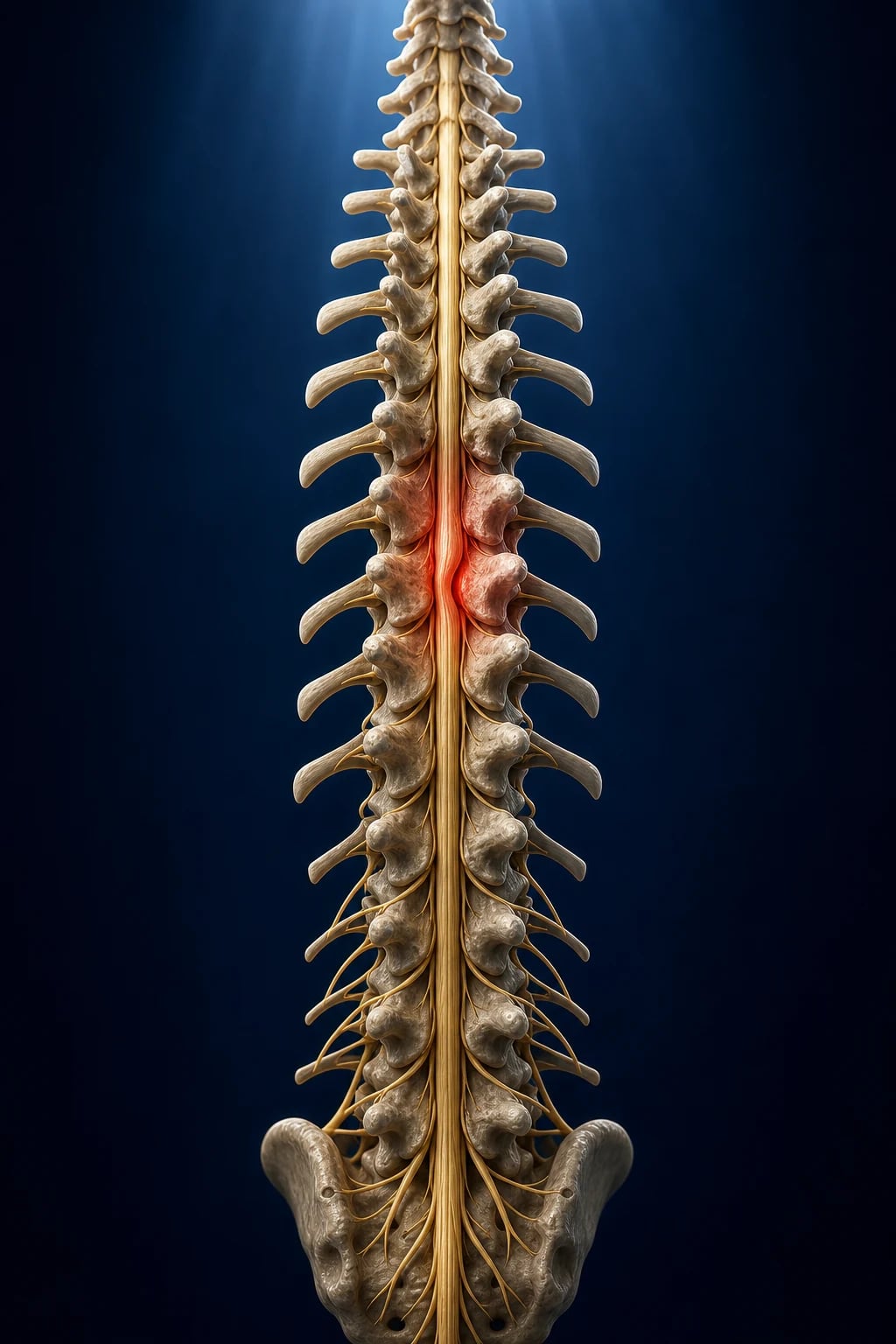
Pathophysiology — why the cord dies, and why it is time-critical
The ischaemic cascade of MSCC — four overlapping phases
| Phase | Mechanism | Reversibility |
|---|---|---|
| 1. Venous congestion | Epidural tumour compresses the epidural venous plexus first → venous outflow obstruction → vasogenic oedema in the cord white matter | Reversible with steroids/decompression — this is the window steroids exploit |
| 2. Vasogenic oedema | Venous back-pressure → breakdown of the blood-spinal cord barrier → interstitial oedema, raised cord tissue pressure | Reversible early; partially reversible later |
| 3. Arterial ischaemia | Rising tissue pressure exceeds arterial inflow → microvascular ischaemia, axonal conduction block → motor/sensory deficit | Largely irreversible beyond 24-48h |
| 4. Infarction | Sustained arterial compromise → cord infarction (grey + white matter necrosis) → permanent paraplegia, sphincter loss | Irreversible — the neurological state after this is fixed |
The clinical correlate is pivotal for the exam: pain (venous/periosteal — early) → weakness/sensory level (oedema + early ischaemia — salvageable) → paralysis/sphincter loss (infarction — fixed). Steroids, decompression and radiotherapy only work in the first two phases. This is why a patient who has been paraplegic for >48h rarely walks again regardless of treatment — the cord has infarcted.[1][2]
Aetiology and causes — the differential of a destructive epidural lesion
Metastatic MSCC (the mechanism to know cold)
Most MSCC is metastatic. Tumour emboli reach the vertebral column preferentially via Batson's vertebral venous plexus — a valveless, low-pressure network of veins running the length of the spinal column that communicates freely with the pelvic, breast, and pulmonary venous beds. Because it is valveless and low-pressure, it permits retrograde flow: rises in intra-abdominal or intra-thoracic pressure (cough, Valsalva) shatter tumour cells from the pelvis, breasts, and lungs backwards into the vertebral bodies. This is why the spine is such a fertile soil for metastasis and why prostate, breast and lung cancers — all draining into Batson's plexus — dominate the causes.[2]
Metastatic (≈97%)
Via Batson plexus
- Breast, lung and prostate are the "big three" primaries (≈60-70% of MSCC combined)
- Multiple myeloma / plasmacytoma — lytic bone disease, high MSCC burden
- Renal cell carcinoma and melanoma — classically RADIORESISTANT → favour surgery
- Lymphoma, small-cell lung cancer, breast and prostate — RADIOSENSITIVE → favour radiotherapy
- Thyroid, gastrointestinal (colon, gastric), unknown primary
- Vertebral body is the seed → tumour grows into the anterior/lateral epidural space → pushes the cord posteriorly/laterally
Non-metastatic mimics
Do not miss
- Intervertebral disc herniation — large central disc, usually lumbar (cauda equina); acute, often with a mechanical event
- Spinal epidural ABSCESS — Staph aureus #1; fever, severe pain, elevated inflammatory markers, rapid progression; a surgical emergency in its own right
- Spinal epidural HAEMATOMA — spontaneous (anticoagulation, coagulopathy) or post-procedural (epidural/spinal anaesthesia, lumbar puncture); sudden onset, severe pain
- Pathological vertebral fracture with retropulsion — cord compression from bone rather than tumour
- Primary spinal cord tumour — ependymoma, astrocytoma, meningioma, neurofibroma (much rarer; intradural on MRI)
- Transverse myelitis / demyelination — no destructive bone lesion; cord signal change only
The exam trap is the radioresistant tumour. Melanoma, renal cell carcinoma, sarcoma and most thyroid cancers respond poorly to radiotherapy — these patients should be referred for surgical decompression (if operable) rather than primary radiotherapy. Conversely, lymphoma, myeloma, seminoma, small-cell lung cancer, and hormone-sensitive breast and prostate cancer are highly radiosensitive and are usually managed with radiotherapy ± systemic therapy.[5]
Clinical features — what to find, and in what order they appear
Early symptoms
Better prognosis
- Back pain (#1 — present in 95%; progressive, mechanical, worse on lying/coughing/straining)
- Pain may precede neurological signs by days to weeks — the best window for salvage
- Nocturnal pain that is NOT relieved by rest (unlike mechanical back pain, which improves with rest)
- Radicular pain (band-like, in a dermatomal distribution) — from nerve root compression
- Lhermitte sign (electric shock down the spine on neck flexion) — cervical cord involvement
Late symptoms
Poor prognosis
- Motor weakness (paraparesis → paraplegia) — usually the sign that triggers urgent imaging
- Sensory loss (numbness below the level of compression — find the sensory level)
- Bowel/bladder dysfunction — URINARY RETENTION is classical; constipation/faecal incontinence; VERY LATE, poor prognosis
- Autonomic dysfunction — orthostatic hypotension, abnormal sweating, priapism
- Gait disturbance, falls — may be the presenting complaint in frail/older patients
The classical sequence (and what each stage means for outcome)
The temporal evolution of MSCC — pain → weakness → paralysis
1. Pain (95%) — earliest and most common
Localised back pain at the level of the lesion, often worse on lying flat, coughing, sneezing or straining (any manoeuvre that raises intra-abdominal/intra-thoracic pressure and distends Batson plexus or the epidural venous plexus). Radicular (band-like) pain follows nerve root irritation. Pain can precede any neurological deficit by days to weeks — this is the salvageable window, and missing it (attributing cancer-patient back pain to "musculoskeletal" causes) is the commonest reason patients present paraplegic.
2. Weakness (≈75% at diagnosis) — the urgency trigger
Motor weakness below the level: UMN signs above the conus (spasticity, hyperreflexia, extensor plantars), LMN signs at the level (flaccid, areflexic, wasted). Weakness is progressive and is THE indication for emergency imaging and surgical referral — a patient who is losing power hour by hour cannot wait for an elective MRI slot.
3. Sensory level (≈50%) — localises the lesion
Pinprick/light-touch level found on careful dermatomal examination LOCALISES the lesion (the cord segment is ~2 vertebral levels ABOVE the sensory level in the thoracic spine). Cervical compression → quadriparesis; thoracic → paraparesis; conus/cauda equina → saddle anaesthesia + lower motor neuron legs.
4. Sphincter dysfunction (≈50%) — the ominous late sign
Urinary retention is the classical finding (check a bladder scan / post-void residual — >100 mL is significant). Faecal incontinence / constipation and loss of anal tone follow. Sphincter involvement = conus or cauda equina involvement, or very severe cord compression, and portends a poor neurological outcome — many of these patients never regain continence or ambulation.
5. Complete paraplegia — the fixed, infarcted state
Once the cord has infarcted (deficit present >24-48h), the neurological state is largely fixed. Fewer than 10% of patients paraplegic for >48h ever walk again. This is why every step above is about reaching the patient in stages 1-2.
Diagnosis — whole-spine MRI is the gold standard
MSCC diagnostic protocol — the first 24 hours
1. Recognise and give steroids IMMEDIATELY (before imaging)
As soon as MSCC is suspected in a cancer patient with new/progressive back pain or any neurological symptom, give DEXAMETHASONE 10-16 mg IV STAT. Do NOT wait for the MRI to give steroids — the oedema-reducing effect is time-dependent and the MRI may be hours away. Steroids improve pain and preserve neurological function while imaging and definitive treatment are arranged.
2. Whole-spine MRI (the gold standard)
MRI of the WHOLE spine (sagittal T1/T2 + STIR, with axial images through any abnormal level) is the gold standard. Whole-spine (not just the symptomatic region) because ~30% of patients have multiple levels of disease, and the symptomatic level may not be the most dangerous on imaging. IV gadolinium helps define the epidural tumour and cord interface, and distinguishes tumour from post-treatment change. Urgent — within 24h if neurological signs, same-day if progressive deficit.
3. If MRI is contraindicated — CT myelography
For pacemaker/ICD, ferromagnetic implant, severe claude, or an unstable patient unsuitable for MRI: CT myelography (intrathecal contrast via lumbar puncture, then CT) demonstrates the block and cord displacement. It is invasive, less sensitive for soft tissue/paraspinal disease, and cannot show intramedullary oedema — but it is the accepted fallback.
4. Grade the compression — the Bilsky ESCC scale
On the axial MRI at the maximally compressed level, assign a Bilsky epidural spinal cord compression (ESCC) grade (0, 1a-c, 2, 3). This is the radiological grading that stratifies who needs surgery (high-grade ESCC 2-3) from who can be managed with radiotherapy alone (low-grade 0-1). It is the imaging scale the spinal oncology MDT uses to make the surgical decision.
5. Assess mechanical stability — the Spinal Instability Neoplastic Score (SINS)
SINS (six components: location, lesion type — lytic/blastic/mixed, spinal alignment, vertebral body collapse, posterolateral involvement, pain) scores 0-18. A SINS of 0-6 = stable (no surgical stabilisation needed); 7-12 = potentially unstable (refer to spine surgery); 13-18 = unstable (surgical stabilisation). Mechanical instability is an indication for surgery independent of cord compression.
6. Stage the disease and estimate prognosis
Identify the primary tumour (if unknown — biopsy), assess burden of metastatic disease (CT chest/abdomen/pelvis), and estimate life expectancy — the Patchell surgical criteria require life expectancy >3 months. A patient with a prognosis measured in weeks is better served by radiotherapy + palliative care than by major spinal surgery.
7. Baseline neurological examination — document the Frankel grade
Document a detailed baseline neurological exam (motor power by myotome, sensory level, reflexes, sphincter tone, ambulatory status) and assign a Frankel (ASIA) grade. Ambulatory status and Frankel grade at presentation are the strongest predictors of outcome and the benchmark against which treatment response is judged.
The Bilsky epidural spinal cord compression (ESCC) scale
The Bilsky ESCC scale is the radiological grading of cord compression on axial MRI at the level of maximal compression. It was developed and validated for inter-observer reliability by Bilsky and the Spine Oncology Study Group, and is the imaging scale that determines the surgical vs radiotherapy threshold.[3]
Bilsky ESCC scale — grade, definition, and management implication
| Grade | Definition (axial MRI) | Cord/canal involvement | Typical management |
|---|---|---|---|
| 0 | Bone-only disease; no epidural disease | Tumour confined to bone | Systemic therapy ± local RT; no emergency surgery |
| 1a | Epidural tumour abutting the cord but no cord deformation | No cord displacement | Radiotherapy (radiosensitive); usually no surgery |
| 1b | Epidural tumour with minimal cord deformation / displacement | Cord touched, minimally deformed | Radiotherapy ± surgery depending on primary and stability |
| 1c | Epidural tumour deforming the cord but no CSF block visible | Cord deformed, some CSF preserved | Radiotherapy; surgery if radioresistant / unstable / progressive |
| 2 | Epidural tumour producing partial CSF block — cord compressed but some CSF visible around it | Substantial compression, partial block | Surgery preferred (decompression) + RT; high-dose steroids |
| 3 | Epidural tumour producing complete CSF block — cord completely surrounded/compressed, no CSF visible | Complete block, severe compression | Surgical emergency — decompression if operable; otherwise emergent RT |
Rule of thumb: Bilsky ESCC 0-1 can usually be managed with radiotherapy ± systemic therapy; 2-3 (high-grade, with cord deformation/CSF block) usually warrants surgical decompression if the patient is operable, because radiotherapy alone seldom relieves a high-grade mechanical compression.[3][5]
The Spinal Instability Neoplastic Score (SINS)
SINS is a parallel scale that grades mechanical (not neurological) stability — because an unstable spine needs surgical fixation even if the cord is not compressed. Six components, each scored, total 0-18.[6]
SINS components and scoring (total 0-18)
| Component | 0 points | 1 point | 2 points | 3 points |
|---|---|---|---|---|
| Location | — | Junctional (occiput-C2, C7-T2, T11-L1, L5-S1) | Mobile spine (C3-C6, L2-L4) | Rigid (T3-T10) |
| Pain | Pain-free | Occasional non-mechanical pain | Mechanical pain (pain on movement/lying) | — |
| Bone lesion | — | Blastic | Mixed | Lytic |
| Alignment | — | — | Subluxation/translation present | — |
| Vertebral body collapse | — | No collapse, >50% body involved | <50% collapse | >50% collapse |
| Posterolateral elements | — | — | Involved (facet/pedicle) | — |
Interpretation: SINS 0-6 = stable; 7-12 = indeterminate/potentially unstable — refer to spine surgery; 13-18 = unstable (surgical stabilisation). Mechanical instability is an independent surgical indication: a SINS of 13-18 mandates a spine surgery opinion regardless of the Bilsky grade or neurological status.[6]
Management — the time-critical protocol
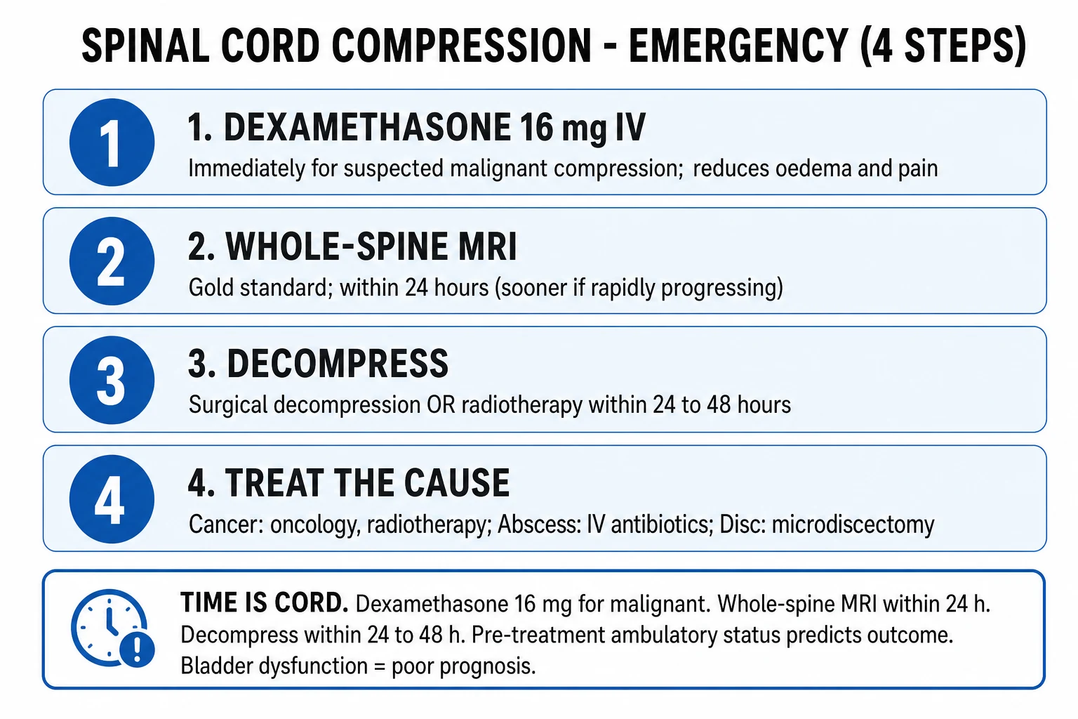
MSCC management protocol — steroids, image, decide, treat
1. Give DEXAMETHASONE 10-16 mg IV IMMEDIATELY on suspicion
On suspicion of MSCC — before imaging or specialist review. Reduces vasogenic spinal cord oedema from tumour compression, improves neurological function and pain, and buys time. Then dexamethasone 4 mg Q6H (16 mg/day), tapering over 2-3 weeks as definitive treatment takes effect. Do NOT delay steroids for MRI. AVOID the old 96-100 mg megadose bolus — the Sørensen trial showed it increases serious side effects (perforation, psychosis, infection) with no outcome benefit over moderate doses. The 10 mg IV STAT then 4 mg Q6H regimen is widely used and supported; some units (NICE) use 16 mg loading.
2. Whole-spine MRI (gold standard) — within 24h, same-day if progressive
MRI is the gold standard — shows exact level(s) of compression, extent of tumour, cord displacement/oedema, and paraspinal disease. Grade the compression (Bilsky ESCC) and stability (SINS). If multiple levels, treat the one causing the neurological deficit. If MRI contraindicated (pacemaker): CT myelography. Plain films/CT show bone destruction but NOT the cord — insufficient alone.
3. Multidisciplinary decision — surgery vs radiotherapy
Convene spinal surgery + radiation oncology + medical oncology urgently. Patchell trial (Lancet 2005): surgery + radiotherapy superior to radiotherapy alone for single-level compression in ambulatory or paraparetic patients with life expectancy >3 months — better ambulation, better continence, better survival. Indications for SURGERY: single-level (or few-level) compression, spinal instability (SINS 13-18), unknown primary needing biopsy, radioresistant tumour (melanoma, renal cell, sarcoma, most thyroid), recurrent compression after prior radiotherapy, or high-grade ESCC (2-3).
4. Surgical decompression ± stabilisation — within 24-48h
Direct decompressive surgery + stabilisation (the Patchell approach) within 24-48h of neurological deficit. The operation relieves mechanical compression immediately, obtains tissue for histology, and stabilises an unstable segment. Delay beyond 48h (especially >72h) progressively erodes the neurological benefit — the window of reversibility closes.
5. Radiotherapy — definitive or post-operative
For MULTIPLE levels, very poor performance status, expected survival <3 months, radiosensitive tumour (lymphoma, myeloma, seminoma, breast, prostate, SCLC), or after surgery. Typical regimens: 8 Gy single fraction (most common, best for short prognosis/pain), 20 Gy in 5 fractions, or 30 Gy in 10 fractions (better local control, longer prognosis). Given within 24-48h of diagnosis when definitive. Stereotactic body radiotherapy (SBRT) for oligometastatic/radioresistant disease.
6. Ongoing supportive care
Physiotherapy (maintain/gain strength, mobility, pressure-area care). Pain management — opioids for nociceptive pain, neuropathic agents (gabapentin/pregabalin, amitriptyline) for radicular pain, plus the steroid for tumour/oedema pain. VTE prophylaxis (cancer + immobility = very high DVT risk — LMWH). Bowel/bladder care (urinary catheter if retention; bowel regimen). Nutritional support. Psychological support and early palliative care involvement for advanced disease.
7. Treat the cancer — systemic therapy
After acute management, treat the underlying malignancy with disease-appropriate systemic therapy (hormonal for breast/prostate, chemotherapy for lymphoma/SCLC, targeted/immunotherapy as indicated). Bisphosphonates (zoledronic acid) or denosumab reduce skeletal-related events (including MSCC) in bone-metastatic breast/prostate/myeloma and are part of secondary prevention.
Surgical vs radiotherapy — the Patchell framework
Surgery vs primary radiotherapy — who gets what
| Favour SURGERY (decompression ± stabilisation) | Favour primary RADIOTHERAPY |
|---|---|
| Single-level (or few-level) compression | Multiple (≥3) levels of cord compression |
| Spinal instability — SINS 13-18 (mechanical indication alone) | Very poor performance status / bed-bound |
| High-grade ESCC (Bilsky 2-3) with cord deformation / CSF block | Expected survival <3 months |
| Unknown primary tumour — need tissue for diagnosis | Radiosensitive tumour (lymphoma, myeloma, seminoma, SCLC, breast, prostate) |
| Radioresistant tumour (renal cell, melanoma, sarcoma, thyroid) | Recurrent compression where surgery is not technically feasible |
| Spinal instability / pathological fracture with retropulsion | Patient declines surgery / not fit for anaesthesia |
| Recurrent compression in a previously irradiated field (cannot re-irradiate safely) | Cervical instability where surgery is very high risk |
| Life expectancy >3 months and ambulatory or paraparetic (Patchell criteria) | Paraplegic >48-72h (cord infarcted — surgery will not restore function) |
The Patchell trial is the cornerstone evidence: in a randomised comparison of direct decompressive surgery + radiotherapy versus radiotherapy alone (both 30 Gy / 10 fractions), the surgery group was more likely to remain ambulatory (84% vs 57%), regain ambulation if initially non-ambulatory (62% vs 19%), and retain continence (156 vs 17 days continent), with improved median survival (126 vs 100 days). The trial was stopped early for benefit. Caveat: it enrolled single-level compression only, in patients fit for surgery and with prognosis >3 months — extrapolation to multifocal or poor-prognosis disease is not supported.[1]
Steroid dose — the evidence and the current practice
Dexamethasone dose regimens — from megadose to moderate
| Regimen | Loading | Maintenance | Evidence / role |
|---|---|---|---|
| Megadose (historical, NOT recommended) | 96-100 mg IV bolus | 24-96 mg/day | Sørensen 1994 RCT: no neurological benefit over moderate doses; significantly MORE serious side effects (GI perforation, sepsis, psychosis). Abandoned. |
| Moderate (NICE / most UK & ANZ units) | 16 mg IV STAT | 8 mg BID (=16 mg/day), taper over 2-3 weeks | The most widely recommended loading dose; balances efficacy and toxicity. NICE CG75. |
| Moderate-low (alternative, widely used) | 10 mg IV STAT | 4 mg Q6H (=16 mg/day), taper over 2-3 weeks | A common pragmatic regimen; equivalent total daily dose; supported for most patients. Lower single-bolus dose may reduce bolus-related toxicity. |
| Low / analgesic | — | 2-4 mg BID | For pain control / mild compression without deficit, or palliative symptom relief |
The take-home for the exam: give a moderate loading dose of dexamethasone IMMEDIATELY on suspicion (before imaging), then continue at 16 mg/day tapering over 2-3 weeks while definitive treatment takes effect. Do NOT give a 96-100 mg megadose — it adds toxicity without benefit. The 10 mg IV STAT → 4 mg Q6H regimen is a perfectly acceptable and widely used alternative to the 16 mg bolus.[2][4][5]
Differential diagnosis — what else causes back pain + deficit in a cancer patient
MSCC vs the other destructive epidural lesions
| Feature | MSCC (metastatic) | Spinal epidural abscess | Spinal epidural haematoma | Cauda equina syndrome | Transverse myelitis |
|---|---|---|---|---|---|
| Onset | Days-weeks (can acutely worsen with fracture) | Days (can be hours once deficit starts) | Sudden (minutes-hours) | Hours-days | Hours-days |
| Pain | Progressive, worse lying/coughing | Severe, fever, night sweats | Sudden severe, often after procedure/anticoagulation | Low back + bilateral sciatica; saddle pain | Back pain + sensory level |
| Fever / sepsis | Absent | Present (sepsis, raised WBC/CRP) | Absent | Absent | Absent (± recent infection/vaccine) |
| Risk factors | Known cancer | IV drug use, immunocompromise, diabetes, bacteraemia, recent spinal procedure | Anticoagulation, coagulopathy, recent epidural/spinal/LP | Large central disc, trauma | Viral infection, vaccination, autoimmune |
| MRI | Destructive vertebral body lesion + epidural soft tissue + cord compression | Ring-enhancing collection, cord displacement | Biconvex epidural T1/T2 hyperintensity, no bone destruction | Large central disc at L4-S1; no cord (below conus) | Cord signal change, no destructive bone lesion, no epidural mass |
| Management | Dexamethasone + surgery/RT | Surgical decompression + IV antibiotics (anti-staph; cover MRSA) | Emergency surgical evacuation (or reversal of anticoagulation if early/minimal) | Emergency surgical decompression (within <24-48h) | MRI to exclude compression, then high-dose steroids/IVIG/plasma exchange as indicated |
The two non-malignant mimics an intensivist must not miss are epidural abscess (fever + sepsis + severe pain — needs surgical decompression + IV antibiotics) and epidural haematoma (anticoagulated/procedural + sudden pain — needs emergency surgical evacuation and anticoagulation reversal). Both present like MSCC but have a normal vertebral body on MRI — the destructive bone lesion is the radiological hallmark of metastatic MSCC.[2]
Prognosis and outcomes
Prognostic factors in MSCC — who walks, who survives
| Factor | Favourable | Unfavourable |
|---|---|---|
| Ambulatory status at presentation | Ambulatory (60-80% remain ambulatory) | Non-ambulatory / paraplegic (10-20% / <5% regain ambulation) |
| Duration of deficit before treatment | <24-48h | >48-72h (cord infarction → fixed deficit) |
| Rate of neurological progression | Slow (days-weeks) | Rapid (hours) |
| Primary tumour | Radiosensitive (myeloma, lymphoma, breast, prostate, SCLC) | Radioresistant (melanoma, renal cell, sarcoma) / poor-prognosis visceral mets |
| Number of compression levels | Single | Multiple |
| Bilsky ESCC grade | Low (0-1) | High (2-3) |
| Visceral / brain metastases | Absent | Present (worse survival) |
| Performance status | Good (ECOG 0-1) | Poor (ECOG 3-4) |
| Sphincter function | Continent | Incontinent / retention (late sign) |
Survival: median survival after MSCC is 3-6 months overall (it is a marker of advanced cancer), but ranges from weeks (visceral metastases, poor performance status) to years (e.g., hormonally-treated breast/prostate, myeloma). The strongest single determinant of functional outcome is ambulatory status at presentation — which is why early recognition and treatment before weakness develops is the whole point.[1][2][5]
Landmark trials and guidelines
Patchell 2005 — direct decompressive surgery + RT vs RT alone (PMID 16112300)
Source
Lancet — the landmark randomised trial that redefined MSCC management
Design
Randomised trial, 101 patients with single-level MSCC from solid tumours (excluded lymphoma/myeloma), all ambulatory or paraparetic, prognosis >3 months
Intervention
Direct decompressive surgical resection + post-op radiotherapy (30 Gy / 10 fractions) vs radiotherapy alone (same regimen)
Primary result
Surgery group significantly more likely to regain/maintain ambulation (84% vs 57%) and continence, with longer median survival (126 vs 100 days)
Key caveat
Trial stopped early for benefit; enrolled only SINGLE-LEVEL compression in FIT patients with prognosis >3 months — does NOT support surgery for multifocal or poor-prognosis disease
Clinical bottom line
For the operable, single-level, >3-month-prognosis patient, surgery + RT is superior to RT alone — the basis for offering surgery to fit patients
Sørensen 1994 — high-dose dexamethasone in MSCC (PMID 8142159)
Source
European Journal of Cancer — the only randomised trial of steroid dose in MSCC
Design
Randomised, double-blind, 57 patients with carcinomatous MSCC; high-dose dexamethasone (96 mg IV bolus then 96 mg/day taper) vs placebo, all receiving radiotherapy
Result
Trend toward better gait function in the steroid group at 3 months (81% vs 63%); small study, wide confidence intervals
Legacy / caveat
The megadose regimen (96 mg) tested here was ADOPTED historically but later ABANDONED — subsequent data showed megadose steroids add serious toxicity (GI perforation, psychosis, infection) without outcome benefit over moderate doses. The trial established that steroids help; the megadose did not survive.
Clinical bottom line
Steroids work — but give a MODERATE dose (10-16 mg loading then 16 mg/day). Do not use the 96-100 mg megadose.
Bilsky 2010 — reliability of the ESCC grading scale (PMID 20809724)
Source
Journal of Neurosurgery: Spine — the Spine Oncology Study Group validation study
What it established
A 6-point epidural spinal cord compression (ESCC) scale (0, 1a, 1b, 1c, 2, 3) graded on axial MRI at the maximally compressed level, with substantial inter- and intra-observer reliability
Clinical use
Stratifies the surgical decision: low-grade (0-1) disease can be managed with radiotherapy; high-grade (2-3) disease with cord deformation / CSF block usually warrants surgical decompression
Clinical bottom line
The imaging language the spinal oncology MDT speaks — know the 0/1a-c/2/3 framework and the surgery threshold at grade 2-3
Fisher 2010 — Spinal Instability Neoplastic Score / SINS (PMID 20562730)
Source
Spine — Spine Oncology Study Group evidence-based expert consensus
What it established
A six-component scoring system (location, pain, bone lesion type, alignment, vertebral collapse, posterolateral involvement) totalling 0-18, grading MECHANICAL (not neurological) instability in neoplastic spinal disease
Clinical use
0-6 stable; 7-12 potentially unstable (refer to spine surgery); 13-18 unstable (surgical stabilisation). Instability is an independent surgical indication — fix the spine even if the cord is not compressed.
Clinical bottom line
SINS complements Bilsky: Bilsky grades cord compression (neurological), SINS grades stability (mechanical) — both drive the surgical decision
Loblaw 2011 / 2012 — systematic review & clinical practice guideline (PMID 22420969)
Source
International Journal of Radiation Oncology, Biology, Physics — Cancer Care Ontario / ASCO-endorsed guideline
What it established
Updated systematic review of MSCC diagnosis and management; recommends urgent imaging, immediate steroids, and a stratified surgery-vs-radiotherapy approach based on Patchell criteria, primary tumour, and prognosis
Key recommendations
Whole-spine MRI as gold standard; dexamethasone on suspicion (moderate dose); surgery for single-level + fit + prognosis >3 months; radiotherapy for multifocal, radiosensitive, or poor-prognosis disease
Clinical bottom line
The pragmatic guideline synthesis — how to apply Patchell, Bilsky and SINS at the bedside
George 2015 — Cochrane review of MSCC interventions (PMID 26337716)
Source
Cochrane Database of Systematic Reviews — the most current comprehensive evidence appraisal
What it established
Systematic review of all randomised interventions for metastatic extradural spinal cord compression in adults; confirms surgery + RT > RT alone (Patchell), and that radiotherapy is effective for radiosensitive tumours and ambulatory patients
Limitations noted
Limited high-quality RCT evidence for steroid dose, radiotherapy fractionation, and the role of surgery in multifocal/poor-prognosis disease
Clinical bottom line
The evidence base is modest but consistent: steroids + urgent imaging + risk-stratified surgery or radiotherapy remains the standard of care
Exam practice
SAQ — Acute paraparesis in known breast cancer
10 minutes · 10 marks
A 62-year-old woman with metastatic breast cancer (bone-only disease, ECOG 1) presents with three weeks of progressive thoracic back pain, worse lying flat, and over the past 24 hours bilateral leg weakness and difficulty voiding. Examination reveals a T8 sensory level, 3/5 power in both legs, and a palpable bladder. MRI has not yet been performed.
SAQ — Radiotherapy timing in radioresistant single-level MSCC
10 minutes · 10 marks
A 58-year-old man with metastatic renal cell carcinoma presents with five days of progressive thoracic back pain and 12 hours of rapidly worsening paraparesis (power 2/5). Whole-spine MRI confirms Bilsky grade 3 ESCC at T6 with cord deformation and a CSF block, single-level disease, SINS 9. The spinal surgeons assess him as fit for surgery.
Clinical pearls [1]
Red flags
[1]Summary algorithm — the one-minute exam answer
MSCC — the entire management in seven moves
SUSPECT
Cancer patient + new/progressive back pain (worse on lying/coughing) or ANY new neurological symptom → MSCC until excluded.
STEROID
Dexamethasone 10-16 mg IV STAT immediately — BEFORE imaging. Then 4 mg Q6H, taper over 2-3 weeks. No megadose.
IMAGE
Whole-spine MRI (gold standard) within 24h, same-day if progressive deficit. CT myelogram if MRI contraindicated. Grade Bilsky ESCC + SINS.
REFER
Urgent spinal surgery + radiation oncology + medical oncology MDT. Parallel referrals — do not serialise.
DECIDE
Surgery if single-level + fit + prognosis >3 months + radioresistant/unstable/high-grade (Bilsky 2-3, SINS 13-18). Radiotherapy if multifocal, radiosensitive, poor prognosis, or not operable.
TREAT
Surgical decompression ± stabilisation within 24-48h, OR radiotherapy (8 Gy single fraction / 20 Gy-5# / 30 Gy-10#) within 24-48h. Then treat the cancer (systemic therapy + bisphosphonate/denosumab).
SUPPORT
Physiotherapy, analgesia (opioid + neuropathic agent), VTE prophylaxis (LMWH), bladder/bowel care, nutrition, early palliative care. Document ambulatory status / Frankel grade as the outcome benchmark.
References
- [1]Patchell RA, Tibbs PA, Regine WF, et al. Direct decompressive surgical resection in the treatment of spinal cord compression caused by metastatic cancer: a randomised trial Lancet, 2005.PMID 16112300
- [2]George R, Jeba J, Ramkumar G, et al. Interventions for the treatment of metastatic extradural spinal cord compression in adults Cochrane Database Syst Rev, 2015.PMID 26337716
- [3]Bilsky MH, Laufer I, Fourney DR, et al. Reliability analysis of the epidural spinal cord compression scale J Neurosurg Spine, 2010.PMID 20809724
- [4]Sørensen S, Helweg-Larsen S, Mouridsen H, Hansen HH. Effect of high-dose dexamethasone in carcinomatous metastatic spinal cord compression treated with radiotherapy: a randomised trial Eur J Cancer, 1994.PMID 8142159
- [5]Loblaw DA, Mitera G, Ford M, Laperriere NJ. A 2011 updated systematic review and clinical practice guideline for the management of malignant extradural spinal cord compression Int J Radiat Oncol Biol Phys, 2012.PMID 22420969
- [6]Fisher CG, DiPaola CP, Ryken TC, et al. A novel classification system for spinal instability in neoplastic disease: an evidence-based approach and expert consensus from the Spine Oncology Study Group Spine (Phila Pa 1976), 2010.PMID 20562730