ICU · Pharmacology
Antiarrhythmics — Vaughan-Williams Classification
Also known as Antiarrhythmics · Vaughan-Williams · Class I sodium channel blockers · Class II beta-blockers · Class III potassium channel blockers · Class IV calcium channel blockers · Amiodarone · Lidocaine · Procainamide · Flecainide · Adenosine · Digoxin · Sicilian Gambit
Antiarrhythmics — the Vaughan-Williams classification: Class I (the sodium channel blockers — Ia quinidine/procainamide [prolong AP + QT], Ib lidocaine/phenytoin [shorten AP, ischaemic-tissue selectivity], Ic flecainide/propafenone [markedly slow conduction, AVOID in structural heart disease — CAST]); Class II (the beta-blockers — metoprolol/esmolol/bisoprolol — slow AV node, reduce sympathetic); Class III (the potassium channel blockers — amiodarone [multichannel, half-life ~60 days, multi-organ toxicity], sotalol, ibutilide, dofetilide, dronedarone); Class IV (the calcium channel blockers — verapamil/diltiazem, non-dihydropyridine only); and the others (digoxin, adenosine, magnesium). Includes the Singh-Vaughan Williams mechanism, the Sicilian Gambit, each drug's indication/dose/adverse effects/contraindications/QT risk, and a full amiodarone toxicity deep dive (pulmonary, thyroid, hepatic, skin).
On this page & tools
Your progress
Saved locally on this device.
8 MCQs with explanations
Target exams
Overview & definition
The antiarrhythmics — the Vaughan-Williams classification (4 classes by the ion channel / receptor target). The Sicilian gambit (the alternative — by the mechanism). The clinical use in the ICU (the AF, the VT, the SVT).[1]
The Vaughan-Williams classification groups antiarrhythmics by the dominant ion channel or receptor the drug acts upon — proposed by EM Vaughan Williams in 1970 and refined in 1984.[1] It is imperfect (many drugs span several classes — amiodarone has Class I, II, III and IV actions — and it ignores clinical context such as structural heart disease), yet it remains the lingua franca of the exam and the bedside. The Sicilian Gambit (1991) is the alternative mechanistic framework: it classifies drugs by their precise molecular target (channel, receptor, pump), the resulting cellular effect (e.g. ↓ automaticity, ↑ refractoriness), and the clinical goal — useful for deeper mechanistic reasoning.[1]
A cardinal principle of ICU antiarrhythmic therapeutics: every antiarrhythmic is also potentially pro-arrhythmic. The very drugs used to suppress arrhythmias can prolong QT (→ torsades), depress conduction (→ heart block), or depress contractility (→ hypotension/heart failure).[12] Hence the standing rule — treat the patient, not the ECG: if the patient is shocked, ischaemic, or in pulmonary oedema, go straight to synchronised electrical cardioversion and reserve pharmacological management for the stable.
Three questions decide which drug (or electricity): (1) is the patient haemodynamically stable or unstable? (unstable → electricity, not drugs); (2) what is the rhythm (atrial vs ventricular, narrow vs broad, regular vs irregular)?; and (3) is there structural heart disease / LV dysfunction? (this bars flecainide and several Class I agents — CAST).[2]
The Vaughan-Williams classification
| Class | Mechanism | Agents | Effect |
|---|---|---|---|
| I | Na channel block (the phase 0) | Ia: quinidine, procainamide; Ib: lidocaine, phenytoin; Ic: flecainide, propafenone | Depress the conduction |
| II | Beta-blockade | Metoprolol, esmolol, bisoprolol | Slow the AV node; reduce the sympathetic |
| III | K channel block (the repolarisation) | Amiodarone, sotalol, ibutilide, dofetilide, dronedarone | Prolong the AP duration / the QT |
| IV | Ca channel block (the AV node) | Verapamil, diltiazem | Slow the AV node |
| Other (V) | — | Digoxin, adenosine, magnesium | See below |
The clinical use
- The amiodarone (class III + I + II + IV — the multichannel): the VT, the AF. The half-life (the weeks). The adverse: the pulmonary fibrosis, the thyroid (both hypo and hyper), the hepatitis, the corneal microdeposits, the photosensitivity, the QT prolongation + the torsades.[1]
- The lidocaine (class Ib): the VT (the post-MI; the ischemic). The - the - the.[1]
- The beta-blockers (class II): the rate control (the AF). The esmolol (the short half-life — the ICU titration).[1]
- The adenosine: the SVT termination (the AV node block). The 6 mg then 12 mg rapid IV push. The - the - the (the — the asthma, the heart transplant).[1]
- The digoxin: the AF rate control (the vagomimetic + the - the - the). The toxicity (the - the - the).[1]
- The magnesium: the torsades (the - the - the).[1]
Red flags
The Singh–Vaughan Williams mechanism & the Sicilian Gambit
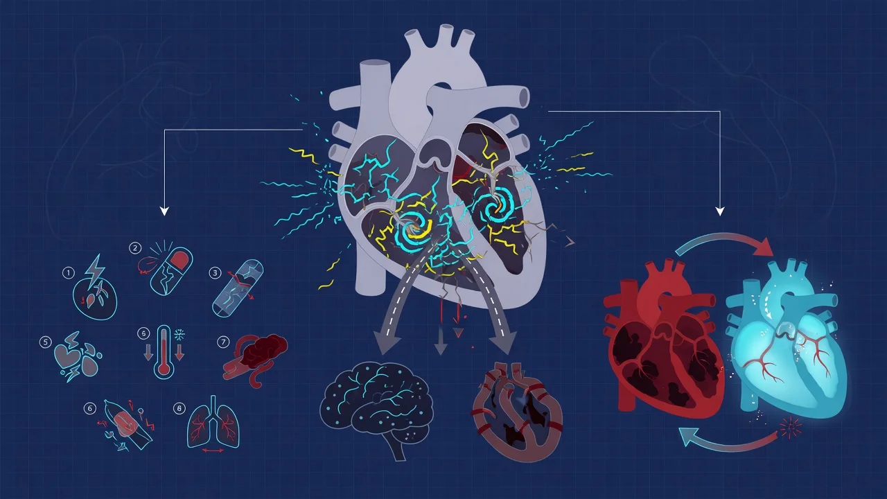
The original Vaughan-Williams scheme classifies by the dominant ion-channel effect, but the Singh–Vaughan Williams refinement and the Sicilian Gambit extend it by mapping each drug to a precise molecular target and the resulting electrophysiological consequence. The cellular effects that antiarrhythmics exploit are:[1]
- Reduce automaticity (phase-4 depolarisation) — by blocking the pacemaker current (I_f) or β-receptors (Class II), or Ca²⁺ channels (Class IV) at the SA/AV node. This slows a rapidly firing focus.
- Reduce conduction velocity (phase-0 depolarisation) — by blocking the fast Na⁺ channel (Class I). This abolishes re-entry by converting unidirectional block into bidirectional block.
- Prolong the refractory period / action-potential duration — by blocking the delayed-rectifier K⁺ current I_Kr (Class III). This lengthens the time a re-entrant wavefront encounters refractory tissue, abolishing the circuit.
- Reduce triggered activity (early and delayed afterdepolarisations) — magnesium suppresses EADs (torsades); calcium blockers and beta-blockers suppress DADs (digoxin toxicity, catecholaminergic VT). [1]
The Sicilian Gambit arranges drugs on a grid of targets (Na⁺, Ca²⁺, K⁺ channels; β-adrenoceptors; muscarinic receptors; the Na⁺/K⁺ pump) crossed with effects (membrane stabilisation, automaticity, conduction, refractoriness, contractility). Its virtue is that it explains why a particular drug suits a particular arrhythmia — and predicts the pro-arrhythmic and toxic profile. Its weakness is complexity at the bedside, which is why the 4-class shorthand endures.[1]
Class-specific ECG signatures: Class Ia prolongs QRS and QT; Class Ib shortens QT (the only group that does); Class Ic widens QRS (marked conduction slowing) with little QT effect; Class II and IV prolong the PR interval (AV-nodal effect); Class III prolongs the QT interval. [1]
Class I — sodium channel blockers (phase 0)
Class I drugs bind the fast voltage-gated Na⁺ channel and slow phase 0 (depolarisation) of the cardiac action potential, depressing conduction velocity. They are sub-classified by their kinetics of binding/unbinding and their effect on action-potential duration (APD). The state-dependent binding concept is exam gold: Class Ib drugs bind preferentially to inactivated channels (which predominate in ischaemic/depolarised tissue), explaining their niche in ischaemic VT.[1]
Class Ia — quinidine, procainamide, disopyramide
- Mechanism: intermediate Na⁺ channel kinetics; moderate phase-0 depression AND K⁺ channel block → prolong APD and QT. The only Class I subgroup that lengthens repolarisation.
- Indications: historically atrial and ventricular arrhythmias; procainamide IV still used for tolerated VT and to terminate WPW+AF; disopyramide for HOCM (negative inotropy reduces outflow gradient) and vagally-mediated AF.
- Doses: procainamide 20–50 mg/min IV until response, hypotension, QRS widens >50%, or 17 mg/kg; then 1–4 mg/min infusion. Quinidine largely historic. Disopyramide 100–200 mg PO.
- Adverse effects: quinidine — cinchonism (tinnitus, headache), thrombocytopenia, torsades ("quinidine syncope"), doubles digoxin levels (displace from tissue binding); procainamide → active metabolite NAPA (N-acetylprocainamide, a Class III), drug-induced lupus (anti-histone antibodies, ~30% chronic) — more common in slow acetylators; disopyramide — potent anticholinergic (urinary retention, glaucoma) and negative inotropy.
- Contraindications: long QT, torsades, digoxin toxicity; disopyramide in HFrEF/AV block.
- QT risk: HIGH. Largely superseded by amiodarone; availability is increasingly limited. [1]
Class Ib — lidocaine, phenytoin, mexiletine
- Mechanism: fast Na⁺ channel kinetics (bind/recover rapidly each cycle) and they shorten APD. Crucially, they preferentially act on ischaemic/depolarised tissue (where Na⁺ channels dwell in the inactivated state that Ib drugs bind) — hence their niche in ischaemic VT.[1]
- Indications: monomorphic VT in acute ischaemia/infarction (first-line); digoxin-toxic arrhythmias (phenytoin); neuropathic pain (mexiletine, oral lidocaine analogue). Ineffective for AF/SVT — do not reach for them in atrial arrhythmia.
- Doses: lidocaine 1–1.5 mg/kg IV bolus (→ 3 mg/kg total, may repeat), then 1–4 mg/min infusion (reduce in HF/hepatitis/age >70). Phenytoin loading 10–15 mg/kg IV slowly.
- Adverse effects: predominantly CNS — perioral tingling, metallic taste, tinnitus, light-headedness, seizures, coma (level-related); hypotension/bradycardia at high dose; phenytoin — gingival hyperplasia, nystagmus, ataxia, infusion-site reactions (purple-glove), hypotension (propylene glycol vehicle).
- Contraindications: atrial arrhythmias (won't work); severe AV block; prior idiosyncrasy. Reduce dose in liver failure/low cardiac output (lidocaine is hepatic-flow dependent).
- QT risk: NONE (shortens QT). This is the safest Class I group for the QT interval.
Class Ic — flecainide, propafenone
- Mechanism: slow Na⁺ channel kinetics → marked depression of conduction velocity (prominent QRS widening), minimal effect on APD/QT. Strong use-dependence (more block at faster rates).
- Indications: pharmacological cardioversion of recent-onset AF ("pill-in-the-pocket" in selected patients); AVNRT/AVRT; ventricular ectopy suppression (historically — see CAST). Propafenone has weak β-blocking activity.
- Doses: flecainide 2 mg/kg IV over 10 min (or 200–300 mg PO); propafenone 2 mg/kg IV over 10 min (or 450–600 mg PO).
- Adverse effects: pro-arrhythmia (incessant VT, organise AF into atrial flutter with 1:1 conduction — so ALWAYS co-prescribe an AV-nodal blocker), negative inotropy, QRS widening.
- Contraindications: any structural heart disease / LV dysfunction / ischaemia — CAST (1989/1991) showed increased mortality; established Brugada; HFrEF; second-/third-degree AV block. Flecainide challenge (provocative test) diagnoses Brugada but is contraindicated once established.[2]
- QT risk: low-moderate (mainly conduction/QRS, not QT) — but the pro-arrhythmic risk is lethal in damaged hearts.
Class II — beta-blockers (β-adrenoceptor antagonists)
- Mechanism: competitive blockade of cardiac β1-adrenoceptors → ↓ phase-4 depolarisation (automaticity), slow SA node rate, slow AV nodal conduction, and reduce the catecholamine-driven triggered activity that drives many ICU arrhythmias. They reduce myocardial oxygen demand and, in ischaemia, reduce infarct size and arrhythmic death.[1]
- Indications: rate control of AF/flutter/SVT (first-line); termination of AVNRT (with vagal manoeuvres); SVT prophylaxis; rate control in thyrotoxicosis/aerobic-exercise arrhythmia; reduces mortality post-MI and in HFrEF (the only antiarrhythmic class proven to improve survival); beta-blocker overdose (treated — see toxicology); congenital long-QT syndrome.
- Agents & doses:
- Metoprolol 5 mg IV slow push, repeat every 5 min up to 15 mg; PO 25–100 mg.
- Esmolol — loading 500 mcg/kg/min over 1 min then 50–300 mcg/kg/min infusion. Half-life ~9 minutes, cleared by plasma esterases (independent of liver/kidney) → ideal for ICU titration; if it causes hypotension/bradycardia it wears off in minutes.
- Landiolol — ultra-short-acting β1-selective (half-life ~4 min), used for critically ill/septic AF in some regions.
- Bisoprolol/carvedilol/nebivolol — oral chronic therapy (HFrEF mortality benefit).
- Adverse effects: bradycardia, AV block, hypotension, negative inotropy (worsens decompensated HF), bronchospasm (non-selective), masking of hypoglycaemia, fatigue, erectile dysfunction, hyperkalaemia (β2 blockade impairs cellular K⁺ uptake).
- Contraindications: decompensated heart failure (acute), severe asthma/bronchospasm (non-selective), advanced AV block (without pacemaker), severe peripheral vascular disease, cocaine intoxication (unopposed α → hypertension — use benzodiazepines first; consider labetalol/carvedilol which block α too).
- QT risk: low (and chronic use SHORTENS QT — used to treat congenital long-QT). Class II is among the safest antiarrhythmics for QT.
Class III — potassium channel blockers (prolong repolarisation)
Class III drugs block the delayed-rectifier potassium current (I_Kr), prolonging phase-3 repolarisation → prolonged APD and refractory period → broadened QT, which suppresses re-entry. They are the most-used antiarrhythmics in modern ICU practice. A unifying caveat: reverse-use dependence — the QT-prolonging effect is greatest at slow heart rates, so bradycardia, hypokalaemia and hypomagnesaemia amplify torsades risk.[1]
Amiodarone — the workhorse ICU antiarrhythmic (full deep dive)
Amiodarone is the single most effective and broadly applicable antiarrhythmic in the ICU. It is genuinely multichannel: predominant Class III (K⁺ block) but also Class I (Na⁺), Class II (non-competitive beta-blockade), and Class IV (Ca²⁺ channel) activity. This breadth explains its efficacy across AF, VT, and VF and the fact that it is safe in heart failure (minimal negative inotropy). [1]
Pharmacokinetics — the problem of the long half-life: amiodarone is enormously lipophilic (accumulates in fat, liver, lung, skin), is highly protein-bound, and has a terminal half-life of ~60 days (range 25–100) after chronic dosing. Effects accumulate and persist for months after cessation. IV onset is faster (minutes) but full tissue loading takes weeks. This long half-life underpins both its smooth efficacy and its toxicity profile.[1]
Dosing:
- Cardiac arrest (VF/pulseless VT): 300 mg IV bolus after the 3rd shock; 150 mg at the 5th shock; then 900 mg infusion over 24 h.
- Stable VT / pharmacological cardioversion of AF: 300 mg IV over 20–60 min (or 5 mg/kg), then 900 mg over 24 h (1 g/day).
- Oral loading: 600–1600 mg/day for 1–3 weeks → maintenance 100–400 mg/day. [1]
The four-pillar toxicity profile — know all four: [1]
- PULMONARY toxicity (amiodarone-induced pulmonary toxicity, APT). The most feared — interstitial pneumonitis / pulmonary fibrosis, occasionally organising pneumonia or ARDS. Risk relates to cumulative dose and duration; onset months-years into therapy (but acute pulmonary toxicity after IV bolus is described). Presents with dry cough, dyspnoea, new infiltrates on imaging. Mortality 10–20% if unrecognised. Diagnosis is one of exclusion; baseline + surveillance chest imaging and DLCO are mandatory. Treatment = stop amiodarone ± corticosteroids.[1]
- THYROID toxicity (both directions). Amiodarone is ~40% iodine by weight — a single 200 mg tablet delivers ~75 mg of organic iodine (50× the daily requirement). This produces both amiodarone-induced thyrotoxicosis (AIT, type 1 iodine-induced and type 2 destructive thyroiditis) and amiodarone-induced hypothyroidism. AIT is harder to treat and carries excess mortality in the ICU. Check TSH at baseline and every 6 months.
- HEPATIC toxicity. Transaminitis (common, often subclinical, up to ~25%), clinically significant hepatitis, and rarely cirrhosis/acute liver failure. The IV formulation (polysorbate 80 vehicle) can cause acute hepatotoxicity. Check LFTs at baseline and every 6 months.
- SKIN & ocular toxicity. Blue-grey (slate-grey) discolouration of sun-exposed skin + photosensitivity (cosmetically distressing, slowly reversible on cessation); corneal microdeposits (virtually universal — vortex keratopathy, usually asymptomatic but rarely cause halos/photophobia — reversible). Less heralded: bluish nail/skin deposits, and chronic peripheral neuropathy, tremor, ataxia, optic neuropathy (rare, serious).[1]
Drug interactions (the exam favourite): amiodarone inhibits CYP3A4, CYP2C9, and P-glycoprotein → halve the dose of warfarin (marked INR rise) and halve the dose of digoxin (P-gp), and increase levels of statins (simvastatin — rhabdomyolysis risk), ciclosporin, flecainide, phenytoin. Additive QT prolongation with all Class Ia/III drugs, macrolides, fluoroquinolones, antipsychotics, methadone. [1]
QT prolongation: amiodarone reliably prolongs the QT — but torsades is uncommon (it homogeneously prolongs repolarisation across the myocardial wall, reducing transmural dispersion — unlike ibutilide/sotalol). Nonetheless, correct K⁺/Mg²⁺ and avoid co-prescription with other QT-prolongers where possible. [1]
Sotalol
- Class III (K⁺ block, I_Kr) plus Class II (non-selective beta-blockade — the racemic drug; the d-isomer is pure Class III but is pro-arrhythmic without the protective beta-block, so racemic sotalol is used). Renal excretion (dose-adjust in renal impairment). Used for AF rhythm control and ventricular ectopy. Reverse-use dependence means its QT-prolonging effect is greatest at slow heart rates → torsades risk, particularly in bradycardia, hypokalaemia, and renal failure; initiate in-hospital with QT monitoring.
- QT risk: HIGH. [1]
Ibutilide
- IV Class III for pharmacological cardioversion of recent-onset AF/flutter, particularly flutter. Carries the highest torsades risk of any antiarrhythmic (≈4–8%) — give with magnesium pre-treatment and continuous ECG monitoring for at least 4 hours after the dose.
- Dose: 1 mg IV over 10 min (0.01 mg/kg <60 kg), repeat once if needed.
- QT risk: HIGHEST. [1]
Dofetilide
- Pure I_Kr blocker; oral, for AF rhythm control. Marked QT/torsades risk → mandatory in-hospital initiation with creatinine-clearance-based dosing and continuous QT monitoring (mandatory 3-day monitored admission in some jurisdictions, with specialist prescriber qualification required). [1]
Dronedarone
- A benzofuran derivative of amiodarone WITHOUT the iodine moiety → less pulmonary/thyroid/skin toxicity but also less effective than amiodarone. Used for paroxysmal/persistent AF rhythm control. ATHENA (2009) showed reduced CV hospitalisation/death in stable AF — the one positive dronedarone trial.[7]
- CONTRAINDICATED in heart failure — ANDROMEDA (Køber 2008) showed increased mortality in decompensated HFrEF (early excess deaths).[9] PALLAS (Connolly 2011) showed increased mortality/stroke/CV hospitalisation in permanent (long-standing persistent) AF → dronedarone is now restricted to paroxysmal/persistent AF that can be cardioverted to sinus rhythm.[8]
- AVOID with strong CYP3A4 inhibitors (severe interaction) and in severe hepatic impairment. QT risk moderate.
Class IV — calcium channel blockers (non-dihydropyridine)
- Mechanism: block the L-type Ca²⁺ channel, principally at the SA and AV nodes (which are Ca²⁺-dependent), slowing conduction and increasing refractoriness. Only the non-dihydropyridines (verapamil, diltiazem) have AV-nodal antiarrhythmic activity; the dihydropyridines (nifedipine, amlodipine, felodipine) are vascular-selective vasodilators with no clinically useful AV-nodal effect.[1]
- Indications: rate control of AF/flutter (when beta-blocker contraindicated); termination of AVNRT (IV verapamil/diltiazem); idiopathic left ventricular fascicular VT (verapamil-sensitive) — the one VT that verapamil treats.
- Doses: diltiazem 0.25 mg/kg IV (~20 mg) over 2 min, repeat 0.35 mg/kg at 15 min, then 5–15 mg/h infusion; verapamil 5–10 mg IV over 2 min (repeat 5–10 mg at 15–30 min if needed). Oral forms for chronic rate control.
- Adverse effects: bradycardia, AV block, hypotension, negative inotropy (worsens HFrEF), constipation (verapamil).
- Contraindications: broad-complex tachycardia of uncertain origin (if it is VT → cardiovascular collapse/death — the classic "verapamil death"); HFrEF (negative inotropy); HOCM (worsens outflow gradient); WPW + AF (→ VF); second-/third-degree AV block; concomitant IV beta-blocker (severe bradycardia/asystole).
- QT risk: none. (These drugs slow the AV node and shorten the ventricular response, not QT.)
"Class V" — adenosine, digoxin, magnesium
Adenosine
- Mechanism: agonist at the A1 adenosine receptor → opens inward-rectifier K⁺ channels + blocks Ca²⁺ entry → transient AV-nodal block. Endogenous adenosine underlies the brief asystole of the diving reflex and of reperfusion. Half-life ~10 seconds (rapidly deaminated/uptaken by RBCs) → effects vanish within one circulation time.
- Indication: termination of regular narrow-complex SVT (AVNRT/AVRT — re-entry involving the AV node). Diagnostic use in stable irregular/broad tachycardia (transient AV block reveals underlying atrial activity). Will NOT cardiovert AF (only transiently slows it).
- Dose: 6 mg rapid IV push followed immediately by a flush, then 12 mg if no response; can repeat 12 mg once. Must warn the patient of the brief unpleasant "doom"/asystole/sense of impending death. Lower dose (3 mg) in denervated (transplanted) hearts, central line administration, or dipyridamole pretreatment.
- Adverse effects: flushing, chest tightness, dyspnoea, brief asystole (obligate — the mechanism); usually resolves within seconds.
- Contraindications: asthma/COPD with bronchospasm (adenosine is a bronchoconstrictor); denervated (transplanted) hearts (denervation hypersensitivity → prolonged asystole — halve the dose); second-/third-degree AV block; sick sinus syndrome. Always have resuscitation gear ready.[1]
Digoxin
- Mechanism: inhibits the Na⁺/K⁺-ATPase → ↑ intracellular Na⁺ → reduced Na⁺/Ca²⁺ exchange → ↑ intracellular Ca²⁺ → positive inotropy; and vagomimetic (parasympathomimetic) action → slowed AV-nodal conduction (the basis of its rate-control use in AF). It has a narrow therapeutic index and accumulates in renal failure.
- Indications: rate control of AF (especially in HF/HOCM where beta-blocker/CCB are problematic — it is the one rate-control drug safe in HFrEF); adjunct in HFrEF (mild symptomatic benefit; neutral mortality). Onset slow (hours) — not for acute rate control alone.
- Dose: 500 mcg IV (repeated every 6–8 h to loading ~1 mg), then 62.5–250 mcg/day (renal-adjusted). Therapeutic level 0.5–0.9 ng/mL (modern low targets).
- Toxicity: atrial tachycardia with block, bidirectional VT (pathognomonic), bradycardia/AV block, premature ventricular complexes, nausea/vomiting, xanthopsia (yellow-green vision), confusion, hyperkalaemia (the ATPase is poisoned → K⁺ leaks out of cells — a high K⁺ suggests severe toxicity). Predisposed by hypokalaemia (digoxin and K⁺ compete for the ATPase — always correct K⁺), renal failure, hypomagnesaemia, hypercalcaemia, hypothyroidism.
- Antidote: digoxin-specific Fab antibody fragments (Digibind/DigiFab) for life-threatening arrhythmia, hyperkalaemia, massive overdose. Do NOT give calcium (theoretical "stone heart" — controversial, but Fab is definitive).[12]
- QT risk: low (digoxin does not prolong QT — at toxic levels it shortens QT and causes the characteristic "digoxin effect" sagging ST segments).
Magnesium
- Mechanism: cofactor for the Na⁺/K⁺-ATPase; suppresses early afterdepolarisations (the trigger for torsades) and L-type Ca²⁺ channels; membrane stabiliser. Effective even when serum Mg²⁺ is "normal" (intracellular depletion matters more than serum).
- Indications: torsades de pointes (first-line); digoxin toxicity adjunct; hypomagnesaemia; polymorphic VT; AF rate control (refractory); eclampsia prophylaxis/treatment.
- Dose: 2 g (8 mmol) IV over 1–2 min (torsades), then 1–2 g/h infusion; 1–2 g IV in cardiac arrest with suspected hypomagnesaemia/torsades.
- Adverse effects: flushing, hypotension, bradycardia, areflexia (dose-related; sign of toxicity — check reflexes), respiratory depression at high levels; potentiates neuromuscular blockers.
- Contraindications: significant renal failure (risk of hypermagnesaemia — reduce dose, monitor levels/reflexes), heart block.
- QT risk: SHORTENS QT (anti-torsadogenic). [1]
QT prolongation risk — ranked
| Risk tier | Drug | Notes |
|---|---|---|
| Highest | Ibutilide | Torsades in ~4–8%; magnesium pre-treatment; monitored |
| High | Sotalol, quinidine, procainamide, disopyramide, dofetilide | Reverse-use dependence; initiate in-hospital |
| Moderate | Amiodarone | Reliably prolongs QT but torsades uncommon (homogeneous repolarisation) |
| Low | Flecainide/propafenone | Mainly QRS widening; lethal via different mechanism in damaged hearts |
| Low | Beta-blockers, verapamil, diltiazem | Safe for QT; beta-blockers TREAT long-QT |
| Shortens QT | Lidocaine/phenytoin (Ib), magnesium | Anti-torsadogenic; ischaemic-VT niche |
Beyond antiarrhythmics, common ICU QT-prolonging culprits (the "hidden killer"): macrolides (azithromycin, clarithromycin), fluoroquinolones (ciprofloxacin, moxifloxacin), antipsychotics (haloperidol — common in ICU delirium), methadone, ondansetron, fluconazole. Risk is additive and amplified by bradycardia, hypokalaemia, hypomagnesaemia. QTc >500 ms or a >60 ms rise from baseline → stop/reduce the culprit and recheck.[12]
Amiodarone toxicity — monitoring schedule
| System / toxicity | Test / action | Frequency |
|---|---|---|
| Pulmonary fibrosis | Baseline + surveillance chest X-ray (or CT) and DLCO; symptom review (cough/dyspnoea) | Baseline, 3-monthly symptom review, 6–12-monthly imaging |
| Thyroid (hypo/hyper) | TSH, free T4 | Baseline, then 6-monthly |
| Hepatic | LFTs (ALT/AST/ALP/bilirubin) | Baseline, then 6-monthly |
| Cardiac (QT, bradycardia) | ECG (QTc, rate), electrolytes (K⁺/Mg²⁺) | Baseline, then periodically |
| Ocular (corneal deposits) | Symptom review (halos/photophobia); slit-lamp if symptomatic | Baseline, then as symptomatic |
| Skin | Sun-protection advice; examine for blue-grey discolouration | At each review |
| Drug interactions | Review warfarin INR (halve dose), digoxin level (halve dose), statin (avoid simvastatin) | At initiation and dose changes |
| Neurological | Screen for tremor, neuropathy, ataxia | At each review |
Compare: Class I sub-groups
Ia — Quinidine/Procainamide
Na block + prolong APD/QT
- Intermediate Na⁺ channel kinetics; moderate phase-0 depression + K⁺ block → PROLONG QT
- Quinidine: cinchonism, thrombocytopenia, torsades ("quinidine syncope"), doubles digoxin levels
- Procainamide → NAPA (active Class III metabolite); IV for tolerated VT; lupus-like syndrome (anti-histone Ab, chronic use)
- Disopyramide: potent negative inotrope (used in HOCM); marked anticholinergic (urinary retention, glaucoma)
- QT risk HIGH; largely superseded by amiodarone; availability limited
Ib — Lidocaine
Shorten APD; ischaemic-tissue selectivity
- Fast Na⁺ channel kinetics; SHORTEN APD; bind preferentially to depolarised/ischaemic tissue (inactivated channels)
- Lidocaine 1–1.5 mg/kg IV bolus → infusion 1–4 mg/min (reduce in HF/hepatic disease/age)
- FIRST-LINE for monomorphic VT in ACUTE ISCHAEMIA/infarction
- Ineffective for AF/SVT (do not use for atrial arrhythmias)
- QT risk NONE (shortens QT — safest Class I for QT); toxicity = CNS: perioral tingling, tinnitus, seizures, coma (level-related)
Ic — Flecainide/Propafenone
Markedly slow conduction; AVOID structural heart disease
- Slow Na⁺ kinetics → MARKED conduction slowing (QRS widening), little APD effect
- Effective for recent-onset AF cardioversion ("pill-in-pocket") in NORMAL hearts only; ALWAYS co-prescribe AV-nodal blocker (organises AF into flutter with 1:1)
- CAST trial: INCREASED MORTALITY post-MI → CONTRAINDICATED in structural/LV dysfunction, ischaemia, HFrEF
- Flecainide challenge diagnoses Brugada; contraindicated in established Brugada
- Propafenone (Ic + weak beta-blockade) shares the same structural-heart prohibition
Compare: AF rate-control vs rhythm-control agents
Rate control (AV node)
Slow ventricular response
- BETA-BLOCKER (metoprolol/esmolol) — first-line; AVOID in decompensated HF/asthma
- NON-DHP CCB (diltiazem/verapamil) — if beta-blocker contraindicated; AVOID in HFrEF/HOCM
- DIGOXIN — when others contraindicated; SAFE in HF/HOCM; slow onset (hours); vagomimetic; low therapeutic index
- AV node blockers NEVER in WPW + AF (accessory pathway → VF)
- Target: HR <110 (lenient, AFFIRM/RACE II) or <80 strict if symptomatic
Rhythm control (cardioversion)
Restore sinus rhythm
- AMIODARONE — safest in structural heart disease/HF; broad-spectrum; long half-life; multichannel
- FLECAINIDE/PROPAFENONE — "pill-in-pocket"; NORMAL heart only (not HF/ischaemia) — CAST
- IBUTILIDE/VERNAKALANT — IV pharmacological cardioversion (torsades risk for ibutilide)
- ELECTRICAL synchronised cardioversion — most effective; REQUIRED if unstable
- Consider catheter ablation for drug-refractory/symptomatic rhythm control
Compare: amiodarone vs lidocaine for VT
Amiodarone
Multichannel Class III ± I/II/IV
- Broad-spectrum: AF, stable VT, VF/pulseless VT (300 mg in arrest after 3rd shock)
- Stable VT: 300 mg IV over 1 h → 900 mg over 24 h infusion
- SAFE in heart failure (minimal negative inotropy)
- Effective in BOTH ischaemic and non-ischaemic VT
- Long half-life (~60 days); multi-organ toxicity (pulmonary/thyroid/hepatic/QT)
- ALPS trial: no overall survival benefit vs placebo in OHCA, but benefit in witnessed/bystander-CPR subgroup
Lidocaine
Class Ib — ischaemic-tissue selectivity
- Preferred for VT in ACUTE ISCHAEMIA / post-MI (ischaemic-tissue selectivity)
- 1–1.5 mg/kg IV bolus (→ 3 mg/kg max) → 1–4 mg/min infusion
- Ineffective for AF/SVT; QT risk none (shortens QT)
- CNS toxicity: seizures, perioral tingling, coma
- ALPS trial: similar (no) overall survival benefit vs placebo in OHCA; historical second-line to amiodarone
Compare: amiodarone vs dronedarone
Amiodarone
Iodine-containing benzofuran
- 40% iodine by weight → thyroid toxicity (both directions)
- Half-life ~60 days; extremely lipophilic (fat/liver/lung/skin accumulation)
- Broadest efficacy: AF, VT, VF; SAFE in heart failure
- Toxicity: pulmonary fibrosis, hepatitis, thyroid, blue-grey skin, corneal deposits, neuropathy
- CYP3A4/2C9 + P-gp inhibition → halve warfarin + digoxin doses
- QT prolongation (torsades uncommon)
Dronedarone
Iodine-free benzofuran derivative
- No iodine → less thyroid/skin/pulmonary toxicity
- Shorter half-life (~24 h); less lipophilic → less tissue accumulation
- LESS effective than amiodarone; oral only
- CONTRAINDICATED in HFrEF (ANDROMEDA — mortality) and permanent AF (PALLAS — stroke/mortality)
- ATHENA: reduced CV hospitalisation/death in STABLE paroxysmal/persistent AF
- AVOID with strong CYP3A4 inhibitors; LFT monitoring (hepatotoxicity — rare liver failure)
FlowSteps: management of new-onset atrial fibrillation in the ICU
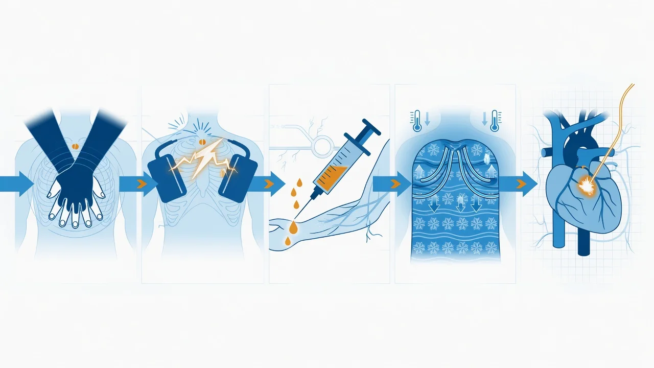
Management of new-onset atrial fibrillation in the ICU
- ASSESS HAEMODYNAMIC STABILITY — this is the decisive branch point. (a) UNSTABLE features: hypotension (SBP <90), shock, ischaemic chest pain, acute pulmonary oedema, altered conscious state → go straight to synchronised DC cardioversion (step 2). (b) STABLE → proceed to rate or rhythm control (steps 3–5). (c) Establish monitoring (continuous ECG, SpO₂, BP — arterial line if unstable), IV access, 12-lead ECG (confirm AF — irregularly irregular, no P waves; check for delta wave = WPW; check QT), bloods (K⁺, Mg²⁺, Ca²⁺, troponin, TSH, FBC, U&E), and SEARCH FOR TRIGGERS (sepsis, electrolyte disturbance, hypoxia, pain/agitation, inotropes/vasopressors, fluid overload, MI, PE, thyroid). Correct any reversible trigger — this alone often terminates the AF.
- SYNCHRONISED DC CARDIOVERSION (if unstable). (a) Sedate/anaesthetise (propofol/etomidate + analgesia). (b) Pads anterior–posterior or anterolateral. (c) SYNCHRONISE to the R wave (avoids delivering the shock on the T wave → VF). (d) 120–200 J biphasic (escalating if needed). (e) If AF >48 h/unknown duration, stroke risk exists — but in the unstable patient cardiovert immediately and anticoagulate after (heparin). (f) If unsuccessful, repeat at higher energy, consider amiodarone 300 mg IV then re-cardiovert.
- RATE CONTROL (stable patient — the default for most). (a) BETA-BLOCKER: metoprolol 5 mg IV slow push (repeat every 5 min to 15 mg) OR esmolol 500 mcg/kg/min ×1 min then 50–300 mcg/kg/min. (b) NON-DHP CCB (if beta-blocker contraindicated): diltiazem 0.25 mg/kg IV, repeat 0.35 mg/kg at 15 min, then 5–15 mg/h. AVOID verapamil/diltiazem in HFrEF, HOCM, broad-complex/WPW. (c) DIGOXIN (if HF/HOCM or others contraindicated): 500 mcg IV (slow onset — hours). (d) Target HR <110 (lenient) or <80–90 if symptomatic. (e) NEVER give AV-nodal blockers in WPW + AF — use amiodarone or cardiovert.
- RHYTHM CONTROL (selected stable patients). Indications: new-onset (<48 h), highly symptomatic despite rate control, younger/first episode, tachycardia-mediated cardiomyopathy. (a) AMIODARONE (ICU default, especially structural heart disease): 300 mg IV over 1 h, then 900 mg over 24 h; effective and safe in HF. (b) FLECAINIDE 2 mg/kg IV over 10 min (or 200–300 mg PO) — ONLY structurally normal heart — CAST. (c) IBUTILIDE 1 mg IV over 10 min (pre-treat with magnesium, monitor). (d) If fails/recurs → elective synchronised cardioversion (after anticoagulation/TOE).
- ANTICOAGULATION + LONG-TERM PLAN. (a) Stroke risk: CHA₂DS₂-VASc (0 male/1 female = omit; ≥2 = anticoagulate; 1 = consider). Bleeding: HAS-BLED. (b) For cardioversion: if AF <48 h → cardiovert then start anticoagulation; if >48 h/unknown → therapeutic anticoagulation 3 weeks before (or TOE) + 4 weeks after. (c) DOAC (apixaban/rivaroxaban/dabigatran) preferred to warfarin for non-valvular AF (warfarin for moderate-severe mitral stenosis/mechanical valve). (d) LONG-TERM: rate control ± rhythm control; consider ablation if drug-refractory/symptomatic. (e) Correct all reversible ICU triggers before labelling "lone AF".
FlowSteps: management of ventricular tachycardia in the ICU
Management of ventricular tachycardia in the ICU
- PULSELESS VT/VF → ARREST ALGORITHM. (a) Confirm no pulse → start CPR (100–120/min, depth 5–6 cm). (b) DEFIBRILLATE (UNSynchronised) 150–200 J biphasic ASAP. (c) 2 min CPR → rhythm check → shock → 2 min CPR → rhythm check → shock → AMIODARONE 300 mg IV after the 3rd shock → 2 min CPR → shock → AMIODARONE 150 mg at the 5th shock. (d) Reversible causes — 4 Hs and 4 Ts (hypoxia, hypovolaemia, hypo/hyperkalaemia, hypothermia; thrombosis [coronary/PE], tension pneumothorax, tamponade, toxins). (e) Post-ROSC: amiodarone infusion 900 mg/24 h, treat cause, targeted temperature management if comatose.
- STABLE MONOMORPHIC VT (broad, regular, with a pulse, tolerated). (a) Confirm VT (broad complex, AV dissociation, capture/fusion beats, concordance) — but treat as VT any broad-complex tachycardia with structural heart disease (do NOT give verapamil). (b) AMIODARONE 300 mg IV over 20–60 min then 900 mg over 24 h — first-line in most ICUs. (c) LIDOCAINE 1–1.5 mg/kg IV (if acute ischaemia/infarction; or amiodarone unavailable). (d) PROCAINAMIDE (where available) 20–50 mg/min to response or 17 mg/kg. (e) If pharmacological fails or patient destabilises → synchronised DC cardioversion (100–200 J).
- TORSADES DE POINTES (polymorphic VT with long QT). (a) Recognise: polymorphic (twisting) VT around a baseline, with a prolonged QT in sinus beats between episodes. (b) STOP all QT-prolonging drugs (antiarrhythmics — sotalol/ibutilide/quinidine/procainamide/amiodarone; antibiotics — macrolides/fluoroquinolones; antipsychotics; methadone; ondansetron). (c) MAGNESIUM 2 g IV bolus over 1–2 min (then 1–2 g/h) — first-line, effective even if Mg²⁺ "normal". (d) Correct K⁺ (target >4.5) and Mg²⁺ (target >1.0). (e) If bradycardia-dependent (pause-induced) → overdrive pacing (transvenous 90–110 bpm) or isoprenaline to increase HR and shorten QT. (f) If pulseless → defibrillate (unsynchronised) + CPR.
- IDENTIFY AND TREAT THE SUBSTRATE. (a) Ischaemia (troponin, angiography) — commonest driver of monomorphic VT (scar-related re-entry) and polymorphic VT. (b) Electrolytes (K⁺, Mg²⁺, Ca²⁺). (c) Drug toxicity (digoxin — Fab fragments; cocaine/sympathomimetics — benzodiazepines ± beta-blockers; QT-prolongers — stop). (d) ICD evaluation for secondary prevention if structurally driven VT. (e) Correct hypoxaemia, acidosis, inotrope excess — all pro-arrhythmic.
FlowSteps: termination of regular narrow-complex SVT in the ICU
Termination of regular narrow-complex SVT (AVNRT/AVRT) in the ICU
- CONFIRM IT IS REGULAR NARROW-COMPLEX SVT. 12-lead ECG: rate 150–250, narrow QRS (<120 ms), regular, P waves buried/retrograde. Exclude AF (irregular) and broad-complex tachycardia (treat as VT). If haemodynamically UNSTABLE → synchronised DC cardioversion (50–100 J biphasic).[10]
- VAGAL MANOEUVRES (first-line if stable). Modified Valsalva (REVERT trial — semi-recumbent, 15 s forced expiration into 20 mL syringe to blow the plunger, then supine with legs elevated 45° for 15 s) improves termination to ~43% vs ~17% standard. Carotid sinus massage (after auscultating for bruits) alternative. Record a continuous rhythm strip throughout.[10]
- ADENOSINE (if vagal manoeuvres fail). (a) Establish IV access in the antecubital fossa; attach a continuous ECG rhythm strip. (b) Warn the patient of the brief unpleasant asystole/sense of doom. (c) 6 mg rapid IV push followed immediately by a 20 mL saline flush; record the strip. (d) If no response in 1–2 min → 12 mg rapid IV push + flush; repeat 12 mg once if needed. (e) Lower dose (3 mg) in central line, denervated heart, or dipyridamole.[1]
- SECOND-LINE / ADENOSINE-CONTRAINDICATED. If adenosine is contraindicated (asthma, transplanted heart) or fails: verapamil 5–10 mg IV over 2 min (AVOID in HFrEF, broad-complex, WPW) OR diltiazem 0.25 mg/kg IV OR esmolol infusion. Amiodarone 300 mg IV if structural heart disease/HFrEF.
- PREVENT RECURRENCE + DEFINE MECHANISM. (a) Once terminated: review 12-lead for WPW (delta wave), concealed accessory pathway. (b) Cardiology referral for electrophysiology study ± catheter ablation (curative for AVNRT/AVRT). (c) Oral prophylaxis (beta-blocker, verapamil, or flecainide if structurally normal) while awaiting ablation. (d) Document that adenosine was given (the brief asystole is therapeutic, not a complication).
SAQ — Amiodarone for new-onset AF in the septic post-operative patient
10 minutes · 10 marks
A 72-year-old man is on ICU day 3 after an emergency laparotomy for a perforated diverticulum. He has been in septic shock, now on 0.2 mcg/kg/min noradrenaline. He develops new fast atrial fibrillation (ventricular rate 142/min, irregularly irregular, no P waves), BP 95/55, mild bilateral pulmonary oedema, K⁺ 3.1 mmol/L, Mg²⁺ 0.55 mmol/L, temperature 38.4°C, troponin minimally raised. He is mildly confused but not acutely shocked. Discuss the role of amiodarone in his management.
SAQ — Lidocaine for monomorphic VT on day 1 post primary PCI
10 minutes · 10 marks
A 58-year-old man is on ICU day 1 after primary PCI and stenting for an anterior STEMI. He is on dual antiplatelet therapy and a low-dose noradrenaline infusion. The monitor shows a regular broad-complex tachycardia at 180/min; BP 102/64, conscious and talking. 12-lead shows AV dissociation and a capture beat. K⁺ 4.0, Mg²⁺ 0.8, QTc 420 ms. Discuss the pharmacological management.
Clinical pearls
Red flags
Evidence and trials
CAST (Echt, NEJM 1991)
RCT: 1498 post-MI patients with asymptomatic ventricular ectopy
Population: Post-myocardial infarction with ≥6 VPCs/hour, LVEF ≤40%
Key finding
INCREASED arrhythmic death (4.5% vs 1.2%) and total mortality in the encainide/flecainide arm. Trial stopped early.
Practice change
Class Ic drugs INCREASE mortality in post-MI/structural heart disease. Flecainide/propafenone are CONTRAINDICATED in any structural heart disease, LV dysfunction, or ischaemia — only for AF cardioversion in a structurally normal heart.
AFFIRM (Wyse, NEJM 2002)
RCT: 4060 patients with AF and risk factors for stroke/death
Population: Adults ≥65 with AF + ≥1 stroke risk factor
Key finding
No significant difference in mortality (rate control 25.9% vs rhythm control 26.7% at 5 years). Rate control non-inferior; fewer hospitalisations and drug adverse effects with rate control.
Practice change
In stable AF, rate control is non-inferior to rhythm control for survival and is the DEFAULT strategy. Rhythm control reserved for symptoms, new-onset, failed rate control, tachycardiomyopathy.
AF-CHF (Roy, NEJM 2008)
RCT: 1376 patients with AF + heart failure
Population: Adults with HFrEF (LVEF ≤35%) + AF
Key finding
No significant difference in cardiovascular mortality (27% vs 25%) or all-cause mortality. Rhythm control did not improve outcomes and had more hospitalisations.
Practice change
In AF + heart failure, a rhythm-control strategy is NOT superior to rate control. Rate control is the default; reserve rhythm control (amiodarone) for symptoms or failed rate control.
ALPS (Kudenchuk, NEJM 2016)
RCT: 3026 patients with out-of-hospital cardiac arrest
Population: Adults with shock-refractory VF/pulseless VT cardiac arrest
Key finding
Neither amiodarone (24%) nor lidocaine (23.7%) significantly improved survival to discharge vs placebo (21.9%) overall. Pre-specified subgroup (witnessed arrest with bystander CPR) showed improved survival with active drugs.
Practice change
Amiodarone/lidocaine do not improve overall OHCA survival, but may benefit witnessed/early arrests. Amiodarone 300 mg after the 3rd shock remains standard for shock-refractory VF/pulseless VT.
RACE II (Van Gelder, NEJM 2010)
RCT: 614 patients with permanent AF
Population: Adults with permanent AF, heart rate ≤110 at rest
Key finding
Lenient rate control was NON-INFERIOR to strict rate control for the primary composite (12.9% vs 14.9%) with fewer clinic visits and fewer treatment changes.
Practice change
A lenient rate-control target (HR <110) is non-inferior to strict (<80) in stable permanent AF. Lenient rate control is the default; tighten only if symptomatic.
ANDROMEDA (Køber, NEJM 2008) & PALLAS (Connolly, NEJM 2011) — dronedarone harm
Two RCTs of dronedarone in high-risk patients
Population: ANDROMEDA: decompensated HFrEF; PALLAS: permanent AF with risk factors
Key finding
ANDROMEDA stopped early — increased mortality in HFrEF (8.1% vs 3.8%). PALLAS stopped early — increased CV death/stroke/systemic embolism/hospitalisation in permanent AF.
Practice change
Dronedarone is CONTRAINDICATED in heart failure (NYHA III–IV or recent decompensation) and in permanent AF. Reserve for paroxysmal/persistent AF that can be cardioverted. ATHENA (Hohnloser 2009) was the only positive trial (stable paroxysmal/persistent AF).
REVERT (Appelboam, Lancet 2015)
RCT: 433 patients with SVT
Population: Adults presenting with supraventricular tachycardia
Key finding
Modified Valsalva achieved sinus rhythm in 43% vs 17% with standard Valsalva (NNT ~4).
Practice change
The modified Valsalva manoeuvre (forced strain then supine legs elevated 45°) more than doubles SVT reversion and should be FIRST-LINE before adenosine in stable patients.
Prognosis & exam framing
- AF in ICU: commonest ICU arrhythmia (10–30% of critically ill, higher post-cardiac surgery); independently associated with longer ICU stay, stroke, and mortality — but mostly a marker of illness severity. Most resolves with correction of the trigger.
- Rate vs rhythm (AFFIRM, AF-CHF, RACE II): rate control is non-inferior to rhythm control for mortality in stable AF and in AF + HFrEF; lenient rate control (HR <110) is non-inferior to strict — rate control is the default.[3][5][4]
- ALPS (2016): amiodarone/lidocaine no overall OHCA survival benefit; benefit in witnessed/bystander-CPR subgroup.[6]
- Amiodarone toxicity: pulmonary fibrosis mortality 10–20% if unrecognised; thyroid dysfunction in ~10–20% on chronic therapy; routine surveillance (TFTs, LFTs, chest imaging, DLCO) is mandatory — the toxicity is the reason amiodarone is not first-line for long-term outpatient AF despite being most effective.[1]
- Torsades: mortality high if untreated; magnesium terminates most episodes; prognosis driven by the underlying QT-prolonging cause and substrate.[11]
- Dronedarone: useful ONLY in stable paroxysmal/persistent AF (ATHENA); harmful in HFrEF (ANDROMEDA) and permanent AF (PALLAS).[7][8][9]
- Drug-induced arrhythmia: pro-arrhythmia from QT-prolonging drugs (antiarrhythmics, antibiotics, antipsychotics, methadone) is a leading iatrogenic cause of sudden death — vigilance for QTc >500 ms and additive risk is essential.[12]
References
- [1]Vaughan Williams EM A classification of antiarrhythmic actions reassessed after a decade of new drugs J Clin Pharmacol, 1984.PMID 6144698
- [2]Echt DS, Liebson PR, Mitchell LB, et al. Mortality and morbidity in patients receiving encainide, flecainide, or placebo. The Cardiac Arrhythmia Suppression Trial N Engl J Med, 1991.PMID 1900101
- [3]Wyse DG, Waldo AL, DiMarco JP, et al. A comparison of rate control and rhythm control in patients with atrial fibrillation N Engl J Med, 2002.PMID 12466506
- [4]Van Gelder IC, Groenveld HF, Crijns HJ, et al. Lenient versus strict rate control in patients with atrial fibrillation N Engl J Med, 2010.PMID 20231232
- [5]Roy D, Talajic M, Nattel S, et al. Rhythm control versus rate control for atrial fibrillation and heart failure N Engl J Med, 2008.PMID 18565859
- [6]Kudenchuk PJ, Brown SP, Daya M, et al. Amiodarone, Lidocaine, or Placebo in Out-of-Hospital Cardiac Arrest N Engl J Med, 2016.PMID 27043165
- [7]Hohnloser SH, Crijns HJ, van Eickels M, et al. Effect of dronedarone on cardiovascular events in atrial fibrillation N Engl J Med, 2009.PMID 19213680
- [8]Connolly SJ, Camm AJ, Halperin JL, et al. Dronedarone in high-risk permanent atrial fibrillation N Engl J Med, 2011.PMID 22082198
- [9]Køber L, Torp-Pedersen C, McMurray JJ, et al. Increased mortality after dronedarone therapy for severe heart failure N Engl J Med, 2008.PMID 18565860
- [10]Appelboam A, Reuben A, Mann C, et al. Postural modification to the standard Valsalva manoeuvre for emergency treatment of supraventricular tachycardias (REVERT): a randomised controlled trial Lancet, 2015.PMID 26314489
- [11]Gowda RM, Khan IA, Wilbur SL Torsade de pointes: the clinical considerations Int J Cardiol, 2004.PMID 15203254
- [12]Heist EK, Ruskin JN Drug-induced arrhythmia Circulation, 2010.PMID 20921449
- [13]Zimetbaum P Antiarrhythmic drug therapy for atrial fibrillation Circulation, 2012.PMID 22249528