ICU · Pharmacology
Electrolyte Therapy — Sodium, Potassium, Magnesium, Calcium & Phosphate
Also known as Electrolyte therapy · Potassium replacement · Hypokalaemia · Hyperkalaemia · Hyponatraemia · Hypernatraemia · Magnesium · Calcium · Phosphate · Hypophosphataemia
Electrolyte therapy in the ICU: sodium (hyponatraemia - 3% saline 100 mL bolus for severe symptomatic, correction <8 mmol/L per 24 h, osmotic demyelination risk; hypernatraemia - free water deficit, correction <0.5 mmol/L/h), potassium (hypokalaemia peripheral 10 mmol/h, central 20 mmol/h, fix Mg first; hyperkalaemia calcium gluconate + insulin/dextrose + salbutamol + dialysis), magnesium (MgSO4 2-4 g IV; hypermagnesaemia calcium antagonist), calcium (ionised; hypocalcaemia calcium gluconate 10%; hypercalcaemia saline + bisphosphonate), phosphate (hypophosphataemia respiratory muscle weakness, K-phos IV, refeeding syndrome).
On this page & tools
Your progress
Saved locally on this device.
8 MCQs with explanations
Target exams
Overview & definition
Electrolyte disturbance is ubiquitous in critical illness and is both a marker of severity and a direct cause of arrhythmia, weakness, and seizures. The five ICU-critical ions are sodium, potassium, magnesium, calcium, and phosphate. Two principles govern therapy: replace the deficit (oral where possible, intravenous when symptomatic or severe), and treat the cause (losses, shifts, refeeding, renal failure). A third principle governs the rate of correction: change the serum concentration slowly — both rapid correction of hyponatraemia (osmotic demyelination syndrome, ODS) and rapid correction of hypernatraemia (cerebral oedema, seizures) are iatrogenic killers.[1][1]

Sodium (Na)
Normal range 135-145 mmol/L. Serum sodium is the dominant determinant of serum osmolality and therefore of brain cell volume. Symptoms relate less to the absolute value than to the rate of change and to the accompanying osmolality. Because sodium disorders are fundamentally disorders of water balance (relative to total body sodium), the approach is: assess volume status, determine the rate of change, calculate the deficit/surplus, and correct at a controlled pace.[2][3]
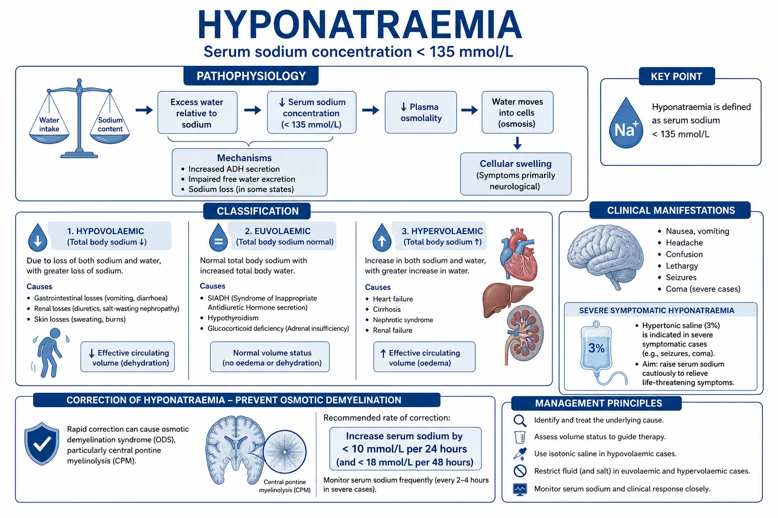
Hyponatraemia (Na < 135 mmol/L) [1]
- Classify by volume status (the deciding question for treatment):[1][2]
- Hypovolaemic (sodium and water loss, more sodium than water): diuretics, vomiting/diarrhoea, third-space losses, adrenal insufficiency. Treat with isotonic saline to restore volume — the ADH drive switches off and sodium corrects.
- Euvolaemic (water excess, normal total sodium): SIADH is the prototype; also glucocorticoid deficiency, hypothyroidism, primary polydipsia, MDMA. Treat with fluid restriction (the cornerstone), salt tablets, urea, or a vasopressin V2-receptor antagonist (tolvaptan). SIADH criteria: hypo-osmolality, urine osmolality >100 (inappropriately concentrated), urine sodium >40, euvolaemia, no diuretics, normal thyroid/adrenal function.
- Hypervolaemic (sodium and water excess, more water): heart failure, cirrhosis, nephrotic syndrome, renal failure. Treat the underlying state plus fluid restriction and loop diuretics; do not give hypotonic fluids.
- Clinical features: nausea, headache, confusion, seizures, coma. Severity tracks the rate of fall — an acute drop (<48 h) is symptomatic at higher concentrations; a chronic fall is often asymptomatic even below 120 mmol/L because the brain has adapted by extruding osmolytes.
- The osmotic demyelination risk: rapid correction of chronic hyponatraemia (>48 h or unknown) shifts water out of adapted brain cells faster than they can reaccumulate osmolytes, causing osmotic demyelination syndrome (ODS, central pontine myelinolysis) — a devastating, often irreversible quadriparesis, pseudobulbar palsy, and locked-in state. Risk is highest in alcoholics, malnourished, hypokalaemic, burns, and hepatic patients.[1][2]
- Correction limits (the single most important number):[1]
- Correct by no more than 8 mmol/L in any 24 hours (some authorities permit 10-12 mmol/L per 24 h for severe symptoms, then hold); aim for <8 mmol/L per 24 h to leave a safety margin.
- The first 4-6 mmol/L rise relieves life-threatening cerebral oedema — beyond that, stop and reassess.
- Treatment of severe symptomatic hyponatraemia (seizures/coma):[1][1]
- Give 3% hypertonic saline 100 mL bolus IV over 10 minutes, repeated up to 3 times (or 150 mL over 20 min), aiming for a 4-6 mmol/L rise. A 100 mL bolus of 3% saline raises serum sodium by approximately 1-2 mmol/L acutely.
- Bolus therapy is preferred over continuous infusion — it is safer (predictable small increments), faster (immediate effect on cerebral oedema), and easier to stop if over-correcting.
- Check sodium every 2-4 hours during active correction.
- If over-correction occurs: stop hypertonic saline, give desmopressin (DDAVP) 1-2 mcg IV and 5% dextrose in water (D5W) free water to relower sodium back into the target range. DDAVP prevents renal free-water excretion, capping the rise.
- SIADH specifics: fluid restrict to 800-1000 mL/day (often poorly tolerated); add high-protein/salt to raise the solute load; urea 15-30 g/day promotes osmotic diuresis. Vaptans (tolvaptan, conivaptan) block the V2 receptor and produce aquaresis — effective but risk rapid over-correction; the SALT trials showed tolvaptan raises sodium by ~4-5 mmol/L over 4 days.[6]
- NEVER give hypotonic fluids to a hyponatraemic patient.
Hypernatraemia (Na > 145 mmol/L) [1]
- Causes: nearly always free water loss out of proportion to sodium loss — diabetes insipidus (central or nephrogenic), osmotic diuresis (mannitol, glucose, urea), inadequate free water intake (intubated, elderly, impaired thirst), diarrhoea, burns, and iatrogenic hypertonic saline or sodium bicarbonate.[3]
- Clinical features: thirst (if alert), lethargy, irritability, hyperreflexia, seizures, coma. The brain adapts over 24-48 h by generating new intracellular osmoles (idiogenic osmoles), so chronic hypernatraemia is better tolerated — but rapid correction causes cerebral oedema and seizures.
- Correction rate: no more than 0.5 mmol/L per hour, and no more than 10-12 mmol/L per 24 hours (slower — 0.25 mmol/L/h — for chronic hypernatraemia >48 h).[3]
- Calculate the free water deficit:[3]
- Water deficit (L) = TBW × [(serum Na / 140) − 1], where TBW = 0.6 × weight (kg) in men, 0.5 × weight in women, 0.5 × weight in elderly men, 0.45 × weight in elderly women.
- Give the deficit as 5% dextrose over 48-72 hours, plus ongoing insensible and urinary losses. Check sodium every 4-6 hours.
- Diabetes insipidus: a water deprivation test and urine osmolality distinguish central (low ADH, responds to desmopressin) from nephrogenic (resistant). Central DI — give desmopressin (DDAVP) 1-2 mcg IV/SC. Nephrogenic DI — treat the cause (lithium, hypercalcaemia, hypokalaemia); give a low-sodium diet and thiazide diuretic (paradoxically reduces polyuria by inducing mild volume depletion).
- Hypovolaemic hypernatraemia: give 0.9% saline first to restore perfusion, then switch to hypotonic/free water once haemodynamically stable.
- NEVER correct hypernatraemia rapidly. Serum sodium must fall slowly to avoid cerebral oedema.
Potassium (K)
Normal range 3.5-5.0 mmol/L. Potassium is overwhelmingly intracellular (98%), so the serum level reflects distribution, not just total body stores. Acidosis shifts K out of cells (0.6 mmol/L rise per 0.1 pH drop); alkalosis and insulin shift it in.[4]
Hypokalaemia
- Causes: diuretics (loop > thiazide), vomiting/nasogastric losses, diarrhoea, amphotericin, secondary hyperaldosteronism, alkalosis (shifts K into cells), beta-agonists, refeeding, and magnesium depletion (the commonest cause of refractory hypokalaemia).[1]
- Features: weakness, ileus, U waves and flattened/merged T waves, ectopy, and a risk of torsades and digoxin toxicity.[1]
- Treatment:[1]
- Oral preferred (slow, safe): 20-40 mmol per dose; potassium chloride as tablets or liquid (liquid causes GI irritation). For each 0.3 mmol/L fall in serum K, total body deficit is approximately 100 mmol — large deficits are common.
- Intravenous replacement — strict rate and concentration limits: 10 mmol/h via a peripheral line (maximum 40 mmol/L concentration), 20 mmol/h via a central line (up to 80 mmol/L in a dedicated central lumen for severe deficits). Higher rates (>20 mmol/h) need cardiac monitoring and a dedicated central line. NEVER give IV potassium as a bolus — it is fatal.
- Replace magnesium first — hypokalaemia is refractory until magnesium is corrected (ROMK channel effect, see Red Flags).[1]
Hyperkalaemia
- Features: peaked (tented) T waves, widened QRS, flattened/lost P waves, then a sine-wave pattern and asystole/VF arrest. Treat K above 6.5 mmol/L or any ECG change urgently — the ECG, not the number, drives urgency.[4]
- Treatment — three sequential goals, in order:[1][4]
- Stabilise the myocardium (does NOT lower potassium): calcium gluconate 10% 10 mL IV over 2-5 min (onset seconds, lasts 30-60 min; repeat if ECG changes persist). Calcium chloride is an alternative (3x more calcium per mL) but MUST be given centrally — it causes severe tissue necrosis if it extravasates peripherally. Calcium antagonises the membrane excitability of hyperkalaemia without changing the serum level.
- Shift potassium intracellularly (temporarily lowers serum K by 0.5-1.5 mmol/L for 4-6 h, does not remove it): insulin 10 units IV with 25-50 g dextrose (e.g. 50 mL of 50% dextrose), onset 15-30 min, peak 30-60 min (monitor blood glucose — risk of hypoglycaemia for hours); salbutamol 10-20 mg nebulised (or 0.5 mg IV), onset 30 min; sodium bicarbonate 8.4% 50-100 mL IV if the patient is acidotic (poorly effective as sole agent, works mainly in acidosis).
- Remove potassium from the body (definitive): loop diuretics (furosemide, if producing urine), calcium/sodium resonium/patiromer (slow, 24 h+ to work; resonium 15-30 g orally or rectally), and renal replacement therapy (haemodialysis — the fastest and definitive removal; CRRT for haemodynamically unstable patients).[4]
Magnesium (Mg)
Normal range 0.7-1.0 mmol/L. Magnesium is the second most abundant intracellular cation and a cofactor for >300 enzymes, including the Na+/K+-ATPase; it gates outward potassium leak through ROMK channels. [1]
Hypomagnesaemia
- Causes: diuretics (loop and thiazide), PPIs (chronic use reduces intestinal absorption), diarrhoea, alcoholism, refeeding, sepsis, and cisplatin/amphotericin (renal wasting). Often coexists with hypokalaemia and hypocalcaemia — and makes both refractory.[1]
- Features: neuromuscular irritability, atrial and ventricular ectopy, torsades de pointes, seizures. Refractory hypokalaemia and hypocalcaemia will not correct until magnesium is repleted.[1]
- Treatment: IV magnesium sulfate 2-4 g (8-16 mmol) over 1 hour for symptomatic/severe depletion; 2 g over 10 minutes for torsades de pointes or seizure. Oral magnesium (glycinate, oxide) for chronic mild depletion — oral causes diarrhoea at high doses. In cardiac surgery and alcohol withdrawal, repletion is routine.[1]
Hypermagnesaemia
- Causes: renal failure, excessive iatrogenic replacement, and pre-eclampsia/eclampsia therapy (magnesium sulfate infusion) — the classic ICU scenario. Loss of deep tendon reflexes precedes hypotension, prolongation of the PR interval and heart block, respiratory depression, and cardiac arrest.[1]
- Treatment: calcium gluconate 10% 10 mL IV (antagonises the membrane effect immediately — the first and most important step in symptomatic hypermagnesaemia); fluids and loop diuretics (or dialysis in renal failure) for removal; ventilatory support as needed. Monitor reflexes and respiratory rate during magnesium infusions.[1]
Calcium (Ca)
Total 2.2-2.6 mmol/L; the ionised fraction (1.1-1.3 mmol/L) is the physiologically active form and the value to use in ICU (albumin-corrected totals mislead in critical illness).[1]
Hypocalcaemia
- Causes: parathyroid issues (post-thyroidectomy/neck surgery, hypoparathyroidism), acute pancreatitis, rhabdomyolysis, citrated blood/FFP/massive transfusion (citrate chelates calcium), sepsis, alkalosis (binds Ca to albumin), and hypomagnesaemia (impairs PTH release).[1]
- Features: perioral tingling, carpopedal spasm, Chvostek (facial nerve tap → twitch) and Trousseau (BP cuff → carpopedal spasm) signs, seizures, laryngospasm, and a prolonged QT with risk of torsades.[1]
- Treatment: calcium gluconate 10% 10 mL (2.2 mmol of Ca) slow IV over 10 min for symptomatic or severe hypocalcaemia; an infusion (10 ampules in 500 mL over 24 h) for sustained correction. Calcium chloride (10%) provides three times the calcium per mL but MUST be given centrally (severe tissue necrosis if extravasated). Correct concurrent magnesium and pH — hypocalcaemia is refractory until both are normal.[1]
Hypercalcaemia
- Causes: malignancy (PTHrP secretion or extensive bony metastases — the commonest inpatient cause), primary hyperparathyroidism (the commonest outpatient cause), granulomatous disease (sarcoid, TB — extra-renal vitamin D activation), thiazides, and milk-alkali syndrome.[1]
- Features: "stones, bones, groans, and psychiatric overtones" — confusion, constipation, polyuria/polydipsia (nephrogenic DI from renal concentrating defect), dehydration, shortened QT, renal stones, and coma. Severe hypercalcaemia (ionised above 1.5 mmol/L or corrected total >3.0-3.5, or symptomatic) is an emergency.[1]
- Treatment:[1]
- Isotonic saline first — patients are volume-depleted from polyuria, and saline restores GFR and promotes calciuria. Give 3-6 L over the first 24 h (often with a loop diuretic once rehydrated — NEVER thiazides, they retain calcium).
- Bisphosphonate — zoledronic acid 4 mg IV (or pamidronate 60-90 mg IV) is the definitive therapy for hypercalcaemia of malignancy; onset 24-72 h, peak at 5-7 days, lasts weeks.
- Calcitonin (4-8 IU/kg SC every 12 h) acts within hours (inhibits osteoclasts) for a temporary fall — a bridge while the bisphosphonate works.
- Glucocorticoids (prednisolone) are specifically effective in granulomatous disease and vitamin-D-mediated hypercalcaemia (lymphoma, sarcoid) — they reduce extra-renal 1-alpha-hydroxylation.
- Dialysis for severe/refractory cases, especially in renal failure.
Phosphate (PO4)
Normal range 0.8-1.5 mmol/L. Phosphate is essential for ATP generation, 2,3-DPG (oxygen unloading), and cell membrane phospholipids. [1]
Hypophosphataemia
- Causes: refeeding syndrome (the paradigm — insulin surge drives phosphate, potassium, and magnesium into cells;[5]), DKA recovery (insulin drives phosphate into cells), sepsis, alcoholism, renal replacement therapy, and burns. Reduced intake (malnutrition, chronic alcoholism) is the usual substrate.
- Features: muscle weakness (including the diaphragm — respiratory failure and failed ventilator weaning is the classic ICU presentation), rhabdomyolysis, haemolysis, impaired myocardial function (reduced stroke volume), and leucocyte dysfunction. Acute severe falls can precipitate acute respiratory failure and a cardiomyopathy.[1][5]
- Treatment: oral or IV sodium/potassium phosphate; correct severe deficits (below 0.3-0.5 mmol/L) intravenously (e.g. potassium phosphate 0.08-0.24 mmol/kg over 4-6 h). Reduce the refeeding risk by starting nutrition slowly (10-15 kcal/kg/day, advancing over a week), giving thiamine before feeds, and monitoring phosphate, potassium, and magnesium daily for the first week.[5]
Hyperphosphataemia
- Causes: renal failure, tumour lysis syndrome, rhabdomyolysis. Calcium-phosphate deposition can cause acute kidney injury and ectopic calcification. Treat with phosphate binders (calcium acetate, sevelamer) and dialysis if severe.[1]
Fellowship SAQs — electrolyte therapy

SAQ — Refractory hypokalaemia, hypomagnesaemia and torsades de pointes in a septic ICU patient
10 minutes · 10 marks
A 68-year-old woman is day 6 in ICU for pyelonephritis with septic shock, now stabilised on low-dose noradrenaline. She has received furosemide 40 mg daily for fluid overload and has been on a proton-pump inhibitor for a year. The nurse calls you because the cardiac monitor shows polymorphic ventricular ectopy and a run of torsades de pointes that self-terminated. Her potassium is 2.9 mmol/L despite 60 mmol of IV potassium chloride over the last 24 hours, and her magnesium is 0.42 mmol/L. ECG shows a corrected QT of 520 ms. She is alert and her blood pressure is 105/60.
SAQ — Severe hypophosphataemia and refeeding syndrome in a malnourished alcoholic
10 minutes · 10 marks
A 52-year-old man with a long history of alcohol dependence is admitted to ICU after a seizure. He has eaten almost nothing for two weeks and his BMI is 15.5. Enteral feeding was started 36 hours ago at 30 kcal/kg/day. He was intubated earlier today for aspiration pneumonia and has just failed a spontaneous breathing trial with a maximal inspiratory pressure of −18 cmH2O. Bloods show phosphate 0.28 mmol/L, potassium 3.0 mmol/L, magnesium 0.5 mmol/L, and a respiratory and metabolic acidosis with a rising lactate.
Red flags
Clinical pearls
Management flowcharts
Severe symptomatic hyponatraemia (seizure/coma) — the first hour
- RECOGNISE — serum Na <125 mmol/L (usually) with seizures, coma, or severe agitation, OR a rapid acute fall. This is a neurosurgical-grade emergency — cerebral oedema is killing the patient.
- GIVE 3% HYPERTONIC SALINE 100 mL IV BOLUS OVER 10 MIN — repeat up to 3 doses (every 10 min) aiming for a 4-6 mmol/L rise. Do NOT use a slow continuous infusion for seizures — bolus therapy is faster and more predictable. Each 100 mL bolus raises Na by ~1-2 mmol/L.
- CHECK SERUM Na EVERY 2 HOURS during active correction. Stop boluses once the seizure stops and Na has risen by 4-6 mmol/L — the cerebral oedema has resolved.
- SWITCH TO MAINTENANCE — stop all hypotonic fluids, commence fluid restriction if SIADH, and continue controlled correction at <8 mmol/L per 24 h with hypertonic saline infusion if needed.
- DIAGNOSE THE CAUSE — volume status, urine osmolality and sodium, cortisol, TSH, drug history. Treat the underlying cause (volume depletion, stop thiazides/SSRIs, adrenal replacement, etc.).
- MONITOR FOR OVER-CORRECTION — if Na rises >8 mmol/L in 24 h, STOP hypertonic saline, give DDAVP 1-2 mcg IV + D5W free water to relower sodium back into the target range.[1][2]
Hyperkalaemia with ECG changes — calcium, shift, remove (in that order)
- STABILISE THE MYOCARDIUM — calcium gluconate 10% 10 mL IV over 2-5 min (onset seconds). Repeat if ECG changes persist after 5 min. This does NOT lower potassium; it raises the arrhythmia threshold. Do not delay for a repeat potassium level.
- SHIFT POTASSIUM INTRACELLULARLY — insulin 10 units IV + 25-50 g dextrose (e.g. 50 mL of 50% dextrose), AND salbutamol 10-20 mg nebulised. Add sodium bicarbonate 8.4% 50-100 mL if acidotic. Onset 15-30 min; monitor blood glucose for 4-6 h (hypoglycaemia is the commonest complication).
- PROMOTE POTASSIUM REMOVAL — furosemide 40-80 mg IV if producing urine; calcium/sodium resonium 15-30 g orally or rectally (slow, 24 h+); and arrange haemodialysis for refractory hyperkalaemia, renal failure, or severe tissue breakdown (rhabdomyolysis, tumour lysis).
- IDENTIFY AND TREAT THE CAUSE — renal failure, ACE inhibitor/ARB/spironolactone, acidosis, rhabdomyolysis, tumour lysis, Addison's disease. Recheck potassium hourly during treatment and 2-hourly for 6 h after, because rebound is common.
- PREVENT RECURRENCE — review the medication list, treat acidosis, ensure adequate dialysis dose if on RRT.[1][4]
Hypernatraemia — calculate the deficit and correct slowly
- DETERMINE THE CAUSE AND VOLUME STATUS — is it water loss (DI, osmotic diuresis, inadequate intake) or sodium gain (hypertonic saline, sodium bicarbonate)? Is the patient hypovolaemic (needs saline first) or euvolaemic (free water)?
- CALCULATE THE FREE WATER DEFICIT — Water deficit (L) = TBW × [(serum Na / 140) − 1], where TBW = 0.6 × wt (men), 0.5 × wt (women/elderly men), 0.45 × wt (elderly women).
- CHOOSE THE REPLACEMENT FLUID — 5% dextrose in water for pure free-water deficit; 0.45% saline or oral water if feasible; 0.9% saline FIRST if hypovolaemic, then switch once haemodynamically stable.
- CORRECT AT <0.5 mmol/L/h (10-12 mmol/L per 24 h; slower — 0.25 mmol/L/h — if chronic >48 h) — spread the deficit over 48-72 h. Check Na every 4-6 hours.
- ADD ONGOING LOSSES — insensible (~10 mL/kg/day) plus measured urine output and other losses. Replace these as free water on top of the deficit.
- TREAT DIABETES INSIPIDUS — central DI: DDAVP 1-2 mcg IV/SC; nephrogenic DI: treat the cause (lithium, hypercalcaemia, hypokalaemia), low-sodium diet + thiazide.
- MONITOR FOR OVER-CORRECTION — if Na falls >0.5 mmol/L/h, slow the free water; rapid fall causes cerebral oedema and seizures.[3]
Refeeding syndrome prevention — the first week of nutrition in a high-risk patient
- IDENTIFY HIGH-RISK PATIENTS — BMI <16, unintentional weight loss >15% in 3-6 months, little/no intake for >10 days, low baseline phosphate/potassium/magnesium, alcoholism, anorexia, chemotherapy.
- GIVE THIAMINE BEFORE THE FIRST FEED — 200-300 mg IV/oral daily for 5-7 days. Thiamine is a cofactor for carbohydrate metabolism and is depleted by the glucose load.
- START FEEDS LOW AND GO SLOW — 10-15 kcal/kg/day, advancing to full target over 5-7 days. Do NOT push to target on day 1.
- REPLACE PHOSPHATE, POTASSIUM, MAGNESIUM BEFORE AND DURING — check all three daily for the first week, more often if IV repleting. Correct any deficit promptly.
- MONITOR — daily weights, fluid balance, electrolytes, ECG (for QT). Restrict sodium and fluid to avoid the refeeding oedema.
- ESCALATE IF IT OCCURS — severe hypophosphataemia (respiratory/cardiac weakness, failed wean) needs IV potassium phosphate 0.08-0.24 mmol/kg over 4-6 h; reduce or pause feeds.[5]
Comparison tables
| Potassium (KCl) | Peripheral: 10 mmol/h, max 40 mmol/L concentration | Safe for mild-moderate deficits | Pain/chemophlebitis; slow |
| Potassium (KCl) | Central: 20 mmol/h, up to 80 mmol/L in dedicated lumen | Fast for severe deficits | Needs central line + cardiac monitor; >20 mmol/h is dangerous |
| Magnesium (MgSO4) | 2-4 g (8-16 mmol) IV over 1 h; 2 g over 10 min for torsades | Rapid effect on arrhythmia/seizure | Flushing, hypotension if given too fast; accumulates in renal failure |
| Calcium (gluconate 10%) | 10 mL = 2.2 mmol Ca, slow IV over 2-10 min; infusion over 24 h | Can be given peripherally | Tissue necrosis if extravasated (less than CaCl2); does not correct the cause |
| Calcium (chloride 10%) | 10 mL = 6.8 mmol Ca (3× gluconate), CENTRAL line only | Most calcium per mL — for arrest | Severe tissue necrosis if extravasated peripherally |
| Phosphate (K-phos / Na-phos) | IV 0.08-0.24 mmol/kg over 4-6 h for severe (PO4 <0.3) | Reverses respiratory/cardiac weakness fast | Can cause hyperkalaemia (K-phos), hyperphosphataemia, hypocalcaemia if too fast |
| Sodium (3% hypertonic saline) | 100 mL bolus over 10 min (×3 max); infusion for controlled correction | Rapid relief of cerebral oedema | Over-correction → osmotic demyelination; check Na every 2 h |
| Calcium gluconate 10% 10 mL IV | Stabilises myocardium (no K change) | Onset seconds; prevents arrhythmia | Lasts 30-60 min; does not remove K |
| Insulin 10 U + dextrose 25-50 g IV | Shifts K into cells (Na/K-ATPase) | Onset 15-30 min; lowers K ~1 mmol/L | Hypoglycaemia for hours; needs glucose monitoring |
| Salbutamol 10-20 mg nebulised | Shifts K into cells (β2 receptor) | Onset 30 min; synergistic with insulin | Tachycardia, tremor; less effective in β-blocked |
| Sodium bicarbonate 8.4% 50-100 mL | Shifts K into cells (alkalinisation) | Helps if acidotic; buffers | Ineffective alone; hypertonic; volume/Na load |
| Furosemide 40-80 mg IV | Removes K (renal excretion) | Definitive if making urine | Needs working kidneys; causes volume depletion |
| Calcium/sodium resonium / patiromer | Binds K in gut (removal) | Oral/rectal; outpatient option | Slow (24 h+); constipation; resonium causes GI necrosis if rectal retained |
| Haemodialysis | Removes K (definitive) | Fastest removal; for renal failure/refractory | Needs access; haemodynamic instability → use CRRT |
| Hypovolaemic | Na + water loss (more Na); urine Na <20 (extra-renal) or >20 (renal) | Responds rapidly to volume replacement | Treat with isotonic saline — ADH switches off, Na corrects |
| Euvolaemic (SIADH) | Water excess, normal total Na; urine osm >100, urine Na >40 | Well-defined criteria; many treatments | Fluid restriction is the cornerstone; vaptans risk over-correction; DDAVP rescue if over-correcting |
| Hypervolaemic | Na + water excess (more water); oedema (heart failure, cirrhosis, renal failure) | Diuretic-responsive | Restrict fluid + loop diuretic; NEVER hypotonic; treat the cause |
| Isotonic saline (3-6 L/24 h) | Restores volume, promotes calciuria | First step; reverses dehydration/DI | Volume overload in heart failure; needs monitoring |
| Loop diuretic (furosemide) | Promotes renal calcium excretion | Add once rehydrated | NEVER thiazide (retains Ca); causes volume/K/Mg depletion |
| Bisphosphonate (zoledronate 4 mg IV) | Inhibits osteoclasts | Definitive for malignancy; lasts weeks | Onset 24-72 h; osteonecrosis of jaw; nephrotoxicity |
| Calcitonin (4-8 IU/kg SC q12h) | Inhibits osteoclasts (rapid) | Bridge — acts within hours | Tachyphylaxis after 48 h; transient effect |
| Glucocorticoids | Reduce extra-renal vitamin D activation | Specific for granulomatous disease/lymphoma | Ineffective in malignancy/hyperparathyroidism |
| Dialysis | Removes calcium directly | For severe/refractory, especially renal failure | Invasive; reserved for crises |
Key trials and guidelines
Spasovski 2014 — European Clinical Practice Guideline on Hyponatraemia (PMID 24562549)
Source
European Journal of Endocrinology 2014; 170(3):G1-G47 (also published in Intensive Care Medicine and four other European societies)
Question
What is the evidence-based diagnosis and treatment of hyponatraemia?
Key recommendations
Classify by symptoms (moderately severe vs severe) AND by onset (acute <48 h vs chronic); hypertonic saline 3% for severe symptomatic; correct by <10 mmol/L in the first 24 h then <8 mmol/L per 24 h thereafter; fluid restriction first-line for euvolaemic/hypervolaemic; vaptans (tolvaptan) for SIADH when fluid restriction fails; DDAVP + water for over-correction
Strength
Multinational evidence-based guideline (European Society of Intensive Care Medicine, European Society of Endocrinology, European Renal Association, European Federation of Endocrine Societies)
Key finding
The danger of sodium correction is OVER-correction (ODS), not under-correction — cap at 10 mmol/L per 24 h, aim <8
Clinical bottom line
The international standard for hyponatraemia — every sodium-correction question in CICM/FFICM/EDIC is answerable from here
Adrogué & Madias 2000 — Hyponatremia and Hypernatremia (PMIDs 10824078 & 10816188)
Source
New England Journal of Medicine 2000; 342(21):1581-1589 (hypo) and 342(20):1493-1499 (hyper) — a matched pair of reviews
Question
What are the causes, consequences, and correct rate of correction of sodium disorders?
Key concept
Sodium disorders are disorders of water balance; the brain adapts over 24-48 h; the rate of correction (not the target) determines outcome
Correction limits
Chronic hyponatraemia: <10-12 mmol/L per 24 h (risk of ODS); chronic hypernatraemia: <10 mmol/L per 24 h or <0.5 mmol/L/h (risk of cerebral oedema)
Key finding
The free-water deficit formula — Water deficit (L) = TBW × [(serum Na / 140) − 1] — and the Adrogué-Madias formula for the change in Na per litre of fluid infused
Clinical bottom line
The classic reference for sodium-correction mathematics — cited in every guideline since
Schrier 2006 — SALT-1 and SALT-2 (PMID 17105757)
Source
New England Journal of Medicine 2006; 355(20):2099-2112 — two randomised double-blind placebo-controlled trials, 448 patients total
Question
Does oral tolvaptan (a V2-receptor antagonist / vaptan) correct euvolaemic/hypervolaemic hyponatraemia?
Primary outcome
Change in serum sodium AUC over 4 days + over 30 days
Key result
Tolvaptan raised serum sodium by ~4-5 mmol/L over 4 days vs placebo (~1 mmol/L); effect maintained at 30 days; fluid restriction was not required
Key finding
Aquaresis via V2-receptor blockade is effective in SIADH and heart failure hyponatraemia — but risk of over-correction means sodium must be checked frequently in the first 24 h
Clinical bottom line
Established vaptans as a treatment for SIADH; clinical use is limited by cost, over-correction risk, and the availability of simpler options (fluid restriction, urea, salt)
Mehanna 2008 — Refeeding Syndrome (PMID 18583681)
Source
BMJ 2008; 336(7659):1495-1498 — narrative review and NICE-aligned guidance
Question
What is refeeding syndrome, who is at risk, and how is it prevented and treated?
Definition
A potentially fatal shift of fluid and electrolytes (phosphate, potassium, magnesium) that occurs on resuming nutrition in a malnourished patient, driven by an insulin surge
Key recommendations
Identify high-risk patients (BMI <16, weight loss >15%, minimal intake >10 days); give thiamine before feeding; start at 10 kcal/kg/day and advance over 5-7 days; replace phosphate, potassium, magnesium before and during feeding; monitor daily
Key finding
Refeeding syndrome is under-recognised and largely preventable with slow initiation of feeding and electrolyte/thiamine supplementation
Clinical bottom line
The bedside reference for refeeding prevention in ICU — know the high-risk criteria and the slow-start + thiamine + electrolyte strategy
Nyirenda 2009 — Hyperkalaemia (PMID 19854840)
Source
BMJ 2009; 339:b4114 — concise clinical review
Question
What is the practical, evidence-based management of hyperkalaemia?
Key framework
Stabilise the myocardium (calcium) → shift potassium into cells (insulin/dextrose, β2-agonists, bicarbonate if acidotic) → remove potassium (diuretics, resonium, dialysis)
Key finding
Calcium does not lower potassium but is given first because it prevents arrhythmia; insulin/dextrose and salbutamol are synergistic; definitive removal requires diuresis, gut binding, or dialysis
Clinical bottom line
The 'calcium-shift-remove' sequence that every ICU exam answer should reproduce
References
- [1]Spasovski G, Vanholder R, Allolio B, et al Clinical practice guideline on diagnosis and treatment of hyponatraemia Intensive Care Med, 2014.PMID 24562549
- [2]Adrogué HJ, Madias NE Hyponatremia N Engl J Med, 2000.PMID 10824078
- [3]Adrogué HJ, Madias NE Hypernatremia N Engl J Med, 2000.PMID 10816188
- [4]Nyirenda MJ, Tang JI, Padfield PL, Seckl JR Hyperkalaemia BMJ, 2009.PMID 19854840
- [5]Mehanna HM, Moledina J, Travis J Refeeding syndrome: what it is, and how to prevent and treat it BMJ, 2008.PMID 18583681
- [6]Schrier RW, Gross P, Gheorghiade M, et al (SALT Investigators) Tolvaptan, a selective oral vasopressin V2-receptor antagonist, for hyponatremia N Engl J Med, 2006.PMID 17105757