ICU · Pharmacology
Therapeutic plasma exchange (plasmapheresis) in ICU
Also known as Plasma exchange (PLEX) · Plasmapheresis · Therapeutic plasma exchange (TPE)
Therapeutic plasma exchange (TPE) removes pathogenic substances (autoantibodies, toxins, immune complexes, abnormal proteins) from plasma by centrifugation or filtration, replacing with donor plasma or albumin. Indications in ICU: Guillain-Barre syndrome (1 — removes anti-ganglioside antibodies), myasthenia gravis crisis, thrombotic thrombocytopenic purpura (TTP — removes anti-ADAMTS13 antibodies), Goodpasture syndrome, ANCA vasculitis, acute disseminated encephalomyelitis, mushroom poisoning, drug overdose (removes protein-bound toxins). Technique: vascular access (central line), anticoagulation (citrate), exchange 1-1.5 plasma volumes per session, 5-10 sessions over 1-2 weeks. Replacement fluid: albumin 5% (most), FFP (for TTP — replaces ADAMTS13). Complications: hypocalcaemia (citrate), hypotension, bleeding (coagulopathy from removal), infection (catheter), allergic reaction (donor plasma).
On this page & tools
Your progress
Saved locally on this device.
Target exams
Red flags
Clinical pearls
Red flags
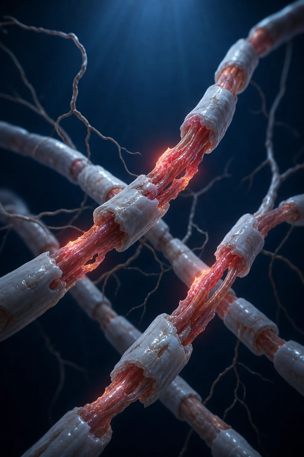
Mechanism of TPE — what the procedure actually removes
Pathogenic substances removed by TPE — and the diseases they cause
| Substance class | Representative molecule | Disease state where removal is therapeutic |
|---|---|---|
| IgG autoantibodies | Anti-AChR, anti-ganglioside (GM1, GQ1b), anti-MAG | Myasthenia gravis crisis, Guillain-Barré syndrome, CIDP |
| IgG autoantibodies (vWF-cleaving) | Anti-ADAMTS13 | Thrombotic thrombocytopenic purpura (TTP) |
| IgG anti-basement membrane | Anti-GBM (anti-α3 chain of type IV collagen) | Goodpasture syndrome |
| IgG anti-neutrophil cytoplasmic | Anti-MPO, anti-PR3 (ANCA) | Granulomatosis with polyangiitis, microscopic polyangiitis |
| IgM monoclonal paraprotein | IgM pentamer (very high MW) | Waldenström macroglobulinaemia, hyperviscosity syndrome |
| IgG/IgA monoclonal | Monoclonal protein | Multiple myeloma (esp. IgA, IgG3), cryoglobulinaemia |
| Immune complexes | Antigen–antibody complexes | SLE nephritis, cryoglobulinaemic vasculitis, post-infectious GN |
| Cold-reactive antibodies | Cold agglutinins (IgM anti-I) | Cold agglutinin disease, cold haemagglutinin syndrome |
| Antiphospholipid antibodies | aPL (lupus anticoagulant, anti-β2-GPI) | Catastrophic antiphospholipid syndrome (CAPS) |
| Bound toxin | Amatoxin (Amanita phalloides), mushroom | Mushroom poisoning (acute liver failure) |
| Protein-bound drug | Highly protein-bound overdose agents | Selected drug overdose (digoxin Fab–resistant, L-thyroxine) |
| Abnormal lipoprotein | LDL | Familial hypercholesterolaemia (homozygous) |
| Inflammatory mediators | Cytokines, complement, DAMPs | Sepsis (investigational), SIRS, transplant rejection |
| Desmoglein antibodies | Anti-desmoglein 1/3 | Pemphigus vulgaris / foliaceus (refractory) |
The three therapeutic effects of TPE — beyond simple removal
| Effect | What happens | Clinical implication |
|---|---|---|
| 1. Mass removal | Pathogenic solute discarded with the plasma | The dominant mechanism — drives the logarithmic exchange-volume logic |
| 2. Replacement | Deficient factors supplied by donor plasma | Use FFP (not albumin) in TTP to replace the missing ADAMTS13 enzyme — the classic exam point |
| 3. Immunomodulation | Removal shifts idiotypic–anti-idiotypic balance; restores reticuloendothelial function | Explains sustained benefit beyond the calculated half-life of removed antibody; synergy with rituximab |
Separation technique — centrifugal vs membrane
Centrifugal vs membrane plasma separation
| Feature | Centrifugation | Membrane filtration |
|---|---|---|
| Principle | Density-gradient separation (g-force) | Porous membrane (sieving, pore ~0.2–0.5 µm) |
| Equipment | Dedicated apheresis machine (Spectra Optia, Amicus, Cobe Spectra) | Plasmafilter on CRRT/dialysis circuit (Plasmaflo) |
| Blood flow required | 30–80 mL/min (lower, tolerates peripheral/PICC access) | 100–150 mL/min (needs robust central access) |
| Extracorporeal volume | ~150–250 mL | ~150–300 mL |
| Plasma separation efficiency | High (also removes some platelets — buffy coat) | High (cells returned intact, no platelet loss) |
| Platelet loss per session | Slight (can be used therapeutically — platelet apheresis) | Negligible |
| Haemolysis risk | Low | Slightly higher if high shear (high blood flow, low Hct) |
| Access requirement | Can use large peripheral cannula or any CVC | Needs high-flow dual-lumen dialysis catheter |
| Familiarity on ICU | Specialist operator (haematology/apheresis nurse) | Any CRRT-trained ICU nurse can run it |
| Common in | Standalone apheresis service, neurology/haematology referrals | ICU where CRRT platform is reused for TPE |
Vascular access for TPE
Vascular access options for TPE
| Access site | Pros | Cons / cautions | When chosen |
|---|---|---|---|
| Internal jugular (RIJ preferred) | Direct route to SVC/RA; compressible if bleeding; avoids citrate-associated subclavian stenosis | Needs sterility; risk of pneumothorax | Default first choice for acute ICU TPE courses |
| Femoral | Bedside insertion; no pneumothorax; usable in coagulopathy | Higher CRBSI risk; patient immobile; DVT risk | Second choice; bedside/emergency; severe coagulopathy |
| Subclavian | Comfort; lowest infection (long term) | AVOID in citrate TPE — mechanical compression + citrate anticoagulation → bleeding; stenosis limits future fistulae | Generally avoided for acute TPE |
| Peripheral (large antecubital, 17 G+) | No central line risk | Only suitable if excellent veins; single-needle flow-limited; painful | Single / few sessions, healthy veins (rare in ICU) |
| Tunnelled dialysis catheter / Tesio | Long-term, low infection, high flow | Insertion complexity; not for acute | Long TPE courses (e.g., transplant desensitisation) |
Anticoagulation — citrate is the workhorse
Anticoagulation options for the TPE circuit
| Anticoagulant | Mechanism | Monitoring | Pros | Cons / when avoided |
|---|---|---|---|---|
| Citrate (trisodium citrate 4%) | Chelates ionised Ca²⁺ in circuit → no clotting; Ca²⁺ returned/repleted | Ionised calcium (patient) + total Ca/iCa ratio; clinical signs | Circuit-only effect; reversible; standard of care | Hypocalcaemia; needs intact hepatic metabolism; avoid/caution in severe liver failure, severe lactic acidosis (citrate accumulation → severe alkalosis + low iCa) |
| Heparin (unfractionated) | Systemic anticoagulation via antithrombin | aPTT/ACT | Familiar; doesn't cause hypocalcaemia | Systemic bleeding; removed by TPE partly; heparin rebound |
| Heparin + protamine (regional) | Heparin in circuit, protamine post-circuit | ACT, aPTT | Circuit-only anticoagulation | Complex; protamine reactions; rarely used in TPE |
| No anticoagulant | None | — | None | High clotting risk — only if severe bleeding + very short session |
Replacement fluid — albumin vs FFP (the single most exam-worthy decision)
Albumin 5% vs fresh frozen plasma (FFP) vs cryosupernatant
| Property | 5% albumin | FFP | Cryosupernatant (cryo-poor plasma) |
|---|---|---|---|
| Composition | Human albumin in saline | All plasma proteins — clotting factors, ADAMTS13, vWF, fibrinogen | Plasma depleted of cryoprecipitate — less fibrinogen, less vWF; still has ADAMTS13 |
| Use as default | YES — most TPE | No — reserved | No — niche |
| Infection risk | Negligible (heat-treated) | Very low (but non-zero — viruses, bacterial, TRALI) | Same as FFP |
| Allergic/TRALI risk | Near zero | Higher — allergic, TRALI, TACO | Similar to FFP |
| Cost / availability | Cheap, abundant | Expensive, scarce, blood-bank dependent | Very scarce |
| ABO compatibility required? | No | Yes — must be ABO-compatible | Yes |
| Replaces ADAMTS13? | No | Yes | Yes (preferred for TTP — less UL-vWF) |
| Indication: TTP | ❌ NEVER alone | ✔ (or cryosupernatant) | ✔ Preferred in some centres |
| Indication: GBS, MG, vasculitis, Goodpasture | ✔ Default | Only if coagulopathy/bleeding | No |
| Indication: coagulopathy / factor depletion | No (worsens dilution) | ✔ | No |
Exchange volume and session frequency
The logarithmic efficiency of plasma exchange (single session)
| Exchange volume (plasma volumes) | Fraction of intravascular solute removed | Residual solute |
|---|---|---|
| 0.5 | ~39% | ~61% |
| 1.0 (standard) | ~63% | ~37% |
| 1.5 (standard) | ~78% | ~22% |
| 2.0 | ~86% | ~14% |
| 3.0 | ~95% | ~5% |
Worked example — calculating a TPE prescription
| Step | Calculation |
|---|---|
| Patient | 70 kg, Hct 0.35 |
| Estimated plasma volume | 40 mL/kg × 70 × (1 − 0.35) = 1820 mL |
| Session target (1.5 PV) | 1820 × 1.5 ≈ 2730 mL removed per session |
| Replacement ratio | ~1:1 (replace ≈ 2700 mL) — albumin 5% (or FFP for TTP) |
| Anticoagulation | Citrate 4% at AC ratio ~1:12 to 1:15 blood:citrate |
| Calcium | Calcium gluconate 10% infusion titrated to iCa (target >1.0 mmol/L) |
| Frequency | Daily (TTP, CAPS) or every other day (GBS, MG) |
| Course length | 5–14 sessions until clinical endpoint (e.g. platelet >150 × 10⁹/L × 2 days for TTP) |
ASFA categories — the framework that organises every indication
ASFA category definitions
| Category | Meaning | ICU example |
|---|---|---|
| I | First-line therapy (standard of care) | GBS, TTP, Goodpasture, MG crisis, hyperviscosity |
| II | Second-line / adjunctive therapy | ANCA vasculitis, SLE nephritis, pemphigus, ABO-incompatible transplant, HELLP |
| III | Optimum role not established — individualise | Sepsis, drug overdose, fulminant hepatic failure |
| IV | No demonstrated benefit — do not offer (outside trial) | — |
ASFA Category I indications in ICU — TPE is first-line
| Disease | Pathogenic substance removed | Replacement fluid | Typical schedule | Key trial / rationale |
|---|---|---|---|---|
| Guillain-Barré syndrome | Anti-ganglioside antibodies (GM1, GQ1b) | Albumin | 5 sessions over 7–10 days | French Cooperative 1987; PE/Sandoglobulin 1997 — equivalent to IVIG |
| Myasthenia gravis crisis | Anti-AChR (or anti-MuSK) antibodies | Albumin | Daily–alternate day, 5–6 sessions | Equivalent to IVIG; faster onset than immunosuppression |
| Goodpasture (anti-GBM) | Anti-GBM antibody | Albumin (FFP if bleeding) | Daily ×14 days or until antibody undetectable | Urgent — delays cause irreversible renal failure |
| TTP (acquired) | Anti-ADAMTS13 antibody | FFP (or cryosupernatant) | Daily until platelet >150 × 2 days, then taper | Rock 1991 — PE superior to plasma infusion; HERCULES caplacizumab 2019 |
| Hyperviscosity syndrome | Monoclonal IgM (Waldenström) | Albumin (FFP if bleeding) | 1–2 sessions urgently; then treat underlying | Emergency — 1–2 exchanges rapidly lower viscosity |
| Catastrophic antiphospholipid syndrome (CAPS) | Antiphospholipid antibodies + cytokines | FFP | Daily until recovery (3–7+) | Combined with glucocorticoids + rituximab/IVIG |
| Cold agglutinin disease | IgM cold agglutinin | Albumin; warm everything | 1–3 sessions, daily | Pre-warm circuit/blood — cold triggers haemolysis |
ASFA Category II indications — TPE as adjunct
| Disease | Role of TPE | Replacement | Schedule | Notes |
|---|---|---|---|---|
| ANCA vasculitis (severe) | Severe renal (dialysis-dependent) or diffuse pulmonary haemorrhage | Albumin | 7 sessions over 14 days | MEPEX positive (renal recovery); PEXIVAS 2020 NEUTRAL overall — role now narrower |
| SLE nephritis (severe) | Adjunct in refractory proliferative lupus nephritis | Albumin | 3–6 sessions | Not routine — selected refractory cases |
| Pemphigus vulgaris (severe) | Adjunct for refractory / rapid control | Albumin | Daily ×3 then taper | Adjuvant; concurrent steroids + rituximab |
| ABO-incompatible transplant | Desensitisation — remove anti-A/B antibodies pre-transplant | Albumin | Pre-transplant to titre target; post-transplant rebound | Combined with rituximab + IVIG |
| HELLP / postpartum TMA | Adjunct in persistent/secondary postpartum HELLP | FFP | 3–6 sessions | Selected persistent cases; not for uncomplicated HELLP |
| Cryoglobulinaemic vasculitis | Remove cryoprecipitating immune complexes | Albumin; warm circuit | 3–8 sessions | Symptomatic severe vasculitis; warm everything |
| Solid organ transplant rejection (humoral) | Remove donor-specific antibodies | Albumin | Variable | Antibody-mediated rejection |
Disease-specific ICU protocols
TTP — the daily-plasma-exchange protocol (highest-yield exam scenario)
- Suspect TTP early — the pentad (MAHA thrombocytopenia, neuro, renal, fever) is rare; the dyad of thrombocytopenia + MAHA (schistocytes) with no other cause is enough to start empirically — do NOT wait for ADAMTS13 result (treat first, draw ADAMTS13 level before first exchange)
- Start TPE within 4–24 h of suspicion — every hour of delay increases mortality; Rock 1991 showed PE superior to simple plasma infusion
- Use FFP (or cryosupernatant) as replacement — albumin alone is a fatal error; FFP replaces the missing ADAMTS13 enzyme
- Exchange 1.0–1.5 plasma volumes DAILY until platelet count >150 × 10⁹/L for 2 consecutive days AND MAHA (LDH, schistocytes) resolves
- Add corticosteroids (methylprednisolone) — suppresses the autoantibody
- Add caplacizumab (anti-vWF nanobody) — HERCULES 2019: faster platelet recovery, fewer TTP-related deaths; bleeds more. Caplacizumab + TPE is now standard for acquired TTP
- Consider rituximab for refractory/relapsing idiopathic TTP — administer AFTER TPE (TPE removes rituximab)
- Taper TPE once platelets stable — reduce frequency (alternate day → every third day) rather than stopping abruptly to avoid rebound
- Monitor — platelets q12–24h, LDH, iCa (citrate), fibrinogen (factor depletion), ADAMTS13 trend
- Beware refractory TTP — no improvement after 4–7 days: intensify (increase volume, add rituximab/bortezomib/cyclophosphamide, reassess for alternate diagnosis — atypical HUS, DIC)
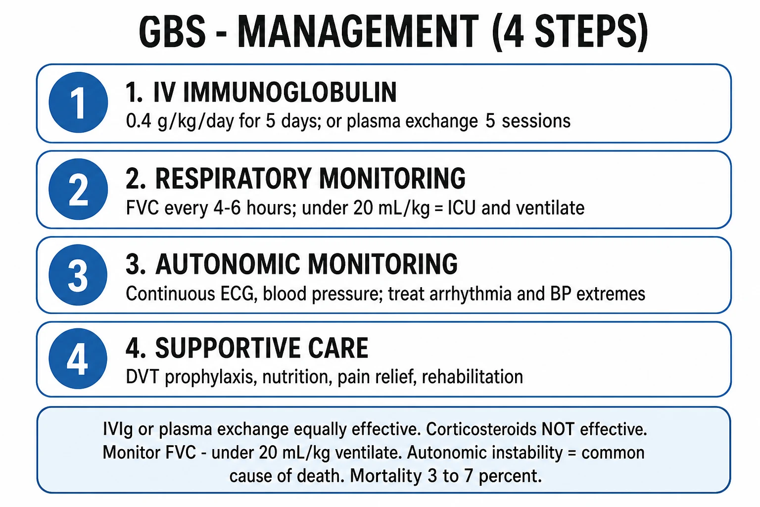
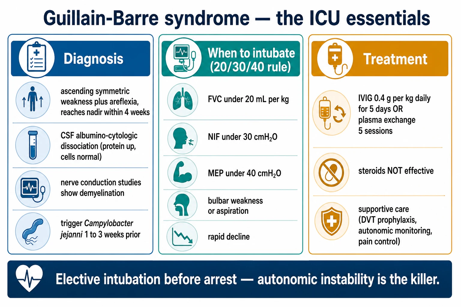
Guillain-Barré syndrome — TPE protocol
- Identify the candidate — GBS within 4 weeks of onset, unable to walk (GBS-DS ≥3), or rapid progression / respiratory decline / autonomic instability
- Choose TPE or IVIG — equally effective (PE/Sandoglobulin 1997); IVIG more convenient and widely used. Choose TPE if IVIG contraindicated, or combine only in non-responders
- Albumin replacement — FFP not needed (no factor to replace)
- 5 sessions, 1.0–1.5 plasma volumes, every other day (or daily) over 7–14 days
- Earliest benefit at 1–2 weeks — motor recovery; do not expect instant improvement
- Time adjunctive drugs AFTER TPE — IVIG, steroids (no benefit alone in GBS) given after the session so they are not removed
Anti-GBM (Goodpasture) disease — urgent TPE
- Treat as an emergency — every day of delay correlates with irreversible glomerular destruction; start at diagnosis if pulmonary haemorrhage or rapidly progressive GN
- Daily TPE, 1.5 plasma volumes, albumin replacement (FFP if active pulmonary bleeding / coagulopathy), for 14 days OR until anti-GBM antibody undetectable
- Combine with high-dose corticosteroids + cyclophosphamide — TPE removes antibody; immunosuppression stops production
- Treat pulmonary haemorrhage aggressively — the leading cause of early death; TPE reduces alveolar haemorrhage within days
- Decision on dialysis-dependent, biopsy-proven crescentic disease — TPE less likely to recover renal function if >85% crescents / oligoanuric at presentation (discuss with nephrology) — but pulmonary haemorrhage alone is an absolute indication regardless
- Do NOT transplant while anti-GBM positive — wait until antibody undetectable >6 months (recurrence risk)
Myasthenia gravis crisis — TPE protocol
- Indication — myasthenic crisis (respiratory failure / bulbar weakness needing ICU), severe refractory exacerbation, pre-thymectomy optimisation in refractory disease
- TPE or IVIG — equivalent efficacy; faster onset with TPE (days vs IVIG 1–2 weeks). Choose TPE for the most rapid effect or when IVIG contraindicated (IgA deficiency, renal failure, hyperviscosity)
- Albumin replacement, 5–6 sessions over 10–14 days
- Coordinate with long-term immunosuppression — TPE is a bridge; rituximab/cyclophosphamide given AFTER TPE (it is removed)
- Beware hypocalcaemia + MG — citrate-induced weakness can mimic worsening myasthenia; maintain iCa >1.0 mmol/L
- Watch for respiratory improvement — predict by NIF/vital capacity trend; expect weaning from NIV/ventilation over days
Catastrophic antiphospholipid syndrome (CAPS)
- Recognise CAPS — ≥3 organs affected by microthrombi within 1 week, histopath confirmation of small-vessel thrombosis, antiphospholipid antibody positivity
- Triple therapy — anticoagulation (heparin) + high-dose glucocorticoids + TPE daily with FFP
- FFP replacement is preferred — restores depleted natural anticoagulants (antithrombin, protein C/S) and removes aPL antibodies + cytokines
- Daily exchanges until multi-organ failure stabilises (typically 3–7+ sessions)
- Add rituximab / IVIG in refractory cases (give IVIG/rituximab AFTER TPE session)
- Treat any precipitant — infection (most common trigger), surgery, malignancy, obstetric event
Hyperviscosity syndrome (Waldenström macroglobulinaemia) — emergency TPE
- Recognise — symptomatic IgM >40 g/L or serum viscosity >4–5 cP: visual disturbance, headache, mucosal bleeding, altered consciousness, retinal vein sausaging
- EMERGENCY 1–2 plasma volume exchanges immediately — IgM is 80% intravascular, so removal is dramatic and rapid; symptoms can resolve within hours
- Albumin replacement (FFP if bleeding)
- Treat underlying clone — rituximab-based chemoimmunotherapy (give rituximab AFTER TPE — it is removed; beware rituximab flare of IgM)
- Beware hyperviscosity-induced anaemia — do NOT transfuse PRBCs until viscosity controlled (raising Hct raises viscosity further)
Adverse effects — the full ICU complication profile
TPE adverse effects — mechanism, frequency, and management
| Adverse effect | Mechanism | Frequency | Recognition | Management / prevention |
|---|---|---|---|---|
| Hypocalcaemia (citrate) | Citrate chelates ionised Ca²⁺; load exceeds hepatic metabolism | Very common (up to 90% have subclinical iCa drop) | Perioral tingling, Chvostek/Trousseau, QT prolongation, hypotension, muscle weakness mimicking MG/GBS | Prophylactic calcium gluconate infusion; monitor iCa continuously; reduce citrate:AC ratio; slow flow |
| Hypotension / volume shift | Reduction in oncotic pressure; fluid removed faster than replaced; vasovagal | Common (~5–10%) | Intra-procedure BP drop | Concurrent 1:1 replacement; albumin not saline; vasopressors if vasoplegic |
| Bleeding / coagulopathy | Albumin replacement depletes clotting factors (no factors in albumin); heparin | After 3–4 daily albumin exchanges; line-site bleeding | Rising PT/aPTT, falling fibrinogen | Factor replacement (FFP for 1 session), fibrinogen check, minimise heparin |
| Allergic / transfusion reaction | Donor plasma proteins (esp. FFP); ethylene oxide; latex | Common with FFP | Urticaria, bronchospasm, anaphylaxis | Pre-medicate (antihistamine); use albumin where possible; washed products; stop session if severe |
| TRALI / TACO | Donor anti-WBC antibodies (FFP); volume overload | Rare but serious | Hypoxaemia during/after FFP TPE (TRALI); pulmonary oedema (TACO) | Stop, respiratory support, diurese (TACO) |
| Infection (catheter-related — CRBSI) | Indwelling central catheter | The commonest serious complication | Exit-site infection, bacteraemia, sepsis | Aseptic insertion, chlorhexidine dressing, remove catheter promptly at course end |
| Citrate accumulation / severe alkalosis | Citrate metabolism → bicarbonate; impaired hepatic clearance | Severe liver failure, severe lactic acidosis | Rising total Ca : iCa ratio (>2.5), metabolic alkalosis, worsening iCa | Switch to heparin anticoagulation; reduce citrate; correct acidosis |
| Haemolysis | Shear in catheter/membrane; ABO-incompatible FFP | Rare | Falling Hb, haemoglobinuria, rising LDH | Check ABO compatibility of FFP; review line; slow flow |
| Thrombocytopenia / bleeding | Platelet loss (centrifugal) + heparin | Variable | Falling platelets | Monitor; platelet support if bleeding |
| Hypothermia | Large extracorporeal volume | Common | Cold patient | Blood warmer on return line |
| Hypokalaemia / metabolic | Electrolyte shifts with replacement fluid | Common | ECG changes | Add KCl to replacement; monitor |
| Bradycardia / hypotension at line disconnect | Volume/return cessation | Occasional | End of session | Flush line carefully; monitor briefly post-session |
Citrate-induced hypocalcaemia — recognition and management
- Anticipate it — virtually every TPE session lowers iCa; the question is whether it becomes symptomatic
- Risk factors — high citrate:AC ratio (small/paediatric patient, low blood flow), severe liver disease (impaired citrate metabolism), severe lactic acidosis, hypothermia, large FFP replacement (FFP is itself citrated), alkalosis
- Recognise early symptoms — perioral/lingual/finger tingling, chest tightness, palpitations; signs: Chvostek, Trousseau, prolonged QT, hypotension, muscle weakness indistinguishable from worsening MG/GBS
- Monitor — ionised calcium at baseline, mid-session, end; trend total Ca : iCa ratio (rising ratio >2.1 = citrate accumulation even if total Ca looks normal — total Ca is falsely reassuring)
- Prophylaxis — continuous calcium gluconate 10% infusion (e.g. 10 mL/hr of 10% calcium gluconate, or calcium chloride via central line) titrated to iCa; slow blood/citrate flow rate; pre-warm patient
- Treatment if symptomatic — pause exchange, give IV calcium gluconate 10% 10–20 mL (or calcium chloride via CVC), restore iCa >1.0 mmol/L before resuming
- If citrate accumulation (total Ca : iCa >2.5, severe alkalosis, persistent low iCa despite replacement) — switch anticoagulation to heparin, reduce citrate, support hepatic metabolism (correct shock/acidosis/hypothermia)
Special situations
TPE in special populations and situations
| Situation | Key adjustment / caution |
|---|---|
| Pregnancy | GBS, TTP, CAPS, myasthenia crisis in pregnancy — TPE is safe; albumin preferred; left lateral tilt; foetal monitoring; access from RIJ |
| Paediatric | Smaller volumes → higher citrate:AC ratio → higher hypocalcaemia risk; keep blood warmer; calculate PV carefully; RIJ access |
| Severe coagulopathy / active bleeding | Use FFP replacement; minimise heparin; correct fibrinogen (cryoprecipitate); consider reduced exchange volume |
| Severe liver failure / citrate intolerance | Switch citrate → heparin; TPE itself (albumin) may support drug/toxin removal in liver failure but evidence weak (Category III) |
| On warfarin / DOAC | INR may be raised by TPE (factor removal) — bridging; DOACs partly removed — dose post-session |
| Concurrent IVIG / rituximab / monoclonal | TPE removes these protein-bound drugs — administer AFTER the TPE session, ideally at least 24 h after |
| Sepsis / unstable haemodynamics | Relative contraindication — stabilise first; if TPE essential (CAPS/TTP), do on vasopressors with careful volume balance |
| Drug/toxin removal | Protein-bound drugs (L-thyroxine, digoxin in Fab-resistant, some beta-blockers) and amatoxin — TPE adjunct; haemodialysis preferred for water-soluble |
| Repeated/long courses | Catheter infection, anaemia, hypogammaglobulinaemia, iron deficiency — monitor and supplement |
Clinical pearls — the deeper exam set
Red flags
Key trials and evidence
French Cooperative Group — Plasma exchange in Guillain-Barré syndrome (PMID 2893583)
Study design
Multicentre randomised controlled trial — 220 patients
Population
Adults with GBS within 17 days of onset, severe enough to need aid walking
Intervention
Plasma exchange (4 sessions over 8 days, with albumin vs FFP as replacement) vs no exchange
Primary outcome
Time to recover walking — significantly shorter with plasma exchange
Key finding
Plasma exchange improved recovery vs control — established TPE as first-line in GBS. Albumin and FFP replacement were equivalent for GBS
Clinical bottom line
Landmark trial establishing TPE as standard of care for severe GBS; albumin replacement is adequate (no factor replacement needed)
Plasma Exchange/Sandoglobulin GBS Trial (PMID 9014908)
Study design
Multicentre randomised trial — 383 patients, 3 arms
Population
Adults with severe GBS (unable to walk unaided) within 2 weeks of onset
Intervention
Plasma exchange vs IVIG (0.4 g/kg/day ×5) vs combined PE + IVIG
Primary outcome
Disability grade at 4 weeks
Key finding
IVIG and plasma exchange EQUALLY effective; combined therapy NO better than either alone
Clinical bottom line
PE and IVIG equivalent in GBS — no benefit to combining. Choose by logistics (IVIG simpler — no central line, no citrate)
Canadian Apheresis Study Group — TPE in TTP (Rock, PMID 2062330)
Study design
Multicentre randomised controlled trial — 102 patients
Population
Adults with TTP
Intervention
Plasma EXCHANGE (with FFP) vs plasma INFUSION (1.5 L/day)
Primary outcome
Response (platelet + LDH normalisation) and mortality at 6 months
Key finding
Plasma EXCHANGE far superior — 6-month mortality 22% vs 37% with infusion; clinical response 78% vs 49%
Clinical bottom line
Established plasma EXCHANGE (not infusion) as the standard for TTP — the mechanism (removing anti-ADAMTS13 antibody) outperforms simply giving more plasma
MEPEX — Plasma exchange in severe ANCA vasculitis (Jayne, PMID 17582159)
Study design
Multicentre randomised trial — 137 patients
Population
Severe ANCA-associated vasculitis with renal involvement (creatinine 500–1100 µmol/L, many dialysis-dependent)
Intervention
Plasma exchange (7 sessions) vs methylprednisolone pulses (3 × 1 g), both with oral cyclophosphamide/steroids
Primary outcome
Alive and OFF dialysis at 3 months
Key finding
Plasma exchange better — 69% vs 49% alive off dialysis at 3 months; reduced ESKD risk
Clinical bottom line
Established TPE for severe (dialysis-dependent) ANCA vasculitis renal disease — the renal-recovery signal that PEXIVAS later tempered
PEXIVAS — Plasma exchange in severe ANCA vasculitis (Walsh, PMID 32053298)
Study design
International multicentre randomised trial — 704 patients (the largest apheresis RCT)
Population
Severe ANCA-associated vasculitis (renal: eGFR <50, many dialysis-dependent; and/or alveolar haemorrhage)
Intervention
Plasma exchange (7–14 sessions) vs no exchange; all received glucocorticoid (reduced-dose vs standard)
Primary outcome
Composite of death, ESKD, or sustained GFR decline at 12 months
Key finding
NO benefit of plasma exchange — primary outcome 34% vs 40% (HR not significant). HIGHER serious infection with TPE
Clinical bottom line
Routine TPE does NOT improve death/ESKD in severe ANCA vasculitis overall. Narrowed the indication to dialysis-dependent renal disease and life-threatening diffuse pulmonary haemorrhage
HERCULES — Caplacizumab in acquired TTP (Scully, PMID 30625070)
Study design
Multicentre randomised double-blind trial — 145 patients
Population
Adults with acquired TTP, treated with daily plasma exchange + immunosuppression
Intervention
Caplacizumab (anti-vWF nanobody) vs placebo, added to TPE
Primary outcome
Time to platelet response (platelet doubling)
Key finding
Faster platelet recovery (2.69 vs 2.88 days; OR ~4), fewer TTP-related deaths (3 vs 1 refractory/recurrence composite), fewer recurrences. BUT more bleeding (caplacizumab is anti-haemostatic)
Clinical bottom line
Caplacizumab + TPE + steroids is now standard for acquired TTP — faster resolution, fewer deaths. Caplacizumab does NOT replace TPE; continue immunosuppression to eradicate the anti-ADAMTS13 antibody
Prognosis and outcomes
TPE outcomes by indication — what the evidence shows
| Indication | Outcome with TPE | Key evidence |
|---|---|---|
| TTP (acquired) | Mortality reduced from ~90% (untreated) to <15% with daily TPE + steroids; caplacizumab further reduces death/recurrence | Rock 1991; HERCULES 2019 |
| Guillain-Barré syndrome | Faster motor recovery vs no treatment; equivalent to IVIG | French Cooperative 1987; PE/Sandoglobulin 1997 |
| Myasthenia crisis | Faster improvement than immunosuppression alone; equivalent to IVIG | Gajdos Cochrane 2012 |
| Goodpasture (anti-GBM) | Pulmonary haemorrhage resolves in days; renal outcome depends on crescent burden at presentation | Kaplan / registry data |
| ANCA vasculitis (severe renal) | Improved renal recovery if dialysis-dependent (MEPEX); NO overall survival/ESKD benefit (PEXIVAS) | Jayne 2007; Walsh 2020 |
| Hyperviscosity (Waldenström) | Symptom resolution within hours of 1–2 exchanges | Observational / consensus |
| CAPS | Mortality reduced with triple therapy (anticoagulation + steroids + TPE) from ~70% to ~30–50% | Registry (Cervera) |
| Cold agglutinin disease | Rapid symptom control in severe haemolysis; transient (treat underlying clone) | Observational |
The exam one-minute summary — write this in the viva
| Component | One-line answer |
|---|---|
| What is TPE? | Extracorporeal removal of pathogenic plasma macromolecules, replaced by donor plasma or albumin |
| Mechanism? | Mass removal (logarithmic) + factor replacement (FFP in TTP) + immunomodulation |
| Technique? | Centrifugal (commonest) or membrane separation; RIJ central line; citrate anticoagulation |
| Volume? | 1.0–1.5 plasma volumes/session; PV = 40 mL/kg × (1 − Hct) |
| Frequency? | Daily–alternate day; 5–14 sessions over 1–2 weeks |
| Replacement? | Albumin default; FFP for TTP (ADAMTS13) and coagulopathy; 1:1 concurrent |
| Category I ICU indications? | GBS, MG crisis, Goodpasture, TTP, hyperviscosity, CAPS, cold agglutinin |
| Category II? | ANCA vasculitis (severe), SLE nephritis, pemphigus, ABO-incompatible transplant, HELLP |
| Top complication? | Citrate hypocalcaemia — monitor IONISED Ca, ratio >2.1 = accumulation |
| Drug interaction? | Removes IVIG/rituximab/monoclonals — administer AFTER session |
| Fatal error? | Albumin-only replacement in TTP |
SAQ — Acquired TTP: empirical plasma exchange with FFP replacement
10 minutes · 10 marks
A 34-year-old previously well woman is admitted with three days of fluctuating confusion, mucosal bleeding, petechiae and dark urine. Hb 68 g/L, platelets 12 × 10⁹/L, creatinine 168 µmol/L, LDH 1850 U/L, INR 1.1, aPTT 32 s. Blood film shows abundant schistocytes. ADAMTS13 activity has been sent but the result is pending. She is orientated to person only, BP 156/92, HR 108, SpO2 97%.
SAQ — Guillain-Barré syndrome: IVIG versus plasma exchange
10 minutes · 10 marks
A 52-year-old man is admitted with progressive ascending weakness over seven days following a diarrhoeal illness three weeks earlier (Campylobacter jejuni positive). On examination he is areflexic, unable to walk, with bilateral facial weakness and bibasal crackles. MRC sum score 32/60, FVC 18 mL/kg, MIP −25 cmH₂O. The neurology registrar asks whether he should receive IVIG or plasma exchange, and how each is given.
References
- [1]Padmanabhan A, Connelly-Smith L, Aqui N, et al. Guidelines on the Use of Therapeutic Apheresis in Clinical Practice - Evidence-Based Approach from the Writing Committee of the American Society for Apheresis: The Eighth Special Issue J Clin Apher, 2019.PMID 31180581
- [2]Kaplan AA. Therapeutic plasma exchange: a technical and operational review J Clin Apher, 2013.PMID 23420589
- [3]The French Cooperative Group on Plasma Exchange in Guillain-Barré syndrome. Efficiency of plasma exchange in Guillain-Barré syndrome: role of replacement fluids. French Cooperative Group on Plasma Exchange in Guillain-Barré syndrome Ann Neurol, 1987.PMID 2893583
- [4]Plasma Exchange/Sandoglobulin Guillain-Barré Syndrome Trial Group. Randomised trial of plasma exchange, intravenous immunoglobulin, and combined treatments in Guillain-Barré syndrome. Plasma Exchange/Sandoglobulin Guillain-Barré Syndrome Trial Group Lancet, 1997.PMID 9014908
- [5]Rock GA, Shumak KH, Buskard NA, et al. (Canadian Apheresis Study Group). Comparison of plasma exchange with plasma infusion in the treatment of thrombotic thrombocytopenic purpura. Canadian Apheresis Study Group N Engl J Med, 1991.PMID 2062330
- [6]Jayne DR, Gaskin G, Rasmussen N, et al. Randomized trial of plasma exchange or high-dosage methylprednisolone as adjunctive therapy for severe renal vasculitis J Am Soc Nephrol, 2007.PMID 17582159
- [7]Walsh M, Merkel PA, Peh CA, et al. Plasma Exchange and Glucocorticoids in Severe ANCA-Associated Vasculitis N Engl J Med, 2020.PMID 32053298
- [8]Scully M, Cataland SR, Peyvandi F, et al. Caplacizumab Treatment for Acquired Thrombotic Thrombocytopenic Purpura N Engl J Med, 2019.PMID 30625070