ICU · Pharmacology
Toxicology Antidotes — ICU
Also known as Antidotes · Naloxone · N-acetylcysteine · Flumazenil · Digoxin Fab · Fomepizole · Methylene blue · Hydroxocobalamin · Lipid emulsion · High-dose insulin euglycaemia therapy · Atropine + pralidoxime · Sodium bicarbonate · Deferoxamine · Succimer · Folinic acid · Andexanet alfa · Idarucizumab · Protamine · Vitamin K · Prothrombin complex concentrate
ICU toxicology antidotes organised by toxin: naloxone (opioids), flumazenil (benzodiazepines - caution seizures), N-acetylcysteine (paracetamol - nomogram-driven), digoxin Fab (life-threatening digoxin toxicity), atropine + pralidoxime (organophosphates - before aging), fomepizole/ethanol (methanol/ethylene glycol - alcohol dehydrogenase inhibition), sodium bicarbonate (TCA Na-channel blockade + salicylate urine alkalinisation), methylene blue (methaemoglobinaemia - NOT G6PD), hydroxocobalamin/sodium thiosulfate/dicobalt EDTA (cyanide), calcium + high-dose insulin euglycaemia therapy + lipid emulsion (CCB/BB), octreotide (sulfonylurea), deferoxamine (iron), succimer/EDTA/BAL (lead), folinic acid/leucovorin (methotrexate), vitamin K/PCC/andexanet/idarucizumab (warfarin and DOAC reversal), protamine (heparin), oxygen/hyperbaric (carbon monoxide). Lipid rescue therapy for lipophilic drug overdose.
On this page & tools
Your progress
Saved locally on this device.
8 MCQs with explanations
Target exams
Red flags
Overview & principle
The poisoned patient is managed by resuscitation, decontamination (activated charcoal within 1 h; whole-bowel irrigation for sustained-release iron/Li/packets), enhanced elimination (haemodialysis for salicylate, metformin, lithium, toxic alcohols), and specific antidotes. An antidote is given when the toxin is known or strongly suspected and the patient meets treatment criteria - empirical shotgun antidoting is avoided.[1]
The hierarchy of the poisoned patient is (1) ABCDE resuscitation → (2) decontamination (activated charcoal, whole-bowel irrigation) → (3) enhanced elimination (urine alkalinisation, haemodialysis) → (4) specific antidote. Resuscitation always comes first — never give an antidote to a patient with an unsecured airway, uncorrected hypoxia, or untreated hypotension. The classic exam framework is the "toxidrome" (toxic syndrome): a constellation of vital signs, pupil size, secretions, bowel sounds, and skin findings that pinpoints the toxin class and drives empirical antidoting before laboratory confirmation. [1]
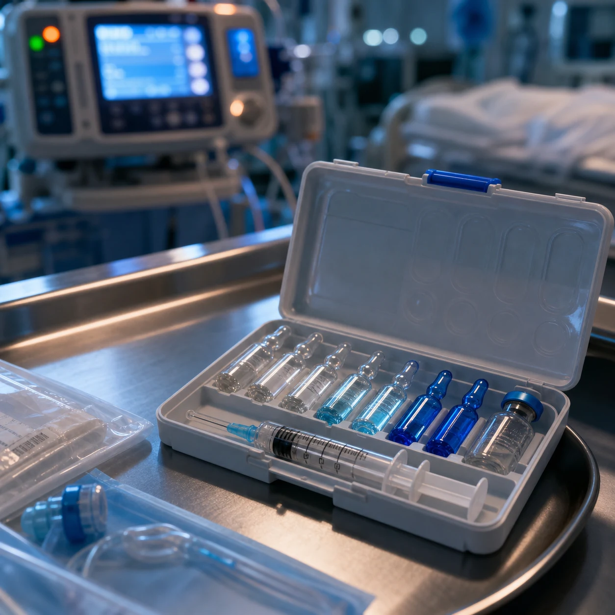
Toxidromes — pattern recognition drives empirical antidoting
The five classic toxidromes — recognise the pattern, give the antidote
| Toxidrome | Vital signs | Pupils | Secretions/skin | Bowel sounds | Toxin class | Antidote |
|---|---|---|---|---|---|---|
| Anticholinergic | Hyperthermia, tachycardia, dry | Mydriasis (dilated) | Dry skin, urinary retention | Decreased (ileus) | Antihistamines, atropine, TCA, antiparkinsonian | Physostigmine (caution) — supportive otherwise |
| Cholinergic (SLUDGE) | Bradycardia (or tachy if nicotinic), wheeze | Miosis (constricted) | Profuse salivation, lacrimation, sweating, diarrhoea, urination | Hyperactive | Organophosphates, carbamates, nerve agents | Atropine + pralidoxime |
| Opioid | Bradypnoea, hypotension, hypothermia, miosis | Miosis | Dry, pulmonary oedema (heroin) | Decreased | Heroin, morphine, fentanyl, oxycodone | Naloxone |
| Sympathomimetic / stimulant | Hypertension, tachycardia, hyperthermia | Mydriasis (dilated) | Diaphoretic, piloerection | Increased (hyperactive) | Cocaine, amphetamine, MDMA, theophylline | Benzodiazepines (control agitation/seizures) — no specific antidote |
| Sedative-hypnotic | Hypotension, bradypnoea | Normal or mild miosis | Normal | Normal | Benzodiazepines, barbiturates, ethanol | Flumazenil (BZD only — caution) |
The exam trap: a mixed toxidrome (e.g. TCA overdose produces anticholinergic + cardiotoxic + sedative features) demands treatment of the most dangerous element first (TCA → NaHCO₃ for the wide QRS), not the sedation. Never give flumazenil to the comatose TCA-overdosed patient.[1]
Specific antidotes — master table
| Toxin | Antidote | Mechanism / note |
|---|---|---|
| Opioids | Naloxone | Mu-receptor antagonist; 0.04-0.4 mg titrated to respiratory rate (avoid sudden withdrawal/pulmonary oedema) |
| Benzodiazepines | Flumazenil | GABA-A antagonist; avoid in mixed overdose or chronic users (seizure risk) |
| Paracetamol | N-acetylcysteine (NAC) | Restores glutathione; give from the nomogram or if unknown timing |
| Digoxin | Digoxin Fab (DigiFab) | Antibody fragments bind digoxin; for life-threatening arrhythmia, K over 5.5, massive ingestion |
| Organophosphates / nerve agents | Atropine + pralidoxime | Atropine for muscarinic; pralidoxime reactivates acetylcholinesterase (before aging) |
| Methanol / ethylene glycol | Fomepizole (or ethanol) | Inhibits alcohol dehydrogenase; plus haemodialysis and cofactors |
| TCAs | Sodium bicarbonate | Sodium loading + alkalinisation overcomes Na-channel blockade; target pH 7.5 |
| Salicylates | Sodium bicarbonate | Urine alkalinisation traps salicylate in tubule; haemodialysis if severe |
| Methaemoglobinaemia | Methylene blue | Reduces metHb via NADPH; avoid in G6PD deficiency |
| Cyanide | Hydroxocobalamin | Binds cyanide to form cyanocobalamin; safe with smoke inhalation |
| CCB / BB overdose | Calcium, HIET, lipid | Calcium for contractility; high-dose insulin euglycaemia; lipid emulsion |
| Sulfonylurea | Octreotide / dextrose | Octreotide suppresses insulin release; avoids dextrose-driven insulin surge |
| Iron | Deferoxamine | Chelates iron |
| Heparin | Protamine sulfate | Neutralises heparin (1 mg per 100 U) |
| Warfarin / rodenticide | Vitamin K + PCC/FFP | Vitamin K1; prothrombin complex concentrate for reversal |
| Local anaesthetic (LAST) | Lipid emulsion 20% | Lipid sink extracts the drug from receptors |
Comprehensive antidote table — every ICU-relevant toxin, antidote, dose and trigger
The complete toxin → antidote → dose → trigger reference (high-yield exam table)
| Toxin | Antidote(s) | Adult dose | Give when |
|---|---|---|---|
| Paracetamol (acetaminophen) | N-acetylcysteine (NAC) | 150 mg/kg IV over 1 h → 50 mg/kg over 4 h → 100 mg/kg over 16 h (21-h regimen); or 200 mg/kg over 4 h → 100 mg/kg over 16 h (20-h) | Level above treatment line on Rumack-Matthew nomogram, or unknown time >150 mg/kg ingested, or ALT rising, or any unknown-timing presentation |
| Opioids | Naloxone | 0.04-0.4 mg IV titrated q2-3 min to respiratory rate >12; infusion 2/3 wake-up dose/h | Respiratory depression (RR <12), pin-point pupils, ↓GCS with known/suspected opioid |
| Benzodiazepines | Flumazenil | 0.2 mg IV over 30 s, repeat 0.3 mg then 0.5 mg q1 min to max 3 mg | Isolated iatrogenic/known pure BZD reversal — NOT mixed/chronic use |
| Beta-blockers | Glucagon, HIET, calcium, lipid | Glucagon 5-10 mg IV bolus then 1-5 mg/h; HIET 1 U/kg + 0.5-1 U/kg/h + dextrose; CaCl₂ 1 g | Symptomatic bradycardia/hypotension unresponsive to atropine, fluids |
| Calcium-channel blockers | Calcium, HIET, lipid, vasopressors | CaCl₂ 1 g (10 mL 10%) or CaGluconate 3 g repeat q10-20 min; HIET as above | Bradycardia/hypotension/AV block; cardiac arrest (CCA — high-dose insulin + calcium + lipid + ECMO) |
| Digoxin / cardiac glycosides | Digoxin-specific Fab (DigiFab) | DigiFab: number of vials = serum digoxin (ng/mL) × weight (kg) / 1000; or empiric 10 vials if unknown/arrest | Life-threatening arrhythmia (VT/VF, complete heart block), K⁺ >5.5 mmol/L, massive ingestion, arrest |
| Iron | Deferoxamine | 15 mg/kg/h IV (max 6 g/24 h); continue until urine iron-clear (vin rose → normal) | Serum iron >500 mcg/dL (90 µmol/L), or symptomatic (shock, vomiting, GI bleed), or pills on X-ray |
| Lead | Succimer (DMSA), CaNa₂-EDTA, BAL (dimercaprol) | Succimer 10 mg/kg PO TDS ×5d then BD; CaNa₂-EDTA 75 mg/kg/h IV ×5d; BAL 75 mg/m² IM q4h | Encephalopathy (BAL + EDTA), level >70 mcg/dL, or symptomatic with level >45 mcg/dL |
| Methanol / ethylene glycol | Fomepizole (or ethanol) + folate/thiamine/pyridoxine + HD | Fomepizole 15 mg/kg IV load then 10 mg/kg q12h (↑ q4h with HD); ethanol target BAC 100-150 mg/dL | Confirmed toxic alcohol, anion gap acidosis + osmolar gap, or level >20 mg/dL |
| Methotrexate | Folinic acid (leucovorin) ± glucarpidase | 15 mg/m² IV/PO q6h ×72h+ until MTX level <0.05 µmol/L; glucarpidase 50 U/kg if renal failure + high level | High-dose MTX with delayed clearance, AKI, level >5 µmol/L at 24h |
| Organophosphates / nerve agents | Atropine + pralidoxime (2-PAM) | Atropine 1.2-2 mg IV doubling q5min until dried secretions (may need >100 mg); 2-PAM 30 mg/kg IV then 8 mg/kg/h | Cholinergic (SLUDGE) presentation — give BEFORE aging |
| Salicylates | Sodium bicarbonate (urine alkalinisation) + haemodialysis | 1-2 mmol/kg bolus then infusion 150 mL 8.4% in 850 mL D5W at 2-3 mL/kg/h; add KCl | Symptomatic, level >30 mg/dL (acute) or >100 mg/dL; HD if >100 acute, >60 chronic with end-organ |
| Tricyclic antidepressants | Sodium bicarbonate | 1-2 mmol/kg IV bolus, repeat to QRS <100 ms and pH 7.50; infusion to maintain | QRS >100 ms, right-axis aVR, hypotension, VT/VF |
| Warfarin / superwarfarin | Vitamin K₁ (phytomenadione) + PCC/FFP | VK₁ 5-10 mg IV slow; 4F-PCC 25-50 IU/kg (INR-based); FFP 15 mL/kg if no PCC | Major bleed (ICH) — PCC + VK; or high INR no bleed — VK only |
| Dabigatran | Idarucizumab | 5 g IV (2 × 2.5 g vials) | Life-threatening bleed, need for urgent procedure, ICH |
| Apixaban / rivaroxaban (anti-Xa DOAC) | Andexanet alfa (or 4F-PCC) | Andexanet low dose 400 mg bolus + 4 mg/min ×15 min; high dose 800 mg + 8 mg/min | Life-threatening bleed (ICH) within 18 h of last dose |
| Heparin (UFH) | Protamine sulfate | 1 mg per 100 U heparin in last 2-3 h (max 50 mg/dose), slow IV | Bleeding, or pre-procedure reversal — over-reversal causes anticoagulation |
| Heparin (LMWH) | Protamine (partial reversal) | 1 mg per 100 anti-Xa U enoxaparin (partial — ~60% for >8 h) | Significant bleeding |
| Cyanide | Hydroxocobalamin ± sodium thiosulfate (or dicobalt EDTA) | Hydroxocobalamin 5 g IV (adult) repeat ×1; Na-thiosulfate 12.5 g IV; dicobalt EDTA 300 mg | Smoke inhalation (soot, lactate >10), industrial, cardiac arrest — give empirically |
| Carbon monoxide | 100% O₂ ± hyperbaric O₂ (HBO) | 100% NRB mask; HBO 2.5-3.0 ATA ×3 sessions | Symptomatic, pregnancy, COHb >25% (HBO criteria) |
| Methaemoglobinaemia | Methylene blue | 1-2 mg/kg IV over 5 min, repeat in 30-60 min (max 7 mg/kg) | Symptomatic (dyspnoea, chest pain), metHb >30%, or >20% with anaemia — NOT G6PD |
| Sulfonylureas | Octreotide ± dextrose | Octreotide 50 mcg SC TDS, or 50 mcg bolus + infusion 1-3 mcg/kg/h | Hypoglycaemia despite dextrose — first-line per Boyle 1993 |
| Insulin | Dextrose ± glucagon ± octreotide | 50% dextrose 50 mL repeat; glucagon 1 mg IM; octreotide if sulfonylurea co-ingestion | Hypoglycaemia |
| Local anaesthetic (LAST) | Lipid emulsion 20% | 1.5 mL/kg bolus, then 0.25 mL/kg/min ×10 min (max 10 mL/kg first 30 min) | LAST — CNS toxicity → cardiac arrest (bupivacaine/ropivacaine) |
Paracetamol (acetaminophen) — N-acetylcysteine (NAC)
Mechanism of hepatotoxicity
Paracetamol is metabolised predominantly by glucuronidation and sulfation to non-toxic conjugates; a small fraction (5-10%) undergoes CYP2E1 oxidation to the hepatotoxic intermediate NAPQI (N-acetyl-p-benzoquinone imine). NAPQI is normally detoxified by conjugation with glutathione. In overdose, glutathione stores are depleted, free NAPQI binds covalently to hepatocyte macromolecules producing zone 3 (centrilobular) necrosis — the classic pattern of acute liver failure. NAC works by (a) replenishing glutathione (it is a cysteine proline, the rate-limiting precursor), and (b) acting as a glutathione substitute directly conjugating NAPQI, plus (c) enhancing microvascular blood flow in established liver injury.[1][2]
The Rumack-Matthew nomogram
Treatment is guided by the Rumack-Matthew nomogram, applied ONLY to a single acute ingestion with a known time of ingestion ≥4 h (allowing for absorption to complete). The treatment line begins at 150 mcg/mL (99 µmol/L) at 4 h post-ingestion and slopes downward to zero at ~15-16 h. A level drawn before 4 h is uninterpretable; if timing is unknown or the ingestion is staggered/repeated, give NAC empirically.[1]
The two IV NAC regimens — 21-h vs 20-h
| Parameter | 21-hour (Prescott) regimen | 20-hour (SNZ/BAT) regimen |
|---|---|---|
| Bag 1 | 150 mg/kg over 60 min | 200 mg/kg over 4 h |
| Bag 2 | 50 mg/kg over 4 h | 100 mg/kg over 16 h |
| Bag 3 | 100 mg/kg over 16 h | — |
| Total dose | 300 mg/kg | 300 mg/kg |
| Anaphylactoid risk | Higher with fast first bag (rate-related) | Lower (slower first infusion) |
| Use | Traditional, widely used (Australia, USA) | Newer, UK SNZ, less anaphylactoid |
Anaphylactoid reaction
The non-IgE-mediated (mast cell activation, complement) anaphylactoid reaction occurs in up to 20-50% (especially with the fast 60-min first bag of the 21-h regimen), peaks at 30-60 min, and is rate-related rather than dose-related. Manifestations: flushing, urticaria, pruritus, mild bronchospasm, rarely hypotension. Management: pause or slow the infusion, give an antihistamine (chlorphenamine), and restart at a slower rate once settled — it does NOT contraindicate completion of the course. True anaphylaxis is rare. [1]
When to stop NAC (recovery criteria)
After the 21-h (or 20-h) course, stop NAC if ALL: ALT falling or normal AND INR <2.0 AND clinically well. If ALT rising or INR ≥2.0, continue NAC (50 mg/kg over 8 h bags) until ALT falls and INR <2.0 — NAC continues to be beneficial even after injury is established (improves microvascular perfusion).[1]
King's College criteria for transplant referral
Refer to a liver transplant unit if arterial pH <7.30 (after fluid resuscitation) regardless of grade, OR all three of: INR >6.5 AND creatinine >300 µmol/L AND grade 3-4 encephalopathy. This single criterion (pH <7.30) carries mortality >90% without transplant. [1]
Paracetamol overdose — the NAC protocol from presentation to transplant referral
- Resuscitate and take history — airway, time of ingestion (single vs staggered), co-ingestants, weight, prior liver disease, alcoholism, malnutrition, enzyme-inducing drugs (these LOWER the nomogram line to 100 mcg/mL — "high-risk" line)
- Draw a 4-h level — never draw before 4 h (uninterpretable). If presentation is >8 h post-ingestion, start NAC empirically while awaiting the level — do not wait
- Plot on the Rumack-Matthew nomogram — if level is above the treatment line (or timing unknown / staggered ingestion), give NAC. If single acute ingestion <4 h ago, the level is not yet interpretable — repeat at 4 h
- Start the 21-h IV regimen — 150 mg/kg over 60 min → 50 mg/kg over 4 h → 100 mg/kg over 16 h. Use the 20-h regimen if your unit prefers (slower first bag, fewer anaphylactoid reactions)
- Manage anaphylactoid reaction — slow/stop the infusion, give chlorphenamine 10 mg IV, restart at 50% rate once settled. Continue the full course — it is non-IgE and self-limiting
- At the end of the course (h 21) — check ALT and INR. If ALT normal and INR <2.0 AND clinically well → STOP. If ALT rising or INR ≥2.0 → CONTINUE NAC (50 mg/kg over 8 h bags) until ALT falls and INR <2.0
- Check INR and ALT every 12 h during the continuation phase — INR peaks at 48-72 h. A rising INR with falling ALT is the hallmark of recovery (hepatocyte regeneration)
- Apply King's College criteria for transplant referral at 24-72 h — arterial pH <7.30 (after fluids), OR INR >6.5 + creatinine >300 µmol/L + grade 3-4 encephalopathy. Refer EARLY — centres prefer to transfer before encephalopathy
- Look for complications of acute liver failure — hypoglycaemia (monitor glucose hourly), cerebral oedema (head elevation, hypertonic saline, mannitol), coagulopathy (PCC/VK if bleeding), AKI (renal replacement), sepsis (broad-spectrum cover)
Opioids — naloxone
Naloxone is a competitive mu-opioid receptor antagonist (also weak kappa and delta) with rapid onset (1-2 min IV) and short half-life (~60-90 min). In opioid overdose the goal is to reverse respiratory depression, not arousal — titrate to a respiratory rate >12/min, accepting that the patient may remain drowsy. The classic error is over-reversal with a large bolus, precipitating acute withdrawal (agitation, sweating, vomiting, pulmonary oedema, catecholamine surge, arrhythmia).[1]
Dose: 0.04 mg IV (or 0.1 mg) increments every 2-3 min, escalating to 0.4 mg and then 2 mg if no response; if no response to a total of 10 mg, reconsider the diagnosis. For long-acting opioids (methadone, sustained-release morphine, fentanyl patches) or large ingestion, start a naloxone infusion at 2/3 of the wake-up dose per hour, titrated. Naloxone has a shorter half-life than most opioids — the patient who wakes up will re-sedate; observe for at least 2 h after the last bolus (longer for methadone/extended-release). [1]
Benzodiazepines — flumazenil (CAUTION: seizures)
Flumazenil is a competitive GABA-A receptor antagonist that reverses benzodiazepine sedation. It is dangerous because chronic benzodiazepine use causes receptor upregulation that has adapted to a tonic inhibitory tone; flumazenil removes this protection and lowers the seizure threshold. In a mixed overdose (especially with a proconvulsant coingestant such as a TCA, theophylline, cocaine, or bupropion), flumazenil can precipitate refractory status epilepticus that is very difficult to control.[1]
Use flumazenil ONLY for: (a) isolated iatrogenic oversedation (e.g. post-procedural midazolam reversal in a non-tolerant patient), or (b) known pure benzodiazepine overdose in a non-tolerant patient. Never use it for the unknown-coma patient, the chronic user, or any co-ingestion with a proconvulsant — support ventilation instead. Dose: 0.2 mg IV over 30 s, repeat 0.3 mg then 0.5 mg q1 min to max 3 mg. Like naloxone, flumazenil has a shorter half-life (~40-80 min) than the benzodiazepines it reverses — re-sedation occurs. [1]
High-dose insulin euglycaemia therapy (HIET)
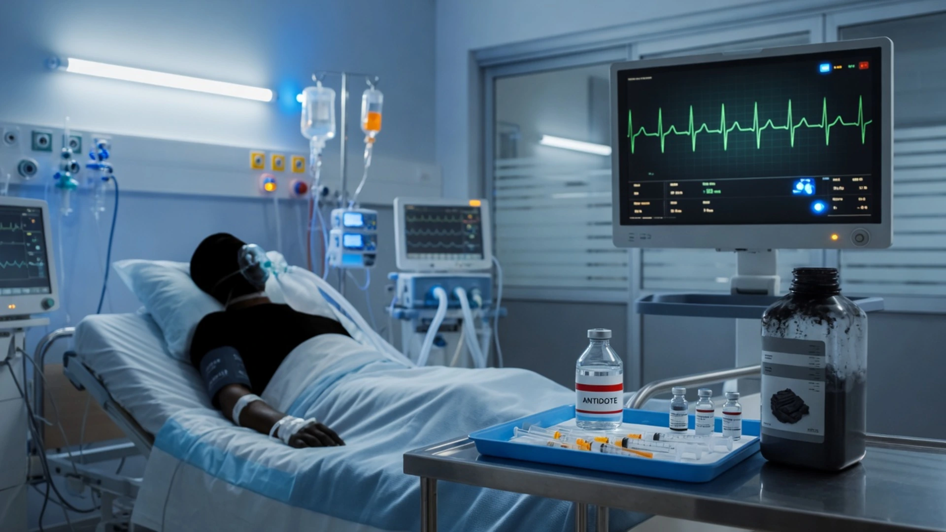
For severe calcium-channel blocker or beta-blocker overdose unresponsive to fluids, calcium, and vasopressors: a regular-insulin bolus (1 U/kg) then infusion (0.5-1 U/kg/h) with concurrent dextrose to keep glucose normal. Insulin shifts myocardial metabolism from free fatty acids to carbohydrate, improving inotropy without much effect on heart rate. Monitor glucose and potassium.[1]
Mechanism and rationale
In CCB/BB overdose, the heart becomes bradycardic, negatively inotropic, and vasodilatory (CCBs) or bradycardic with AV block (BBs). Conventional catecholamines often fail because the receptor/pathway is blocked. HIET exploits a non-adrenergic inotropic mechanism: under stress the myocardium preferentially uses free fatty acids; insulin forces a shift to carbohydrate metabolism which is more efficient per mole of oxygen, restoring inotropy. It also improves intracellular calcium handling and microvascular perfusion.[12]
Protocol
- Bolus regular insulin 1 U/kg IV
- Infusion 0.5-1 U/kg/h (titrate to effect, up to 10 U/kg/h in refractory cases)
- Concurrent dextrose — 25 g (50 mL of 50%) bolus, then 0.5 g/kg/h; monitor glucose q1h initially
- Potassium — keep K⁺ >2.5 mmol/L (insulin drives K⁺ intracellularly); replace 10-20 mmol/h as needed
- Continue 12-24 h after haemodynamic stability then taper over 12-24 h (rebound toxicity possible)[13]
The two big mistakes: (1) letting glucose fall (insulin without enough dextrose — euglycaemia, not hypoglycaemia, is the goal), and (2) stopping too early (CCBs redistribute from tissue stores; wean slowly). [1]
Intravenous lipid emulsion
A 20% lipid bolus (1.5 mL/kg) then infusion (0.25 mL/kg/min) creates a "lipid sink" that sequesters lipophilic drugs, used for local anaesthetic systemic toxicity and refractory overdose with lipophilic agents (TCAs, CCBs, bupropion).[1]
Mechanism — more than a "lipid sink"
Three complementary mechanisms are now recognised:[14]
- Lipid sink / partitioning — the expanded intravascular lipid phase extracts lipophilic drug from aqueous plasma and target-tissue receptors, lowering the effective free-drug concentration
- Cardiac fatty acid metabolism — lipid provides a high substrate load of fatty acids, bypassing CCB/BB-induced metabolic blockade and directly fuelling myocardial contraction
- Ion channel trafficking / cytoprotection — accelerated removal of drug from cardiac sodium channels and post-conditioning cytoprotective effects
Indications
- Local anaesthetic systemic toxicity (LAST) — bupivacaine/ropivacaine cardiac arrest (the original indication)
- Refractory cardiac arrest / cardiotoxicity from lipophilic drugs — TCA, CCB (verapamil/diltiazem), BB (propranolol), bupropion, lamotrigine, quetiapine, after standard therapy (HIDT, bicarb, calcium, vasopressors) has failed
- Lipid rescue is NOT first-line for most overdoses — escalate through standard therapy first, add lipid when refractory [1]
Protocol
- Bolus 20% lipid emulsion 1.5 mL/kg IV over 2-3 min (lean body weight)
- Infusion 0.25 mL/kg/min for 10 min (or until stable); maximum 10 mL/kg in first 30 min
- Repeat bolus if still unstable
- Complications — ARDS, pancreatitis, fat overload syndrome, lipaemia interfering with lab assays; avoid in pregnancy, fat metabolism disorders, egg allergy [1]
Severe calcium-channel blocker overdose — the full escalation ladder (verapamil/diltiazem)
- Recognise the lethal toxidrome — bradycardia, hypotension, AV block (CCBs) ± hyperglycaemia (verapamil blocks insulin release), often after a few hours of GI symptoms. Verapamil sustained-release is the deadliest — delayed peak at 6-12 h
- ABCDE resuscitation — airway, 100% O₂, two large-bore cannulae, IV fluids (cautious — these patients often have cardiogenic shock and pulmonary oedema), continuous cardiac monitoring
- Decontamination — activated charcoal 50 g if <1-2 h and airway protected; whole-bowel irrigation for sustained-release formulations with polyethylene glycol 1-2 L/h via NG until rectal effluent clear
- First-line antidote: calcium — CaCl₂ 1 g (10 mL of 10%) slow IV (central line preferred) OR CaGluconate 3 g (30 mL of 10%) repeat every 10-20 min up to 3-4 doses; assess for rise in BP/HR
- Add atropine 0.5-1 mg IV for symptomatic bradycardia/AV block — often ineffective (the bradycardia is not vagally mediated); do not persist if no response
- Start high-dose insulin euglycaemia therapy (HIET) — regular insulin 1 U/kg IV bolus then 0.5-1 U/kg/h with concurrent dextrose (25 g bolus then 0.5 g/kg/h); monitor glucose q1h and K⁺ (replace to keep >2.5)
- Vasopressors — noradrenaline ± adrenaline/vasopressin for MAP <65 mmHg; high doses often needed
- Escalate to lipid emulsion if refractory — 20% lipid 1.5 mL/kg bolus then 0.25 mL/kg/min ×10 min (lipophilic CCB)
- Consider cardiac pacing for symptomatic high-grade AV block unresponsive to drugs (capture is often poor)
- VA-ECMO as salvage for refractory cardiac arrest / cardiogenic shock — bridge to drug clearance; refer to ECMO centre early
- Continue HIET 12-24 h after stability then wean slowly over 12-24 h — verapamil redistributes from tissue, rebound toxicity is common
- Monitor glucose (HIET), K⁺, lactate, calcium (avoid hypercalcaemia from Ca boluses), mental state
Digoxin — Fab antibody fragments
Digoxin-specific Fab antibody fragments (DigiFab in most of the world; Digibind discontinued) are ovine immunoglobulin fragments that bind digoxin with >1000× the affinity of the cardiac Na-K-ATPase, forming an inert complex excreted by the kidney. Indications for Fab in acute or chronic digoxin toxicity:[5]
- Life-threatening arrhythmia — ventricular tachycardia/fibrillation, complete heart block, asystole
- Hyperkalaemia — K⁺ >5.5 mmol/L in acute overdose (this is a marker of toxicity, NOT to be treated with calcium — the "calcium in digoxin toxicity" prohibition is now debated but traditional teaching still cautions)
- Massive ingestion — known >10 mg in an adult, >4 mg in a child
- Cardiac arrest attributed to digoxin — give empirically [1]
Dose calculation
- From serum concentration (steady-state, ≥6 h post-ingestion): vials = [serum digoxin (ng/mL) × weight (kg)] / 100 (DigiFab) — then round up
- From ingested dose: vials = total mg ingested × 0.8 (bioavailability) × 1.25 (DigiFab) — round up
- If unknown: give empiric 5-10 vials in acute overdose, 3 vials in chronic toxicity
- In cardiac arrest: give 10 vials empirically [1]
Pitfalls
- Serum digoxin level becomes uninterpretable after Fab — most assays measure total (bound + free); free digoxin is the meaningful measure but few labs offer it. Do NOT re-dose based on a "high" level post-Fab.
- Re-distribution of digoxin from tissue stores can cause re-toxicity 8-24 h later; the Fab-digoxin complex is renally excreted, so renal failure prolongs the need for monitoring.
- Hypokalaemia, hypomagnesaemia, hypercalcaemia develop as Fab reverses the digoxin effect — replete K⁺.
- Saturation kinetics — at very high digoxin concentrations Fab may not bind all of it; expect a partial response. [1]
Organophosphates — atropine + pralidoxime
Organophosphates (pesticides such as parathion, malathion, chlorpyrifos; nerve agents such as sarin, VX) irreversibly inhibit acetylcholinesterase by phosphorylating the serine hydroxyl at the active site, causing acetylcholine accumulation at muscarinic, nicotinic, and CNS receptors. The clinical picture is the cholinergic toxidrome: SLUDGE (Salivation, Lacrimation, Urination, Defecation, GI distress, Emesis) plus miosis, bradycardia, bronchospasm, bronchorrhoea, fasciculations, weakness, seizures, coma. Death is from bronchorrhoea/bronchospasm (muscarinic) and respiratory muscle paralysis (nicotinic). [1]
Aging
After phosphorylation, the enzyme undergoes aging — the loss of an alkyl group that makes the bond irreversible. Once aged, pralidoxime cannot reactivate the enzyme — newly synthesised AChE (over weeks) is the only recovery. Aging time is toxin-dependent: soman (nerve agent) ages in minutes, sarin in 3-4 h, tabun ~14 h, diethyl organophosphates (parathion) hours, dimethyl (dichlorvos) in minutes. This is why pralidoxime must be given EARLY.[1]
Atropine vs pralidoxime — divide the labour
- Atropine blocks muscarinic effects only (dries secretions, reverses bradycardia/bronchospasm/bronchorrhoea). It does NOT reverse nicotinic effects (muscle weakness, fasciculations) or CNS effects.
- Pralidoxime (2-PAM) reactivates AChE (before aging), reversing nicotinic effects and restoring muscle strength. It is the only agent that treats the paralysis. [1]
Atropine vs pralidoxime in organophosphate poisoning
| Feature | Atropine | Pralidoxime (2-PAM) |
|---|---|---|
| Receptor action | Muscarinic antagonist | Reactivates AChE (dephosphorylates — before aging) |
| Reverses secretions/bronchospasm/bradycardia | YES (muscarinic) | No (indirectly, slowly) |
| Reverses muscle weakness/fasciculations | No | YES (nicotinic) |
| Reverses CNS effects | Partial | Yes |
| Titration endpoint | Dried secretions, HR >80, SBP >80 | Resolution of fasciculations/weakness |
| Dose | 1.2-2 mg IV doubling q5min (may need >1000 mg total) | 30 mg/kg IV bolus then 8 mg/kg/h infusion ×24-48 h |
| Time-critical | Give immediately | Give BEFORE aging (minutes-hours) |
Methanol / ethylene glycol — fomepizole
Methanol and ethylene glycol are toxic alcohols that are themselves relatively non-toxic but are metabolised by alcohol dehydrogenase (ADH) to highly toxic acids: methanol → formaldehyde → formic acid (blindness, basal ganglia injury); ethylene glycol → glycoaldehyde → glycolic acid and oxalic acid (severe metabolic acidosis, AKI from calcium oxalate crystals). The antidote strategy is to block ADH with fomepizole (or ethanol) so the parent alcohol is excreted unchanged by the kidney, then remove it with haemodialysis and provide cofactors (folate for methanol → CO₂ + H₂O; thiamine + pyridoxine for ethylene glycol → less-toxic metabolites). [1]
Fomepizole vs ethanol for ADH blockade
| Feature | Fomepizole | Ethanol |
|---|---|---|
| Mechanism | Competitive ADH inhibitor (8000× affinity of ethanol) | Competitive ADH substrate |
| Ease of use | Easy — fixed dose, no monitoring | Difficult — IV infusion, frequent BAC monitoring (target 100-150 mg/dL) |
| Adverse effects | Nausea, headache, rash | Intoxication, hypoglycaemia, hepatitis, CNS depression |
| Cost | Expensive (~$1000+/dose) | Cheap |
| Availability | Variable worldwide | Universally available |
| Preferred | YES when available | When fomepizole unavailable |
Fomepizole dose: 15 mg/kg IV loading dose, then 10 mg/kg q12h ×4 doses, then 15 mg/kg q12h until toxic alcohol level <20 mg/dL and acidosis resolved. Increase dosing frequency to q4h during haemodialysis (fomepizole is dialysable).[3][4]
Suspected toxic alcohol ingestion (methanol / ethylene glycol)
- Recognise the biochemical signature — severe high-anion-gap metabolic acidosis WITH a concomitant osmolar gap (early, before metabolism), normal lactate (or falsely elevated lactate with ethylene glycol on some analysers), normal glucose. Visual symptoms (methanol) or flank pain/AKI (ethylene glycol) support the diagnosis
- Resuscitate — airway, IV access, fluids; sodium bicarbonate to correct severe acidosis (target pH >7.20)
- Give fomepizole 15 mg/kg IV STAT — do not wait for confirmatory levels if the picture is suggestive; fomepizole is safe and time-critical
- Add cofactors — folinic acid (leucovorin) 1 mg/kg IV q4-6h for methanol (accelerates formate → CO₂ + H₂O); thiamine 100 mg IV and pyridoxine 50 mg IV q6h for ethylene glycol (diverts glyoxylate to non-toxic metabolites)
- Arrange haemodialysis — indicated for confirmed diagnosis with level >50 mg/dL, severe acidosis (pH <7.25-7.30), AKI, or visual symptoms. Dialysis removes both parent alcohol and acid metabolites
- Increase fomepizole dosing during HD — q4h instead of q12h (fomepizole is dialysable)
- Continue fomepizole + cofactors until level <20 mg/dL AND acidosis resolved AND clinically improving
- Look for end-organ injury — ophthalmology review for methanol (retinal/optic nerve toxicity), nephrology for ethylene glycol (AKI, oxalate crystal deposition); MRI basal ganglia (methanol causes putaminal necrosis)
- Search for co-ingestants and intent — psychosocial assessment, consider concurrent suicide attempt
Methotrexate — folinic acid (leucovorin)
High-dose methotrexate (used in oncology) is rescued with folinic acid (leucovorin, 5-formyl-tetrahydrofolate), the reduced folate that bypasses the dihydrofolate reductase blockade. This rescues normal cells (bone marrow, gut mucosa) from methotrexate's anti-folate effect. Folinic acid is NOT the same as folic acid — folic acid must be reduced by DHFR (the very enzyme MTX blocks), so it is ineffective. Give folinic acid 15 mg/m² q6h IV/PO starting 24-42 h after high-dose MTX, continuing until MTX level <0.05 µmol/L. Glucarpidase (carboxypeptidase G2) cleaves MTX to inactive metabolites and is used for MTX-induced AKI with delayed clearance (level >5 µmol/L at 24 h). Hydration, alkalinisation of urine (NaHCO₃ to keep urine pH >7), and avoidance of nephrotoxins are supportive. [1]
Salicylates — sodium bicarbonate + haemodialysis
Salicylate poisoning causes respiratory alkalosis (direct stimulation of medullary respiratory centre) followed by high-anion-gap metabolic acidosis (uncoupling of oxidative phosphorylation → lactate + ketones + salicylate anion). The mixed picture of respiratory alkalosis + metabolic acidosis with a normal/anion-gap is the classic ABG. The key to management is urine alkalinisation: salicylate is a weak acid (pKa 3.0); in alkaline urine it becomes ionised (trapped) and is excreted. Target urine pH >7.5 with IV sodium bicarbonate.[18]
Sodium bicarbonate in TCA vs salicylate — same drug, different goals
| Parameter | TCA | Salicylate |
|---|---|---|
| Goal of NaHCO₃ | Overcome Na-channel blockade | Trap salicylate in tubule (ion trapping) |
| Target | Serum pH 7.45-7.55 AND QRS <100 ms | Urine pH >7.5 |
| Mechanism | Na⁺ load + alkalinisation | HCO₃⁻ in tubule → salicylate ionised → trapped |
| Bolus vs infusion | Bolus 1-2 mmol/kg, repeat to QRS | Infusion 1-2 mmol/kg/h + KCl |
| Hypokalaemia | Less central | CRITICAL — K⁺ must be >4 for urine alkalinisation to work (hypokalaemia causes H⁺/K⁺ exchange in tubule, acidifying urine) |
| Endpoint | QRS narrows, haemodynamics improve | Urine pH >7.5, serum level falling |
| Haemodialysis | Only for refractory | Indicated early (level >100 acute, >60 chronic, end-organ) |
Haemodialysis for salicylate: indicated for acute level >100 mg/dL (or >120 in some guidelines), chronic level >60 mg/dL with end-organ toxicity (altered mental state, AKI, pulmonary oedema), refractory acidosis, or deterioration despite alkalinisation. HD is highly effective (clearance ~LD) but salicylate redistributes from tissue post-HD — watch for rebound, consider continuous RRT.[18]
Tricyclic antidepressants — sodium bicarbonate
TCA overdose is lethal via sodium-channel blockade (cardiotoxicity — wide QRS, right-axis deviation of the terminal 40 ms in aVR, ventricular arrhythmias, hypotension) plus alpha-1 blockade (hypotension), anticholinergic effects, and seizures. Sodium bicarbonate is the antidote: it works by (a) providing a sodium load that overcomes the Na-channel blockade (mass-action), (b) alkalinising the blood (pH 7.50) which increases binding of the drug to albumin and reduces free-drug concentration, and (c) overcoming the K⁺ channel blockade. Dose: 1-2 mmol/kg IV bolus, repeat to QRS <100 ms and pH 7.50; an infusion of 100-150 mmol in 850 mL D5W maintains the effect.[6][7]
Do NOT use class Ia (quinidine, procainamide, disopyramide) or class Ic (flecainide) antiarrhythmics — they worsen the Na-channel blockade. For refractory cardiotoxicity use lidocaine (class Ib, does not prolong QRS) or lipid emulsion. Magnesium is reasonable for TCA-induced torsades. Hypotension refractory to fluids + bicarbonate → noradrenaline (alpha-1 agonist overcomes alpha-1 blockade) or HIET. [1]
Warfarin and DOACs — reversal agents
Warfarin
Warfarin inhibits vitamin K epoxide reductase (VKORC1), depleting reduced vitamin K needed for gamma-carboxylation of factors II, VII, IX, X, and proteins C/S. Reversal depends on urgency:[19]
- Major bleed (ICH): 4-factor prothrombin complex concentrate (4F-PCC) 25-50 IU/kg + vitamin K₁ 10 mg IV slow (over 20 min). PCC works in minutes; vitamin K takes 6-12 h. FFP 15 mL/kg is an alternative if PCC unavailable (more volume, slower).
- High INR, no major bleed: vitamin K₁ 1-5 mg PO/IV (oral preferred, lower anaphylactoid risk). For INR >10 with no bleed, 2.5-5 mg PO; for INR 5-10, hold warfarin ± 1-2.5 mg PO.
- Superwarfarin rodenticides (brodifacoum): massive vitamin K doses (50-100 mg/day for weeks-months) — these have 100× the half-life of warfarin. [1]
DOAC reversal
DOAC reversal agents — dabigatran vs anti-Xa (apixaban/rivaroxaban/edoxaban)
| Feature | Dabigatran (direct thrombin inhibitor) | Apixaban / rivaroxaban / edoxaban (anti-Xa) |
|---|---|---|
| Specific reversal agent | Idarucizumab (humanised monoclonal Fab) | Andexanet alfa (recombinant modified FXa, decoy) |
| Dose | 5 g IV (2 × 2.5 g vials) | Low dose 400 mg bolus + 4 mg/min ×15 min; high dose 800 mg + 8 mg/min (for >10 mg dose or <8 h) |
| Onset | Immediate | Immediate |
| Duration | ~12-24 h | ~1-2 h (anti-Xa activity returns) |
| Clinical trial | RE-VERSE AD (Pollack 2015) | ANNEXA-4 (Connolly 2016) |
| Practical alternative | None (or aPCC) | 4F-PCC 25-50 IU/kg (cheaper, immediate, off-label) |
| Special caution | — | Andexanet interferes with heparin and anti-Xa assays for ~24 h (spurious anti-Xa) |
Idarucizumab binds dabigatran with >350× the affinity of thrombin, forming an inert complex; RE-VERSE AD showed >90% immediate reversal of anticoagulant effect. No prothrombin risk inherent (idarucizumab has no intrinsic haemostatic activity).[17] Andexanet alfa is a modified recombinant FXa that lacks catalytic activity but retains the binding site for anti-Xa inhibitors — it acts as a decoy, sequestering the DOAC away from endogenous FXa. ANNEXA-4 demonstrated rapid anti-Xa reversal but thrombotic events (~10%) are the trade-off.[16] Because andexanet is expensive and has a long procurement window, many centres use 4F-PCC 25-50 IU/kg off-label for anti-Xa DOAC-associated ICH with comparable effectiveness.[19]
Cyanide — hydroxocobalamin / sodium thiosulfate / dicobalt EDTA
Cyanide binds cytochrome a3 (cytochrome oxidase) in mitochondrial complex IV, halting oxidative phosphorylation — cells switch to anaerobic glycolysis producing severe lactic acidosis, and venous blood remains oxygenated (so the AV oxygen difference narrows and venous blood looks arterially red). Sources: smoke inhalation (burning plastics/wool release HCN), industrial (electroplating, mining, jewellery), nitroprusside infusion (releases cyanide), and suicide. [1]
The three antidotes
Cyanide antidotes — three options, different settings
| Antidote | Mechanism | Dose (adult) | Setting / advantage | Disadvantage |
|---|---|---|---|---|
| Hydroxocobalamin (Cyanokit) | Binds cyanide → cyanocobalamin (vitamin B12) | 5 g IV over 15 min, repeat ×1 | Smoke inhalation (safe with CO co-poisoning); does not interfere with tissue oxygenation | Turns skin, urine, plasma red-orange; interferes with co-oximetry and many labs |
| Sodium thiosulfate | Donates sulfur → rhodanese/thiosulfate sulfurtransferase converts CN → thiocyanate (renally excreted) | 12.5 g IV over 10 min | Cheap; synergistic with hydroxocobalamin or sodium nitrite | Slow onset (rhodanese rate-limiting); ineffective alone in severe poisoning |
| Dicobalt EDTA (Kelocyanor) | Chelates cyanide directly | 300 mg IV over 1 min, repeat | Rapid | Severe hypotension if no cyanide present — give ONLY with confirmed cyanide poisoning; cardiac arrest risk |
| (Sodium nitrite / amyl nitrite) | Oxidises Hb → methaemoglobin, which binds cyanide preferentially | Amyl nitrite inhalation; Na-nitrite 300 mg IV | Traditional "Cyanide Antidote Kit" | Methaemoglobinaemia — dangerous if concomitant CO (smoke inhalation — blood cannot carry O₂). Now largely abandoned |
Empirical treatment rule: in the smoke inhalation victim with soot in the mouth, depressed GCS, and lactate >10 mmol/L, give hydroxocobalamin empirically — do not wait for a cyanide level (levels are rarely available in time). The combination hydroxocobalamin + sodium thiosulfate is recommended in many smoke-inhalation protocols. Avoid sodium nitrite in suspected CO co-poisoning (it generates methaemoglobin, compounding the hypoxia).[8][9]
Carbon monoxide — oxygen / hyperbaric
Carbon monoxide binds haemoglobin with 240× the affinity of oxygen, forming carboxyhaemoglobin (COHb) and shifting the oxyhaemoglobin dissociation curve leftward (impairing tissue offloading). CO also binds myoglobin (cardiac dysfunction) and cytochrome a3 (mitochondrial poisoning — analogous to cyanide). Treatment: 100% oxygen via non-rebreather mask (reduces COHb half-life from ~320 min on room air to ~80 min on 100% O₂). Hyperbaric oxygen (HBO) at 2.5-3.0 ATA reduces the half-life to ~20-30 min and may reduce delayed neurologic sequelae. Indications for HBO (controversial): syncope, seizure, coma, pregnancy with COHb >15%, COHb >25%, or persistent symptoms after normobaric O₂. Controversy: the rigorously designed randomised trials have not consistently shown benefit, but most toxicology guidelines still recommend HBO for severe poisoning. [1]
Heparin — protamine sulfate
Protamine is a strongly basic polypeptide (derived from salmon sperm) that binds and neutralises heparin (a strongly acidic polysaccharide), forming an inactive complex. Dose: 1 mg protamine per 100 units of UFH in the last 2-3 h (heparin half-life ~60-90 min, so older doses need less reversal), max 50 mg per dose, slow IV over 1-3 min (rapid infusion causes hypotension, anaphylaxis). For LMWH (enoxaparin): protamine gives only ~60% reversal if given within 8 h, less if delayed. Pitfalls: (a) over-reversal — protamine itself is a weak anticoagulant (excess unbound protamine interferes with platelets and thrombin); give only enough to neutralise the residual heparin, (b) HIT — never use protamine to "reverse" suspected HIT; stop the heparin and use a non-heparin anticoagulant (argatroban, bivalirudin), (c) allergy in patients with fish allergy, prior protamine exposure (insulin formulations), or vasectomy. [1]
Methaemoglobinaemia — methylene blue
Methaemoglobin (metHb) is haemoglobin in which the iron is in the ferric (Fe³⁺) state — it cannot bind oxygen and shifts the dissociation curve leftward, impairing oxygen release. Causes: nitrites (amyl/sodium nitrite, nitrate-contaminated water in infants), dapsone, benzocaine (topical anaesthetic spray), primaquine, nitroprusside, aniline dyes, and inborn errors (cytochrome b5 reductase deficiency). Pulse oximetry plateaus at ~85% (does not distinguish metHb from oxyHb well) — diagnosis requires co-oximetry showing metHb >1-2% (normal <1%). Symptoms: cyanosis (does not improve with O₂), "chocolate brown" blood, dyspnoea, headache; at metHb >30% fatigue, confusion, chest pain; >50-70% seizures, coma, arrhythmia, death.[10]
Methylene blue mechanism
Methylene blue acts as an electron carrier via NADPH-methaemoglobin reductase (the alternative pathway, normally minor): methylene blue → leukomethylene blue → reduces Fe³⁺ → Fe²⁺. Dose: 1-2 mg/kg IV over 5 min, repeat in 30-60 min if needed (max 7 mg/kg). Effect within 30-60 min. [1]
The two absolute cautions
- G6PD deficiency — methylene blue requires NADPH (regenerated via the hexose monophosphate shunt, which is G6PD-dependent); in G6PD deficiency methylene blue is ineffective AND causes oxidant-induced haemolysis. Screen before giving if possible; alternatives are ascorbic acid (1 g IV q6h, slower) or exchange transfusion.
- Serotonin syndrome / MAOI — methylene blue is a weak MAO inhibitor and can precipitate serotonin syndrome in patients on SSRIs/MAOIs; weigh risk vs benefit. [1]
Paradoxically, methylene blue itself is an oxidant and at high doses (>7 mg/kg) causes methaemoglobinaemia — do not exceed the cumulative dose. [1]
Lead — succimer / EDTA / BAL
Lead poisoning (occupational — battery recycling, demolition, soldering; folk remedies; retained bullets; leaded paint in old housing) causes abdominal pain, encephalopathy, peripheral neuropathy (wrist drop), nephropathy, and anaemia (basophilic stippling). Chelation options:[1]
- Succimer (DMSA, dimercaptosuccinic acid) — oral chelator, first-line for mild-moderate (level >45 mcg/dL, asymptomatic-mild); 10 mg/kg PO TDS ×5 days then BD ×14 days. Better tolerated than parenteral agents.
- CaNa₂-EDTA (calcium disodium edetate) — IV chelator for moderate-severe (level >70 mcg/dL, symptomatic); 75 mg/kg/h IV ×5 days. Note: NEVER use Na₂-EDTA (without calcium) — it causes fatal hypocalcaemia.
- BAL (dimercaprol, British anti-lewisite) — IM chelator, given FIRST in encephalopathy to mobilise lead from the brain before EDTA (which can redistribute lead into the brain); 75 mg/m² IM q4h ×5 days, combined with CaNa₂-EDTA after the first dose. [1]
Encephalopathy protocol: BAL first → then CaNa₂-EDTA → then transition to succimer once stable. Supportive care includes seizure control, control of cerebral oedema, and removing the source. [1]
Iron — deferoxamine
Iron overdose (children's prenatal vitamins, adult haemochromatosis overdose) follows four stages: (1) GI toxicity (0-6 h — vomiting, diarrhoea, GI bleed — direct corrosive effect), (2) apparent recovery (6-24 h — deceptive "quiescent" phase), (3) shock / metabolic acidosis / hepatic failure (6-48 h — mitochondrial poisoning, systemic vasodilation), (4) GI obstruction (2-6 weeks — pyloric stricture from scarring). Deferoxamine is a specific iron chelator (siderophore derived from Streptomyces); it binds ferric iron to form ferrioxamine (water-soluble, excreted renally — turns urine "vin rosé"). Dose: 15 mg/kg/h IV (initial rate; may start at 5 mg/kg/h and titrate to avoid hypotension), max 6 g/24 h; continue until urine returns to normal colour and serum iron is normal. Indications: serum iron >500 mcg/dL (90 µmol/L), symptoms of systemic toxicity, or pills visible on abdominal X-ray. [1]
Lipid rescue therapy — when and how
Lipid emulsion therapy has expanded beyond local anaesthetic toxicity to become a rescue therapy for refractory cardiac toxicity from any lipophilic drug. The decision to escalate to lipid follows failure of standard therapy (decontamination, specific antidote, HIET, bicarbonate, calcium, vasopressors). High-yield lipophilic drugs for which lipid rescue has reported success:[14][15]
Drugs amenable to lipid rescue (lipophilicity-driven)
| Drug | LogP (lipophilicity) | Evidence | Typical context |
|---|---|---|---|
| Bupivacaine / ropivacaine | High (3.4-3.7) | Strong (the original indication) | LAST after regional anaesthesia |
| Verapamil / diltiazem | High | Moderate-strong (case series) | CCB overdose, refractory shock |
| Propranolol | High (3.5) | Moderate (case reports) | BB overdose with cardiac toxicity |
| Tricyclic antidepressants | High | Moderate (rescue after bicarb fails) | TCA overdose refractory to NaHCO₃ |
| Bupropion / lamotrigine / quetiapine | High | Case reports | Refractory seizures/QRS widening |
| Cocaine / cocaethylene | High | Animal studies | Refractory cardiotoxicity |
| Moxidectin / ivermectin | High | Veterinary origin | Limited human data |
The "no-harm" position — give lipid early in refractory cardiac arrest from a lipophilic drug; the risk of ARDS/pancreatitis is small and the patient is dying. Reversibility is the rationale: many of these patients arrest not from irreversible organ destruction but from circulating drug — extracting it restores the heart.[15]
SAQ — NAC for a staggered paracetamol overdose in a high-risk patient
10 minutes · 10 marks
A 24-year-old woman (60 kg) is brought to ED ~6 hours after an intentional overdose of 30 g of paracetamol (sixty 500 mg tablets) co-ingested with alcohol. She is nauseated but alert, RR 16, SpO2 98 percent on room air, HR 96, BP 118/72, GCS 15. She has a history of alcohol misuse and is on rifampicin for pulmonary tuberculosis. A 4-hour post-ingestion paracetamol level is 250 mg/L (above the treatment line on the Rumack-Matthew nomogram). ALT 45 U/L, INR 1.1, creatinine 80 micromol/L, venous pH 7.36. You are asked to manage her antidotal therapy.
SAQ — Naloxone for an opioid overdose from long-acting methadone
10 minutes · 10 marks
A 34-year-old man is found unconscious in a public toilet with a syringe and needle nearby. GCS 6 (E1V1M4), RR 4/min with shallow breaths, SpO2 86 percent on room air, pinpoint pupils, HR 58 (sinus bradycardia), BP 112/68, temperature 36.4 degrees C, blood glucose 6.2 mmol/L. He has fresh needle-track marks. His partner reports he uses prescribed long-acting methadone 80 mg daily for opioid dependence and has missed several doses of his usual supervision. You secure the airway with bag-valve-mask ventilation.
Clinical pearls — the exam-distinguishing points
Trial cards — the landmark evidence
Antman et al 1990 — Digoxin-specific Fab for life-threatening digitalis intoxication (PMID 2188752)
Study design
Multicentre open-label case series — 150 patients with life-threatening digitalis toxicity
Population
Adults with life-threatening digoxin/digitoxin toxicity (ventricular arrhythmia, high-grade AV block, hyperkalaemia ≥5.5)
Intervention
Digoxin-specific Fab antibody fragments (ovine), dose calculated from serum level and weight
Primary outcome
Clinical response (resolution of life-threatening toxicity)
Key findings
90% complete response within minutes to hours; hyperkalaemia resolved rapidly; survival in patients who had reached cardiac arrest was lower but still meaningful. Few allergic reactions (~6%, mostly mild)
Clinical bottom line
Digoxin-specific Fab is highly effective and safe for life-threatening digitalis toxicity — established Fab as the standard of care, supplanting conservative management
Brent et al 1999 — Fomepizole for ethylene glycol poisoning (MEPED study) (PMID 10080845)
Study design
Prospective multicentre open-label trial — 19 patients with ethylene glycol poisoning
Population
Adults with ethylene glycol level >20 mg/dL, or strong suspicion with anion-gap acidosis and osmolar gap
Intervention
Fomepizole (loading 20 mg/kg, then 10 mg/kg q12h) ± haemodialysis, with thiamine and pyridoxine cofactors
Primary outcome
Change in ethylene glycol level and metabolic acidosis
Key findings
Fomepizole inhibited ethylene glycol metabolism (no rise in glycolate/oxalate), normalised anion gap and pH, no patient developed renal failure when fomepizole started before AKI; minimal adverse effects
Clinical bottom line
Fomepizole is effective and safe for ethylene glycol poisoning — established fomepizole as the standard ADH-inhibitor, replacing the harder-to-titrate ethanol infusion
Brent et al 2001 — Fomepizole for methanol poisoning (PMID 11172179)
Study design
Prospective multicentre open-label trial — 11 patients with methanol poisoning
Population
Adults with methanol level >20 mg/dL, or strong suspicion with anion-gap acidosis and osmolar gap
Intervention
Fomepizole (same MEPED regimen) ± haemodialysis, with folinic acid cofactor
Primary outcome
Change in methanol level and metabolic acidosis, visual outcomes
Key findings
Fomepizole halted methanol metabolism, normalised anion gap and pH; patients treated before visual symptoms had no visual loss; no new morbidity attributable to fomepizole
Clinical bottom line
Fomepizole is effective and safe for methanol poisoning; combined with the 1999 MEPED data, fomepizole became the standard first-line antidote for all toxic alcohols
Connolly et al 2016 — Andexanet alfa for anti-Xa inhibitor bleeding (ANNEXA-4) (PMID 27573206)
Study design
Multicentre prospective single-arm open-label study — 67 patients (initial report; expanded subsequently)
Population
Adults with acute major bleeding within 18 h of apixaban or rivaroxaban
Intervention
Andexanet alfa (low or high dose based on drug/dose) bolus + infusion
Primary outcome
Change in anti-Xa activity; clinical haemostasis at 12 h
Key findings
Anti-Xa activity reduced 89% (apixaban) and 93% (rivaroxaban); excellent/good haemostasis in 79%; thrombotic events ~10% within 30 days
Clinical bottom line
Andexanet alfa rapidly reverses anti-Xa DOACs and achieves clinical haemostasis in most patients — the trade-off is a 10% thrombotic risk. Cost and procurement mean many centres still use off-label 4F-PCC
Pollack et al 2015 — Idarucizumab for dabigatran reversal (RE-VERSE AD) (PMID 26095746)
Study design
Multicentre prospective single-arm open-label study — 90 patients (initial cohort; expanded to ~500)
Population
Adults on dabigatran with uncontrolled bleeding or needing urgent procedure
Intervention
Idarucizumab 5 g IV (2 × 2.5 g boluses)
Primary outcome
Maximum percentage reversal of dabigatran anticoagulant effect (diluted thrombin time or ecarin clotting time)
Key findings
Immediate reversal of >90% in 88-98% of patients; clinical haemostasis achieved in the majority; thrombotic events ~5% (related to underlying disease)
Clinical bottom line
Idarucizumab rapidly and completely reverses dabigatran — the first specific DOAC reversal agent; established the proof-of-concept for targeted monoclonal reversal
Knudsen & Abrahamsson 1997 — Epinephrine + sodium bicarbonate in experimental amitriptyline poisoning (PMID 9142034)
Study design
Randomised controlled animal study (pigs) — experimental amitriptyline toxicity
Population
Anaesthetised pigs infused with amitriptyline to circulatory collapse
Intervention
Epinephrine alone, sodium bicarbonate alone, or the combination, vs placebo
Primary outcome
Survival and haemodynamic recovery
Key findings
Both epinephrine and sodium bicarbonate independently improved survival vs placebo; the COMBINATION was additive (best survival); neither alone was as good as both
Clinical bottom line
Provides the mechanistic rationale for combined sodium bicarbonate + vasopressor (adrenaline/noradrenaline) in TCA cardiotoxicity — both the Na⁺ load/alkalinisation AND the inopressor are needed
Boyle et al 1993 — Octreotide for sulfonylurea-induced hypoglycaemia (PMID 8445035)
Study design
Randomised placebo-controlled crossover trial — 7 healthy volunteers
Population
Healthy subjects rendered hypoglycaemic by glyburide (glibenclamide)
Intervention
Octreotide vs placebo, with dextrose as needed
Primary outcome
Glucose levels, insulin suppression, dextrose requirement
Key findings
Octreotide reversed hyperinsulinaemia, reduced the dextrose infusion rate needed to maintain euglycaemia, and shortened the duration of hypoglycaemia
Clinical bottom line
Established octreotide as first-line adjunct (with dextrose) for sulfonylurea overdose — breaks the dextrose → insulin release → hypoglycaemia cycle that dextrose alone perpetuates
Weinberg 2006 — Lipid rescue from local anaesthetic cardiac toxicity (PMID 17192120)
Study design
Comprehensive review of animal data and case reports
Population
Local anaesthetic systemic toxicity (LAST) — predominantly bupivacaine cardiac arrest
Intervention
20% lipid emulsion 1.5 mL/kg bolus then 0.25 mL/kg/min infusion
Primary outcome
Return of spontaneous circulation and survival
Key findings
Lipid emulsion resuscitated animals and patients unresponsive to prolonged ACLS — case series of dramatic recoveries after bupivacaine arrest; recommended into practice guidelines worldwide
Clinical bottom line
Lipid emulsion is the antidote for LAST — established the 'lipid sink' (and now metabolic/ionic) mechanisms, and opened the door to lipid rescue for other lipophilic drug overdoses
FlowSteps — protocolised antidote delivery
Sodium bicarbonate for TCA cardiotoxicity — when and how
- Recognise TCA cardiotoxicity on ECG — QRS >100 ms, right-axis deviation of terminal 40 ms (R in aVR ≥3 mm), or any ventricular arrhythmia/hypotension after TCA ingestion
- Establish IV access and continuous monitoring — 12-lead ECG, BP, telemetry; prepare for intubation if GCS depressed
- Give 8.4% sodium bicarbonate 1-2 mmol/kg IV BOLUS (50-100 mL of 8.4% for a 70 kg adult) over 1-2 min
- Reassess QRS within 5 min — narrow the QRS to <100 ms; if still wide, repeat the bolus
- Target serum pH 7.45-7.55 — venous/arterial gas; alkalinisation increases drug binding to albumin and reduces free-drug concentration
- Start a maintenance infusion — 100-150 mmol NaHCO₃ in 850 mL D5W with 30 mmol KCl at 2-3 mL/kg/h; titrate to QRS <100 ms and pH 7.50
- Correct hypokalaemia — K⁺ must be >4 mmol/L for alkalinisation to hold (hypokalaemia drives H⁺/K⁺ exchange, opposing alkalinisation)
- Add vasopressor for hypotension — noradrenaline (alpha-1 agonist overcomes TCA alpha-1 blockade) ± adrenaline; consider HIET if refractory
- For refractory cardiotoxicity — escalate to lipid emulsion (1.5 mL/kg bolus + infusion); consider lidocaine (class Ib) for ventricular arrhythmia — AVOID class Ia/Ic (worsen Na-channel blockade)
- Continue infusion 4-6 h after QRS normal and stable, then wean; observe 12-24 h (TCA redistributes from tissue)
Organophosphate poisoning — atropine + pralidoxime protocol
- Protect the staff FIRST — wear PPE (organophosphates are absorbed through skin); decontaminate the patient (remove clothing, wash with soap and water — do NOT use bleach on skin, it can convert organophosphates to more toxic forms)
- ABCDE — secure airway, give 100% O₂, suction secretions (often copious) — have intubation ready; bronchorrhoea can be torrential
- Give atropine 1.2-2 mg IV STAT (or IM if no IV), then DOUBLE the dose every 5 min until the endpoint: dried secretions, HR >80, SBP >80, clear chest. Do NOT use pupil size as the endpoint (pupils lag). Total doses of hundreds to thousands of mg may be needed
- Start an atropine infusion once the endpoint is reached — 10-20% of the total loading dose per hour, titrated to maintain dried secretions; continue 24-48 h (lipophilic redistribution)
- Give pralidoxime (2-PAM) 30 mg/kg IV bolus over 15-30 min, then 8 mg/kg/h infusion for 24-48 h — give EARLY (before aging). Reverses muscle weakness/fasciculations (nicotinic)
- Reassess for resolution of nicotinic features — fasciculations should resolve, muscle strength improve, respiratory effort strengthen; if not, the enzyme may have already aged (more pralidoxime will not help, support ventilation)
- Treat seizures with benzodiazepines (diazepam 10 mg IV) — not anticholinergics; seizures worsen brain injury
- Look for the intermediate syndrome (24-96 h) — recurrent weakness, neck flexor weakness, cranial nerve palsies, respiratory failure; requires prolonged ventilation
- Watch for OP-induced delayed polyneuropathy (OPIDP 1-3 weeks) — distal sensorimotor neuropathy, distal weakness; no specific treatment
- Psychiatric and social assessment — intentional overdose is common; remove access to organophosphates
Cyanide toxicity — empirical antidoting in smoke inhalation
- Recognise the scenario — enclosed-space fire with smoke inhalation, soot in the mouth/nose, depressed GCS, lactate >10 mmol/L (a surrogate for cyanide; lactate >10 in a fire victim is highly suggestive), cardiovascular instability
- ABCDE — 100% O₂, secure airway (may need intubation for GCS) — fluid resuscitation for shock
- Draw blood for COHb (co-oximetry), lactate, cyanide level (specialist lab, results delayed) — do NOT wait for the cyanide level to treat
- Give hydroxocobalamin 5 g IV over 15 min (5 g in 200 mL saline) — repeat ×1 if no response or severe poisoning. SAFE in concomitant CO poisoning (does not affect oxygen carriage)
- Add sodium thiosulfate 12.5 g IV over 10 min — synergistic (donates sulfur to rhodanese, accelerating CN → thiocyanate conversion)
- AVOID sodium nitrite in suspected CO co-poisoning — it generates methaemoglobin, compounding the impaired oxygen delivery
- Warn staff that hydroxocobalamin turns skin, urine, plasma, and mucous membranes red-orange for 2-7 days — not an allergic rash; also interferes with co-oximetry, bilirubin, creatinine, magnesium, iron assays
- If cardiac arrest — continue CPR for at least 30-60 min after antidote — cyanide-poisoned arrest may recover with prolonged resuscitation after the antidote takes effect
- Look for co-carbon monoxide poisoning — treat with 100% O₂; consider HBO once stabilised (controversial)
- Investigate the source and notify public health — industrial, suicide, or mass casualty (terrorism)
Warfarin-related major bleed (ICH) — PCC + vitamin K reversal
- Confirm major bleed and immediately stop warfarin — ICH, GI bleed with haemodynamic compromise, retroperitoneal bleed; send INR urgently
- Give 4-factor prothrombin complex concentrate (4F-PCC) 25-50 IU/kg IV (INR-based dosing per local protocol — typically 25 IU/kg for INR 2-4, 35 for INR 4-6, 50 for INR >6) — works in MINUTES; faster and less volume than FFP
- Give vitamin K₁ (phytomenadione) 5-10 mg IV SLOWLY over 20 min — onset 6-12 h, peak 24 h; sustained reversal (lasts days-weeks depending on dose). NEVER give IV fast (anaphylactoid)
- Recheck INR at 30 min and 2-4 h — target INR <1.5 for neurosurgery; if still high, give more PCC
- If PCC unavailable, give FFP 15 mL/kg — more volume, slower, less complete reversal
- Treat the bleed — neurosurgery for ICH evacuation if indicated; endoscopy for GI bleed; transfuse for haemodynamic instability
- Assess thrombotic risk — PCC (and vitamin K) carry a thrombotic risk; weigh against the bleeding risk (usually bleeding wins acutely)
- Plan anticoagulation resumption — typically 7-14 days post-ICH for atrial fibrillation, sooner for mechanical valves; the decision balances bleed recurrence vs thrombosis
- For superwarfarin (brodifacoum) — high-dose oral vitamin K 50-100 mg/day for WEEKS-MONTHS — these have 100× the half-life of warfarin; relapse is common if vitamin K stopped too early
- Investigate cause of supra-therapeutic INR — drug interaction (antibiotics, amiodarone), dietary change, hepatic dysfunction, non-adherence — to prevent recurrence
Red flags
References
- [1]Rumack BH, et al. Acetaminophen overdose. 662 cases with evaluation of oral acetylcysteine treatment Arch Intern Med, 1981.PMID 7469629
- [2]Prescott LF, et al. The disposition and kinetics of intravenous N-acetylcysteine in patients with paracetamol overdosage Eur J Clin Pharmacol, 1989.PMID 2598989
- [3]Brent J, et al. Fomepizole for the treatment of ethylene glycol poisoning. Methylpyrazole for Toxic Alcohols Study Group N Engl J Med, 1999.PMID 10080845
- [4]Brent J, et al. Fomepizole for the treatment of methanol poisoning N Engl J Med, 2001.PMID 11172179
- [5]Antman EM, et al. Treatment of 150 cases of life-threatening digitalis intoxication with digoxin-specific Fab antibody fragments. Final report of a multicenter study Circulation, 1990.PMID 2188752
- [6]Knudsen K, Abrahamsson J Epinephrine and sodium bicarbonate independently and additively increase survival in experimental amitriptyline poisoning Crit Care Med, 1997.PMID 9142034
- [7]Knudsen K, et al. Effects of magnesium sulfate and lidocaine in the treatment of ventricular arrhythmias in experimental amitriptyline poisoning in the rat Crit Care Med, 1994.PMID 8125001
- [8]Baud FJ, et al. Elevated blood cyanide concentrations in victims of smoke inhalation N Engl J Med, 1991.PMID 1944484
- [9]Borron SW, Baud FJ Antidotes for acute cyanide poisoning Curr Pharm Biotechnol, 2012.PMID 22352728
- [10]Clifton J 2nd, Leiken JB Methylene blue Am J Ther, 2003.PMID 12845393
- [11]Boyle PJ, et al. Octreotide reverses hyperinsulinemia and prevents hypoglycemia induced by sulfonylurea overdoses J Clin Endocrinol Metab, 1993.PMID 8445035
- [12]Krenz JR, et al. An Overview of Hyperinsulinemic-Euglycemic Therapy in Calcium Channel Blocker and β-blocker Overdose Pharmacotherapy, 2018.PMID 30141827
- [13]Schult RF, et al. Evaluation of high-dose insulin/euglycemia therapy for suspected β-blocker or calcium channel blocker overdose following guideline implementation Am J Health Syst Pharm, 2022.PMID 34957477
- [14]Fettiplace MR, et al. The Mechanisms Underlying Lipid Resuscitation Therapy Reg Anesth Pain Med, 2018.PMID 29356774
- [15]Weinberg G Lipid rescue resuscitation from local anaesthetic cardiac toxicity Toxicol Rev, 2006.PMID 17192120
- [16]Connolly SJ, et al. Andexanet Alfa for Acute Major Bleeding Associated with Factor Xa Inhibitors N Engl J Med, 2016.PMID 27573206
- [17]Pollack CV Jr, et al. Idarucizumab for Dabigatran Reversal N Engl J Med, 2015.PMID 26095746
- [18]Dargan PI, et al. An evidence based flowchart to guide the management of acute salicylate (aspirin) overdose Emerg Med J, 2002.PMID 11971828
- [19]Christensen H, et al. European Stroke Organisation Guideline on Reversal of Oral Anticoagulants in Acute Intracerebral Haemorrhage Eur Stroke J, 2019.PMID 31903428